Review

Metallography of Quasicrystals in Al-Alloys

Faculty of Mechanical Engineering, University of Maribor, Smetanova ulica 17, SI-2000 Maribor, Slovenia
*
Author to whom correspondence should be addressed.
Materials 2025, 18(19), 4575; https://doi.org/10.3390/ma18194575
Submission received: 4 August 2025 / Revised: 29 September 2025 / Accepted: 30 September 2025 / Published: 1 October 2025
(This article belongs to the Section Advanced Materials Characterization)

Abstract

Quasicrystals are ordered phases without periodicity. They are often found in aluminium and other alloys. They can appear in different sizes. Therefore, several metallographic and characterisation techniques are required to fully determine their shape, size, crystallography, and chemical composition. This review paper gives special attention to identifying quasicrystals in aluminium alloys using classical metallographic techniques, such as etching, deep etching, and particle extraction, which allow the investigation of larger areas by light and scanning electron microscope, giving additional information by combining with complementary high-resolution techniques.

1. Introduction

It has been 40 years since Shechtman et al. [1] published the paper “Metallic phase with long-range orientational order and no translational symmetry” after discovering a new phase in Al-Mn melt-spun ribbons. Later, this fascinating metallic phase obtained the name “quasicrystal” [2], which is widely used. Electron diffraction in TEM was used for determining its structure [3,4]. Therefore, TEM and its complementary techniques (EDS and EELS) still play a vital role in quasicrystal characterisation. The quasicrystals embedded in a metallic matrix can have sizes as large as a few millimetres, so they could be resolved by the naked eye or as small as a few nanometres, requiring high-resolution microscopy techniques [5]. The wide range of sizes requires other characterisation techniques besides TEM.

1.1. The Significance of Metallography

Metallography is a scientific discipline that deals with the metallographic preparation of samples, their analysis, and the identification of microstructural constituents in metals, alloys, and other materials [6]. Classical metallographic preparation and microscopy of the metallic sample have remained the primary methods for characterising and developing new metallic materials and for the quality control of materials in the metallurgical and metal processing industries [7].
A landmark year for metallography was 1863, when Henry Clifton Sorby succeeded in preparing a metallic sample by grinding, polishing, and etching. It was suitable for the analysis with a light microscope. He stated that he opened a new field of research, namely “microscopic metallurgy”. He wrote in his diary that he had discovered the structure of steel. Later, the method was developed for all metals, including aluminium, and is still the primary method for determining 2D microstructures. Metallographic research has played an essential role in industrial development, then and now [8]. Probably the first comprehensive review of the metallography of aluminium alloys was prepared by Anderson in 1919 [9].
Several research methods have been applied to metallography. Exner systematically described a list of metallographic methods [10]. He divided the methods into metallographic sample preparation, optical, quantitative, chemical, surface and dynamic metallographic methods, and visual metallography. A significant milestone in metallography was the invention of scanning electron microscopes, which allowed for better resolution, higher magnification, and an extraordinary depth of field compared to light microscopy [11]. However, metallographic preparation for scanning electron microscopy has not changed a lot. The grinding, polishing and etching methods are still widely used in scientific research and industrial materials control [12].
Advances in electron microscopy, with the introduction of field-emission scanning microscopes with a resolution of less than 0.5 nm, have brought a broader awareness of the adverse effects of rough grinding and aggressive etching on the original material’s microstructure. This notion indicated the necessity for changing established sample preparation methods and analytical procedures. The latter is significant when revealing and identifying nanoparticles by SEM [13].

1.2. Quasicrystal Structure

Quasicrystal is a unique state of matter exhibiting an ordered structure without periodicity. Quasicrystals were discovered in the 1980s [1]. Their finding resulted in a considerable paradigm shift regarding the structure of the solids [14]. Mathematicians discovered aperiodic tiling in the early 1960s [15], but nobody believed they would appear in the physical world. Sharma [16] briefly described the history of quasicrystal research. Even after their discovery, Linus Pauling, a famous scientist and a two-time Nobel winner, tried to disprove their existence [17].
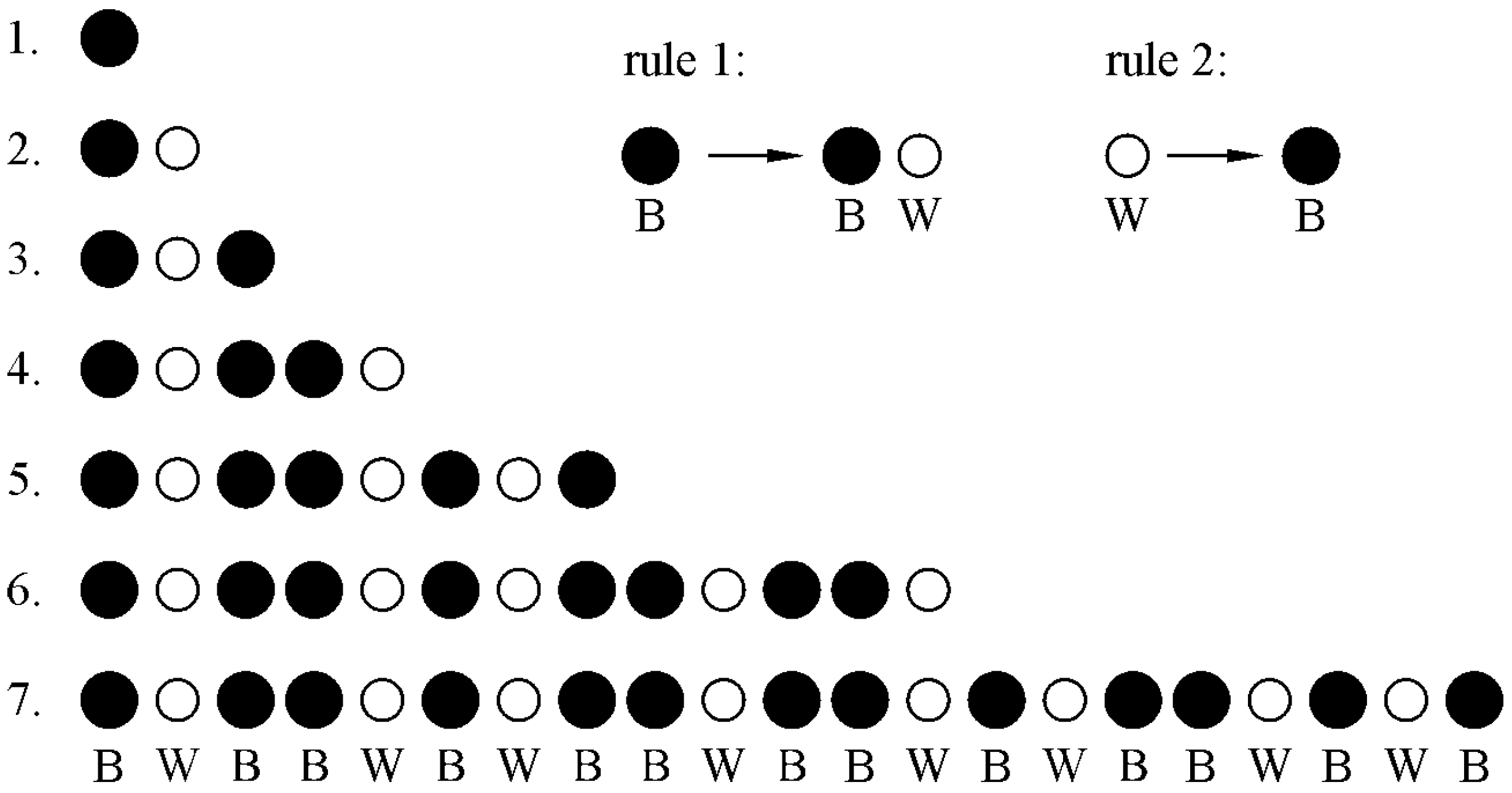
For most non-mathematicians, the basic characteristics of quasicrystals are hard to understand when dealing with them for the first time. They can be easily comprehended by making sequences of black (B) and white (W) circles using two simple rules (Figure 1). We start with just one black circle in the first generation. The sequence in the next generation can be obtained by changing one black circle to a black and white circle (rule 1) and a white circle to a black circle (rule 2). We can see that the sequence is not periodic, but the colour of a circle at any position can be unambiguously predicted. The sequence has a long-range order without periodicity. The sequence S at generation n is the sum of the sequences of the two previous generations:
Sn = Sn−2 + Sn−1
The number of circles in each generation follows the Fibonacci sequence: 1, 2, 3, 5, 8, 13, 21, 34…
Figure 1. Quasiperiodicity is shown as a sequence of black and white circles (Fibonacci chain). The distribution in the next generation is the sum of the two previous generations. The sequence has a long-range order without periodicity [18].
Materials 18 04575 g001
Periodic crystals have a repeating unit cell extending in all three spatial dimensions, creating a long-range order. They have two-, three-, four-, or sixfold rotational symmetries. Tiles with these symmetries can completely cover the surface (Figure 2). They have sharp diffraction patterns with the same symmetries. On the contrary, amorphous materials lack any long-range order. However, they have a short-range order. Their diffraction patterns are typically diffuse, without sharp peaks.
Quasicrystals have non-crystallographic symmetries. Tiles with such symmetries cannot cover the whole surface or overlap (Figure 3).
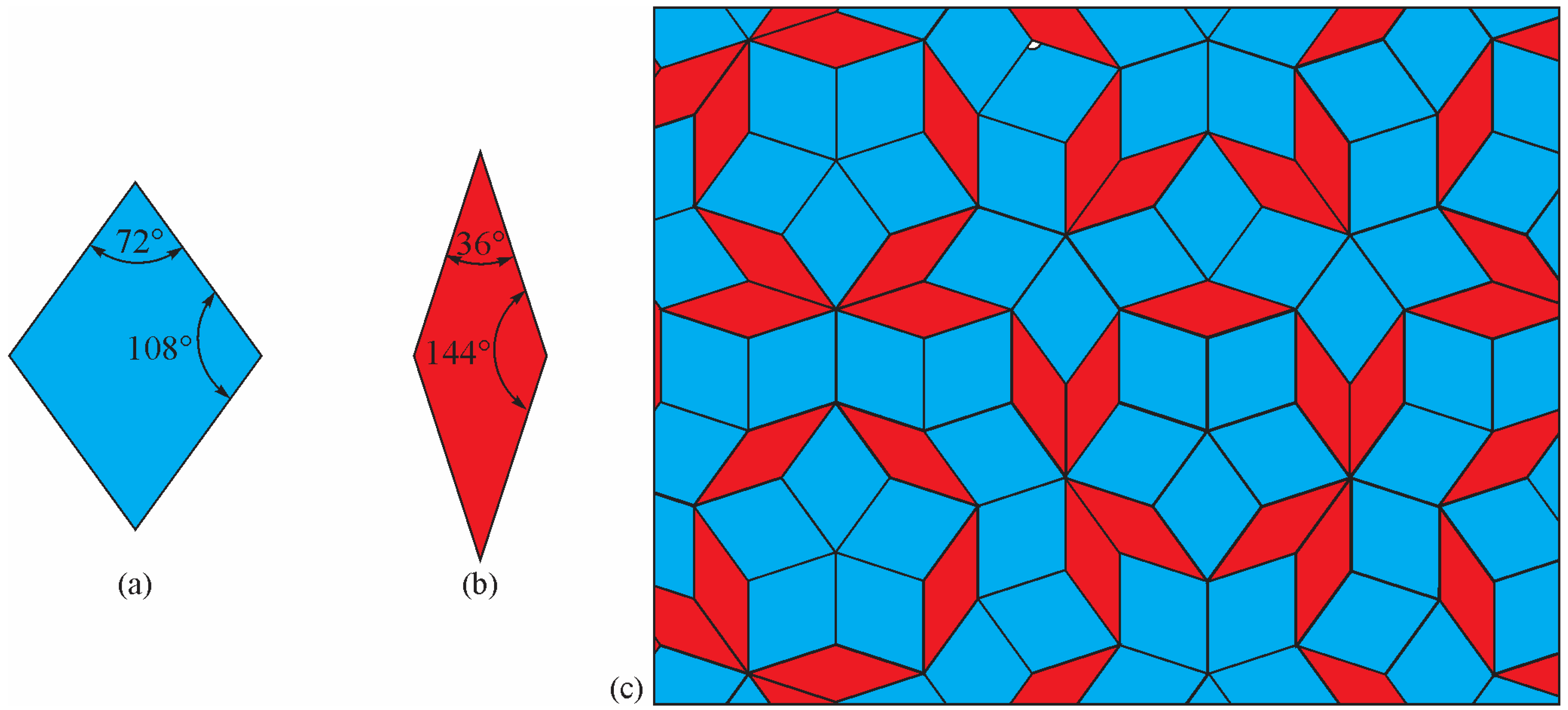
Since non-crystallographic shapes cannot cover a surface completely, Penrose [19] used two types of rhombi to achieve full coverage (Figure 4). He obtained a 2D quasiperiodic structure by applying special matching rules.
The Penrose 2D quasicrystal tiling displays a non-repeating, aperiodic pattern that can fill space without gaps. It also has a sharp diffraction pattern but non-crystallographic or forbidden symmetries. Experimentally, quasicrystals with five-, eight-, ten-, or twelvefold symmetries were discovered [20,21,22,23]. Mackay [24] experimentally proved that Penrose tiling has a sharp diffraction pattern and provided a formalism for its description.
The real quasicrystals consist of aperiodic arrangements of clusters. On the other hand, quasicrystalline approximants (QAs) consist of the periodic arrangement of the same clusters, enabling crystallographers to identify their structure. The usual classification of quasicrystal structures is based on the prevalent cluster type in the structure [14]. This can be the Mackay or pseudo-Mackay cluster (type A) [25,26,27], the Bergman cluster (type B or Frank–Kasper type) [28,29], and the Tsai (type C) [30] cluster. However, icosahedral quasicrystals of type A cannot be adequately described based on Mackay clusters alone. They can be slightly better described with Bergman clusters (type B). When only one of these two cluster types is used, only ≈75% of the structure is covered, but it is increased to ≈98% when both types are used simultaneously. The remainder represents glue atoms [31].
Real quasicrystals also have different defects, such as phasons [32,33,34,35], dislocations [36,37,38], vacancies [39,40], antiphase boundaries [41], stacking faults [42,43], cracks [44,45,46], and growth defects [46]. Defects affect quasicrystals’ physical, chemical, and mechanical properties, like in periodic crystals. Each defect type has its specific effect on properties. We can mention phason defects, which are specific defects in quasicrystals, arising from rearrangements in quasiperiodic atomic order. They, for example, reduce electric conductivity and affect plastic deformation and magnetic properties [47].
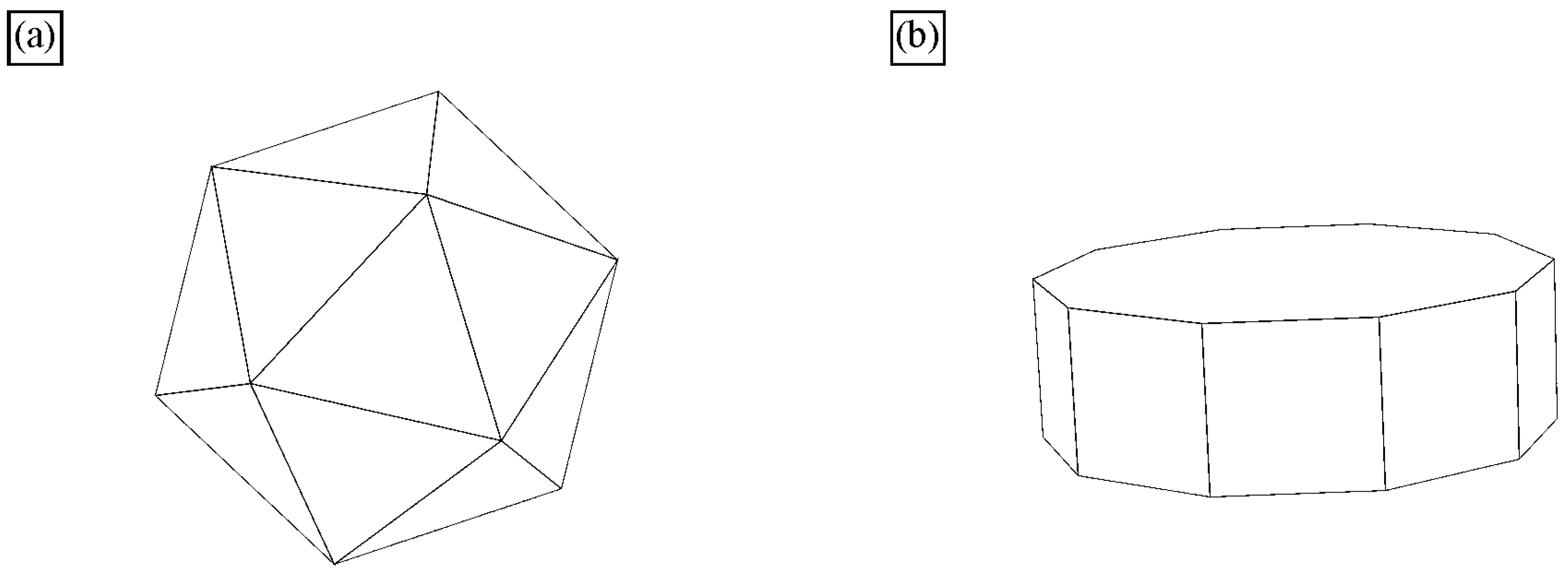
All quasicrystals were made artificially, while natural quasicrystals were discovered later [48,49]. The quasicrystals in the Al-Mn system are metastable and transform into stable phases during annealing [50]. On the other side, many systems have stable quasicrystals, including Al-Cu-Fe [51]. Thus, the quasicrystalline phase appears in the appropriate phase diagram [50]. Stable quasicrystals can be grown from the melt. Such single quasicrystals are typically faceted and possess shape characteristics for five-, eight-, ten-, and fivefold symmetries. The most typical shapes are icosahedra, pentagonal dodecahedra, and decagonal prisms (Figure 5). Their sizes are between mm and cm. They can be observed by the naked eye or with a magnifying glass. They are used to determine their electronic, mechanical, physical, and other properties. The examples of some quasicrystals are given in Table 1. The quasicrystals normally form in binary and multicomponent alloys. Single-element quasicrystals can form under special conditions. For example, plasmonic silver nanoparticles can have a shape of icosahedron [52], silicon quasicrystal can form on glass [53], while quasicrystalline monolayers of Sb and Bi form on the icosahedral Al-Pd-Mn and decagonal Al-Ni-Co surfaces [54].
There are two large types of quasicrystals: 2D and 3D quasicrystals [14]. The former has three groups, i.e., eightfold octahedral, tenfold decagonal, and twelvefold dodecagonal. However, only the fivefold icosahedral type is found as the latter. Icosahedral quasicrystals are quasiperiodic in all directions. Their structure is based on an icosahedron, a body with twenty faces, all triangles (Figure 5a). An icosahedron can be regarded as a projected polyhedron of a 6D cube into the 3D space. It has 12 vertices with fivefold symmetry, 20 triangular facets with threefold symmetry and 30 edges with twofold symmetry. Since the icosahedron is centrally symmetrical, the number of independent symmetric axes is half of the above numbers, i.e., 6 for fivefold, 10 for threefold, and 15 for twofold. This makes the icosahedron a body with a very high degree of symmetry. Decagonal quasicrystals (DQCs) are 2D quasicrystals. They are quasiperiodic in one plane, thus in two directions, but periodic in the third direction. The basic unit is a decagonal prism (Figure 5b). The decagonal prism has a tenfold axis perpendicular to the basal plane and ten twofold axes. A set of fivefold axes passes through the centres of the opposite vertical facets, while a set of other fivefold axes goes through the centres of opposite vertical edges [56].
The best way to identify quasicrystals is through electron diffraction in TEM (Figure 6). The diffraction patterns of an IQC particle with the zone axes parallel to two-, three-, and fivefold axes correspond to Figure 6b–d, respectively. A system with six indices is used to index icosahedral quasicrystals. For details, a reader should consult, for example, references [14,57,58]. In order to be able to index the reciprocal lattice spots as a combination of integers, at least six basis vectors are required for the icosahedral phase, and five for the decagonal phase. Elser [57] selected the six vectors for indexing the IQC diffraction pattern, which can be obtained by a projection of a cube in six dimensions to three dimensions. The Bragg vector for each diffraction peak is expressed as a linear combination of basis vectors multiplied by integer indices, scaled by a factor called the quasilattice parameter. The observable reciprocal space spot nearest to the transmitted beam along a fivefold direction (vertex vector) is indexed as (100,000).
Figure 7 shows that these axes also appear in its stereographic projection. The periodic crystals also have two- and threefold axes; hence, their diffraction pattern is periodic. However, quasicrystals do not have periodic diffraction patterns. The distances to the main peaks typically increase with the golden mean τ, which is the ratio between a diagonal and an edge of a pentagon:
τ = 1 + 5 2
Twofold diffraction patterns also allow the calculation of a quasicrystalline constant [57,58].
Figure 7. Stereographic projection of (a) the icosahedral space group along a fivefold axis and (b) the decagonal space group along the tenfold axis [18].
Materials 18 04575 g007
Figure 8 shows the image, the diffraction pattern of DQC in a periodic α-Al matrix, and the presence of the ordered L12 structure. The quasiperiodic structure is evident (Figure 8a), and the diffraction pattern (Figure 8b) shows the tenfold rotational symmetry.

1.3. Stability of Quasicrystals

The quasicrystal can be thermodynamically stable or metastable. Here are some examples of stable quasicrystals: Al-Cu-Fe [61,62], Al-Mn-Pd, Al-Cu-(Co-Rh), Ag-In-Ca [63], Al-Ni-Co [64], Cd-Mg-Dy [65], Cd-Ca [30], Al-Ni-Rh [66], Al-Cu-Co [67], Al-Co-Ni [68], Al-Pd-RE (RE—rare-earth metal) [69], and metastable ones, Al-Mn [70], Al-Fe [71], Al-Cr [72,73], Al-V [74], and Al-Ni [75]. The readers can obtain much more detailed information about stability and properties of quasicrystals in “Comprehensive experimental datasets of quasicrystals and their approximants” [55].
Quasicrystals are thermodynamically stable when their free energy is lower than that of competing phases. This is achieved through contributions from harmonic and anharmonic vibrations, chemical substitution disorder, and electronic excitations [76]. Thermodynamic stability of quasicrystals often depends on the electron per atom ratio (e/a), typically around 1.7 to 1.8 for stable phases [61,77], the atomic size factor [61,76], and entropy contribution. In the Al-Mn system, the entropy contribution due to the presence of icosahedral short-range order in the liquid phase can facilitate the formation of quasicrystals upon cooling. Generally, it prevents the transition of quasicrystals to more stable crystalline phases [78,79]. The work of Mihalkovic and Widom [76] suggests that the Al-Cu-Fe quasicrystal can be a truly stable phase even at 0 K; other stable quasicrystals can be stable at least at higher temperatures due to entropy stabilisation. The stability of a quasicrystal can be influenced by alloying. So et al. [69] studied the (Al, Ga)-Pd-RE systems, using the rare-earth elements Yb, Tm, and Er. They found that a stable IQC phase is at 2.10 valence electrons per atom, and that the stability of IQC phases becomes lower with increasing atomic radii of the RE metals.
According to stability, quasicrystals can be formed by different manufacturing processes. The most typical are rapid solidification [62,80,81], mechanical alloying [82,83,84,85], powder metallurgy and sintering [86,87,88,89,90,91], arc melting [92], additive manufacturing [93,94,95,96], precipitation hardening [97,98], thermal spray coating [99,100,101,102], and casting [103,104,105,106,107].

1.4. Properties and Applications of Quasicrystals

Quasicrystals have a long-range quasiperiodicity and, at the same time, a complex local atomic order independent of their long-range structure. Thus, there is often a controversy about which effect is most important for the properties of quasicrystals [108].
In the electronic structure of quasicrystals, there is a pseudogap at the Fermi level, indicating that the density of electronic states is zero. The presence of a pseudogap is a key factor influencing the electron transport. It strongly reduces electrical conductivity; thus, electron transport resembles semiconductor-like transport, not metallic conductivity [109].
The pseudogap also determines the magnetic behaviour of quasicrystals. Some quasicrystals exhibit temperature-dependent susceptibility consistent with pseudogap-induced electronic states near the Fermi level [110,111,112,113]. In magnetic quasicrystals, the spin-glass effect is often observed [114]. It arises in systems with competing interactions—for example, when some neighbouring spins prefer ferromagnetic alignment and others prefer antiferromagnetic alignment, leading to frustration. The system cannot settle into a single ground state but becomes trapped in many nearly degenerate configurations [114].
Thermal conductivity exhibits unusually low values with pronounced anisotropy linked to stacking periodicity and cluster-mode phonon scattering [115,116].
Quasicrystals also have a very low friction coefficient, often lower than conventional metals or ceramics, making them promising for wear-resistant coatings and tribological applications [117]. The unusual friction behaviour arises from their electronic structure (pseudogap reduces their metallic nature and adhesion), atomic surface arrangement, lack of translational periodicity (they contain phasons, which can accommodate atomic rearrangements under stress), high hardness, and formation of thin oxide films on the surface [118].
QCs can also be used for thermoelectric applications. Thermoelectric behaviour is explained by their cluster hierarchy and covalent chemical bonding in clusters [119,120].
The quasiperiodic lattice and cluster structures lead to high hardness, elastic moduli, and complex deformation behaviours [121]. Elastic and plastic deformation are influenced by phason strain fields and dislocation glide mechanisms [122]. Dislocations also cause plastic deformation of single quasicrystals. However, quasicrystals show their brittle character at low temperatures, but at higher temperatures, their ductility increases [123,124]. Nanostructured quasicrystalline coatings exhibit enhanced mechanical performance and damping capacities [125]. Observations of inverse Hall–Petch behaviour in nanophase quasicrystals highlight novel grain size effects on mechanical stability [126]. In situ formed and ex situ added quasicrystals can considerably strengthen the metallic [127,128,129] and polymeric [130] matrices. In Al-alloys strengthened by quasicrystals, adding rare-earth elements considerably enhances mechanical properties. Ce [101,127,128] and mischmetal [129] were the most investigated additions. Mechanical strength exceeding 1000 MPa was attributed to grain refinement, precipitation hardening, and load transfer from nanoscale quasicrystals, although ductility decreases at higher rare-earth contents [131].
Possible applications of quasicrystals are presented in many references [117,132,133,134,135]. Yet, challenges remain in synthesising defect-free, large-scale QCs for industrial use [117]. Table 2 gives some of the most important properties of quasicrystalline alloys.

1.5. The Aim of This Review

Quasicrystals are rarely present as a single-phase material. They are usually embedded in the matrices of different metals. They can appear in various sizes, including the nanoscale and macroscale regions. Analytical TEM is primarily used for its characterisation since it allows the detailed determination of quasicrystals’ crystallographic, chemical, and geometrical characteristics. However, TEM samples are very small and embrace only a small part of a specimen, e.g., a cast part. Therefore, it is also convenient to use other methods to help identify quasicrystals and their distribution in much larger areas than those investigated by TEM. This review paper gives special attention to identifying quasicrystals in aluminium alloys using classical metallographic techniques, such as etching, deep etching, and particle extraction, which allow the investigation of larger areas by light and scanning electron microscope, giving additional information by combining with complementary high-resolution techniques. The first part deals with the preparation of samples for light microscopy and scanning electron microscopy, as well as identifying quasicrystals from 2D cross-sections. Special attention is given to identifying quasicrystals formed during solidification and precipitation from the solid solution. The second part is devoted to direct 3D characterisation of quasicrystals using deep etching and particle extraction methods. The aim is also to give information regarding alternative techniques.

2. Light and Scanning Electron Microscopy of Polished and Lightly Etched Samples

2.1. Classical Metallographic Preparation of Aluminium Alloys

Classical metallographic preparation consists of grinding, polishing, and etching [143]. The main goal of the preparation steps is to reveal the sample’s actual microstructure. It has been a principal method for characterising and developing new metallic materials. Metallographic preparation is also widely applied for the quality control of materials in the metallurgical and metal processing industries [144]. Chemical etching is a controlled process that selectively removes material from a solid surface by chemical reactions, often using acids, bases, or reactive gases [143]. When applied at the micro- and nanoscale, chemical etching can alter the surface’s topography, roughness, and chemical composition. Light microscopy is mainly used to observe microstructures, which normally requires relatively strong etching to reveal microstructural constituents [143,145]. Due to strong etchants, some microstructural constituents are likely attacked too much, but this cannot be visible in a light microscope due to rather low magnifications (up to 2500×). Some etchants can dissolve Mg2Si in Al-Mg-Si alloys [146], while others can dissolve iron-rich phases [147], leaving holes on the surface, interpreted as black-coloured phases. The advent of field-emission scanning microscopes with a resolution of less than 1 nm and the application of excellent backscattered electron detectors have considerably reduced the need for etching [148].
Most etchants for metals were developed by the 1980s and are described in the following books:
  • George, F., Vander, Voort. Metallography Principles and Practice [143] and
  • ASM Handbook, Volume 9, Metallography and Microstructures [149].
They are used in many studies on quasicrystalline alloys. The most used etchants are given in Table 3.
The metallographic preparation of aluminium alloys usually follows general procedures. It often starts with sawing the samples to appropriate sizes, hot or cold mounting into a polymeric resin, grinding with SiC papers, and polishing with different diamond pastes or alumina suspensions (usually 9, 6, and 3 µm), a colloidal suspension of 0.06 µm SiO2, and etching [144]. By mechanically preparing aluminium samples, one should avoid grinding with the finest grinding papers and diamond paste, since SiC and diamond particles can be incorporated into a soft aluminium matrix. The deformation layer is progressively thinned with sequential grinding and polishing steps [143]. Vibrational polishing can effectively remove the deformation layer, making samples appropriate for HR SEM, EDS, and EBSD investigations [156]. In our time, ions can also remove the deformation layer (e.g., argon ions in cross-section polishers). However, parameters should be carefully optimised to avoid the appearance of ion-induced artefacts [157]. Nevertheless, chemical etching is commonly used for light microscopy observations because the microstructure details are hardly visible without etching [158]. On the contrary, polishing without etching can give convenient results when using scanning electron microscopy [94,159]. Examining a polished surface before etching is crucial since a general overview of the microstructural constituents can be obtained. Slight etching can remove a surface deformation layer. A shallow groove formed at grain boundaries and a thin step formed at the particle/matrix interface can increase contrast and make identifying microstructural constituents easier when using a secondary electron detector. Additionally, polished surfaces allow for the most reliable microchemical analysis [160,161]. A classical strong etchant is not always appropriate for scanning electron microscopy, since it can dissolve some phases and deteriorate microstructural and chemical analyses. Thus, modification of established procedures is required [162]. Sometimes, metallographic preparation is taken for granted, and the preparation details are not given. Nevertheless, the inconvenient use of etchants can be identified by inspecting micrographs (look at SEMs in Ref. [163]). Thus, comparing polished and etched specimens is advisable to reveal potential etching artefacts. Etching is unnecessary when using a backscattered electron detector, and the phases have different backscattering coefficients (having elements with different atomic numbers Z) [148]. Thus, microstructural constituents of varying chemical compositions can be distinguished with slight etching or even without it.
This section deals with light and scanning electron microscopy of polished and lightly etched samples of aluminium alloys containing quasicrystals. This is, in fact, 2D metallography. Light microscopy can identify quasicrystals and their geometrical features when quasicrystalline particles are sufficiently large, e.g., above 5 μm. On the other hand, scanning electron microscopy can identify nanoscale quasicrystalline particles (even as small as 20 nm), with additional information obtained by EDS, WDS, or EBSD.
The basis for identifying quasicrystalline particles in a 2D section is based on the fact that the experimentally observed quasicrystals have five-, eight-, ten-, and twelvefold rotational symmetries, which are different from those of periodic crystals [164]. The shape of a crystal, as well as a quasicrystal, depends on its point group symmetry. Several shapes are possible in any point group symmetry, depending on the facets’ surface energy, growth conditions, and presence of surface-active elements. The most often observed shapes of icosahedral quasicrystals are the shapes of a pentagonal dodecahedron [165], icosahedron [166], and rhombohedral triacontahedron [167]. In this way, a well-faceted quasicrystal and non-faceted quasicrystalline dendrites can be unambiguously identified by their shape and “non-crystallographic” symmetry.

2.2. Quasicrystal in Melt-Spun Ribbons

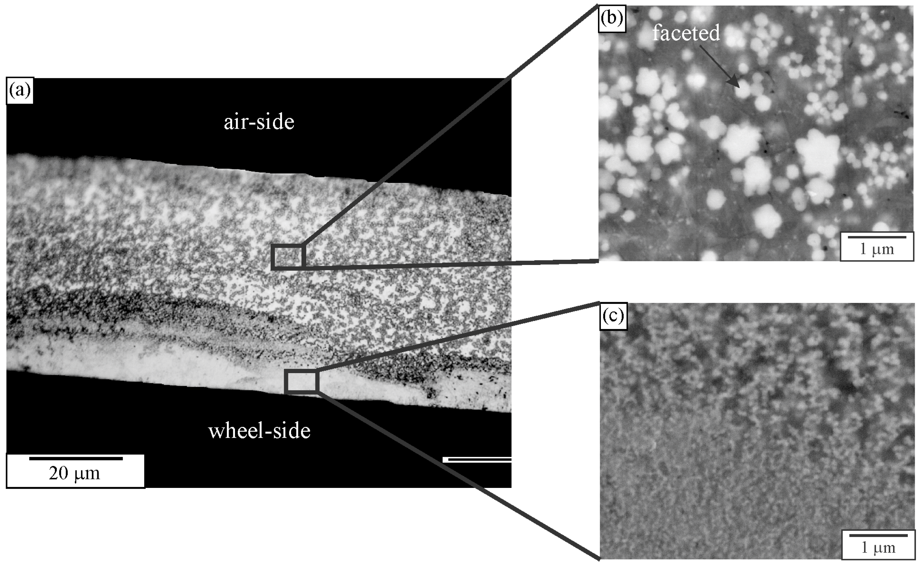
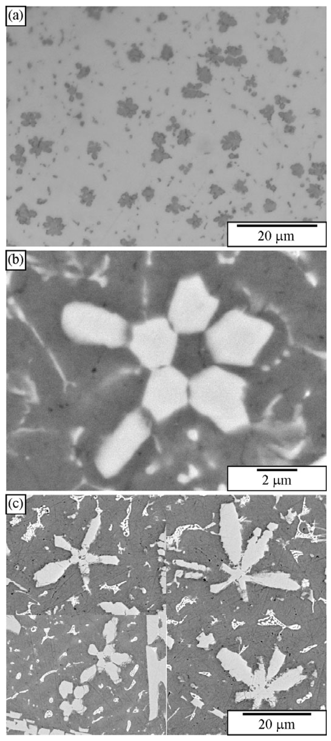
Quasicrystals were first identified in melt-spun ribbons [1]. The microstructure typically consists of quasicrystalline particles embedded in an Al-rich solid solution, denoted by α-Al. The cooling rate by melt spinning can attain 104–106 K/s, enabling copious nucleation of tiny quasicrystalline precipitates [168]. They are too small to be resolved by light microscopy (Figure 9a). Even high-resolution SEM hardly resolves particles formed next to the wheel side (Figure 9c). The faceting tendency can be seen in particles as small as 300 nm. However, it is impossible to determine their exact shape according to their 2D cross-sections (Figure 9b). Thangaraj et al. [166] tried to distinguish quasicrystalline shapes (triacontahedron, icosahedron, and pentagonal dodecahedron) by imaging quasicrystalline particles in TEM along two-, three-, and fivefold axes. They found the shapes indistinguishable when projected along higher symmetry axes, like threefold and fivefold axes. However, the projection along the twofold axes enabled distinction between different shapes. Ishimasa and Nissen [169] found out by the same method that the faceted quasicrystalline particles had a form of pentagonal dodecahedron. The quasicrystals can also have a dendritic shape [169,170,171]. The determination of quasicrystal shapes in melt-spun ribbons by deep etching is given in Section 3, Deep Etching and Particle Extraction.

2.3. Quasicrystals in As-Cast Samples

Quasicrystals in Al-Mn alloys can form at very high cooling rates (105 K/s). Casting into metallic moulds cannot produce such cooling rates; the highest cooling rates can be a few thousand K/s. Under such conditions, quasicrystals do not form, but the orthorhombic Al6Mn phase does [173]. Adding several alloying elements can reduce the critical cooling rate for quasicrystal formation. Kim et al. [174] obtained a quasicrystal phase in a bulk Al93Fe3Cr2Ti2 alloy. The quasicrystalline phase also appeared in the Al-Mn-Be alloy [163,175,176,177], Al-Mn-Fe-Si-V [178], and Al-Mn-Ce alloy [179]. Stan-Głowińska [180] discovered quasicrystals in Al-Mn base alloys alloyed with Cr, Co, Ni, and Cu additions. Leskovar et al. [181] created primary metastable quasicrystals in Al-Mn-Si alloys alloyed with Zn, Ca, and Sr. Primary quasicrystalline phases are also formed in magnesium alloys [182]. Thus, quasicrystals appeared in different sizes and shapes, which can be identified using metallography, as explained in detail in Ref. [183]. The primary quasicrystals can have polygonal or dendritic shapes [167,169,171,184,185]. The same or similar shapes can also appear in the other alloys.
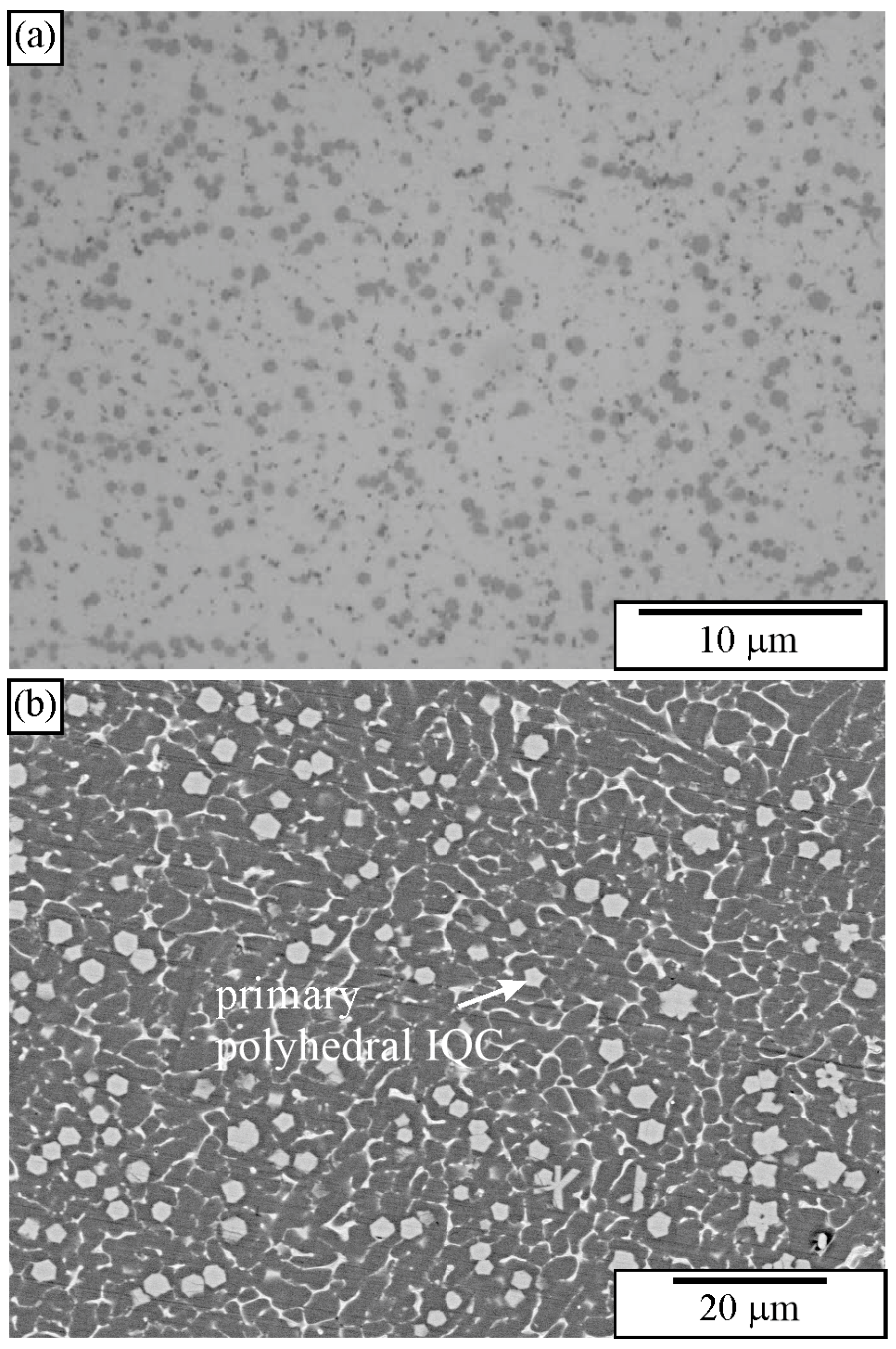
Figure 10 shows the microstructure of an Al-Cu-Mn-Be alloy in the as-cast state. The sample was investigated in a polished condition, without any etching. The sizes of polygonal quasicrystalline particles are 2–5 μm. The light micrograph (Figure 10a) does not have sufficient resolution to reveal the characteristics of the quasicrystalline particles. On the other hand, the backscattered electron micrograph reveals some features of quasicrystalline particles (Figure 10b). Some particles are pentagonal, whereas most of them are hexagonal.
Figure 11 shows a microstructure of the Al-Cu-Fe alloy with several intersecting quasicrystalline particles. The particles are much larger than the previous ones, so their shapes can be seen. The presence of the icosahedral quasicrystalline phase was determined using other methods, such as XRD and TEM [183,187].
To understand the shapes of particles in 2D, sections should be made through a pentagonal dodecahedron (Figure 12) and other possible shapes of primary icosahedral quasicrystals (Figure 13). In Figure 12, the intersecting planes are perpendicular to a pentagonal dodecahedron’s two-, three-, and fivefold axes. Intersections have different shapes, triangles, rectangles, pentagons, hexagons, octagons, and decagons, depending on where the metallographic plane intersects a polyhedron.
A quasicrystalline particle can have any orientation relative to the intersecting (metallographic) plane in a real material. It is impossible to investigate any orientation. We have selected seven different orientations and illustrated them within a stereographic triangle (Figure 13). The triangle’s vertices represent a fivefold, threefold, and twofold axis, and the possible intersections are the same as in Figure 12. In addition, the shapes along the four other axes were examined, which are indicated by numbers 1–4:
  • Between the fivefold and threefold axes;
  • Between the fivefold and twofold axes;
  • Between the twofold and threefold axes;
  • Within the stereographic triangle.
The analysis of all shapes showed that intersecting all polyhedra with a plane produces triangles, rectangles, pentagons, hexagons, octagons, and decagons, and their distorted and truncated variants. When a pentagon appears on a metallographic cross-section, it is impossible to state that a quasicrystal’s basic shape is the pentagonal dodecahedron. Several other characteristics should be considered when discriminating between different shapes.
At first sight, it is hard to determine the 3D shape of the particles in Figure 11 because they can be distorted due to the growth in the preferred directions, or their growth can be distorted by the presence of other particles. However, inspecting selected particles can eliminate some shapes, as shown in Figure 13. The trapezoidal shape, indicated by the arrow in (Figure 11b), seems possible only if the quasicrystal has the shape of a pentagonal dodecahedron. The pentagonal dodecahedron was observed in the same alloy [188,189]. This conclusion can be drawn when the specimen contains the IQC phase. Otherwise, faceted particles of crystalline phases can, in cross-section, have very different shapes, some closely similar to the shapes of quasicrystals [52].
The growth of quasicrystals often takes place in preferred directions. As a result, dendritic arms can appear (Figure 14). Their maximum number is equal to the number of equivalent directions in the crystallographic system. The icosahedral system could have up to 12 fivefold, 20 threefold, and 30 twofold arms. The number of arms does not depend on the equilibrium shape of the icosahedral quasicrystal. The intersecting plane can cut one or more arms. The positions and dimensions of intersections depend on the orientation of the intersection plane, its distance from the centre of a dendrite, the shape of the dendrite arms, and the preferred growth directions. In the microstructure, differently developed dendrite arms can appear. In Figure 14a, short arms are shown. Nissen et al. [185] found faceted dendrites in the Al-Mn alloy; each dendrite arm was faceted and shaped like a pentagonal dodecahedron (Figure 14b). Otherwise, dendrite arms can show a slight tendency to faceting [148,157,158], with very long and well-developed dendrite arms (Figure 14c). A procedure for determining the preferred growth direction from a 2D cross-section is given in Ref. [183].

2.4. Identification of Quasicrystalline Precipitates in Al-Alloys

IQC precipitates can form in many Al-alloys. They precipitate from the supersaturated solid solution. Hansen and Gjonnes [190] found a metastable IQC phase in a heat-treated Al-Mn alloy, which subsequently transformed to the cubic α-AlMnSi phase. Li and Arnberg [191] discovered IQC dispersoids in a commercial 3003 alloy, which precipitated at the early precipitation stage. Quasicrystalline precipitates also formed during ageing in Al-Mn-based alloys fabricated by SLM [150]. Mochugovskiy et al. [192,193,194] discovered IQC precipitates in some Al-Mg-Mn alloys during annealing. Zhan et al. [195] studied nucleation and growth of quasicrystal-related precipitates in Al-Er-Fe alloys. Yang et al. [5] found quasicrystalline clusters in Al-Mg-Zn alloys. Quasicrystalline precipitates also occur in commercial Al-Cu-Li alloys [196,197]. Quasicrystalline precipitates also formed as one of the three strengthening precipitates in an Al-Zr-Gd(Yb) alloy [198]. Zr, Gd, and Yb formed L12 precipitates, but the Fe and Si impurities formed IQC precipitates.
Partly coherent IQC precipitates predominantly formed on the dislocations and dislocation walls. IQC precipitates were also discovered in an Al94Mn2Be2Cu2 alloy during an in situ study of the temperature stability of the IQC phase formed during solidification [199]. They were found in samples heat-treated at 300 and 400 °C [200]. IQC precipitates in this alloy have a primitive icosahedral structure (space group P m 35 ¯ ). They form at least a semi-coherent interface with an aluminium matrix, and both phases have a specific mutual orientation relationship. All aluminium fourfold axes are parallel to the three twofold axes of IQC, and at the same time, threefold aluminium axes are parallel to the threefold axes of IQC. Stan-Głowińska et al. [97,201] discovered nanoscale IQC precipitates in Al-Mg-Zn alloys containing Ga. Zhang et al. found that small amounts of iron in Al-V, Al-Mo, and Al-Cr alloys can give rise to a fine dispersion of P-phase precipitate at the grain centres. The P-phase is a quasicrystalline phase and has an orientation relationship with the Al matrix as i2\\[001]Al, i3\\[111]Al, and i5\\[t10]Al, t = (1 + square-root 5)/2 [202].
Icosahedral quasicrystalline precipitates can also form in Mg alloys [203,204], stainless steel [205], and Zr-amorphous alloy [206,207].
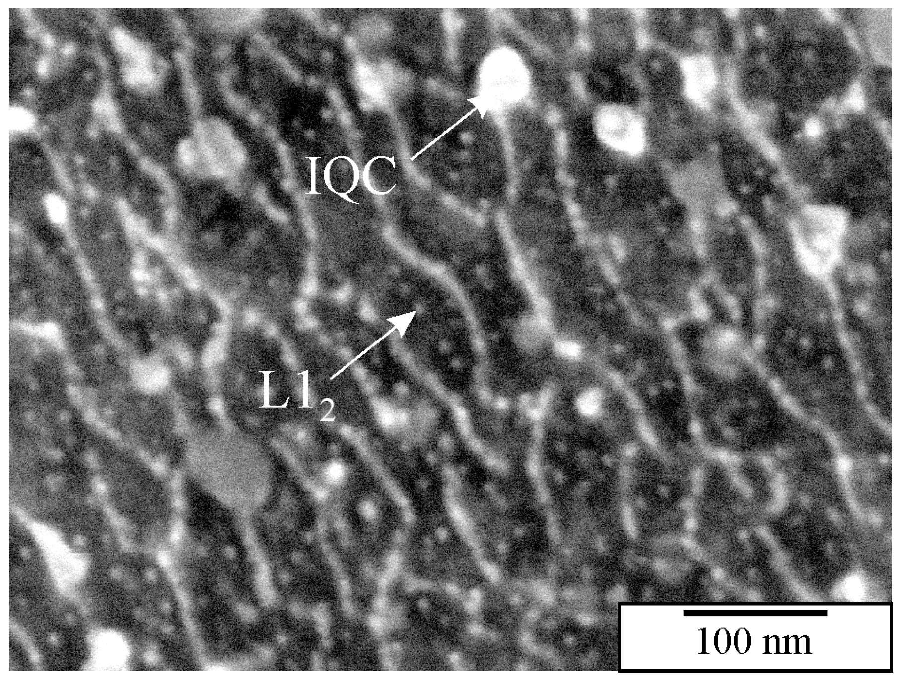
IQC and other quasicrystalline precipitates are relatively small, ranging from 10 to 100 nm. They can be thoroughly investigated in TEM. However, TEM samples are small, and the distribution of precipitates can be determined in a relatively small area. Thus, developing a metallographic procedure that enables observing as many tiny particles as possible in HR SEM is imperative. Österreicher et al. [162] developed a procedure for revealing the Mg2Si precipitates in Al-Mn-Si alloys using 30% nitric acid in methanol. The same method dissolved quasicrystalline precipitates in the authors’ laboratory. However, there is an ongoing investigation regarding the development of metallographic methods to identify small nanosized quasicrystalline particles. The preliminary findings using mechanical polishing with colloidal SiO2 and etching using diluted hydrogen peroxide, H2O2 show promising results (Figure 15). Colloidal silica with a granulation of 0.06 µm and 30% hydrogen peroxide in water with a ratio of 5:1 was used. Polishing with small abrasive grains and low surface tension removes the oxide layer and slightly embosses the surface. The procedure takes 1 to 2 min, with mandatory ultrasonic cleaning in distilled water without using cleaning alcohol. Quasicrystalline particles with a size of 30–50 nm and L12 precipitates with a size of 10–20 nm can be seen. The method needs considerable improvement since several artefacts appear at the nanoscale (bright ridges in Figure 15) [208].
The second approach uses physical polishing and etching with Ar ions. The samples can be prepared by conventional mechanical polishing and finally exposed to Ar ions. Ion polishing improves the surface quality of metallographic samples by using broad-beam argon ion milling, which removes material at the atomic level without causing deformation or disturbance to the microstructure. This method is particularly effective for high-technology materials and complex assemblies, producing flat surfaces convenient for light microscopy, SEM, or EBSD [209,210,211].
By changing operation parameters, such as accelerating voltage, sputtering angle, and duration, it was possible to prepare the surface with visible microstructural constituents without a deformation layer. Figure 16 compares the same alloy’s HR SEM and TEM micrographs, showing the extraordinary degree of resemblance. It is possible to recognise particles as small as 10 nm. In the actual sample, L12 precipitates were present, with sizes around 5 nm, which could not be recognised despite some brighter areas in the α-Al matrix.

2.5. Alternative Methods for the Identification of Quasicrystalline Phases Using Metallography

Previous sections dealt with identifying quasicrystalline particles based on their morphology using light and scanning electron microscopy. SEM still needs an adequately metallographically prepared sample; however, the nature of interactions of electrons with the sample offers several ways to identify crystals.
The electron channelling effect in SEM is a phenomenon where the orientation of the crystal lattice influences the interaction of an electron beam with a crystalline material. When the electron beam is aligned with specific crystallographic planes (usually low index), the penetration depth can be significantly larger; thus, fewer electrons are backscattered. It increases the contrast in SEM images. The contrast level is weak, necessitating a minimum beam current of 5 nA for visibility [212]. It can enable the visualisation of crystallographic defects and crystal orientations.
Shindo et al. [213] found a strong electron channelling effect in a quasicrystal of an Al-Cu-Fe alloy, which has a near-perfect icosahedral symmetry and low phason density. When the electron beam was parallel with one of the high symmetry zone axes, such as the fivefold and twofold, the intensities of the characteristic X-ray of Fe and Cu relative to that of Al were much stronger than under non-channelling conditions. Thus, this method could identify quasicrystals in SEM’s without EBSD.
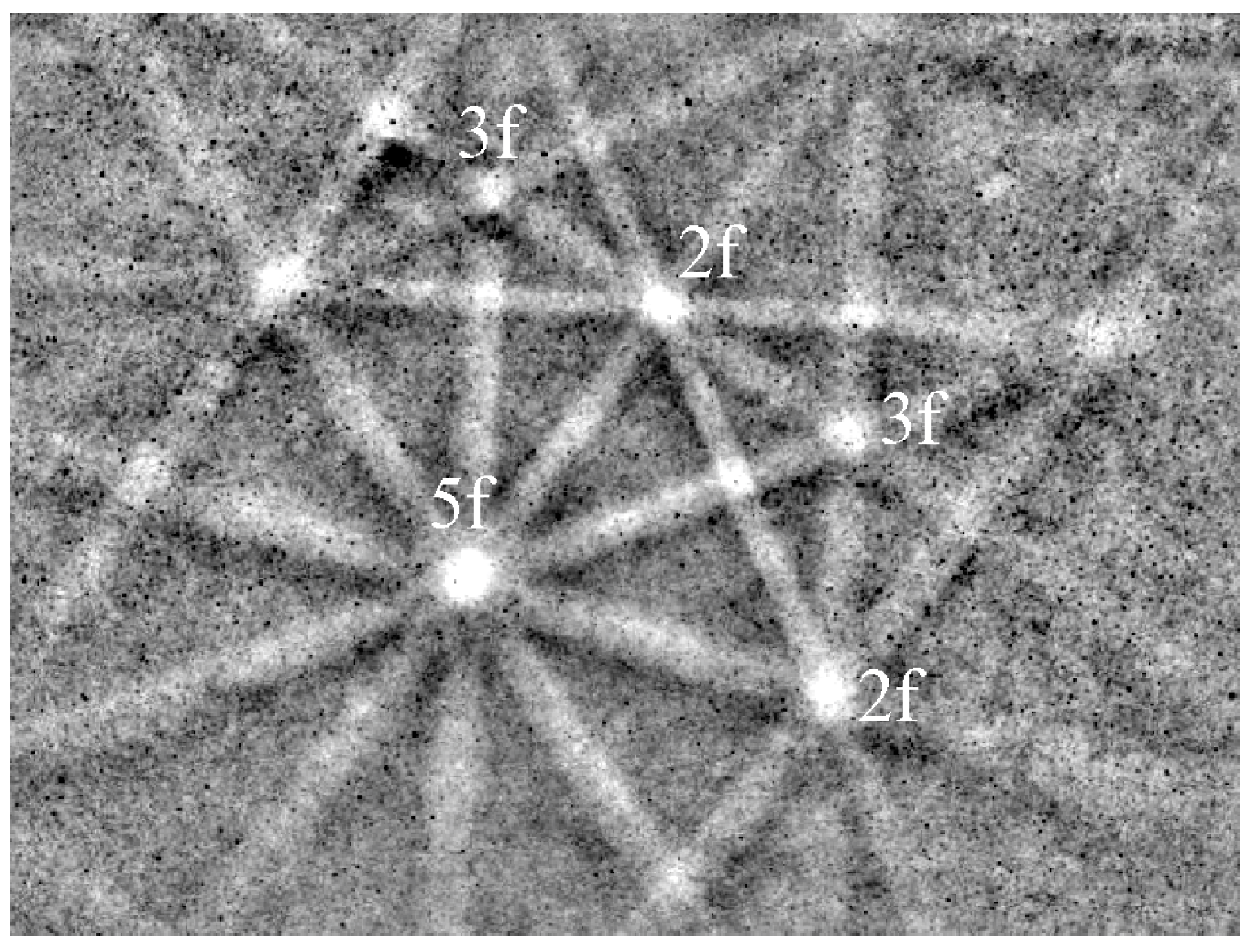
EBSD (Electron Backscatter Diffraction) is an advanced microstructural characterisation technique used in SEM to determine the crystallographic orientation of materials. It is especially powerful for studying crystalline solids at the microscale. A polished crystalline sample is tilted at a high angle (~70°) inside an SEM chamber. The electron beam interacts with the tilted surface, causing backscattered electrons to diffract. These diffracted electrons form a pattern of lines called a Kikuchi pattern on a phosphor screen. The pattern is recorded and analysed to determine the crystal orientation at that specific point (Figure 17). By scanning across an area, EBSD can build up orientation maps that show the crystallographic structure of the sample at high spatial resolution, typically for bulk samples above 100 nm.
EBSD has often been used to characterise quasicrystals. A systematic orientation analysis for icosahedral and decagonal quasicrystals was provided by Winkelmann et al. [214]. Labib et al. [215] used EBSD to characterise IQC and quasicrystalline approximant in Cd-Mg-RE (RE = Y, Sm, Gd, Tb, Dy, Ho, Er, Tm) systems. Kikuchi patterns revealed a high resemblance between the two structures. Winkelmann et al. [214] mapped the locally varying orientations in samples of icosahedral quasicrystals observed in a Ti40Zr40Ni20 alloy and AlNiCo decagonal quasicrystals. Tanaka et al. [216] developed a new analysis system for identifying the orientation of an IQC in an SEM with an EBSD instrument. The system was successfully applied to determine the orientation of any given IQC grain by reading and assigning the Kikuchi bands obtained with the EBSD. Naglič et al. [217] identified IQC and DQC in Al-Mn-Ga alloys. The quasicrystalline phases larger than 100 nm could be identified using bulk specimens, not nanoscale precipitates.
Recently, a new approach to SEM-based diffraction has been developed [218,219]. It is called transmission EBSD (t-EBSD) or transmission Kikuchi diffraction (TKD). TKD improves spatial resolution, enabling effective characterisation of nanocrystalline materials because resolution could be below 10 nm [220,221]. The final spatial resolution depends on the sample atomic number, beam energy, sample thickness, and tilt angle. TKD samples can be prepared using the standard transmission electron microscopy preparation (TEM) methods. Electropolishing 3 mm diameter metallic discs is often the most effective because it can produce a large electron-transparent area. However, FIB-SEM can also be used. The optimum thickness depends on the material and lies between 50 and 100 nm [222].
Transmission scanning electron microscopy on the TKD samples allows microchemical analysis at a very high spatial resolution. In conventional energy-dispersive spectroscopy/scanning-electron microscopy (EDS/SEM) analysis, the pear-shaped interaction volume of incident electrons in the bulk sample determines spatial resolution; thus, the actual scope of EDS analysis is usually a few cubic microns. Lowering the accelerating voltage can effectively reduce the interaction volume of the electron beam. Liu et al. [223] used this method to further simulate EDS mapping of Ni nanoparticles.
Atom probe tomography (APT) is an advanced microscopy technique that allows 3D reconstruction of a material at near-atomic resolution and elemental identification of each atom. Quasicrystalline precipitates were analysed with this technique by Farkoosh et al. [224] in an age-hardened and creep-resistant Al-0.5Mn-0.3Si (at.%) alloy by Sn inoculation. The nanoscale quasicrystalline precipitates were also revealed in a Mg–6Zn–4Al–1Sn–0.5Mn alloy [225].

3. Deep Etching and Particle Extraction

The icosahedral quasicrystalline particles rarely exhibited a pentagonal symmetry on the polished surface [183]. It is even harder for the decagonal quasicrystals that mainly appear as platelike particles [70], which can be found in many crystal systems. Consequently, it is challenging to identify quasicrystalline particles only from 2D sections prepared by traditional metallographic techniques. Therefore, there is a great interest in developing methods for obtaining 3D microstructures, especially when explaining the morphology evolution at different processing techniques [226,227]. The commonly used methods are deep etching and particle extraction from the aluminium matrix [228]. Many researchers have dealt with the methods of deep aluminium etching [143,228,229,230]. A review article on this topic was published by Gupta et al. in 1996 [228]. In the article, they collected the methods and etchants known at that time. It is frequently desirable that the Al matrix be removed in a controlled way [231]. In the first stage, the surface oxide layer should be removed or at least considerably thinned by mechanical and electrochemical polishing or electrolysis. The Pourbaix diagram for the system Al-H2O at 25 °C shows that the oxide layer is stable for a pH between 5 and 8.5 [232], and this range is to be considered by the selection of the solution for chemical removal of the oxide layer.
During deep etching, the matrix should be removed around the particles, while the particles should be retained unchanged. In addition, they remain at their original position. Less material must be removed by deep etching than by particle extraction. When the etching depth approaches the typical particle sizes, deep etching can be stopped because the morphology of particles becomes visible [143]. Longer etching times are required when revealing larger particles in the microstructure. It is thus extremely important that a reagent does not dissolve quasicrystalline and other phases during particle extraction so that they still exhibit their original shape after removing the matrix [233]. The particles should be cathodic relative to the matrix. In the opposite case, if the particles are anodic, they will dissolve, leaving a cavity at the site of the particle [185]. Table 4 gives characteristics of chemical extraction methods for deep etching and extraction of particles, mainly in the aluminium alloys.
The first attempt to reveal the quasicrystalline phase using deep etching in rapidly quenched Al-5.3 at.% Mn alloy was performed by Csanady et al. [170]. They used a slightly acidic medium (the composition is not given in the article), which nicely preserved the crystalline Al6Mn and Al4Mn particles; however, the icosahedral quasicrystalline phase completely dissolved. Nissen et al. [185] studied two rapidly solidified alloys with the nominal compositions Al6Mn (14.3 at.% Mn) and Al17Mn (5.6 at.% Mn). The melt-spun ribbons were etched in a 250 mL methyl alcohol solution with 2 wt.% KI. The alloy sample was suspended in the centre of a 400 mL beaker containing the etchant and surrounded by a high-purity Al or Pt cathode. The etching lasted one hour, during which the voltage applied was 2–5 V and the current was 0.1–0.3 A. This etching process resulted in an almost complete isolation of all quasicrystalline particles from the surrounding crystalline Al solid solution matrix of Al. They also electropolished melt-spun ribbons at −20 °C in a solution consisting of a mixture of perchloric acid (HClO4) and 20% methanol. After this treatment, the quasicrystal particles dissolved and appeared as cavities in the Al-rich matrix.
Kang et al. [226] determined the icosahedral quasicrystalline phase’s 3D morphology and formation process in rapidly solidified Al–Mn alloy. The ribbons were electro-etched in a 250 mL methyl alcohol solution with 2 wt.% KI. The voltage, temperature, and current density for electrolytic etching were 10 V, 25 °C, and 0.1 A/cm2, respectively. They revealed the morphology of the quasicrystalline phase in melt-spun ribbons.
Kang et al. [234] used a NaOH aqueous solution to dissolve the eutectic matrix and preserve the primary IQC particles in the Al–6Mn–2.5Be alloy. They did not give the exact composition of the etchant. However, they revealed the three-dimensional morphological evolution and growth mechanisms of primary IQC particles during directional solidification. On the contrary, Bončina et al. [201] did not reveal the quasicrystalline phase in an Al-Mn-Be-Cu alloy using 5% aqueous solution of NaOH for 30 s. The presence of Cu in the IQC phase may change the potential of the IQC phase relative to the matrix.
Wang et al. [235] deep etched AZ magnesium alloy using 10% nitric acid in ethanol for 5–10 min. They could dissolve the magnesium-rich matrix to reveal an Al-Mn-based decagonal quasicrystal and some other crystalline phase.
Zhu et al. [167] found that a single icosahedral quasicrystal can be obtained in the Mg-Al-Zn alloys. The icosahedral quasicrystals possessed three morphologies: well-developed dendrites and coral-like and petal-like quasicrystals. Samples were mechanically polished and etched in a 5% nitric acid solution and 95% ethanol.
Bončina et al. [236] tested several etchants to reveal the morphology of quasicrystals and other phases in Al-Mn-Be and Al-Mn-Be-Cu alloys. They found that a solution of strong acids (2.5 mL HCl, 5 mL HNO3, and 1 mL HF) slightly attacks the Al matrix, while intermetallic phases were attacked much more strongly (Figure 18). The IQC phase was dissolved entirely, and only its silhouette remained (Figure 18). This indicates that the IQC and other intermetallic phases acted as anodes and the Al matrix as a cathode.
Dilute solutions made of HCl and HNO3 have been extensively used to etch aluminium alloys deeply [228,231]. When utilised to etch the aluminium quasicrystalline alloys containing higher volume fractions of intermetallic phases, etching with a solution of 5 mL of HNO3, 2.5 mL of HCl, and 70 mL of alcohol for 30 s resulted in almost complete dissolution of quasicrystalline particles, and the aluminium matrix remained intact (Figure 19) [236].
Bončina et al. [237] give special attention to improving the procedure of dynamic deep etching and particle extraction from aluminium alloys, especially dedicated to quasicrystalline particles. The basis represents the iodine methanol solution (10 g of iodine and 25 g of tartaric acid in 250 mL of methanol; the ratio of iodine to metal should be greater than 4.7), which was developed by Guzowski et al. [230] to determine the morphology of grain-refining particles in Al-Ti-alloys. The process is not straightforward for aluminium alloys containing quasicrystals; thus, it was modified [237]. The main characteristics of the modified process are presented in more detail below.
There are two main problems. Firstly, the process does not start just by immersing samples into a solution; secondly, iodine or CuI remains in the sample after the process. Several steps should be taken to obtain excellent results. An aluminium sample is initially rinsed into a solution containing 3 to 10 g of iodine and 10 to 17 g of tartaric acid in 100 mL of methanol and put into an ultrasonic cleaner. The sample is sequentially exposed to ultrasonic vibrations and resting, lasting from 10 min up to 24 h. The sequence of rinsing and dissolving, followed by exposure to ultrasonic action, should be alternately repeated 10 to 50 times. After deep etching, the sample is put into the ultrasonic bath to remove the reaction products, washed with alcohol, and dried. The sample is then broken to expose a clean fracture surface with a partly removed aluminium matrix and undissolved particles.
The solution was filtered using a glass filter to obtain extracted particles. The filter retained particles larger than 300 nm. The water-soluble iodides are dissolved by washing the sediment in alcohol and water. The smaller nanoparticles can be collected from the clear alcohol–water solution by the grid and air drying.
The etchant composition is influential, but implementing all etching stages is more critical. The oxide surface layer is not removed, but the ultrasonic vibrations cause a local disruption of the oxide layer (Figure 20a), enabling the penetration of the etchant into the sample (Figure 20b).
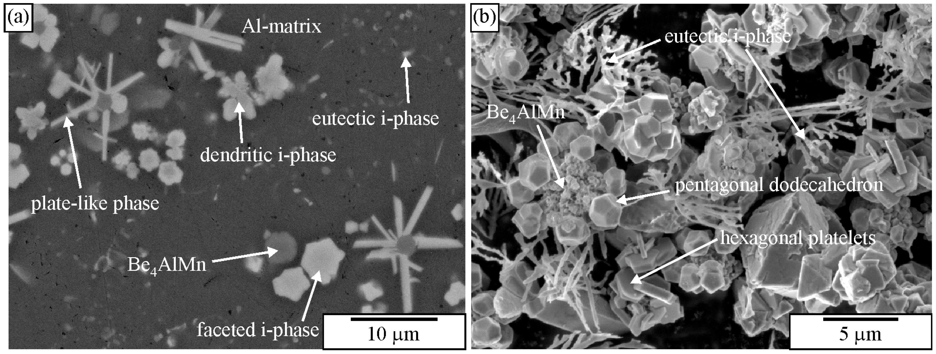
Figure 21 shows images of an as-cast Al-Mn-Be alloy before and after etching with an iodine-methanol solution. The alloy was exposed to the solution for several days. It is evident that with this solution, all phases present in the microstructure were observed. It seems that only Be4AlMn was partly dissolved. A fine, branched structure of the quasicrystalline eutectic phase also remained unchanged.
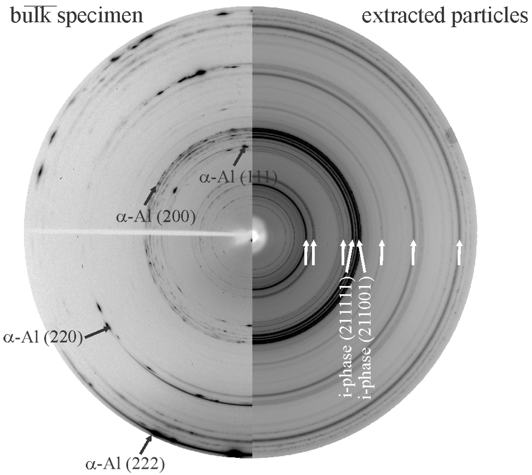
Figure 22 shows 2D diffraction patterns of an Al-Mn-Be alloy in the as-cast condition. It was also confirmed that all reaction products formed during dissolution were removed after applying the iodine-methanol solution. There were no rings corresponding to iodine or other iodides. Removing the Al matrix results in much better visibility of rings of minor phases. In this case, rings belonging to IQC become visible and allow unambiguous identification.

3.1. Deep Etching of Melt-Spun Ribbons

Melt spinning enables the study of equilibrium shape and the roughening behaviour of the quasicrystals. The rapid solidification of dilute Al-Mn-Be and Al-Mn-Be-B alloys makes possible substantial undercooling, thus promoting nucleation of the quasicrystal in the melt [11]. Later, the Al grains nucleate, and their very fast growth enables the trapping of small quasicrystals.
Figure 23a shows a microstructure typical of 30–90 μm thick ribbons. This is an enlarged image of Figure 9. These ribbons possessed a relatively uniform distribution of tiny spherical quasicrystalline particles with sizes between 50 nm and 500 nm in diameter in an α–Al matrix. Backscattered electron images indicated that some particles had spherical shapes, while some showed a tendency for faceting. However, the resolution was usually too low to determine their shape precisely.
During deep etching with the iodine-methanol solution, the matrix was removed around particles, revealing their pentagonal dodecahedron shape. On the particle indicated by the arrow, pentagon facets can be easily recognised. Thus, SEM of deep-etched melt-spun ribbons unambiguously revealed the shape of faceted IQC particles [227].
Transmission electron microscopy allows the determination of quasicrystalline shapes. We can use foils or extracted particles. Figure 24a shows that IQC particles with a size of less than 100 nm ribbons had rounded edges. Particles with sizes up to 200 nm can be almost ideal spheres, while some have bumps (Figure 24b). A particle in Figure 24c is faceted. By comparison of the projection with Figure 25, it is clear that the particle has the shape of a pentagonal dodecahedron, which was observed in SEM after deep etching.
The possible shapes of IQC particles formed upon the primary crystallisation of melt-spun ribbons are shown in Figure 26. The evolution of shapes depends on the undercooling. A larger undercooling can be obtained at the wheel side, resulting in spherical particles because of dynamic roughening [164]. With the decreasing cooling rate, the particles possess some flat faces with rounded corners. Constitutional undercooling can lead to morphological instability of the solid–liquid interface, resulting in bump formation. Such development can lead to the formation of large dendrites with rounded arms. At a smaller undercooling, the effect of dynamic roughening diminishes, resulting in the formation of almost ideal pentagonal dodecahedra.

3.2. Deep Etching of Alloys with Polygonal and Dendritic Icosahedral Quasicrystals

Figure 27a shows an IQC quasicrystal after deep etching in an as-cast Al-Mn-Be-Cu alloy. The backscattered electron image of the same sample is given in Figure 10b. Figure 28b shows an IQC quasicrystal after deep etching in an as-cast Al-Cu-Fe alloy. In both cases, the shape of the IQC was a pentagonal dodecahedron.
Figure 28 shows IQC dendrites. Recognition of the quasicrystalline nature is apparent in the extracted sample.
Figure 29a shows an extracted icosahedral quasicrystalline dendrite covered with small particles. Its shape can be recognised immediately, indicating the strength of the particle extraction technique. On the other side, there is no indication of how deep the particles penetrate the quasicrystal. This information can be gathered from 2D metallographic sections. In our time, the entire quasicrystal can be sectioned with FIB (Figure 29b). Thus, one can simultaneously see the phases’ morphology and internal structure. Making FIB cross-sections of the extracted particles can be much faster than making cross-sections in bulk material since much less material must be removed.
The IQC was also a part of two-phase eutectic cells (α-Al + IQC). Extraction of particles revealed the rodlike structure of the eutectic IQC phase (Figure 30). The rods evolved from a common centre. As the distance from the centre increased, frequent branching occurred. Branching keeps the distance between branches as constant as possible. Detailed TEM analysis of the eutectic IQC showed that the whole rodlike structure is a single quasicrystal. No sharp orientation changes were observed, not even at branching positions. This indicated that branching did not occur by, e.g., twinning, which is typical for the Si phase in the Al-Si eutectic. Still, growth direction can be quickly changed to one of the other preferred directions due to the very high symmetry of the icosahedral phase. Furthermore, it was observed that orientation was changing uniformly along each rod. A rotation of 2.7° around a fivefold zone axis occurred when moving from position 1 to position 2. This kind of lattice rotation might indicate the presence of quasilattice defects, such as phason strains [238]. This was confirmed by peer examination of the diffraction patterns since many diffraction spots were deflected from their ideal positions (significantly weaker ones), and the shapes of some spots showed strong anisotropy.
Particle extraction can also provide advantages by applying characterisation techniques other than SEM-related ones. For instance, the extracted particles can be reliably examined with TEM. Thin particles are placed on a holey carbon grid, allowing TEM examination without other preparation steps (Figure 31). Also, HRTEM and analytical examinations, such as EDS and EELS, can be performed on the extracted particles [186].

3.3. Alternative Methods for Determining 3D Morphologies of Quasicrystals

The first large stable quasicrystals were obtained by slow growth of the quasicrystals from the melt. During solidification, the melt disappeared, leaving holes in which the grown quasicrystals were present. The morphology of the quasicrystal was revealed without any need for metallographic preparation. This way, Beeli and Nissen [184] determined the growth morphology of icosahedral Al-Mn-Pd single quasicrystals. Al-Fe-Cu single crystals can also obtain their equilibrium shape in the cavities during heat treatment [188]. These are special cases that can be applied to stable quasicrystals.
Kral et al. [239] obtained 3D shapes of phases in steels using computer-aided visualisation of 3D reconstructions from serial section images. They gradually removed approximately 0.2 μm of material step-by-step metallographically and took photos of the section after each step. This is rather a slow procedure, which is used rarely. No such attempt was reported in connection with quasicrystals.
Nowadays, sequential cross-sectioning is performed using FIB (usually dual-beam SEM/FIB systems are used). FIB-SEM tomography has proven to be a mature and reliable technique, which can be applied to different materials and research fields [240]. A FIB/SEM system combines focused ion beam milling with high-resolution electron imaging to prepare, image, and analyse cross-sections of materials. It is possible to obtain detailed morphological, compositional, and crystallographic information with spatial resolution in the nanometre range (1–10 nm for imaging; ~10–20 nm for chemical mapping). A cross-section is produced by FIB milling. After FIB removes a slice of material, the electron beam scans the surface. Secondary electrons (SEs) and backscattered electrons (BSEs) provide high-resolution topographic and compositional contrast. The electron and ion beams are arranged at a fixed angle, enabling simultaneous imaging and milling of cross-sections. FIB can reveal a subsurface microstructure, which is imaged by SEM at the same time. By sequentially milling thin slices with FIB and imaging each slice with SEM, a 3D reconstruction of the internal microstructure can be created. Lasagni et al. [241] used FIB-SEM tomography to characterise as-cast and solution-treated AlSi12(Sr). Singh et al. [242] determined nanoscale precipitates in an AA7075-T651 alloy. However, no reports have been found regarding the shape of quasicrystalline phases.
It would be possible to obtain the morphology of large quasicrystalline particles by 3D reconstruction with sub-micro or micro-CT imaging or 3D X-ray microscopes. Both methods require expensive equipment, which is becoming increasingly more available but is often time-consuming. In most cases, these methods do not reach the necessary resolution for metallography [243]. X-ray microscopy (XRM) is very promising in materials science. X-ray microscopy is a technique with the resolution between optical and electron microscopy, offering substantial penetration depths that enable non-destructive imaging of thick and complex specimens. Spatial resolution spans from sub-10-nanometre scales achievable through advanced synchrotron-based methods to sub-micrometre levels in full-field and structured illumination approaches [244]. The characteristics depend highly on the X-ray energy range used; soft X-rays facilitate higher spatial resolution but with limited penetration [245], whereas hard X-rays allow imaging of thicker samples at modestly coarser resolutions. Depth of field is a significant strength of X-ray microscopy. X-ray microscopy enables several contrast mechanisms (absorption contrast for elemental and chemical sensitivity, phase contrast for visualisation of weakly absorbing specimens) while fluorescence and diffraction contrasts give chemical or magnetic information.
Atom probe tomography works by field-evaporating atoms one by one from a sharp needle-shaped specimen, identifying them using time-of-flight mass spectrometry, and reconstructing their original positions into a 3D atomic map. The specimen is prepared into a very sharp needle shape with a tip radius of ~50–100 nm using focused ion beam (FIB) or electrochemical polishing. Then a strong electric field (tens of V/nm) is applied to the tip, and short voltage pulses or laser pulses are used to trigger evaporation in a controlled way. The evaporated ions are accelerated toward a position-sensitive detector. Their time-of-flight (TOF), i.e., the time to reach the detector, depends on the ion’s mass-to-charge ratio. The original position of each atom on the tip is reconstructed using the detector impact position and the evaporation sequence. A 3D atom-by-atom map of the sample is created by combining millions of such evaporation events. APT has a spatial resolution of ~0.2–0.3 nm in depth and ~0.3–0.5 nm laterally. It can detect elements at a ppm-level concentration. It can analyse up to ~100 million atoms in a single dataset. However, conductive or laser-assisted evaporation is required for insulating materials, and the needle shape restricts sample geometry. APT can provide shapes of clusters and nanosized quasicrystalline precipitates [224].

4. Conclusions

This review considers different aspects of the metallography of quasicrystals, mainly focusing on the classical metallographic procedures and discussing alternative methods.
Much information about quasicrystalline phases can be obtained using light and scanning electron microscopy on metallographic samples (2D cross-sections of the microstructure). Adequately oriented quasicrystalline particles can show their characteristic shapes (pentagonal shape for icosahedral quasicrystals) or specific growth angle of eutectic quasicrystals. The possible shapes for faceted and dendritic icosahedral quasicrystals have already been presented in a systematic way. However, there is a research perspective to make similar systems for other types of quasicrystals.
For metallographically prepared samples, the application of EBSD provides information regarding the crystal structure and orientation of crystals. Although there are existing mathematical models that enable identification of quasicrystals and their orientation according to their Kikuchi lines, the main producers of EBSD equipment still do not have quasicrystals in their databases. Thus, the quasicrystalline phase is still not automatically identified, which makes its identification difficult for users who are not familiar with quasicrystals. However, for the skilled users, geometrical, chemical, and crystallographic information of particles larger than 100 nm can be obtained.
Hence, there is still an open question of whether the observation and reliable identification of quasicrystalline particles smaller than 100 nm on bulk samples is viable. The topic is not important just for quasicrystals but also for other phases. There is a research task of how to find an appropriate etchant that would uniformly remove a few nanometres of the matrix, exposing nanometre-sized particles.
Transmission scanning electron microscopy at low voltages (in typical SEM instruments) on thin lamellas (50–100 nm), prepared by FIB instruments, represents a possibility for geometrical, chemical, and crystallographic characterisation of nanoscale precipitates (10 nm or even less). Combined with transmission Kikuchi diffraction, it can also provide crystallographic information. However, it is not a standard method yet. Thus, advancing this method should be an important research direction.
Deep etching and particle extraction provide an excellent possibility of revealing quasicrystals’ morphology and their identification. In this area, the iodine-methanol solution was effective in some alloys. Here, there is still an open question of whether this method is applicable to other alloy systems. It is also rather slow; therefore, more systematic work in improving its behaviour is required. In the field of metallography, the selection of etchants is still mainly empirical. There is a lack of theory that can predict the phase dissolution in multi-phase alloys and especially forecast the retention of their 3D shapes.
Alternative methods that can reveal the 3D shapes of quasicrystals are rather rarely used (FIB/SEM 3D reconstruction, microCT, X-ray microscopy, and APT), so their more frequent application could reveal finer details in the shape and structure of quasicrystals that can disclose additional information regarding their formation and growth mechanisms.
Nevertheless, the most important method for analysing quasicrystalline structures, especially at the atomic resolution, will remain analytical TEM, providing reliable information on the detailed quasicrystalline structure, defects, composition, and geometrical properties.

Author Contributions

Conceptualization, T.B. and F.Z.; methodology, T.B.; writing—original draft preparation, T.B. and F.Z.; writing—review and editing, T.B. and F.Z.; visualisation, F.Z. All authors have read and agreed to the published version of the manuscript.

Funding

The authors acknowledge the financial support from the Slovenian Research Agency (research core funding No. P2-0120 and I0-0029).

Data Availability Statement

No new data were created or analyzed in this study. Data sharing is not applicable to this article.

Conflicts of Interest

The authors declare no conflicts of interest.

Abbreviations

QAQuasicrystalline Approximant
EELSElectron Energy Loss Spectroscopy
HAADFHigh-Angle Annular Dark Field
TEMTransmission Electron Microscopy
IQCIcosahedral Quasicrystal
DQCDecagonal Quasicrystal
STEMScanning TEM
EDSEnergy-Dispersive X-Ray Spectroscopy
WDSWavelength Dispersive X-Ray Spectroscopy
LMLight Microscopy
SEMScanning Electron Microscopy
FIBFocus Ion Beam
EBSDElectron-BackScattered Diffraction
t-EBSDtransmission EBSD
HR SEMHigh-Resolution SEM
XRDX-ray Diffraction
APTAtom-Probe Tomography
TKDtransmission Kikuchi diffraction
RErare-earth metal

References

  1. Shechtman, D.; Blech, I.; Gratias, D.; Cahn, J.W. Metallic phase with long-range orientational order and no translational symmetry. Phys. Rev. Lett. 1984, 53, 1951. [Google Scholar] [CrossRef]
  2. Levine, D.; Steinhardt, P.J. Quasicrystals—A new class of ordered structures. Phys. Rev. Lett. 1984, 53, 2477. [Google Scholar] [CrossRef]
  3. Chattopadhyay, K.; Ranganathan, S.; Subbanna, G.N.; Thangaraj, N. Electron microscopy of quasicrystals in rapidly solidified Al-14% Mn alloys. Scr. Metall. 1985, 19, 767–771. [Google Scholar] [CrossRef]
  4. Cahn, J.W. Quasicrystals. J. Res. Natl. Inst. Stand. Technol. 2001, 106, 975. [Google Scholar] [CrossRef]
  5. Yang, T.; Kong, Y.; Li, K.; Lu, Q.; Wang, Y.; Du, Y.; Schryvers, D. Quasicrystalline clusters transformed from C14-MgZn2 nanoprecipitates in Al alloys. Mater. Charact. 2023, 199, 112772. [Google Scholar] [CrossRef]
  6. Gokhale, A.B.; Banerjee, S. Sample Preparation For Metallography. In Characterization of Materials; Wiley Online Library: Hoboken, NJ, USA, 2002; p. 1. [Google Scholar]
  7. Zhang, L.; Wu, C. Preparation Method for Metallographic Specimen of Iron-Carbon and Silicon-aluminium Alloy. J. Phys. Conf. Ser. 2022, 2338, 012047. [Google Scholar] [CrossRef]
  8. Edyvean, R.H. Christofer, The metallurgical work of Henry Clifton Sorby and an annotated catalogue of his extant metallurgical specimens. Hist. Metall. 2022, 31, 54. [Google Scholar]
  9. Anderson, R.J. The metallography of aluminum. J. Frankl. Inst. 1919, 187, 1. [Google Scholar] [CrossRef]
  10. Exner, H.E. Metallography: Technique of metallurgical investigation or the science of describing metals? Prakt. Metallogr.-Pract. Metallogr. 1999, 36, 115. [Google Scholar] [CrossRef]
  11. McCall, J.L. Scanning Electron Microscopy for Microstructural Analysis. In Microstructural Analysis: Tools and Techniques; McCall, J.L., Mueller, W.M., Eds.; Springer: Boston, MA, USA, 1973; p. 93. [Google Scholar]
  12. Leonard, D.N.; Chandler, G.W.; Seraphin, S. Scanning Electron Microscopy. In Characterization of Materials; John Wiley & Sons: Hoboken, NJ, USA, 2012; p. 1. [Google Scholar]
  13. Hodoroaba, V.D.; Rades, S.; Salge, T.; Mielke, J.; Ortel, E.; Schmidt, R. Characterisation of nanoparticles by means of high-resolution SEM/EDS in transmission mode. IOP Conf. Ser. Mater. Sci. Eng. 2016, 109, 012006. [Google Scholar] [CrossRef]
  14. Steurer, W.; Deloudi, S. Crystallography of Quasicrystals, Concepts, Methods and Structures; Springer: Berlin/Heildelberg, Germany, 2009. [Google Scholar]
  15. Wang, H. Proving theorems by pattern recognition—II. Bell Syst. Tech. J. 1961, 40, 1. [Google Scholar] [CrossRef]
  16. Sharma, B.G. An introductory note on Quasicrystals. Recent Res. Sci. Technol. 2012, 4, 82–84. [Google Scholar]
  17. Pauling, L. So-called icosahedral and decagonal quasicrystals are twins of an 820-atom cubic crystal. Phys. Rev. Lett. 1987, 58, 365. [Google Scholar] [CrossRef] [PubMed]
  18. Bončina, T. Stabilisation and Identification of Quasicrystalline Phases in Al-Mn-Be Based Alloys; University of Ljubljana: Ljubljana, Slovenians, 2010. [Google Scholar]
  19. Penrose, R. The role of aesthetics in pure and applied mathematical research. Bull. Inst. Math. Appl. 1974, 10, 266. [Google Scholar]
  20. Elenius, M.; Zetterling, F.H.M.; Dzugutov, M.; Dzugutov, M.; Fredrickson, D.C.; Lidin, S. Structural model for octagonal quasicrystals derived from octagonal symmetry elements arising in beta-Mn crystallization of a simple monatomic liquid. Phys. Rev. B 2009, 79, 10. [Google Scholar] [CrossRef]
  21. Baipaywad, P.; Wi, J.-S.; Park, H.; Paik, T. Tailoring cubic and dodecagonal quasicrystalline mesophases of mesoporous organosilica nanoparticles and core/shell structure. Mater. Sci. Eng. C-Mater. Biol. Appl. 2019, 98, 666. [Google Scholar] [CrossRef]
  22. Krumeich, F.; Muller, E.; Wepf, R.A.; Conrad, M.; Reich, C.; Harbrecht, B.; Nesper, R. The structure of dodecagonal (Ta,V)(1.6)Te imaged by phase-contrast scanning transmission electron microscopy. J. Solid State Chem. 2012, 194, 106. [Google Scholar] [CrossRef]
  23. Ma, H.; You, L.; He, Z. Stable quaternary Al59Cr23Fe8Si10 decagonal quasicrystal. Mater. Charact. 2020, 166, 10424. [Google Scholar] [CrossRef]
  24. Mackay, A.L. Crystallography and the Penrose pattern. Phys. A Stat. Mech. Its Appl. 1982, 114, 609. [Google Scholar] [CrossRef]
  25. Boudard, M.; Deboissieu, M.; Janot, C.; Heger, G.; Beeli, C.; Nissen, H.U.; Vincent, H.; Ibberson, R.; Audier, M.; Dubois, J.M. Neutron and X-ray single-crystal study of the AlPdMn icosahedral phase. J. Phys.-Condes. Matter 1992, 4, 10149. [Google Scholar] [CrossRef]
  26. Mackay, A.L. A Dense Non-Crystallographic Packing of Equal Spheres. Acta Crystallogr. A-Found. Adv. 1962, 15, 916. [Google Scholar] [CrossRef]
  27. Sirindil, A.; Quiquandon, M.; Gratias, D. Mackay clusters and beyond in icosahedral quasicrystals seen from 6D space. Struct. Chem. 2017, 28, 123. [Google Scholar] [CrossRef]
  28. Bergman, G.; Waugh, J.L.T.; Pauling, L. The crystal structure of the metallic phase Mg32(Al, Zn)49. Acta Crystallogr. 1957, 10, 254. [Google Scholar] [CrossRef]
  29. Sun, W.; Lincoln, F.J.; Sugiyama, K.; Hiraga, K. Structure refinement of (Al, Zn)49Mg32-type phases by single-crystal X-ray diffraction. Mater. Sci. Eng. A 2000, 294–296, 327–330. [Google Scholar] [CrossRef]
  30. Guo, J.Q.; Abe, E.; Tsai, A.P. Stable icosahedral quasicrystals in binary Cd-Ca and Cd-Yb systems. Phys. Rev. B 2000, 62, R14605. [Google Scholar] [CrossRef]
  31. Loreto, L.; Farinato, R.; Catallo, S.; Janot, C.; Gerbasi, G.; Angelis, G.D. Polyhedral and chemical orders in icosahedral Al–Pd–Mn quasicrystals. Phys. B Condens. Matter 2003, 328, 193. [Google Scholar] [CrossRef]
  32. Lyonnard, S.; Coddens, G.; Calvayrac, Y.; Gratias, D. Atomic (phason) hopping in perfect icosahedral quasicrystals Al70.3Pd21.4Mn8.3 by time-of-flight quasielastic neutron scattering. Phys. Rev. B 1996, 53, 3150. [Google Scholar] [CrossRef]
  33. Lyonnard, S.; Coddens, G.; Hennion, B.; Calvayrac, Y. Dynamics of phason hopping in AlFeCu and AlMnPd quasicrystals. Phys. B 1997, 234, 28. [Google Scholar] [CrossRef]
  34. Hiraga, K.; Hirabayashi, M. Quenched phason strains in Al-Mn-Si icosahedral quasicrystal studied by high-resolution electron-microscopy. J. Electron Microsc. 1987, 36, 353. [Google Scholar]
  35. Kiani, M.; Samavat, F.; O’Connor, J. Phason mechanism for diffusion in quasicrystalline structures. Solid State Commun. 2020, 321, 14017. [Google Scholar] [CrossRef]
  36. Feuerbacher, M. Dislocations in icosahedral quasicrystals. Chem. Soc. Rev. 2012, 41, 6745. [Google Scholar] [CrossRef]
  37. Mompiou, F.; Caillard, D. Dislocations and mechanical properties of icosahedral quasicrystals. C. R. Phys. 2014, 15, 82. [Google Scholar] [CrossRef]
  38. Ramaswamy, S. The nature of dislocation-motion in quasicrystals. Bull. Mat. Sci. 1988, 10, 75. [Google Scholar] [CrossRef]
  39. Mehrer, H.; Galler, R. Vacancy-mediated diffusion in quasicrystalline alloys. J. Alloys Compd. 2002, 342, 296. [Google Scholar] [CrossRef]
  40. Cizek, J.; Vlcek, M.; Smola, B.; Stulikova, I.; Prochazka, I.; Kuzel, R.; Jager, A.; Lejcek, P. Vacancy-like defects associated with icosahedral phase in Mg-Y-Nd-Zr alloys modified by the addition of Zn. Scr. Mater. 2012, 66, 630. [Google Scholar] [CrossRef]
  41. Heggen, M.; Feuerbacher, M.; Schall, P.; Urban, K.; Wang, R. Antiphase domains in plastically deformed Zn-Mg-Dy single quasicrystals. Phys. Rev. B 2001, 64, 014202. [Google Scholar] [CrossRef]
  42. Wang, R.H.; Feuerbacher, M.; Yang, W.G.; Urban, K. Stacking faults in high-temperature-deformed Al-Pd-Mn icosahedral quasicrystals. Philos. Mag. A-Phys. Condens. Matter Struct. Defect Mech. Prop. 1998, 78, 273. [Google Scholar] [CrossRef]
  43. Klein, H.; Feuerbacher, M. Structure of dislocations and stacking faults in the complex intermetallic ξ′-(Al-Pd-Mn) phase. Philos. Mag. 2003, 83, 4103. [Google Scholar] [CrossRef]
  44. Mikulla, R.; Stadler, J.; Krul, F.; Trebin, H.R.; Gumbsch, P. Crack propagation in quasicrystals. Phys. Rev. Lett. 1998, 81, 3163. [Google Scholar] [CrossRef]
  45. Trebin, H.R.; Mikulla, R.; Stadler, J.; Schaaf, G.; Gumbsch, P. Molecular dynamics simulations of crack propagation in quasicrystals. Comput. Phys. Commun. 1999, 122, 536. [Google Scholar] [CrossRef]
  46. Ronchetti, M.; Bertagnolli, M.; Jarić, M.V. Generation and Dynamics of Defects in Two-Dimensional Quasicrystals. In Geometry and Thermodynamics: Common Problems of Quasi-Crystals, Liquid Crystals, and Incommensurate Systems; Tolédano, J.C., Ed.; Springer: Boston, MA, USA, 1990; p. 141. [Google Scholar]
  47. Janot, C.; Loreto, L.; Farinato, R. Special defects in quasicrystals. Phys. Status Solidi B-Basic Res. 2000, 222, 121. [Google Scholar] [CrossRef]
  48. Bindi, L.; Steinhardt, P.J.; Yao, N.; Lu, P.J. Icosahedrite, Al63Cu24Fe13, the first natural quasicrystal. Am. Miner. 2011, 96, 928. [Google Scholar] [CrossRef]
  49. Bindi, L.; Yao, N.; Lin, C.; Hollister, L.S.; Andronicos, C.L.; Distler, V.V.; Eddy, M.P.; Kostin, A.; Kryachko, V.; MacPherson, G.J.; et al. Natural quasicrystal with decagonal symmetry. Sci. Rep. 2015, 5, 5. [Google Scholar] [CrossRef]
  50. Grushko, B.; Velikanova, T.Y. Stable and metastable quasicrystals in Al-based alloy systems with transition metals. J. Alloys Compd. 2004, 367, 58. [Google Scholar] [CrossRef]
  51. Hiraga, K.; Zhang, B.P.; Hirabayashi, M.; Inoue, A.; Masumoto, T. Highly ordered icosahedral quasicrystal of Al-Cu-Fe alloy studied by electron-diffraction and high-resolution electron-microscopy. Jpn. J. Appl. Phys. Part 2—Lett. 1988, 27, L951. [Google Scholar] [CrossRef]
  52. González, A.L.; Noguez, C.; Beránek, J.; Barnard, A.S. Size, Shape, Stability, and Color of Plasmonic Silver Nanoparticles. J. Phys. Chem. C 2014, 118, 9128. [Google Scholar] [CrossRef]
  53. Middya, A.R.; Ghosh, K. Quasicrystalline Phase of Silicon on Glass. MRS Online Proc. Libr. 2012, 1493, 24. [Google Scholar] [CrossRef]
  54. Franke, K.J.; Sharma, H.R.; Theis, W.; Gille, P.; Ebert, P.; Rieder, K.H. Quasicrystalline Epitaxial Single Element Monolayers on Icosahedral Al-Pd-Mn and Decagonal Al-Ni-Co Quasicrystal Surfaces. Phys. Rev. Lett. 2002, 89, 156104. [Google Scholar] [CrossRef]
  55. Fujita, E.; Liu, C.; Ishikawa, A.; Mato, T.; Kitahara, K.; Tamura, R.; Kimura, K.; Yoshida, R.; Katsura, Y. Comprehensive experimental datasets of quasicrystals and their approximants. Sci. Data 2024, 11, 1211. [Google Scholar] [CrossRef]
  56. Meisterernst, G.; Bauer, B.; Gille, P. Czochralski Growth of Decagonal AlCoNi Quasicrystals from Al-rich Solution. Acta Phys. Pol. A 2013, 124, 344. [Google Scholar] [CrossRef]
  57. Elser, V. Indexing problems in quasicrystal diffraction. Phys. Rev. B 1985, 32, 4892. [Google Scholar] [CrossRef]
  58. Singh, A.; Ranganathan, S. On the indexing and reciprocal space of icosahedral quasicrystal. J. Mater. Res. 1999, 14, 4182. [Google Scholar] [CrossRef]
  59. Zupanic, F.; Boncina, T.; Krizman, A.; Grogger, W.; Gspan, C.; Markoli, B.; Spaic, S. Quasicrystalline phase in melt-spun Al-Mn-Be ribbons. J. Alloys Compd. 2008, 452, 343. [Google Scholar] [CrossRef]
  60. Zupanič, F.; Gspan, C.; Burja, J.; Bončina, T. Quasicrystalline and L12 precipitates in a microalloyed Al-Mn-Cu alloy. Mater. Today Commun. 2020, 22, 100809. [Google Scholar] [CrossRef]
  61. Inoue, A.; Tsai, A.-P.; Masumoto, T. Stable decagonal and icosahedral quasicrystals. J. Non-Cryst. Solids 1990, 117–118, 824. [Google Scholar] [CrossRef]
  62. Tsai, A.P.; Inoue, A.; Masumoto, T. Preparation of a new Al-Cu-Fe quasicrystal with large grain sizes by rapid solidification. J. Mater. Sci. Lett. 1987, 6, 1403. [Google Scholar] [CrossRef]
  63. Guo, J.Q.; Tsai, A.P. Stable icosahedral quasicrystals in the Ag-In-Ca, Ag-In-Yb, Ag-In-Ca-Mg and Ag-In-Yb-Mg systems. Philos. Mag. Lett. 2002, 82, 349. [Google Scholar] [CrossRef]
  64. Abe, E.; Tsai, A.P. Structure of a metastable Al3Ni decagonal quasicrystal: Comparison with a highly perfect Al72Ni20Co8. J. Alloys Compd. 2002, 342, 96. [Google Scholar] [CrossRef]
  65. Guo, J.Q.; Abe, E.; Tsai, A.P. A new stable icosahedral quasicrystal in the Cd-Mg-Dy system. Philos. Mag. Lett. 2001, 81, 17. [Google Scholar] [CrossRef]
  66. Terauchi, M.; Ueda, H.; Tanaka, M.; Tsai, A.P.; Inoue, A.; Masumoto, T. Electron-energy-loss spectroscopy study of a stable decagonal quasicrystal Al70Ni20Rh10. Philos. Mag. B-Phys. Condens. Matter Stat. Mech. Electron. Opt. Magn. Prop. 1998, 77, 1625. [Google Scholar] [CrossRef]
  67. Tsai, A.P.; Inoue, A.; Masumoto, T. A stable decagonal quasicrystal in the Al-Cu-Co system. Mater. Trans. JIM 1989, 30, 300. [Google Scholar] [CrossRef]
  68. Tsai, A.P.; Inoue, A.; Masumoto, T. Stable decagonal Al-Co-Ni and Al-Co-Cu quasicrystals. Mater. Trans. JIM 1989, 30, 463. [Google Scholar] [CrossRef]
  69. So, Y.-G.; Yoshikawa, T.; Saruhashi, F.; Edagawa, K.; Tamura, R. Formation of Icosahedral Quasicrystals and 1/1 Crystal Approximants in Al-Pd-RE (RE: Rare Earth Metals) Systems. Mater. Trans. 2011, 52, 2011. [Google Scholar] [CrossRef]
  70. Naglič, I.; Samardžija, Z.; Delijić, K.; Kobe, S.; Dubois, J.-M.; Leskovar, B.; Markoli, B. Metastable quasicrystals in Al–Mn alloys containing copper, magnesium and silicon. J. Mater. Sci. 2017, 52, 13657. [Google Scholar] [CrossRef]
  71. Pedrazzini, S.; Galano, M.; Audebert, F.; Collins, D.M.; Hofmann, F.; Abbey, B.; Korsunsky, A.M.; Lieblich, M.; Garcia Escorial, A.; Smith, G.D.W. Strengthening mechanisms in an Al-Fe-Cr-Ti nano-quasicrystalline alloy and composites. Mater. Sci. Eng. A 2016, 672, 175. [Google Scholar] [CrossRef]
  72. Zhang, H.; Wang, D.H.; Kuo, K.H. Quasicrystals, crystalline phases, and multiple twins in rapidly solidified Al-Cr alloys. Phys. Rev. B 1988, 37, 6220. [Google Scholar] [CrossRef]
  73. Swamy, V.T.; Ranganathan, S.; Chattopadhyay, K. Rapidly solidified Al-Cr alloys—Crystalline and quasicrystalline phases. J. Mater. Res. 1989, 4, 539. [Google Scholar] [CrossRef]
  74. He, G.; Shek, C.H.; Lai, J.K.L.; Bian, Z.; Hui, X.D.; Chen, G.L. Microstructure investigation of rapidly solidified Al-V-Fe alloys. J. Mater. Res. 2002, 17, 814. [Google Scholar] [CrossRef]
  75. Yuan, Y.; Hou, H.; Zhao, Y.H.; Yan, F.; Zhang, Y. Microstructure and mechanical properties of an aluminum alloy reinforced by Al-Ni-Co-Mn quasi-crystalline. Mater. Res. Express 2019, 6, 56529. [Google Scholar] [CrossRef]
  76. Mihalkovic, M.; Widom, M. Spontaneous formation of thermodynamically stable Al-Cu-Fe icosahedral quasicrystal from realistic atomistic simulations. Phys. Rev. Res. 2020, 2, 013196. [Google Scholar] [CrossRef]
  77. Tsai, A.P.; Yokoyama, Y.; Inoue, A.; Masumoto, T. Chemistry of Stable Quasi-Crystals. In Physics and Chemistry of Finite Systems: From Clusters to Crystals; Jena, P., Khanna, S.N., Rao, B.K., Eds.; Springer: Dordrecht, The Netherlands, 1992; p. 177. [Google Scholar]
  78. Zaitsev, A.I.; Zaitseva, N.E.; Shakhpazov, E.K.; Arutyunyan, N.A.; Dunaev, S.F. The conditions of formation and stability of quasi-crystalline phases in aluminum-manganese alloys. Russ. J. Phys. Chem. 2006, 80, 869. [Google Scholar] [CrossRef]
  79. Zaitsev, A.I.; Zaitseva, N.E.; Shimko, R.Y.; Arutyunyan, N.A.; Dunaev, S.F.; Kraposhin, V.S.; Lam, H.T. Thermodynamic properties of Al-Mn, Al-Cu, and Al-Fe-Cu melts and their relations to liquid and quasicrystal structure. J. Phys. Condens. Matter 2008, 20, 114121. [Google Scholar] [CrossRef] [PubMed]
  80. Inoue, A.; Kimura, H.M.; Sasamori, K.; Masumoto, T. Microstructure and novel properties of nanocrystalline and nanocrystalline alloys prepared in Al-based systems by rapid solidification. Sci. Rep. Res. Inst. Tohoku Univ. Ser. A-Phys. Chem. Metall. 1996, 42, 165. [Google Scholar]
  81. Nayak, S.S.; Murty, B.S.; Pabi, S.K. Structure of nanocomposites of Al-Fe alloys prepared by mechanical alloying and rapid solidification processing. Bull. Mater. Sci. 2008, 31, 449–454. [Google Scholar] [CrossRef]
  82. Novák, P.; Kubatík, T.; Vystrčil, J.; Hendrych, R.; Kříž, J.; Mlynár, J.; Vojtěch, D. Powder metallurgy preparation of Al–Cu–Fe quasicrystals using mechanical alloying and Spark Plasma Sintering. Intermetallics 2014, 52, 131. [Google Scholar] [CrossRef]
  83. Hosseini, S.; Novak, P. On the Formation of A1NiCo Nano-Quasicrystalline Phase during Mechanical Alloying through Electroless Ni-P Plating of Starting Particles. Materials 2019, 12, 2294. [Google Scholar] [CrossRef]
  84. Hosseini, S.M.; Novak, P.; Vesely, M. Positive temperature dependence of compressive properties in an AlNiCo poly-quasicrystal fabricated by mechanical alloying and spark plasma sintering. Scr. Mater. 2020, 187, 169. [Google Scholar] [CrossRef]
  85. Michalcova, A.; Vojtech, D.; Schumacher, G.; Novak, P.; Plizingrova, E. Effect of iron and cerium additions on rapidly solidified Al-Tm-Ce alloys. Mater. Tehnol. 2013, 47, 757. [Google Scholar]
  86. Li, R.; Wang, Z.; Li, Z.; Khor, K.A.; Dong, Z. Dynamic Fracture Mechanism of Quasicrystal-Containing Al-Cr-Fe Consolidated Using Spark Plasma Sintering. Crystals 2018, 8, 385. [Google Scholar] [CrossRef]
  87. Shadangi, Y.; Sharma, S.; Shivam, V.; Basu, J.; Chattopadhyay, K.; Majumdar, B.; Mukhopadhyay, N.K. Fabrication of Al-Cu-Fe quasicrystal reinforced 6082 aluminium matrix nanocomposites through mechanical milling and spark plasma sintering. J. Alloys Compd. 2020, 828, 54258. [Google Scholar] [CrossRef]
  88. Li, R.T.; Dong, Z.L.; Khor, K.A. Al-Cr-Fe quasicrystals as novel reinforcements in Ti based composites consolidated using high pressure spark plasma sintering. Mater. Des. 2016, 102, 255. [Google Scholar] [CrossRef]
  89. Laplanche, G.; Gadaud, P.; Bonneville, J.; Joulain, A.; Gauthier-Brunet, V.; Dubois, S.; Jay, F. Spark plasma sintering synthesis and mechanical spectroscopy of the omega-Al0.7Cu0.2Fe0.1 phase. J. Mater. Sci. 2012, 47, 169. [Google Scholar] [CrossRef]
  90. Takagiwa, Y.; Kamimura, T.; Hosoi, S.; Okada, J.T.; Kimura, K. Thermoelectric properties of Al-Pd-Re quasicrystal sintered by Spark Plasma Sintering (SPS): Effect of improvement of microstructure. Z. Für Krist.-Cryst. Mater. 2009, 224, 79–83. [Google Scholar] [CrossRef]
  91. Fleury, E.; Lee, J.H.; Kim, S.H.; Kim, W.T.; Kim, J.S.; Kim, D.H. Spark plasma sintering of Al-Si-Cu-Fe quasi-crystalline powder. Metall. Mater. Trans. A-Phys. Metall. Mater. Sci. 2003, 34A, 841. [Google Scholar] [CrossRef]
  92. Li, C.F.; Carey, C.; Li, D.Q.; Caputo, M.; Bouch, R.; Hampikian, H. A study on spherical particles in Al65Cu20Fe15 alloy prepared by arc melting. Mater. Charact. 2018, 140, 162. [Google Scholar] [CrossRef]
  93. Iams, A.D.; Weaver, J.S.; Lane, B.M.; Giannuzzi, L.A.; Yi, F.; LaPlant, D.L.; Martin, J.H.; Zhang, F. Microstructural features and metastable phase formation in a high-strength aluminum alloy fabricated using additive manufacturing. J. Alloys Compd. 2025, 1025, 180281. [Google Scholar] [CrossRef]
  94. de Araujo, A.P.M.; Pauly, S.; Batalha, R.L.; Coury, F.G.; Kiminami, C.S.; Uhlenwinkel, V.; Gargarella, P. Additive manufacturing of a quasicrystal-forming Al95Fe2Cr2Ti1 alloy with remarkable high-temperature strength and ductility. Addit. Manuf. 2021, 41, 101960. [Google Scholar] [CrossRef]
  95. Wang, Y.; Li, J.; Xia, X.; Zou, L.; Yuan, T.; Liu, X.; Lai, D.; Deng, S.; Li, R. A novel Al-Cr-Sc-Zr alloy additively manufactured via laser directed energy deposition: Microstructure, phase analysis and mechanical properties. J. Mater. Process. Technol. 2023, 322, 118204. [Google Scholar] [CrossRef]
  96. Kairy, S.K.; Gharbi, O.; Nicklaus, J.; Jiang, D.; Hutchinson, C.R.; Birbilis, N. On the Characterization of a Hitherto Unreported Icosahedral Quasicrystal Phase in Additively Manufactured Aluminum Alloy AA7075. Metall. Mater. Trans. A-Phys. Metall. Mater. Sci. 2019, 50A, 529. [Google Scholar] [CrossRef]
  97. Stan-Głowińska, K.; Zięba, A.; Rogal, Ł. Characterization of quasicrystalline precipitates in artificially aged Al–Mg–Zn alloy with Ga addition. J. Mater. Res. Technol. 2022, 21, 1749. [Google Scholar] [CrossRef]
  98. Chen, Z.W.; Tang, M.J.; Zhao, K. Effect of rare earth samarium addition on the kinetics of precipitation in Al-Cu-Mn casting alloy. Int. J. Miner. Metall. Mater. 2014, 21, 155. [Google Scholar] [CrossRef]
  99. Mora, J.; Garcia, P.; Muelas, R.; Aguero, A. Hard Quasicrystalline Coatings Deposited by HVOF Thermal Spray to Reduce Ice Accretion in Aero-Structures Components. Coatings 2020, 10, 290. [Google Scholar] [CrossRef]
  100. Fu, Y.Q.; Peng, T.X.; Yang, D.M.; Sun, C.Q.; Chen, Y.Z.; Gao, Y. HVOF sprayed Al-Cu-Cr quasicrystalline coatings from coarse feedstock powders. Surf. Coat. Technol. 2014, 252, 29. [Google Scholar] [CrossRef]
  101. Lepeshev, A.; Sordelet, D.J.; Rozhkova, E.; Ushakov, A. Modification of Structure and Physico-Mechanical Properties of Al-Cu-Fe Quasicrystal Alloy at Plasma Spraying. J. Clust. Sci. 2011, 22, 289. [Google Scholar] [CrossRef]
  102. Fleury, E.; Kim, Y.C.; Kim, J.S.; Ahn, H.S.; Lee, S.M.; Kim, W.T.; Kim, D.H. Sliding friction and wear behavior of Al-Ni-Co-Si quasicrystalline coatings deposited by the high-velocity oxy-fuel spraying technique. J. Mater. Res. 2002, 17, 492. [Google Scholar] [CrossRef]
  103. Zupanič, F.; Macerl, M.; Haga, T.; Bončina, T. Microstructure and Indentation Properties of Single-Roll and Twin-Roll Casting of a Quasicrystal-Forming Al-Mn-Cu-Be Alloy. Metals 2022, 12, 187. [Google Scholar] [CrossRef]
  104. Wang, B.; Zhao, Y.-h.; Hou, H. Study on microstructure and mechanical properties of Mg-xY-6xZn by squeeze casting. Mater. Res. Express 2019, 6, 66574. [Google Scholar] [CrossRef]
  105. Koga, G.Y.; Branquinho e Silva, A.M.; Wolf, W.; Kiminami, C.S.; Bolfarini, C.; Botta, W.J. Microstructure and mechanical behavior of Al92Fe3Cr2X3 (X = Ce, Mn, Ti, and V) alloys processed by centrifugal force casting. J. Mater. Res. Technol-JMRT 2019, 8, 2092. [Google Scholar] [CrossRef]
  106. Chen, Z.; Zhang, Q.; Dong, B.; Liu, Z. Microstructure and Thermal Stability of Icosahedral Quasicrystals in Suction Casting Al-6 at.%Mn(-2 at.%Be) Alloys. J. Mater. Eng. Perform. 2020, 29, 447. [Google Scholar] [CrossRef]
  107. Fleury, E.; Lee, S.M.; Choi, G.; Kim, W.T.; Kim, D.H. Comparison of Al-Cu-Fe quasicrystalline particle reinforced Al composites fabricated by conventional casting and extrusion. J. Mater. Sci. 2001, 36, 963. [Google Scholar] [CrossRef]
  108. Tamura, R.; Asao, T.; Takeuchi, S. Roles of Quasiperiodicity and Local Environment in the Electronic Transport of the Icosahedral Quasicrystals in Al-Pd-TM (TM = Fe, Ru, Os) Systems. Mater. Trans. 2001, 42, 928. [Google Scholar] [CrossRef]
  109. Maniraj, M.; Rai, A.; Barman, S.R.; Krajci, M.; Schlagel, D.L.; Lograsso, T.A.; Horn, K. Unoccupied electronic states of icosahedral Al-Pd-Mn quasicrystals: Evidence of image potential resonance and pseudogap. Phys. Rev. B 2014, 90, 5. [Google Scholar] [CrossRef]
  110. Dolinsek, J.; Vrtnik, S.; Klanjsek, M.; Jaglicic, Z.; Smontara, A.; Smiljanic, I.; Bilusic, A.; Yokoyama, Y.; Inoue, A.; Landauro, C.V. Intrinsic electrical, magnetic, and thermal properties of single-crystalline Al64Cu23Fe13 icosahedral quasicrystal: Experiment and modeling. Phys. Rev. B 2007, 76, 054201. [Google Scholar] [CrossRef]
  111. Ivkov, J.; Stanic, D.; Popcevic, P.; Smontara, A.; Dolinsek, J.; Gille, P. Hall effect in the crystalline orthorombic o-Al13Co4 approximant to the decagonal quasicrystals. Mater. Tehnol. 2010, 44, 9. [Google Scholar]
  112. Matsuo, S.; Nakano, H.; Motomura, S.; Ishimasa, T. Ising spin orders conforming to local matching rule and long-range regularity of interactions in penrose lattice. J. Phys. Soc. Jpn. 2005, 74, 1036. [Google Scholar] [CrossRef]
  113. Shalaeva, E.V.; Prekul, A.F.; Shchegolikhina, N.I.; Medvedeva, N.I. Curie Temperature and Density of States at the Fermi Level for Al-Cu-Fe Phases: Beta-Solid State. Acta Phys. Pol. A 2014, 126, 572. [Google Scholar] [CrossRef]
  114. Hattori, Y.; Niikura, A.; Tsai, A.P.; Inoue, A.; Masumoto, T.; Fukamichi, K.; Arugakatori, H.; Goto, T. Spin-glass behavior of icosahedral Mg-Gd-Zn and Mg-Tb-Zn quasi-crystals. J. Phys.-Condes. Matter 1995, 7, 2313. [Google Scholar] [CrossRef]
  115. Takeuchi, T. Thermal conductivity of the Al-based quasicrystals and approximants. Z. Für Krist.-Cryst. Mater. 2009, 224, 35–38. [Google Scholar] [CrossRef]
  116. Takagiwa, Y.; Maeda, R.; Ohhashi, S.; Tsai, A.-P. Reduction of Thermal Conductivity for Icosahedral Al-Cu-Fe Quasicrystal through Heavy Element Substitution. Materials 2021, 14, 5238. [Google Scholar] [CrossRef]
  117. Dubois, J.M. Potential and marketed applications of quasicrystalline alloys at room temperature or above. Rend. Lincei.-Sci. Fis. Nat. 2023, 34, 689. [Google Scholar] [CrossRef]
  118. Yadav, T.P.; Mukhopadhyay, N.K. Quasicrystal: A low-frictional novel material. Curr. Opin. Chem. Eng. 2018, 19, 163. [Google Scholar] [CrossRef]
  119. Cyrot-Lackmann, F. Quasicrystals as potential candidates for thermoelectric materials. Mater. Sci. Eng. A 2000, 294–296, 611. [Google Scholar] [CrossRef]
  120. Takagiwa, Y.; Kimura, K.; Sawama, K.; Hiroto, T.; Nishio, K.; Tamura, R. Thermoelectric properties of Tsai-type Au-Al-RE (RE: Yb, Tm, Gd) quasicrystals and approximants. J. Alloys Compd. 2015, 652, 139. [Google Scholar] [CrossRef]
  121. Takeuchi, S.; Shinoda, K.; Edagawa, K. Dislocation migration and phason strain relaxation in a quasicrystal. Philos. Mag. A-Phys. Condens. Matter Struct. Defect Mech. Prop. 1999, 79, 317. [Google Scholar] [CrossRef]
  122. Zou, Y.; Kuczera, P.; Sologubenko, A.; Sumigawa, T.; Kitamura, T.; Steurer, W.; Spolenak, R. Superior room-temperature ductility of typically brittle quasicrystals at small sizes. Nat. Commun. 2016, 7, 7. [Google Scholar] [CrossRef] [PubMed]
  123. Feuerbacher, A.; Schall, P. Plastic behaviour of decagonal Al-Ni-Co single quasicrystals. Scr. Mater. 2003, 49, 25. [Google Scholar] [CrossRef]
  124. Heggen, M.; Feuerbacher, M.; Schall, P.; Klein, H.; Fisher, I.R.; Canfield, P.C.; Urban, K. Plasticity of icosahedral Zn-Mg-Dy single quasicrystals. Mater. Sci. Eng. A-Struct. Mater. Prop. Microstruct. Process. 2000, 294, 781. [Google Scholar] [CrossRef]
  125. Ustinov, A.I.; Polishchuk, S.S.; Skorodzievskii, V.S.; Bliznuk, V.V. Effect of grain size on the damping capacity of quasicrystalline Al-Cu-Fe materials. Surf. Coat. Technol. 2008, 202, 5812. [Google Scholar] [CrossRef]
  126. Mukhopadhyay, N.K.; Ali, F.; Scudino, S.; Khoshkhoo, M.S.; Stoica, M.; Srivastava, V.C.; Uhlenwinkel, V.; Vaughan, G.; Suryanarayana, C.; Eckert, J. Inverse Hall Petch Like Mechanical Behaviour in Nanophase Al-Cu-Fe Quasicrystals: A New Phenomenon. Acta Phys. Pol. A 2014, 126, 543. [Google Scholar] [CrossRef]
  127. Boncina, T.; Albu, M.; Zupanic, F. Ageing of Al-Mn-Cu-Be Alloys for Stimulating Precipitation of Icosahedral Quasicrystals. Metals 2020, 10, 937. [Google Scholar] [CrossRef]
  128. Murty, B.S.; Hono, K. Al-Mg- and Fe-based nanocomposites by rapid solidification processing. Trans. Indian Inst. Met. 2005, 58, 769. [Google Scholar]
  129. Kenzari, S.; Weisbecker, P.; Curulla, M.; Geandier, G.; Fournee, V.; Dubois, J.M. Formation and properties of Al composites reinforced by quasicrystalline AlCuFeB particles. Philos. Mag. 2008, 88, 755. [Google Scholar] [CrossRef]
  130. Uflyand, I.E.; Drogan, E.G.; Burlakova, V.E.; Kydralieva, K.A.; Shershneva, I.N.; Dzhardimalieva, G.I. Testing the mechanical and tribological properties of new metal-polymer nanocomposite materials based on linear low-density polyethylene and Al65Cu22Fe13 quasicrystals. Polym. Test. 2019, 74, 178. [Google Scholar] [CrossRef]
  131. Inoue, A. Amorphous, nanoquasicrystalline and nanocrystalline alloys in Al-based systems. Prog. Mater. Sci. 1998, 43, 365. [Google Scholar] [CrossRef]
  132. Dubois, J.M.; Lifshitz, R. Quasicrystals: Diversity and complexity. Philos. Mag. 2011, 91, 2971. [Google Scholar] [CrossRef]
  133. Dubois, J.M. Properties- and applications of quasicrystals and complex metallic alloys. Chem. Soc. Rev. 2012, 41, 6760. [Google Scholar] [CrossRef]
  134. Sekhar, J.A.; Mantri, A.S.; Saha, S.; Balamuralikrishnan, R.; Rao, P.R. Photonic, Low-Friction and Antimicrobial Applications for an Ancient Icosahedral/Quasicrystalline Nano-composite Bronze Alloy. Trans. Indian Inst. Met. 2019, 72, 2105. [Google Scholar] [CrossRef]
  135. Guo, Y.; Bataev, I.; Georgarakis, K.; Jorge, A.M.; Nogueira, R.P.; Pons, M.; Yavari, A.R. Ni- and Cu-free Ti-based metallic glasses with potential biomedical application. Intermetallics 2015, 63, 86. [Google Scholar] [CrossRef]
  136. Maciá Barber, E. Chemical Bonding and Physical Properties in Quasicrystals and Their Related Approximant Phases: Known Facts and Current Perspectives. Appl. Sci. 2019, 9, 2132. [Google Scholar] [CrossRef]
  137. VanCleve, J.E.; White, B.E.; Pohl, R.O. Elastic properties of quasicrystals. Phys. B Condens. Matter 1996, 219–220, 345. [Google Scholar] [CrossRef]
  138. Shaĭtura, D.S.; Enaleeva, A.A. Fabrication of quasicrystalline coatings: A review. Crystallogr. Rep. 2007, 52, 945. [Google Scholar] [CrossRef]
  139. Louzguine-Luzgin, D.V.; Inoue, A. Formation and Properties of Quasicrystals. Annu. Rev. Mater. Res. 2008, 38, 403. [Google Scholar] [CrossRef]
  140. Hippert, F.; Préjean, J.J. Magnetism in quasicrystals. Philos. Mag. 2008, 88, 2175. [Google Scholar] [CrossRef]
  141. Texier, M.; Joulain, A.; Bonneville, J.; Thilly, L.; Rabier, J. Al-Pd-Mn icosahedral quasicrystal: Deformation mechanisms in the brittle domain. Philos. Mag. 2007, 87, 1497. [Google Scholar] [CrossRef]
  142. Wollgarten, M.; Saka, H.; Inoue, A. Microstructural investigation of the brittle-to-ductile transition in Al-Pd-Mn quasicrystals. Philos. Mag. A-Phys. Condens. Matter Struct. Defect Mech. Prop. 1999, 79, 2195. [Google Scholar] [CrossRef]
  143. Vander Voort, G.F. Metallography Principles and Practice; McGraw-Hill: Columbus, OH, USA, 1984. [Google Scholar]
  144. Kraner, J.; Kevorkijan, V.; Godec, M.; Paulin, I. Metallographic methods for determining the quality of Aluminium alloys. Mater. Technol. 2021, 55, 541. [Google Scholar] [CrossRef]
  145. Spaić, S. Metalografska Analiza; Fakulteta za naravoslovje in tehnologijo, Oddelek za montanistiko: Ljubljana, Slovenia, 1993. [Google Scholar]
  146. Jin, Z.; Cai, C.; Hashimoto, T.; Yuan, Y.; Kang, D.; Hunter, J.; Zhou, X. Alkaline etching and desmutting of aluminium alloy: The behaviour of Mg2Si particles. J. Alloys Compd. 2020, 842, 155834. [Google Scholar] [CrossRef]
  147. Zhu, H. Etching Behavior of Aluminum Alloy Extrusions. JOM 2014, 66, 2222. [Google Scholar] [CrossRef]
  148. Zupanic, F. Extracting electron backscattering coefficients from backscattered electron micrographs. Mater. Charact. 2010, 61, 1335. [Google Scholar] [CrossRef]
  149. Vander Voort, G.F. (Ed.) Metallography and Microstructures; ASM Internationals: Science Park, OH, USA, 2004; Volume 9. [Google Scholar]
  150. Zhao, J.; Wang, B.; Liu, T.; Luo, L.; Wang, Y.; Zheng, X.; Wang, L.; Su, Y.; Guo, J.; Fu, H.; et al. Study of in situ formed quasicrystals in Al-Mn based alloys fabricated by SLM. J. Alloys Compd. 2022, 909, 164847. [Google Scholar] [CrossRef]
  151. Tiwari, S.; Kesharwani, R.; Jha, K.K.; Mishra, R.; Gope, P.C. Microstructural, texture, and crystallographic analysis of SiC particle incorporation in double-sided friction stir welding of dissimilar aluminium alloys. Mater. Today Commun. 2024, 40, 109543. [Google Scholar] [CrossRef]
  152. Gao, L.; Harada, Y.; Kumai, S. Microstructural characterization of aluminum alloys using Weck’s reagent, part I: Applications. Mater. Charact. 2015, 107, 426. [Google Scholar] [CrossRef]
  153. Gao, L.; Harada, Y.; Kumai, S. Microstructural characterization of aluminum alloys using Weck’s reagent, part II: Coloring mechanism. Mater. Charact. 2015, 107, 434. [Google Scholar] [CrossRef]
  154. Ma, C.; Zhao, Q.; Liu, X.; Li, Y.; Jiang, Q. Introducing high-density growth twins in aluminum alloys by laser surface remelting via templated nucleation of grains. J. Mater. Sci. Technol. 2025, 213, 315. [Google Scholar] [CrossRef]
  155. Zhao, Y.; He, W.; Zhao, F.; Song, C.; Zhang, W.; Song, D.; Tang, Y.; Sun, Z.; Yin, W.; Xue, Y.; et al. Insight into the Fe-rich phases strengthening mechanisms of non-heat-treatable Al-Mg-Mn-Fe-Cu alloys. J. Mater. Sci. Technol. 2025, 205, 232. [Google Scholar] [CrossRef]
  156. Möbius, J.; Lippmann, S.; Seyring, M. Vibratory polishing of multiphase CuZn30//CuZn80 diffusion pairs for electron backscatter diffraction (EBSD) characterization. Pract. Metallogr. 2023, 60, 363. [Google Scholar] [CrossRef]
  157. Wang, Y.Z.; Wu, W.; Liu, Z.W.; Zeng, Y.; Ding, M.J.; Zhang, C.G. Application of Argon Ion Beam Cross Section Polishing in Material Microstructure Research. Key Eng. Mater. 2012, 492, 419. [Google Scholar] [CrossRef]
  158. Vandervoort, G.F. Imaging by Light Optical Microscopy. In Images Of Materials; Williams, D.B., Pelton, A.R., Gronsky, R., Hirsch, P.B., Eds.; Oxford University Press: Oxford, UK, 1992. [Google Scholar]
  159. DeVries, R.C.; Robertson, C. The microstructure of ballas (polycrystalline diamond) by electrostatic charging in the SEM. J. Mater. Sci. Lett. 1985, 4, 805. [Google Scholar] [CrossRef]
  160. Petersson, P.; Bergsåker, H.; Possnert, G.; Coad, J.P.; Koivuranta, S.; Likonen, J. Ion beam micro analysis of deposits at tokamak divertor surfaces. Nucl. Instrum. Methods Phys. Res. Sect. B Beam Interact. Mater. At. 2010, 268, 1838. [Google Scholar] [CrossRef]
  161. Remond, G.; Nockolds, C.; Phillips, M.; Roques-Carmes, C. Implications of Polishing Techniques in Quantitative X-Ray Microanalysis. J. Res. Natl. Inst. Stand. Technol. 2002, 107, 639. [Google Scholar] [CrossRef]
  162. Österreicher, J.A.; Kumar, M.; Schiffl, A.; Schwarz, S.; Hillebrand, D.; Bourret, G.R. Sample preparation methods for scanning electron microscopy of homogenized Al-Mg-Si billets: A comparative study. Mater. Charact. 2016, 122, 63. [Google Scholar] [CrossRef]
  163. Oz, T.; Karakose, E.; Keskin, M. Impact of beryllium additions on thermal and mechanical properties of conventionally solidified and melt-spun Al-4.5 wt.%Mn-x wt.%Be (x = 0, 1, 3, 5) alloys. Mater. Des. 2013, 50, 399. [Google Scholar] [CrossRef]
  164. Chattopadhyay, K.; Ravishankar, N.; Goswami, R. Shapes of quasicrystals. Prog. Cryst. Growth Charact. Mater. 1997, 34, 237. [Google Scholar] [CrossRef]
  165. Ingersent, K.; Steinhardt, P.J. Quasicrystals with dodecahedral equilibrium faceting. Phys. Rev. Lett. 1988, 60, 2444. [Google Scholar] [CrossRef] [PubMed]
  166. Thangaraj, N.; Subbanna, G.N.; Ranganathan, S.; Chattopadhyay, K. Electron-microscopy and diffraction of icosahedral and decagonal quasi-crystals in aluminum manganese alloys. J. Microsc.-Oxf. 1987, 146, 287. [Google Scholar] [CrossRef]
  167. Zhu, M.; Jian, Z.Y.; Liu, C.X.; Yang, G.C.; Zhou, Y.H. Characterization of the Icosahedral Quasicrystal in the Rapidly Solidified Mg-Al-Zn Ternary Alloy. Appl. Mech. Mater. 2011, 71–78, 822. [Google Scholar] [CrossRef]
  168. Michalcova, A.; Vojtech, D.; Schumacher, G.; Novak, P.; Klementova, M.; Serak, J.; Mudrova, M.; Valdaufova, J. Influence of cooling rate and cerium addition on rapidly solidified Al-TM alloys. Kov. Mater.-Met. Mater. 2010, 48, 1. [Google Scholar] [CrossRef]
  169. Ishimasa, T.; Nissen, H.U. Electron Microscopy and Image Contrast Calculation of Al-Mn Quasicrystal. Phys. Scr. 1986, 1986, 291. [Google Scholar] [CrossRef]
  170. Csanády, Á.; Wessicken, R.; Nissen, H.U.; Albert, B.; Griger, A.; Stefániay, V. Rapidly Quenched Al-5.3 at.% Mn Alloy Containing Quasicrystal Units. Mater. Sci. Forum 1987, 13–14, 559. [Google Scholar] [CrossRef]
  171. Chen, Z.; Hou, Y.; Xie, B.; Zhang, Q. Dendrite Morphology Evolution of Al6Mn Phase in Suction Casting Al-Mn Alloys. Materials 2020, 13, 2388. [Google Scholar] [CrossRef]
  172. Zupanic, F.; Boncina, T.; Sustarsic, B.; Anzel, I.; Markoli, B. Microstructure of Al-Mn-Be melt-spun ribbons. Mater. Charact. 2008, 59, 1245. [Google Scholar]
  173. Stan-Glowinska, K.; Rogal, L.; Goral, A.; Wierzbicka-Miernik, A.; Wojewoda-Budka, J.; Schell, N.; Litynska-Dobrzynska, L. Formation of a quasicrystalline phase in Al-Mn base alloys cast at intermediate cooling rates. J. Mater. Sci. 2017, 52, 7794. [Google Scholar] [CrossRef]
  174. Kim, K.B.; Xu, W.; Tomut, M.; Stoica, M.; Calin, M.; Yi, S.; Lee, W.H.; Eckert, J. Formation of icosahedral phase in an Al93Fe3Cr2Ti2 bulk alloy. J. Alloys Compd. 2007, 436, L1. [Google Scholar] [CrossRef]
  175. Kim, S.H.; Song, G.S.; Fleury, E.; Chattopadhyay, K.; Kim, W.T.; Kim, D.H. Icosahedral quasicrystalline and hexagonal approximant phases in the Al-Mn-Be alloy system. Philos. Mag. A-Phys. Condens. Matter Struct. Defect Mech. Prop. 2002, 82, 1495. [Google Scholar] [CrossRef]
  176. Boncina, T.; Markoli, B.; Zupanic, F. Characterization of cast Al86Mn3Be11 alloy. J. Microsc.-Oxf. 2009, 233, 364. [Google Scholar] [CrossRef]
  177. Chang, H.J.; Fleury, E.; Song, G.S.; Kim, W.T.; Kim, D.H. Formation of quasicrystalline phases in Al-rich Al-Mn-Be alloys. J. Non-Cryst. Solids 2004, 334, 12. [Google Scholar] [CrossRef]
  178. Ferreira, T.; Koga, G.Y.; Gargarella, P.; Kiminami, C.S.; Botta, W.J.; Bolfarini, C. New Al-alloys with dispersed stable quasicrystal approximant phases: Overcoming the barrier of conventional casting processing and microstructure design. J. Manuf. Process. 2023, 98, 196. [Google Scholar] [CrossRef]
  179. Schurack, F.; Eckert, J.; Schultz, L. Al-Mn-Ce quasicrystalline composites: Phase formation and mechanical properties. Philos. Mag. 2003, 83, 807. [Google Scholar] [CrossRef]
  180. Stan-Głowińska, K. Formation of Quasicrystalline Phases and Their Close Approximants in Cast Al-Mn Base Alloys Modified by Transition Metals. Crystals 2018, 8, 61. [Google Scholar] [CrossRef]
  181. Leskovar, B.; Samardzija, Z.; Koblar, M.; Naglic, I.; Markoli, B. Development of an Al-Mn-Si-Based Alloy with an Improved Quasicrystalline-Forming Ability. JOM 2020, 72, 1533. [Google Scholar] [CrossRef]
  182. Tahreen, N.; Zhang, D.F.; Pan, F.S.; Jiang, X.Q.; Li, C.; Li, D.Y.; Chen, D.L. Influence of yttrium content on phase formation and strain hardening behavior of Mg-Zn-Mn magnesium alloy. J. Alloys Compd. 2014, 615, 424. [Google Scholar] [CrossRef]
  183. Zupanic, F.; Boncina, T. Determination of shapes and preferred growth directions of icosahedral quasicrystals from the 2D-sections. J. Alloys Compd. 2016, 681, 532. [Google Scholar] [CrossRef]
  184. Beeli, C.; Nissen, H.U. Growth-morphology of icosahedral Al-Mn-Pd single quasi-crystals. Philos. Mag. B-Phys. Condens. Matter Stat. Mech. Electron. Opt. Magn. Prop. 1993, 68, 487. [Google Scholar]
  185. Nissen, H.U.; Wessicken, R.; Beeli, C.; Csanady, A. Al-Mn quasicrystal aggregates with icosahedral morphological symmetry. Philos. Mag. B-Phys. Condens. Matter Stat. Mech. Electron. Opt. Magn. Prop. 1988, 57, 587. [Google Scholar] [CrossRef]
  186. Zupanic, F.; Boncina, T.; Rozman, N.; Anzel, I.; Grogger, W.; Gspan, C.; Hofer, F.; Markoli, B. Development of an Al-Mn-Be-Cu alloy with improved quasicrystalline forming ability. Z. Kristall. 2008, 223, 735. [Google Scholar] [CrossRef]
  187. Rozman, N.; Boncina, T.; Zupanic, F. Analytical TEM of i-phase in an Al94Mn2Be2Cu2 alloy. Prakt. Metallogr.-Pract. Metallogr. 2010, 47, 388. [Google Scholar] [CrossRef]
  188. Boncina, T.; Markoli, B.; Anzel, I.; Zupanic, F. Phases in a quasicrystalline alloy Al(64,4)Cu(23,5)Fe13,1. Mater. Tehnol. 2007, 41, 271. [Google Scholar]
  189. Zupanič, F.; Bončina, T.; Rozman, N.; Markoli, B. Morphologies of icosahedral quasicrystals in Al-Mn-Be-(Cu) alloys. In Quasicrystals: Types, Systems, and Techniques; Nova Science Publisher, Inc.: New York, NY, USA, 2011; p. 195. [Google Scholar]
  190. Hansen, V.; Gjønnes, J. Quasicrystals in an aluminium alloy matrix and the transformation to α–AlMnSi via intermediate stages. Philos. Mag. A 1996, 73, 1147. [Google Scholar] [CrossRef]
  191. Li, Y.J.; Arnberg, L. Quantitative study on the precipitation behavior of dispersoids in DC-cast AA3003 alloy during heating and homogenization. Acta Mater. 2003, 51, 3415. [Google Scholar] [CrossRef]
  192. Mochugovskiy, A.; Tabachkova, N.; Mikhaylovskaya, A. Annealing induced precipitation of nanoscale icosahedral quasicrystals in aluminum based alloy. Mater. Lett. 2019, 247, 200. [Google Scholar] [CrossRef]
  193. Mochugovskiy, A.G.; Tabachkova, N.Y.; Ghayoumabadi, M.E.; Cheverikin, V.V.; Mikhaylovskaya, A.V. Joint effect of quasicrystalline icosahedral and L12-strucutred phases precipitation on the grain structure and mechanical properties of aluminum-based alloys. J. Mater. Sci. Technol. 2021, 87, 196. [Google Scholar]
  194. Mochugovskiy, A.G.; Tabachkova, N.Y.; Mikhaylovskaya, A.V. Nanodispersoids of the quasicrystalline I-phase in Mn- and Mg-bearing aluminum-based alloys. Mater. Lett. 2021, 284, 128945. [Google Scholar]
  195. Zhan, S.; Xiang, X.M.; Lai, Y.X.; Hu, X.J.; He, Z.B.; Chen, J.H. Nucleation and growth of quasicrystal-related precipitates within the Al-matrix of AlEr(Fe) alloys. Mater. Today Nano 2024, 28, 100522. [Google Scholar] [CrossRef]
  196. Yoshimura, R.; Konno, T.J.; Abe, E.; Hiraga, K. Transmission electron microscopy study of the evolution of precipitates in aged Al–Li–Cu alloys: The θ′ and T1 phases. Acta Mater. 2003, 51, 4251. [Google Scholar] [CrossRef]
  197. Lv, P.; Wang, R.; Peng, C.; Cai, Z.; Feng, Y.; Peng, X. The evolution of precipitates and mechanical properties in rapidly solidified 2195 Al–Cu–Li alloy during thermal exposure. J. Mater. Res. Technol. 2023, 27, 6159. [Google Scholar] [CrossRef]
  198. Gorlov, L.E.; Mosleh, A.O.; Ryzhakova, O.M.; Glavatskikh, M.V.; Barkov, R.Y.; Pozdniakov, A.V. High hardening effect and formation of three types precipitates in the Al-0.3Zr-0.3Gd(Yb) with Fe and Si impurities. J. Alloys Compd. 2025, 1021, 179724. [Google Scholar] [CrossRef]
  199. Boncina, T.; Zupanic, F. In situ TEM study of precipitation in a quasicrystal-strengthened AL-alloy. Arch. Metall. Mater. 2017, 62, 5. [Google Scholar] [CrossRef]
  200. Zupanic, F.; Wang, D.; Gspan, C.; Boncina, T. Precipitates in a quasicrystal-strengthened Al-Mn-Be-Cu alloy. Mater. Charact. 2015, 106, 93. [Google Scholar]
  201. Stan-Głowińska, K.; Zięba, A.; Chulist, R.; Czaja, P.; Shell, N.; Rogal, Ł. Formation of crystalline and quasicrystalline phases in as-cast and aged Al-Mg-Zn and Al-Mg-Zn-Ga alloys. J. Alloys Compd. 2025, 1016, 178948. [Google Scholar]
  202. Zhang, X.D.; Bi, Y.J.; Loretto, M.H. Structure and stability of the precipitates in melt spun ternary Al-transition-metal alloys. Acta Metall. Et Mater. 1993, 41, 849. [Google Scholar] [CrossRef]
  203. Singh, A.; Tsai, A.P.; Nakamura, M.; Watanabe, M.; Kato, A. Nanoprecipitates of icosahedral phase in quasicrystal-strengthened Mg-Zn-Y alloys. Philos. Mag. Lett. 2003, 83, 543. [Google Scholar] [CrossRef]
  204. Ohhashi, S.; Saitoh, K.; Kato, A.; Tsai, A.-P. HAADF-STEM observation of unique quasicrystalline precipitates from solid solution in Mg-Cd-Yb system. Mater. Charact. 2021, 172, 10705. [Google Scholar] [CrossRef]
  205. Nilsson, J.O.; Stigenberg, A.H.; Liu, P. Isothermal formation of quasi-crystalline precipitates and their effect on strength in a 12Cr-9Ni-4Mo maraging stainless-steel. Metall. Mater. Trans. A-Phys. Metall. Mater. Sci. 1994, 25, 2225. [Google Scholar] [CrossRef]
  206. Lu, L.; Xiong, D.X.; Wang, J.B.; Zhao, D.S.; Sun, Y.F. Varied linear phason strain and its induced domain structure in quasicrystalline precipitates of Zr-Al-Ni-Cu-Nb bulk metallic glass matrix composites. J. Mater. Res. 2012, 27, 3041. [Google Scholar] [CrossRef]
  207. Kuhn, U.; Eckert, J.; Schultz, L. Annealing-induced phase transitions in a Zr-Ti-Nb-Cu-Ni-Albulkmetallic glass matrix composite containing quasicrystalline precipitates. Int. J. Mater. Res. 2006, 97, 996. [Google Scholar]
  208. Bončina, T.; Zupanič, F. Metallographic Techniques for analysing nanoparticles in high-strength Al-Mn-Cu-X based alloys. In Proceedings of the Aluminium Two Thousand—ICEB: 12th Aluminium Two Thousand World Congress—7th ICEB International Congress, Bologna, Italy, 19–23 September 2023; Interall: Bologna, Italy, 2023. [Google Scholar]
  209. Hanke, L.D.; Schenk, K.H.; Scholz, D.R. Broad Beam Ion Milling for Microstructure Characterization. Mater. Perform. Charact. 2016, 5, 767. [Google Scholar] [CrossRef]
  210. Barienti, K.; Kahra, C.; Herbst, S.; Nürnberger, F.; Maier, H.J. Ion Beam Processing for Sample Preparation of Hybrid Materials with Strongly Differing Mechanical Properties. Metallogr. Microstruct. Anal. 2020, 9, 54. [Google Scholar] [CrossRef]
  211. Hanke, L.D.; Scholz, D. Microstructure Enhancement Using Ion Beam Milling. Microsc. Microanal. 2015, 21, 597. [Google Scholar] [CrossRef]
  212. Naresh-Kumar, G.; Trager-Cowan, C.; Mingard, K.P. Electron Channelling Contrast Imaging in a Low Voltage Scanning Electron Microscope. Microsc. Microanal. 2019, 25, 504. [Google Scholar] [CrossRef]
  213. Shindo, D.; Hiraga, K.; Williams, T.; Hirabayashi, M.; Inoue, A.; Masumoto, T. Electron channeling effect in an Al-Fe-Cu quasicrystal. Jpn. J. Appl. Phys. Part 2—Lett. 1989, 28, L688. [Google Scholar] [CrossRef]
  214. Winkelmann, A.; Cios, G.; Tokarski, T.; Nolze, G.; Hielscher, R.; Koziel, T. EBSD orientation analysis based on experimental Kikuchi reference patterns. Acta Mater. 2020, 188, 376. [Google Scholar] [CrossRef]
  215. Labib, F.; Ohhashi, S.; Tsai, A.P. Formation and crystallographic orientation study of quasicrystal, 2/1 and 1/1 approximants in Cd-Mg-Y system using electron backscatter diffraction (EBSD). Philos. Mag. 2019, 99, 1528. [Google Scholar]
  216. Tanaka, R.; Ohhashi, S.; Fujita, N.; Demura, M.; Yamamoto, A.; Kato, A.; Tsai, A.P. Application of electron backscatter diffraction (EBSD) to quasicrystal-containing microstructures in the Mg-Cd-Yb system. Acta Mater. 2016, 119, 193. [Google Scholar] [CrossRef]
  217. Naglič, I.; Leskovar, B.; Samardžija, Z.; Markoli, B. Influence of Ga on the formation of phases in cast Al–Mn-based alloys. Intermetallics 2021, 136, 107263. [Google Scholar]
  218. Pinard, P.; Hovington, P.; Lagacé, M.; Gauvin, R. Development of Tools to Increase the Spatial Resolution of EBSD Maps. Microsc. Microanal. 2009, 15, 422. [Google Scholar] [CrossRef]
  219. Brodusch, N.; Demers, H.; Trudeau, M.; Gauvin, R. Acquisition parameters optimization of a transmission electron forward scatter diffraction system in a cold-field emission scanning electron microscope for nanomaterials characterization. Scanning 2013, 35, 375. [Google Scholar] [CrossRef]
  220. Brodusch, N.; Demers, H.; Gauvin, R. Nanometres-resolution Kikuchi patterns from materials science specimens with transmission electron forward scatter diffraction in the scanning electron microscope. J. Microsc. 2013, 250, 1. [Google Scholar] [CrossRef] [PubMed]
  221. Shen, Y.; Xu, J.; Zhang, Y.; Wang, Y.; Zhang, J.; Yu, B.; Zeng, Y.; Miao, H. Spatial Resolutions of On-Axis and Off-Axis Transmission Kikuchi Diffraction Methods. Appl. Sci. 2019, 9, 4478. [Google Scholar] [CrossRef]
  222. Babinsky, K.; De Kloe, R.; Clemens, H.; Primig, S. A novel approach for site-specific atom probe specimen preparation by focused ion beam and transmission electron backscatter diffraction. Ultramicroscopy 2014, 144, 9. [Google Scholar] [CrossRef] [PubMed]
  223. Liu, Z.W.; Sun, C.; Gauvin, R.; Wu, W.; Zeng, Y.; Demers, H. High Spatial Resolution EDS Mapping of Nanoparticles at Low Accelerating Voltage. J. Test. Eval. 2016, 44, 2285. [Google Scholar]
  224. Farkoosh, A.R.; Dunand, D.C.; Seidman, D.N. Enhanced age-hardening response and creep resistance of an Al-0.5Mn-0.3Si (at.%) alloy by Sn inoculation. Acta Mater. 2022, 240, 118344. [Google Scholar] [CrossRef]
  225. Shao, X.H.; Yang, H.J.; Jin, Q.Q.; Song, Y.Y.; Wang, J.; Zhang, B.; Ma, X.L. Nanoscale precipitates with quasicrystal domains enhanced strength-ductility synergy in a Mg–6Zn–4Al–1Sn–0.5Mn alloy. Mater. Sci. Eng. A 2022, 848, 143425. [Google Scholar] [CrossRef]
  226. Kang, H.-J.; Hu, Z.-L.; Gao, X.-X.; Chen, Z.-N.; Lu, Y.-P.; Jie, J.-C.; Guo, J.-J. 3D Morphology and Formation Process of the Icosahedral Quasicrystalline Phase in Rapidly Solidified Al–Mn Alloy. Acta Metall. Sin. (Engl. Lett.) 2016, 29, 28. [Google Scholar] [CrossRef]
  227. Boncina, T. Shapes of the icosahedral quasicrystalline phase in melt-spun ribbons. Metalurgija 2013, 52, 65. [Google Scholar]
  228. Gupta, A.K.; Marois, P.H.; Lloyd, D.J. Review of the techniques for the extraction of second-phase particles from aluminum alloys. Mater. Charact. 1996, 37, 61. [Google Scholar] [CrossRef]
  229. Milenkovic, S.; Dalbert, V.; Marinkovic, R.; Hassel, A.W. Selective matrix dissolution in an Al–Si eutectic. Corros. Sci. 2009, 51, 1490. [Google Scholar] [CrossRef]
  230. Guzowski, M.M.; Sigworth, G.K.; Sentner, D.A. The role of boron in the grain Refinement of Aluminum with Titanium. Metall. Trans. A 1987, 18, 603. [Google Scholar] [CrossRef]
  231. Durinikova, E.; Tillová, E.; Chalupová, M.; Hurtalová, L. Use of unconventional methods of etching in the analysis of intermetallic phases in secondary aluminium alloy. MTM Int. Vurtual J. 2012, 9, 7–10. [Google Scholar]
  232. Davis, J.R. Corrosion of Aluminium and Aluminium Alloys; Butterworth-Heinemann Ltd.: Oxford, UK, 1995. [Google Scholar]
  233. Boncina, T.; Markoli, B.; Anzel, I.; Zupanic, F. Metallographic techniques for the characterization of quasicrystalline phases in aluminium alloys. Z. Kristall. 2008, 223, 747. [Google Scholar] [CrossRef]
  234. Kang, H.; Wang, T.; Lu, Y.; Jie, J.; Li, X.; Su, Y.; Guo, J. Controllable 3D morphology and growth mechanism of quasicrystalline phase in directionally solidified Al–Mn–Be alloy. J. Mater. Res. 2014, 29, 2547. [Google Scholar] [CrossRef]
  235. Wang, D.; Peng, L.; Gourlay, C.M. Al-Mn-based decagonal quasicrystal in AZ magnesium alloys and its nucleation on Al8Mn5 during solidification. Scr. Mater. 2024, 241, 115886. [Google Scholar] [CrossRef]
  236. Bončina, T.; Zupanič, F. Methods of Deep etching and particle extracting for controlling of aluminium alloys. Mater. Today Proc. 2019, 10, 248. [Google Scholar] [CrossRef]
  237. Bončina, T.; Zupanič, F.; Markoli, B. Procedure of Dynamic Deep Etching and Particle Extraction from Aluminium Alloys. EP2458033A1, 12 September 2013. [Google Scholar]
  238. Matsuo, Y.; Yamamoto, K.; Ishii, Y. Investigation of phason strains in decagonal Al-Pd-Mn single quasicrystals by means of x-ray diffraction. J. Phys.-Condes. Matter 1998, 10, 983. [Google Scholar] [CrossRef]
  239. Kral, M.V.; Mangan, M.A.; Spanos, G.; Rosenberg, R.O. Three-dimensional analysis of microstructures. Mater. Charact. 2000, 45, 17. [Google Scholar] [CrossRef]
  240. Mura, F.; Cognigni, F.; Ferroni, M.; Morandi, V.; Rossi, M. Advances in Focused Ion Beam Tomography for Three-Dimensional Characterization in Materials Science. Materials 2023, 16, 5808. [Google Scholar] [CrossRef] [PubMed]
  241. Lasagni, F.; Lasagni, A.; Marks, E.; Holzapfel, C.; Mücklich, F.; Degischer, H.P. Three-dimensional characterization of ‘as-cast’ and solution-treated AlSi12(Sr) alloys by high-resolution FIB tomography. Acta Mater. 2007, 55, 3875. [Google Scholar] [CrossRef]
  242. Singh, S.S.; Loza, J.J.; Merkle, A.P.; Chawla, N. Three dimensional microstructural characterization of nanoscale precipitates in AA7075-T651 by focused ion beam (FIB) tomography. Mater. Charact. 2016, 118, 102. [Google Scholar] [CrossRef]
  243. Hu, Y.; She, Y.; Wu, S.; Kan, Q.; Yu, H.; Kang, G. Critical physics-informed fatigue life prediction of laser 3D printed AlSi10Mg alloys with mass internal defects. Int. J. Mech. Sci. 2024, 284, 109730. [Google Scholar] [CrossRef]
  244. Matsuyama, S.; Yasuda, S.; Yamada, J.; Okada, H.; Kohmura, Y.; Yabashi, M.; Ishikawa, T.; Yamauchi, K. 50-nm-resolution full-field X-ray microscope without chromatic aberration using total-reflection imaging mirrors. Sci. Rep. 2017, 7, 46358. [Google Scholar] [CrossRef]
  245. Kleibert, A.; Vaz, C.A.F.; Erni, R. Correlative transmission electron and soft x-ray microscopy for bridging length scales and functional properties in nanoscience and materials research. Nano Today 2025, 63, 102732. [Google Scholar] [CrossRef]
Figure 2. Periodic tiling (a) with triangles (threefold rotational symmetry), (b) squares (fourfold rotational symmetry), and (c) hexagons (sixfold rotational symmetry) [18].
Materials 18 04575 g002
Figure 3. Aperiodic tiling with (a) pentagons (they fail to cover the whole area) and (b) septagons (they overlap) [18].
Materials 18 04575 g003
Figure 4. Tiling by two types of rhombi using (a) a thick rhombus and (b) a thin rhombus. (c) Penrose quasiperiodic tiling with local fivefold symmetries was obtained using thick and thin rhombi, as shown in (a,b), with special matching rules [18].
Materials 18 04575 g004
Figure 5. Basic symmetry units of IQC and DQC. (a) An icosahedron and (b) a decagonal prism [18].
Materials 18 04575 g005
Figure 6. Identification of IQC using electron diffraction in TEM [59]. (a) A quasicrystalline particle in α-Al (Al-rich solid solution). (b) Twofold, (c) threefold, and (d) fivefold diffraction patterns. The most prominent spots are indicated.
Materials 18 04575 g006
Figure 8. Identification of DQC in a multiphase microstructure using electron diffraction in TEM. (a) A STEM high-resolution HAADF micrograph, (b) DQC tenfold FFT, (c) α-Al matrix fourfold FFT, and (d) L12 precipitate fourfold FFT [60].
Materials 18 04575 g008
Figure 9. Microstructure of a melt-spun Al-Mn-Be alloy; (a) light micrograph and (b,c) backscattered electron micrographs (SEMs) [172].
Materials 18 04575 g009
Figure 10. Microstructure of an Al-Mn-Be alloy after casting into a copper mould; (a) light micrograph and (b) backscattered scanning electron micrograph. Stress unetched [186].
Materials 18 04575 g010
Figure 11. Microstructure of the alloy Al-Cu-Fe with a thermodynamically stable icosahedral quasicrystalline phase. (a) The area with several intersected particles; (b) a particle with a trapezoidal shape, indicated by the arrow, is possible only if the quasicrystalline particle has the shape of a pentagonal dodecahedron. The equilibrium shape of the quasicrystal in this alloy is the pentagonal dodecahedron [183].
Materials 18 04575 g011
Figure 12. Intersections through a pentagonal dodecahedron by intersecting planes whose normal is perpendicular to (a) fivefold, (b) threefold, or (c) twofold axes [183].
Materials 18 04575 g012
Figure 13. Presentation of possible 2D (a) pentagonal dodecahedron, (b) icosahedron, and (c) triacontahedron. Intersections at seven different quasicrystalline intersecting planes give many possible 2D shapes [183]. Numbers indicate the orientation of a quasicrystal; orientation 4 is indicated by the arrow.
Materials 18 04575 g013
Figure 14. Quasicrystalline dendrites: (a) only small arms appear, (b) faceted dendrite arms, and (c) well-developed dendrite arms [186].
Materials 18 04575 g014
Figure 15. Quasicrystalline particles and L12 precipitates in the α-Al matrix. Bright ridges are preparation artefacts that were not confirmed by TEM investigations. Mechanical polishing and etching using SiO2 and H2O2 were applied [208].
Materials 18 04575 g015
Figure 16. Comparison of HRSEM and TEM micrographs in an Al-Mn-Cu-Be-Sc-Zr alloy prepared by physical polishing and etching with Ar ions. (a) HRSEM and (b) TEM [208].
Materials 18 04575 g016
Figure 17. EBSD Kikuchi pattern of an icosahedral particle in an Al-Mn-Cu-Be alloy with indicated twofold (2f), threefold (3f), and fivefold (5f) poles [208].
Materials 18 04575 g017
Figure 18. The scanning electron micrographs of microstructures after 30 s etching with the etchant consisting of 2.5 mL of HCl, 5 mL of HNO3, and 1 mL of HF in the as-cast alloy Al86Mn3Be11 after vacuum melting and slow cooling during solidification [236].
Materials 18 04575 g018
Figure 19. The scanning electron micrographs of the microstructure of alloy Al94Mn2Be2Cu2 with a footprint of the primary quasicrystalline particles after etching for 30 s in a solution of 5 mL of HNO3, 2.5 mL of HCl, and 70 mL of alcohol [236].
Materials 18 04575 g019
Figure 20. Characteristic of deep etching in iodine-methanol solution. (a) The scanning electron micrographs of the hole on the surface of the sample. (b) A schematic presentation of the deep etching of an aluminium alloy, which contains an intermetallic phase [236]. Arrows indicate the movements of the ions.
Materials 18 04575 g020
Figure 21. Microstructure of an Al-Mn-Be alloy in the as-cast condition. (a) Backscattered electron micrograph of the polished sample before etching and (b) secondary electron micrograph after etching with an iodine-methanol solution [236].
Materials 18 04575 g021
Figure 22. Two-dimensional diffraction patterns of a multiphase Al-Mn-Be alloy in the as-cast condition after using synchrotron radiation. (Left) In the bulk specimen, the powder pattern shows incomplete Al rings, indicating a small number of Al grains, and the rings of other phases are relatively weak. (Right) Extracted particles. The Al-rich matrix is dissolved, and lines of minor phases, indicated by the arrows, become more pronounced. Only the most significant rings of the IQC phase are indicated [233].
Materials 18 04575 g022
Figure 23. Quasicrystals in melt-spun ribbons of an Al-Mn-Be-B alloy. (a) Backscattered electron image before deep etching. The micrograph is similar to Figure 9. (b) Secondary electron image of deep-etched quasicrystalline particles using an iodine-methanol solution. Quasicrystalline particles with the shape of the pentagonal dodecahedron, the arrow indicates the pentagonal facet [227].
Materials 18 04575 g023
Figure 24. IQC particles in melt-spun ribbons (bright-field TEM micrographs). (a) Extracted particles of the IQC phase in the alloy Al-Mn; the image was taken along a twofold axis; (b) bright-field micrograph of the alloy Al-Mn-Be; and (c) bright-field electron micrograph of the alloy Al-Mn-Be-B; the image was taken along a twofold axis [227].
Materials 18 04575 g024
Figure 25. Projection of the (a) triacontahedron, (b) icosahedron, and (c) pentagonal dodecahedron along their twofold axis in TEM. The projections are considerably different; thus, it is possible to determine the shapes of icosahedral quasicrystalline particles in TEM [227].
Materials 18 04575 g025
Figure 26. The effect of undercooling on shapes of IQC particles in melt-spun Al-Mn-Be-B ribbons: (a) sphere, (b) particle with rounded edges, (c) particle with bumps (initiation of dendritic growth), and (d) pentagonal dodecahedron. Shape (a) is mainly present at the wheel side, while shape (d) can be observed at the air side, where the cooling rates are lower [227].
Materials 18 04575 g026
Figure 27. Secondary electron micrographs of IQC particles with pentagonal dodecahedral morphology are presented in Figure 10b. (a) Alloy Al-Mn-Be, the yellow regular pentagon fits on the particle facet, and the ideal angle between two pentagon edges, and (b) alloy Al-Cu-Fe [188].
Materials 18 04575 g027
Figure 28. Microstructure of the alloy Al94Mn2Be2Cu2 in the as-cast condition. (a) A backscattered electron micrograph shows IQC dendrites and two-phase (IQC + α-Al) and (α-Al + Al2Cu) eutectics. (b) Secondary electron micrograph of extracted primary IQC, rod-like eutectic IQC, and eutectic Al2Cu phase [186].
Materials 18 04575 g028
Figure 29. Extracted icosahedral dendrite with the arms in threefold directions. (a) A secondary electron SEM micrograph of an extracted quasicrystalline particle (alloy Al-Mn-Be-Cu after annealing at 500 °C for 5 h) is covered by reaction products. (b) Extracted quasicrystalline particle after FIB cross-sectioning (ion-induced secondary electrons). Internal structure is revealed, with the particles of a new phase appearing on the surface of the quasicrystalline particle and inside it [233].
Materials 18 04575 g029
Figure 30. Alloy Al94Mn2Cu2Be2 phase in the as-cast condition. (a) Backscattered electron micrograph of the polished specimen. (b) Secondary electron micrographs of the colonies of the eutectic icosahedral quasicrystalline phase [233].
Materials 18 04575 g030
Figure 31. Eutectic IQC extracted by iodine-methanol solution. (a) The bright-field micrograph in TEM. (b) Selected area diffraction patterns taken along a fivefold axis at position one are indicated in (a). Arrows indicate the rotation direction of the diffraction patterns when moving from point 1 to 2. (c) Selected area diffraction patterns taken along a fivefold axis at position 2 indicated in (a) [186].
Materials 18 04575 g031
Table 1. Structure of selected quasicrystals. The dataset of more than 1400 entries can be downloaded from reference [55].
Alloy SystemQuasicrystal Type
Al86Mn14icosahedral
Al4Mndecagonal
Al70Mn10Pd20icosahedral
Al70Mn15Pd15decagonal
Al3Mn82Si15octagonal
Al65Cu25Fe5V5dodecagonal
Al55Si25Mn20icosahedral
Al63Cu25Fe12icosahedral
Al54Pd30Yb16icosahedral
Al70Ni20Rh10icosahedral
Al6Li3Auicosahedral
Al70Pd20Tc10icosahedral
Al73Pd18Re9icosahedral
Al92Fe3Cr2Ti3icosahedral
Table 2. Some typical properties of quasicrystals. The dataset of more than 1400 entries can be downloaded from reference [55].
PropertyTypical CharacteristicsReference
Electrical conductivityExtremely low: ~10−3 to 10−5 Ω−1·cm−1 (semiconductor-like)[136]
Thermal conductivity1–2 W·m−1·K−1—low due to phonon scattering[119]
Elastic moduliYoung’s modulus: 60–140 GPa[137]
HardnessVickers hardness: 5–9 GPa[138]
Friction coefficientExtremely low: ~0.05
Thermoelectric power (Seebeck coefficient)Up to 200–400 µV/K[119]
Internal frictionLow at T < 4 K; resembles amorphous solids[137]
Corrosion and oxidation resistanceHigh, similar to stainless steel[139]
Magnetic propertiesLow concentrations of magnetic atoms (e.g., Mn, rare-earth elements) and spin-glass behaviour are due to the random distribution of localised moments Competing Kondo and RKKY interactions without long-range magnetic order. Unusual antiferromagnetic correlations influenced by aperiodicity[140]
Fracture behaviourbrittle[141,142]
Table 3. The list of most used etchants by characterisation of quasicrystals in Al-alloys.
EtchantEtching ConditionsReference
Keller’s reagent (1 mL of HF, 1.5 mL of HCl, 2.5 mL of HNO3, 95 mL of distilled water)Room temperature, 10–20 s[150,151]
Kroll’s reagent (10 mL of HNO3, 5 mL of HF, 85 mL of H2O)Room temperature, 5–15 s[82]
NaOH, 0.83 mol/L Room temperature[70]
Weck’s reagent
4 g of KMnO4, 1 g of NaOH, and 100 mL of distilled water for colour etching
Room temperature, 15–30 s[152,153]
Barker’s reagent (5 mL HBF4 (40%) in 200 mL of distilled water)Electrolytic etching, 20–30 V, direct current, 30 s, room temperature[154,155]
Table 4. Characteristics of chemical extraction methods for deep etching and extraction of particles in aluminium alloys.
Chemical Extraction MethodAlloyCharacteristicsReference
250 mL methyl alcohol solution with 2 wt.% KI, electrolytic etching, 2–5 V, 0.1–0.3 AAl6Mn (14.3 at.% Mn) and Al17Mn (5.6 at.% Mn)Preservation of quasicrystalline particles[185]
Slightly acidic mediumAl-5,3 at.% MnPreservation of Al6Mn and Al4Mn, but dissolved IQC[170]
perchloric acid (HClO4) and 20% methanol, electropolishing at −20 °C Al6Mn (14.3 at.% Mn) and Al17Mn (5.6 at.% Mn)Complete dissolution of IQC[185]
NaOH aqueous solution (undefined composition) Al–6Mn–2.5BePreservation of primary IQC-particles [234]
5% aqueous solution of NaOH for 30 sAl-Mn-Be-Cu alloy Dissolution of IQC[201]
electro-etched in a 250 mL methyl alcohol solution with 2 wt.% % KI; 10 V, 25 °C, 0.1 A/cm2Melt-spun Al-Mn3D morphology of IQC was preserved[226]
10% nitric acid in ethanol for 5–10 min. They could dissolve the magnesium-rich matrix to AZ magnesium alloyPreservation of Al-Mn-based decagonal quasicrystals and some other crystalline phases[235]
5% nitric acid solution in 95% ethanolMg-Al-Zn alloysPreservation of IQC[167]
2.5 mL HCl, 5 mL HNO3, and 1 mL HFAl-Mn-Be and Al-Mn-Be-Cu alloysStrong attack on the intermetallic phases[236]
5 mL HNO3, 2,5 mL HCl, and 70 mL alcohol for 30 s Al-Mn-Be and Al-Mn-Be-Cu alloysComplete dissolution of IQC[236]
10 g of iodine and 25 g of tartaric acid in 250 mL of methanolAl-Ti-BPreservation of TiB2 and Al3Ti[230]
Variable contents of iodine and tartaric acid in methanolAl-Mn-Be and Al-Mn-Be-Cu alloysPreservation of IQC and other intermetallic phases[237]
Disclaimer/Publisher’s Note: The statements, opinions and data contained in all publications are solely those of the individual author(s) and contributor(s) and not of MDPI and/or the editor(s). MDPI and/or the editor(s) disclaim responsibility for any injury to people or property resulting from any ideas, methods, instructions or products referred to in the content.