Article

Preparation and Characterization of Materials Based on Graphene Oxide Functionalized with Fe, Mn, Ni, and Cu Oxides and Their Testing for the Removal of Water Pollutants

National Institute for Research and Development of Isotopic and Molecular Technologies, 67-103 Donat, 400293 Cluj-Napoca, Romania
*
Author to whom correspondence should be addressed.
Materials 2025, 18(12), 2735; https://doi.org/10.3390/ma18122735
Submission received: 29 April 2025 / Revised: 3 June 2025 / Accepted: 4 June 2025 / Published: 11 June 2025

Abstract

:
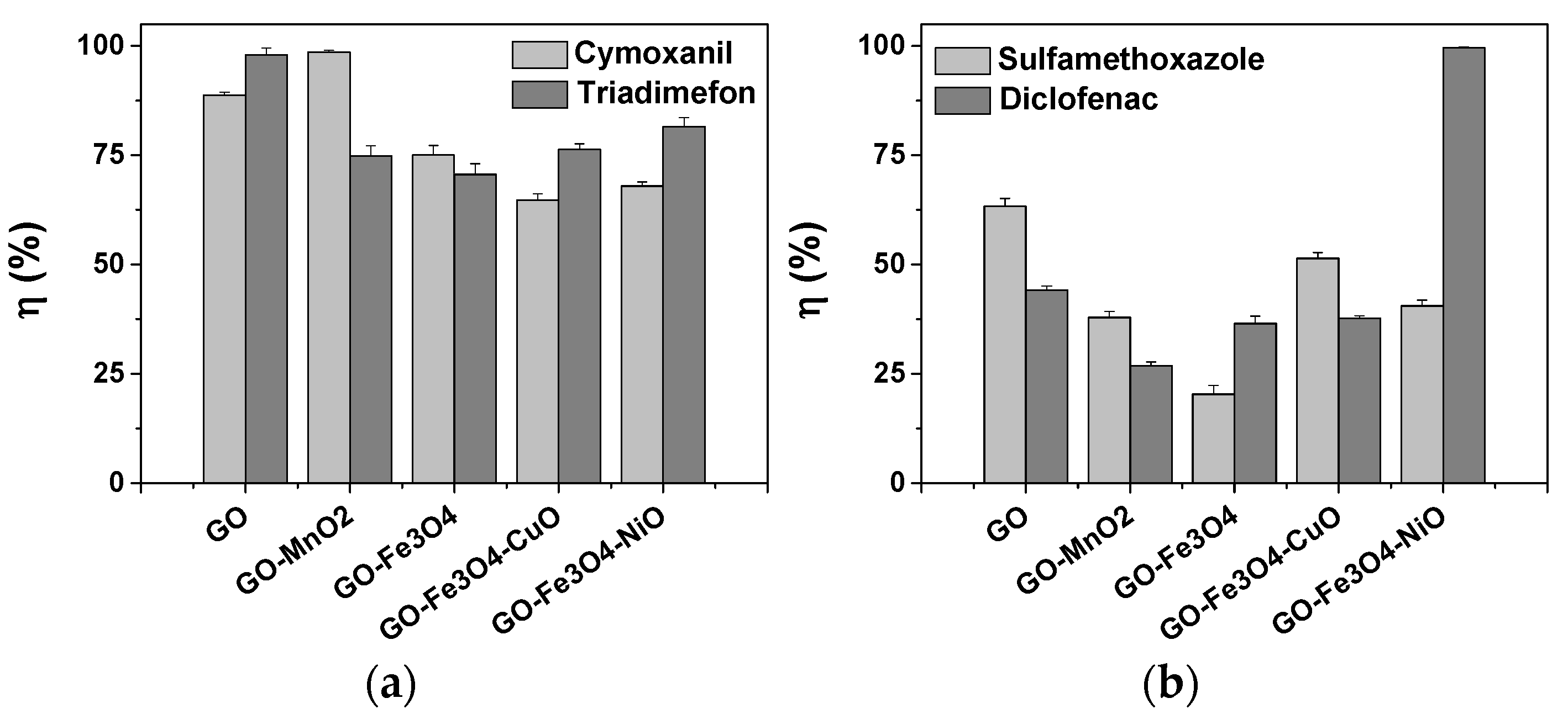
Nanotechnology has emerged as a highly focused field of research due to the unique properties of nanometric materials, particularly their large specific surface areas and excellent adsorption capabilities. This study investigated the synthesis of materials based on graphene oxide (GO) functionalized with different metal oxides (MnO2, Fe3O4, CuO, NiO), with potential applications in water decontamination. The morphological, structural, and compositional properties of these nanocomposites were extensively characterized using different experimental techniques, including X-ray diffraction (XRD), transmission electron microscopy (TEM), Fourier transform infrared spectroscopy (FTIR), X-ray photoelectron spectroscopy (XPS), and vibrating sample magnetometry (VSM) for magnetic property evaluation. Preliminary adsorption tests were performed for the removal of pesticides and drugs from aqueous solutions. The synthesized materials demonstrated a higher affinity for selected pesticides compared to drugs. The best removal efficiencies were 98.59% for cymoxanil, 97.93% for triadimefon, 63.33% for sulfamethoxazole, and 99.59% for diclofenac. The results indicate that the functionalization of GO with metal oxides modifies the material’s structure, increasing its potential for environmental applications such as water purification.

1. Introduction

Water pollution continues to be a major global issue, threatening both human health and environmental sustainability. The presence of different organic and inorganic contaminants in water sources presents significant risks due to their high toxicity, persistence, and concentrations. These pollutants can have wide-ranging effects on ecosystems, public health, and economic stability [1]. Effective solutions, methods, and technologies are being explored to address the issues caused by these pollutants. An intensively addressed field for this purpose is nanotechnology because nanometric materials have large specific surfaces with good adsorption properties. Nanoparticles, nanopowders, and nanomembranes are commonly used for the efficient chemical or biological removal of pollutants from contaminated wastewater.
Graphene is a two-dimensional, very well-known, and valuable material made up of a single layer of carbon atoms arranged in a hexagonal lattice. It has attracted considerable interest due to its important properties, which include exceptional electrical and thermal conductivity, excellent mechanical strength, and an extensive surface area [2,3,4]. However, even with these important advantages, pure graphene faces several limitations. It tends to aggregate due to strong van der Waals forces, exhibits low dispersibility in solvents, and lacks a band gap—factors that restrict its use in multiple applications [5,6]. Moreover, graphene’s chemical stability can be affected under specific environmental conditions, reducing its long-term performance [7].
To manage these limitations, graphene is frequently combined with inorganic nanoparticles, such as metal oxides (MOx) or metal sulfides, resulting in graphene-based composites with increased functionalities [8,9]. These composites showed improved dispersion, more efficient charge separation, and increased mechanical and thermal stability [10,11,12]. Therefore, graphene–inorganic nanocomposites offer a promising approach to enhancing the functionalities of graphene for advanced technological applications [7].
MOx presented great potential in the field of the adsorption of pollutants [13,14]. Thus, MOx can play important roles in the areas of science and technology. Among them, CuO has been exploited in the last few years for applications in catalysts, semiconductors, sensors, or field transistors. CuO nanoparticles alone can also act as an effective adsorbent for water contaminants. Furthermore, these nanoparticles are also used to modify adsorbents such as activated carbon, silica, graphene, etc., which act as support for the nanoparticles to remove pollutants. The presence of CuO nanoparticles on activated carbon improves chemical adsorption [15]. MnO2 is known to present good catalytic and absorption properties, and research has started to focus on wastewater treatment by using nanostructured MnO2 [16]. NiO is a widely studied material with applications in lithium-ion batteries, catalysis, and in adsorption processes. Its applications are determined by the surface area, the trapping of generated charge carriers, or by the interplay between its optical bandgap energy. Despite its low cost, it is not an efficient photocatalyst alone due to its wide bandgap and quick recombination of photogenerated charge carriers. NiO has also been applied in water depollution efforts due to its good affinity towards dyes, but its selective adsorption property against dyes or antibiotics is still much less explored [17]. These deficits, observed in the pristine MOx, can be easily improved by combining these MOx with carbon-based nanostructures [18]. Based on the literature, it is possible to say that carbon nanostructures with MOx represent a new generation of materials that have new properties due to their interaction between the carbon nanostructures properties and MOx properties. Due to these interactions, new properties (e.g., large surface areas, chemically inert surfaces, uniform structures, multiple adsorption sites) can be generated. Along with these properties, by including some magnetic nanoparticles in the materials (e.g., iron-based nanoparticles), we have the advantage that these materials can be recovered easily with a magnet and reused. Alicanoglu and Sponza, in 2015, demonstrated that magnetite nanoparticles have the potential to remove drugs from residual waters [19].
Graphene oxide (GO) is one of the most used graphene derivatives [20]. GO is an oxidized form of graphene that features oxygen-containing functional groups, including carboxyl, hydroxyl, and epoxy groups on its surface [21,22]. These functional groups give hydrophilic properties to GO, enabling it to form stable aqueous suspensions, which facilitates processing and functionalization.
The combination of GO with MOx has demonstrated synergistic properties that improve performance across a range of applications [7], particularly in the environmental remediation, sensing, and biomedical domains. An important application of these materials is water purification. In this case, graphene-based materials function as highly effective adsorbents for the removal of pollutants such as heavy metals, organic dyes, and pharmaceutical residues. These promising results were obtained due to their large surface area and strong adsorption capabilities [23,24]. Furthermore, its excellent photocatalytic properties allow the degradation of organic contaminants under light irradiation, positioning them as highly promising tools for advanced wastewater treatment technologies [25,26,27].
Pesticides are a major class of persistent organic pollutants that are commonly released into environmental water sources because of agricultural practices [28]. Because pesticides a pose high toxicity risk, the World Health Organization (WHO) and the European Union (EU) have established the maximum allowable concentrations in water: 0.5 μg L−1 for surface water and 0.1 μg L−1 for drinking water [29]. Therefore, monitoring and development of removal methods for pesticides in water sources is essential for food safety and human health protection [30].
The removal of pesticides can be facilitated using nanoparticles [31]. Nanoparticle-assisted photocatalysis represents an efficient approach for the degradation of various pesticide compounds. Additionally, nanotechnology based on green chemistry principles has been shown to enhance the decomposition rates of numerous environmental pollutants, including pesticides [32]. Nanomaterials, such as TiO2, ZnO, and GO nanosheets, have been demonstrated to have strong photocatalytic performance, to rapidly degrade pesticides (atrazine and chlorpyrifos). Compared to conventional techniques, this approach offers significant advantages, including greater efficiency, lower costs, and the absence of secondary pollution, making it well-suited for applications such as drinking water purification, wastewater treatment, agricultural runoff control, and surface water remediation [33]. GO synthesized with silver nanoparticles using Cucurbita maxima (pumpkin) leaves effectively degraded chlorpyrifos under the presence of sunlight [34]. A ZnO/α-Fe2O3 nano photocatalyst was demonstrated to remove 89% of carbamate pesticides under optimal pH and temperature conditions [35]. The developed GO–TiO2 nanocomposite achieved a degradation efficiency exceeding 80% of organophosphate pesticides, specifically dichlorvos and malathion [36].
Antibiotics are a significant group of emerging environmental contaminants. Among them, sulfonamide antibiotics are extensively used both for disease treatment and preventive measures. Due to their anionic nature, sulfonamides are not efficiently removed by conventional wastewater treatment processes, thus increasing attention has been directed toward their presence in natural water bodies and water treatment systems. Research into nanomaterial-based composites for water purification is ongoing, but it remains in an early developmental stage. Designing cost-effective synthesis methods for these nanomaterials continues to be a significant challenge [37,38,39,40,41]. Materials based on graphene have shown effectiveness in adsorbing antibiotics from wastewater, leading to a significant reduction in their concentration. The high surface area and customizable properties of these materials improve their removal efficiency, positioning them as promising options for advanced water treatment technologies [42]. The CuO–GO composite demonstrated high antibiotic removal efficiencies, with adsorption capacities of 405 mg g−1 for amoxicillin and 552 mg g−1 for tetracycline. The developed material showed an 80% regeneration efficiency and maintained 82% of its adsorption capacity after five reuse cycles [43].
Diclofenac is a very well-known and often-used pharmaceutical, and is an environmental contaminant that enters water bodies due to its widespread use as a non-steroidal anti-inflammatory drug [44,45]. The global consumption of diclofenac is increasing due to the growth of populations and access to healthcare in many countries [46]. Therefore, the concentration of diclofenac in the environment can be up to 110 ng L−1 [47], presenting potential toxicity risks to the environment and global ecosystems. Consequently, diclofenac is considered an emerging environmental contaminant, and its removal from the environment represents an important global concern [48]. GO demonstrated a removal efficiency of nearly 100% for concentrations of diclofenac ranging from 50 to 450 mg L−1. For these good results, the necessary GO amounts of 0.46 and 1.38 g L−1, respectively, were used to remove the diclofenac from an aqueous solution [49].
The present study aimed to obtain materials based on graphene oxide (GO) functionalized with transition metal oxides (MOx: MnO2, Fe3O4, CuO, NiO) and test their use in water decontamination. The investigation of the morphological, structural, and compositional properties of these materials was carried out using experimental techniques, such as X-ray diffraction (XRD), transmission electron microscopy (TEM), scanning electron microscopy (SEM), Fourier transform infrared spectroscopy (FTIR), X-ray photoelectron spectroscopy (XPS), and the magnetic properties of the nanocomposites were determined using the vibrating sample magnetometry (VSM) method. We hypothesized that the functionalization of the GO with selected MOx leads to improved adsorption performance for the removal of organic pollutants, particularly pesticides and drugs, from aqueous solutions. According to our knowledge, we can say that they are new combinations of materials with unique physicochemical characteristics, and these specific MOx combinations are expected to significantly increase the affinity and removal efficiency of the materials, making them suitable candidates for advanced water decontamination applications.

2. Materials and Methods

2.1. Chemicals

Graphite powder (96–98%) was purchased from KOH-I-NOOR HARDTMUTH a.s. (České Budějovice, Czech Republic). Sulfuric acid (H2SO4, 97%) was purchased from Chomchim Chemical (Râmnicu Vâlcea, Romania). Potassium permanganate (KMnO4, 99%) was bought from Roth (Karlsruhe, Germany) and hydrogen peroxide (H2O2, 30%) from Chimopar Trading (Bucureşti, Romania). MnO2 was prepared according to Lung et al. [50]. Iron (III) chloride hexahydrate (FeCl3•6H2O, 97%), iron (II) sulfate heptahydrate (FeSO4•7H2O, 99%), potassium bromide (KBr, 99%), sulfamethoxazole (98%), sodium nitrate (NaNO3, 99%), and hydrochloric acid (HCl, 37%) were purchased from Sigma–Aldrich (Taufkirchen, Germany). Ammonium hydroxide (NH4OH, sol. 25%), nickel chloride hexahydrate (NiCl2•6H2O, 98%), L-ascorbic acid (99%), cetyltrimethylammonium bromide (CTAB, 99%), cymoxanil (99%), and triadimefon (99%) were purchased from Merck (Taufkirchen, Germany). Sodium hydroxide (NaOH, 97%) and acetonitrile graded for high-performance liquid chromatography (HPLC purity, 99.9%) was purchased from VWR Chemicals (Vienna, Austria). Diclofenac was bought from Refen (Hemofarm, Cluj-Napoca, Romania), and formic acid was purchased from Cristal R Chim (Bucureşti, Romania). In all the experiments, ultrapure water was produced by a Direct-Q 3 UV Water Purification System (Merck, Taufkirchen, Germany).

2.2. Materials Synthesis

2.2.1. GO Synthesis

Into a 500 mL flask, 2.5 g of graphite powder (purity 96–98%) was added, which was mixed with 1.25 g of NaNO3 and 118 mL of H2SO4 (97%), followed by magnetic stirring at 500 rpm in an ice bath for 30 min (˂20 °C). After this step, 7.5 g of KMnO4 was added (in small portions) while stirring vigorously (1000 rpm). The mixture was stirred for 2 h at a temperature below 20 °C. The flask was then heated in a water bath at 35 °C and kept under stirring conditions for 30 min at this temperature, and then 115 mL of deionized water was added, heating the water bath to 98 °C and keeping it that way for 15 min. The obtained paste was diluted by adding 350 mL of deionized water. After this step, 30% H2O2 was added dropwise until a bright yellow color was obtained. The obtained suspension was filtered and washed with 800 mL of 5% HCl and 300 mL of deionized water. The precipitate was dried by lyophilization.

2.2.2. GO-MnO2 Synthesis

To synthesize GO-MnO2, 0.02 g of GO was shaken for 30 min with 20 mL of bidistilled water. In parallel, 0.1 g of MnO2 was shaken with 25 mL of bidistilled water for 30 min. The MnO2 suspension was added to the GO suspension, and stirring was continued on the shaker for another 6.5 h. The precipitate formed was washed by centrifugation with 3 portions of bidistilled water and then with 3 portions of ethanol. The obtained material was dried overnight in an oven at 60 °C.

2.2.3. GO-Fe3O4 Synthesis

To synthesize GO-Fe3O4, 0.06 g of GO was stirred in an ultrasonic bath for 20 min with 36.12 mL of bidistilled water, and then stirring was continued on a magnetic plate, under argon, for 30 min at 60 °C. Further, 91.8 mg of FeCl3•H2O was added to the obtained suspension, and stirring was continued for another 30 min, after which 48 mg of FeSO4•7H2O was added, and stirring was then continued for another 30 min. Finally, 18 mL of 6% NH4OH was added in thin drops. The reaction mixture was left to stir for another 2 h. The obtained material was washed by centrifugation with bidistilled water until a neutral pH was obtained, and then dried overnight in an oven at 60 °C.

2.2.4. GO-Fe3O4-NiO Synthesis

To synthesize GO-Fe3O4-NiO, 0.02 g of GO-Fe3O4 was ultrasonicated for 30 min in 12 mL of bidistilled water. To this suspension were added freshly prepared solutions of 0.1249 g NiCl2•6H2O in 50 mL of bidistilled water, and 0.9687 g of ascorbic acid in 50 mL of bidistilled water. After adding 0.5467 g of CTAB to the previously obtained suspension, 50 mL of bidistilled water was added and the mixture was brought to a pH of 6.5 with a NaOH solution, after which it was heated to 85 °C and stirring was continued for 3 h. The sample was washed by centrifugation with bidistilled water and dried in an oven at 75 °C.

2.2.5. GO-Fe3O4-CuO Synthesis

To synthesize GO-Fe3O4-CuO, 0.02 g of GO-Fe3O4 was ultrasonicated for 30 min in 12 mL of bidistilled water. To this suspension were added freshly prepared solutions of 0.1249 g of CuSO4•5H2O in 50 mL of bidistilled water, and 0.9687 g of ascorbic acid in 50 mL of bidistilled water. After adding 0.5467 g of CTAB to the previously obtained suspension, 50 mL of bidistilled water was added and the mixture was brought to a pH of 6.5 with a NaOH solution, after which it was heated to 85 °C and stirring was continued for 3 h. The sample was washed by centrifugation with bidistilled water and dried in an oven at 75 °C.

2.3. Materials Characterization

2.3.1. Powder X-Ray Diffraction (XRD)

X-ray diffractograms were made with a Bruker D8 Advance diffractometer with a Cu tube CuKα1 radiation. The samples were scanned using the DIFFRAC Plus XRD Commander program (Karlsruhe, Germany). For monochromatizing radiation, a Ge(111) monochromator was used, and a fast LynxEye position detector (Karlsruhe, Germany) was used for the detection of diffracted radiation. Bragg–Brentano reflective geometry was used.

2.3.2. Morphological Characterization

Determination of the morphology of the samples was performed by transmission electron microscopy (TEM) and reflection electron microscopy (REM) measurements using a HITACHI HD-2700 microscope (Tokyo, Japan). The microscope was coupled with an elemental X-ray diffraction detector (EDX) (Tokyo, Japan).

2.3.3. FTIR Analysis

The FTIR spectroscopic analysis of the samples was performed with a JASCO FTIR-6100 spectrometer (JASCO International Co., Ltd., Tokyo, Japan) in the 4000 to 400 cm−1 spectral domain, with a 4 cm−1 resolution. For sample preparation, the KBr pellet technique was used. Each sample was dispersed in about 300 mg of anhydrous KBr and mixed in an agate mortar, then the prepared mixtures were pressed into an evacuated die. The collection and analysis of spectral data were carried out using JASCO Spectra Manager v.2 software.

2.3.4. X-Ray Photoelectron Spectroscopy (XPS) Analysis

The qualitative and quantitative composition of the samples were investigated using an XPS with a custom-built SPECS spectrometer, (SPECS, Berlin, Germany). An Al anode (1486.71.6 eV) was used as the X-ray source. Sample preparation was done by casting onto a sample holder using ethanol. Argon ion sputtering was performed at 1000 V/10 mA. CasaXPS version 2.3.14 was used for spectral analysis by using the relative sensitivities, transmission factors, and electronic mean free path factors. A Shirley background was extracted from the core-level spectra.

2.3.5. Vibrating-Sample Magnetometry (VSM) Analysis

The room temperature magnetic hysteresis measurements were performed using a Vibrating Sample Magnetometer produced by Cryogenic, (London, UK).

2.4. Preliminary Adsorption Tests

The obtained materials were preliminarily tested for the removal of pesticides (cymoxanil and triadimefon) and drugs (sulfamethoxazole and diclofenac) from aqueous solutions. In the case of pesticides, a removal method described by Lung et al. (2023) was used [51]. The initial concentration of cymoxanil was 7 mg L−1, brought to a pH of 4 with an HCl solution. It was combined with an adsorbent dose of 1 g L−1, a temperature of 20 °C, a contact time of 5 min, and stirring at 300 rpm. For the testing of the triadimefon removal from aqueous solution using the obtained materials, the experimental conditions were described by Lung et al. (2022) [52]. In this case, the initial concentration of triadimefon was 20 mg L−1, brought to a pH of 2 with an HCl solution, and then combined with the adsorbent dose of 1 g L−1, a temperature of 20 °C, a contact time of 4 min, and stirring at 300 rpm.
The conditions for removal of the selected drugs using the prepared materials followed those described by Lung et al., 2021: an initial drug concentration of 40 mg L−1, a pH of 2, 20 min of contact time, stirring at 300 rpm, and the same adsorbent dose (1 g L−1) and temperature (20 °C) [50].
All the samples were filtered using nylon syringe filters (13 mm × 0.22 µm) and analyzed using HPLC with a photodiode array detector PDA (Shimadzu LC-2010, Tokyo, Japan).
The HPLC analysis conditions used for the determination of the pesticides were as follows: column Nucleosil 100-5 C18 EC 250 × 4.6 mm (Machery-Nagel, Germany), thermostated at 40 °C, isocratic elution with the mobile phase consisting of acetonitrile (A):ultrapure water with 0.1% formic acid (B), 60:40 (v/v), a flow of 0.4 mL min−1, and an injection volume of 10 µL [52]. For the HPLC analysis of the drugs, the same chromatographic conditions were used, except for the composition of the mobile phase A, which consisted of 90:10 (v/v) acetonitrile:ultrapure water. A known concentration of 0.01 mg L−1 pesticide/drug was added to each sample, and all were analyzed in triplicates.
The removal efficiency (η %) of the selected pollutants from the aqueous solution was calculated by subtracting the pollutant concentration at time t (4, 5, or 20 min, depending on the pollutant) from the concentration at time 0. This difference was then divided by the concentration at time 0 and multiplied by 100.

3. Results and Discussion

3.1. XRD Characterization

Figure 1 shows the X-ray diffractograms for GO, GO-Fe3O4, GO-Fe3O4-CuO, and GO-Fe3O4-NiO. It is observed that the diffraction line appears at the characteristic 12° of GO. From the half-width of this diffraction line, the crystallite sizes for GO were evaluated as 182Å. For GO-Fe3O4, it is observed that the diffraction peak corresponding to GO fades and moves to higher angles, namely 15.5°. There is probably a tendency for GO to transform into graphene. For this sample, all the diffraction lines characteristic of the Fe3O4 phase were present. For the GO-Fe3O4-CuO sample, the following crystalline phases were present: Cu2O, FeO, and Fe2O3. There is also a diffraction halo centered at an angle of 21.4° that lost some of the oxygen characteristics of GO because of its tendency to transform into graphene. GO-Fe3O4-NiO is characterized by two diffraction maxima. One of them has the low-intensity characteristics of the initial GO, and the second one has a more intense halo centered at approximately 20°, which probably indicates the existence of oxygen-depleted GO.

3.2. TEM Characterization

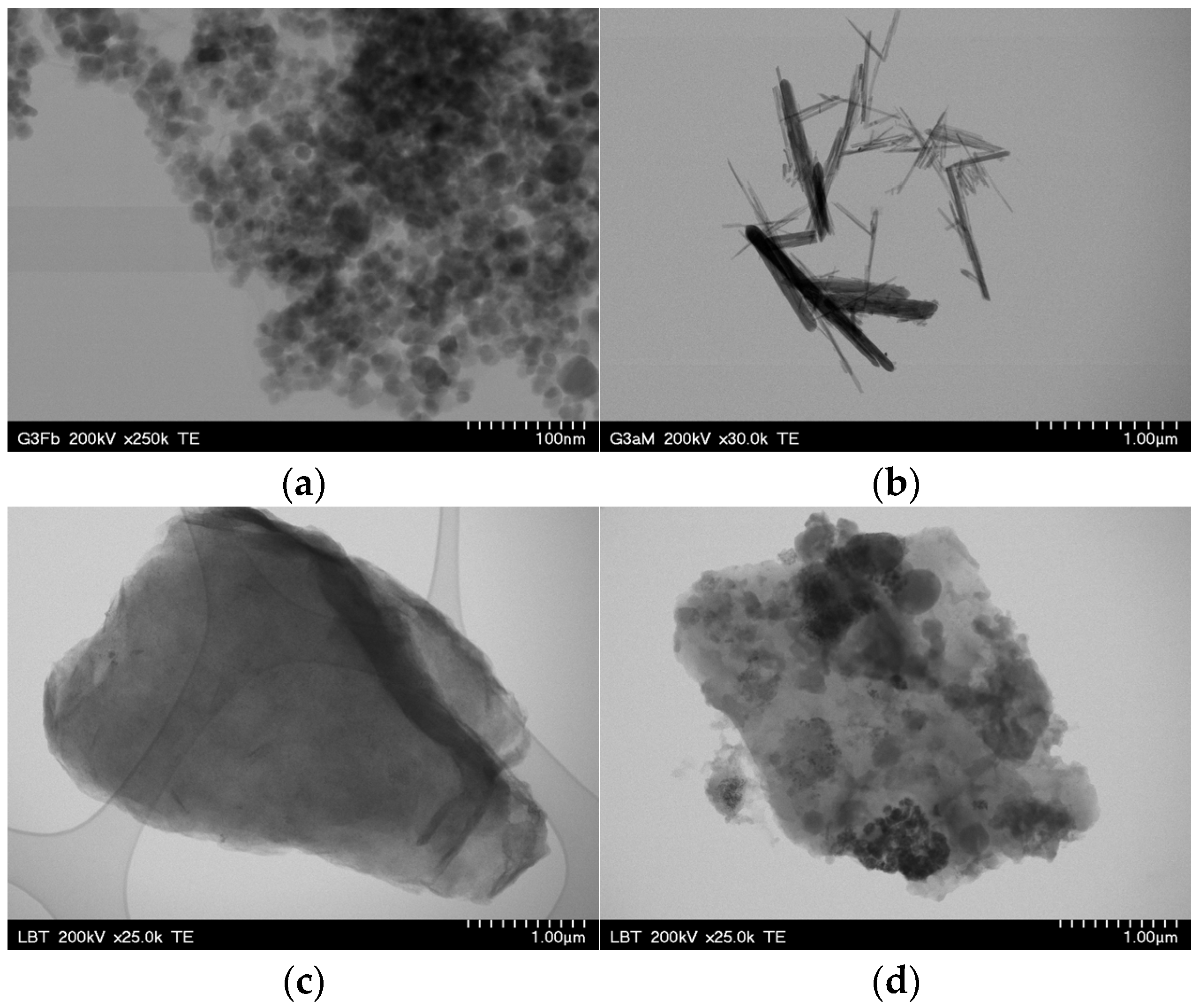
Figure 2 shows the TEM images of the GO-Fe3O4 (a), GO-MnO2 (b), GO-Fe3O4-NiO (c), and GO-Fe3O4-CuO (d) nanocomposites. It is observed that the shape of the Fe3O4 nanoparticles is spherical, that of the MnO2 nanoparticles is acicular, and that both are up to approximately 50 nm in size. A variation in the size of the MnO2 needles is observed, as well as their welding. With the addition of the second phase—CuO and NiO, respectively—to the compound, the embedding of the nanoparticles in a matrix of GO nanoparticles is observed.
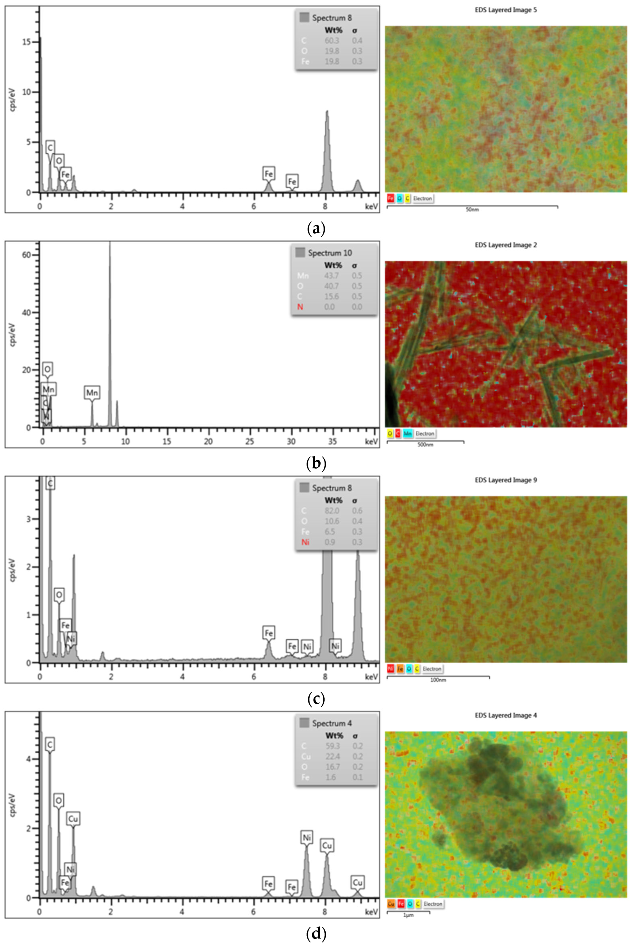
Compositional determination by EDX in the marked areas indicates the presence of elements belonging to all the component phases of the nanocomposites (Figure 3). The element distribution maps show the distribution of the elements and, implicitly, the component phases in the chosen sample. The presence of all the elements of both metallic and organic component phases was found.

3.3. FTIR Characterization

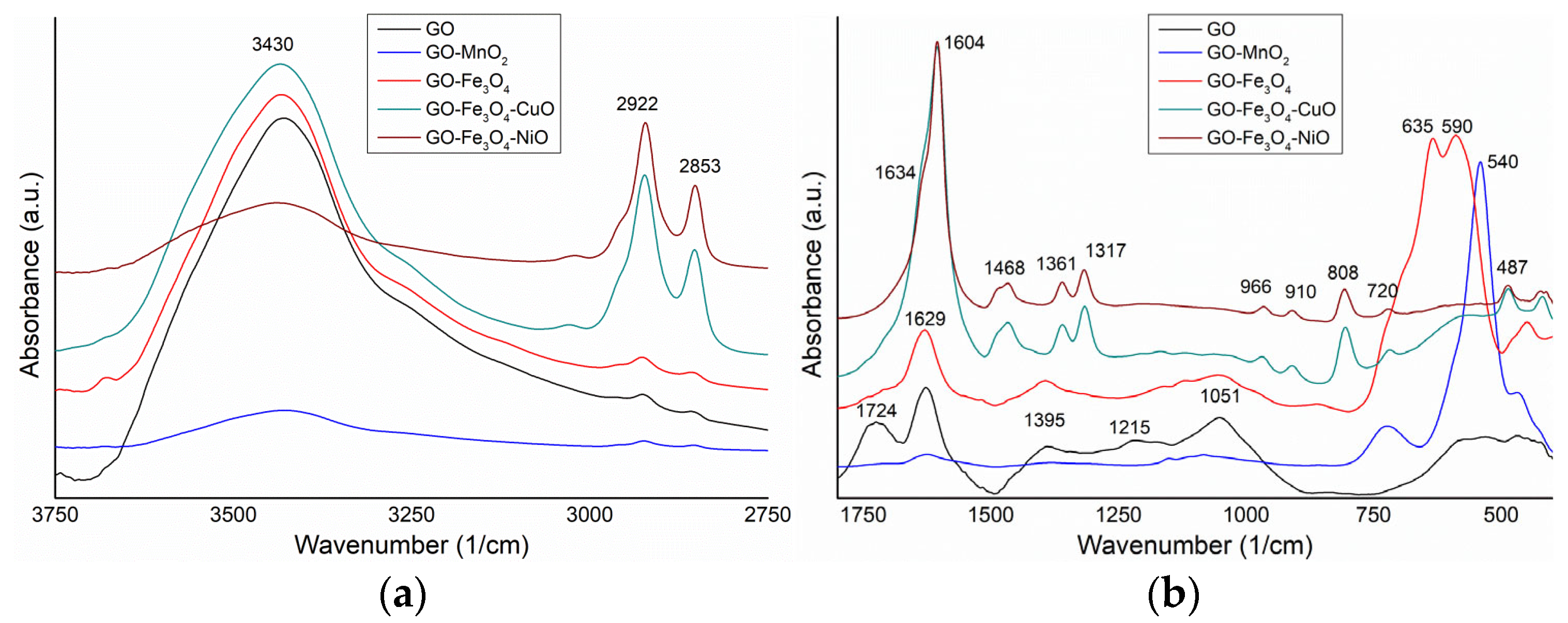
The spectra of the analyzed samples are comparatively presented in Figure 4a,b. The characteristic vibrational bands of GO were identified as follows: 3430 cm−1 (-OH stretching vibrations), 2925 and 2855 cm−1 (-CH2 and -CH3 groups asymmetric and symmetric stretching), 1724 and 1627 cm−1 (-C=O and aromatic -C=C- stretching), 1395 and 1215 cm−1 (C-OH stretching and -O-H deformation), 1051 (C-O bonds stretching) and broadband with a weak intensity of 690–400 cm−1 [53,54,55].
The spectrum of the GO-MnO2 sample presents the following vibrational bands: 3428 cm−1 (O-H stretching), 2923 and 2854 cm−1 (C-H stretching), 1705 and 1624 cm−1 (-C=O and aromatic C=C stretching), 1385 cm−1 (O-H deformation), 1151 and 1082 cm−1 (C-O stretching), 724 cm−1 (Fe-O stretching), and a strong band at 540 cm−1 (Mn-O stretching from MnO2) [56].
In the spectrum of the GO-Fe3O4 sample, the following vibrational bands can be identified: 3430 cm−1 (O-H stretching); 2922 and 2853 cm−1 (C-H stretching); 1623 cm−1 (aromatic C=C stretching); 1392 cm−1 (O-H deformation); 1160, 1118, and 1053 cm−1 (C-O stretching); 635 and 590 cm−1 (Fe-O stretching from Fe2O3, and Fe-O stretching from Fe3O4); and 450 cm−1 (Fe-O stretching from Fe2O3) [57].
The spectrum of the GO-Fe3O4-CuO sample shows the following vibrational bands: 3433 cm−1 (O-H stretching); 2919 and 2849 cm−1 (C-H stretching); 1604 cm−1 (C=C stretching); 1466, 1361 and 1317 cm−1 (O-H deformation); a broad and weak band between 1250 and 1000 cm−1 (C-OH and C-O stretching); 969, 908, 805, and 719 cm−1 (Fe-O stretching from Fe2O3); a broad band between 670 and 530 cm−1 with maxima at 561 cm−1 (Fe-O stretching from Fe3O4) [57]; 486 cm−1 (Cu-O bond stretching from CuO) [58]; and 420 cm−1 (Fe-O stretching of Fe2O3).
In the spectrum of the GO-Fe3O4-NiO sample, vibration bands were identified at the following wavenumbers: 3438 cm−1 (O-H stretching); 2922 and 2853 cm−1 (C-H stretching); 1631 and 1604 cm−1 (-C=O and aromatic C=C stretching); 1482, 1468, and 1361 cm−1 (O-H stretching); 1317, 966, 910, 808, and 720 cm−1 (Fe-O stretching from Fe2O3); a weak and broad band between 680 and 500 cm−1, with small maxima at 610, 580, and 541 cm−1 (Ni-O-H and Fe-O stretching from Fe3O4); and 486, 421, and 412 cm−1 (Ni-O stretching) [57,59].
Using comparative analysis of the spectra, a weak intensity band of O-H stretching vibrations was observed at 3430 cm−1 for the GO-MnO2 and GO-Fe3O4-NiO samples. Also, the stretching vibrations of C-H bonds between 2922 and 2853 cm−1, and aromatic C=C stretching from 1604 cm−1, have a much higher intensity in the case of GO-Fe3O4-CuO and GO-Fe3O4-NiO compared to the other samples. It was observed that the intensity of the vibrations of Fe-O bonds was greatly reduced as a result of the addition of Cu-O and Ni-O.

3.4. XPS Characterization

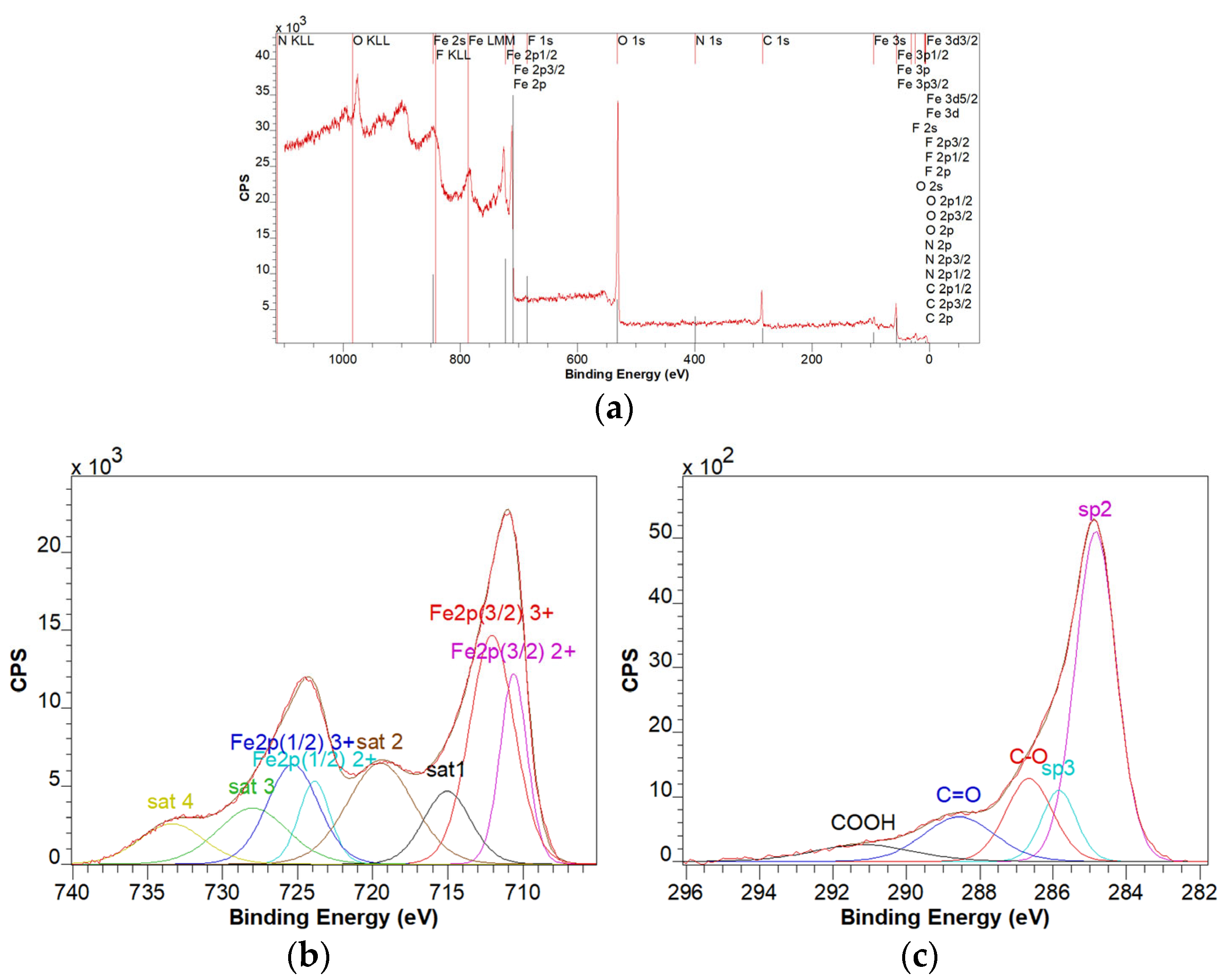
For the GO-Fe3O4 sample, from the analysis of the survey spectrum (Figure 5a), it was found that the Fe, F, N, O, and C elements are present. The multiplet structure of the Fe 2p spectrum (Figure 5b) is characteristic of the 2+ and 3+ oxidation states corresponding to the Fe3O4 compound. The C 1s spectrum (Figure 5c) deconvolution was done by considering the specific lines for the graphene oxide C=C sp2, C-C sp3, C-O/C-OH, C=O, and COOH [60]. The sp3/sp2 ratio was 0.18. One possible source of fluorine contamination was the Teflon stirrer.
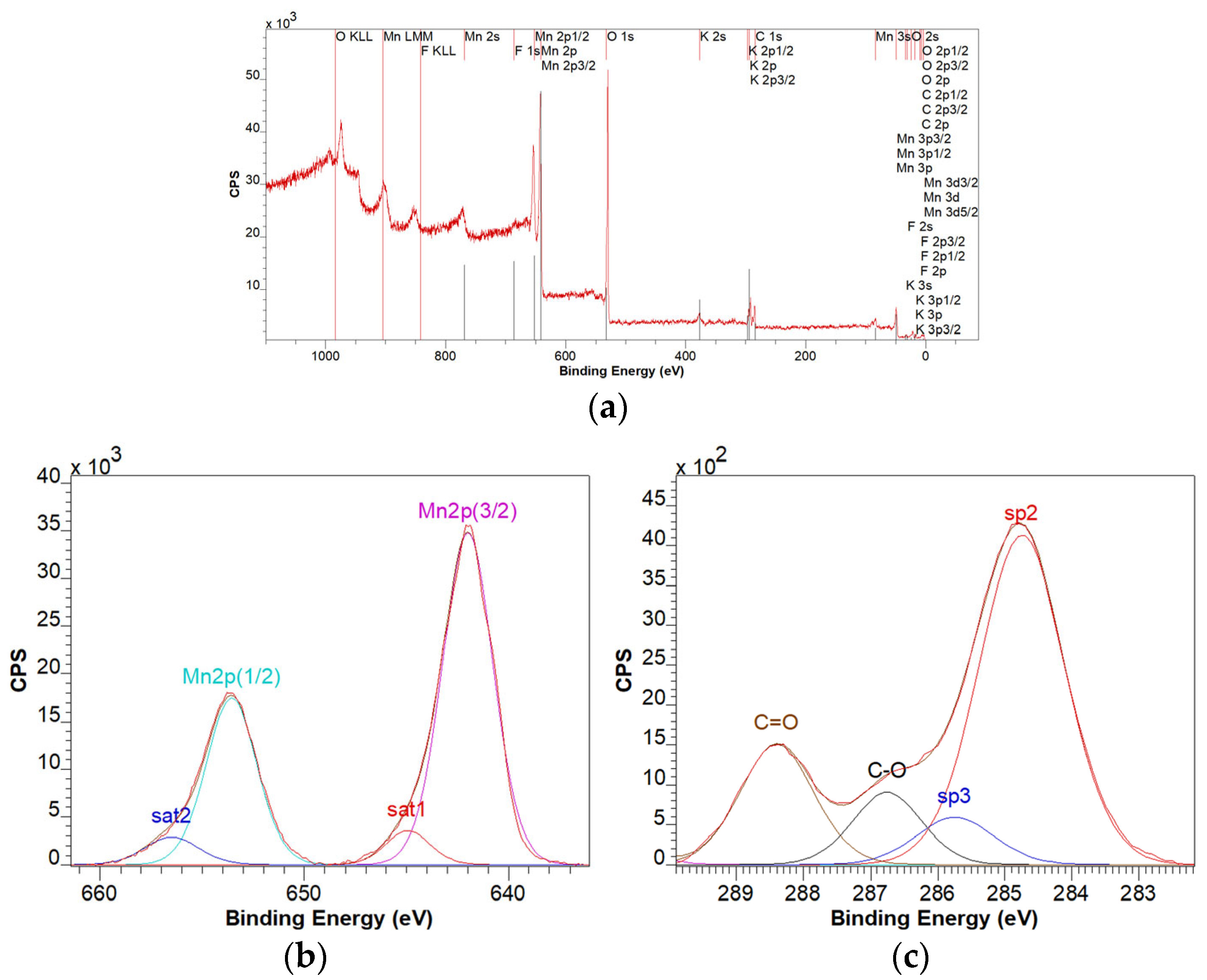
For the GO-MnO2 sample, from the analysis of the survey spectrum (Figure 6a), it was found that the elements Mn, F, K, O, and C are present. The multiplet structure of the Mn 2p spectrum (Figure 6b) is characteristic of the 4+ oxidation state, corresponding to the MnO2 compound. The analysis of the C1s spectrum reveals the specific lines for the graphene oxide with a sp3/sp2 ratio of 0.13.
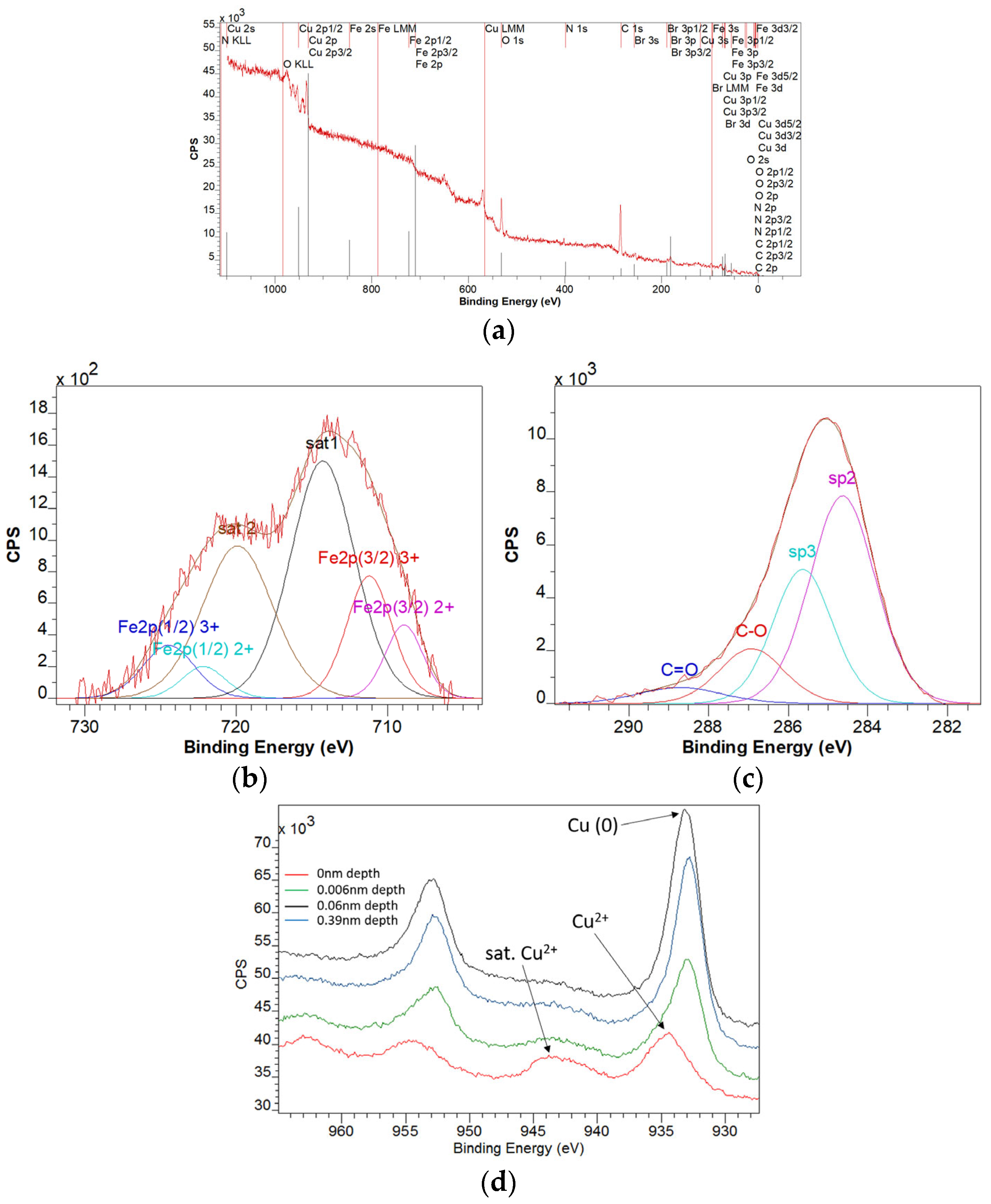
For the GO-Fe3O4-CuO sample, from the survey spectrum analysis (Figure 7a), it was found that the elements Fe, Cu, Br, N, O, and C are present. The multiplet structure of the Fe 2p spectrum (Figure 7b) is characteristic of the 2+ and 3+ oxidation states corresponding to the Fe3O4 compound. The depth profile analysis indicates that Fe is only in the oxidized state. The analysis of the C1s spectrum (Figure 7c) reveals the specific lines for the graphene oxide with a sp3/sp2 ratio of 0.58. The multiplet structure of the Cu 2p spectrum (Figure 7d) is characteristic of the 2+ and 0 oxidation states. The depth profile analysis indicates that the Cu oxidation state is (0), and there was a superficial oxide layer on the surface. At a depth of 0.39 nm, the ratio of Cu(0)/Cu(ox) = 5.39. Analysis of the depth profile of the Br 3p line indicated a homogeneous distribution of Br in the sample. Residual Br comes from CTAB precursor.
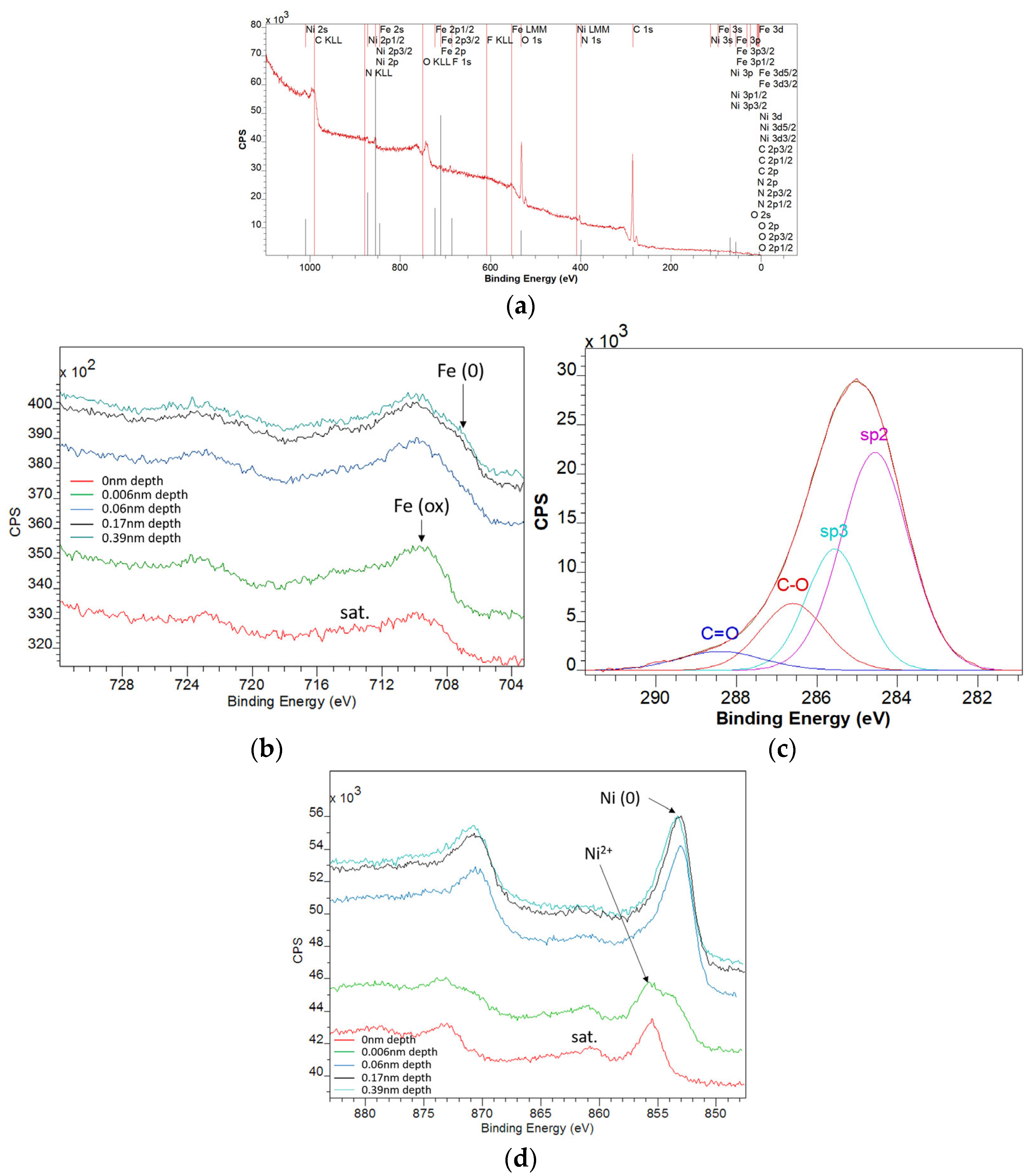
For the GO-Fe3O4-NiO sample, from the analysis of the survey spectrum (Figure 8a), it was found that the elements Fe, Ni, F, N, O, and C are present. The analysis of the F 1s line indicated surface contamination with organic fluorine. The multiplet structure of the Fe 2p spectrum (Figure 8b) is characteristic of the 2+ and 3+ oxidation states corresponding to the Fe3O4 compound. At depths greater than 0.1 nm, the Fe (0) oxidation state appears. At a depth of 0.39 nm, the ratio of Fe(0)/Fe(ox) = 0.2. The analysis of the C1s spectrum (Figure 8c) revealed the specific lines for the graphene oxide with a sp3/sp2 ratio of 0.46. The multiplet structure of the Ni 2p spectrum (Figure 8d) was characteristic of the 2+ and 0 oxidation states. Analysis of the depth profile indicated that the oxidation state of Ni was (0), and there was a superficial oxide layer on the surface. At a depth of 0.39 nm, the ratio of Ni(0)/Ni(ox) = 2.19.

3.5. VSM Characterization

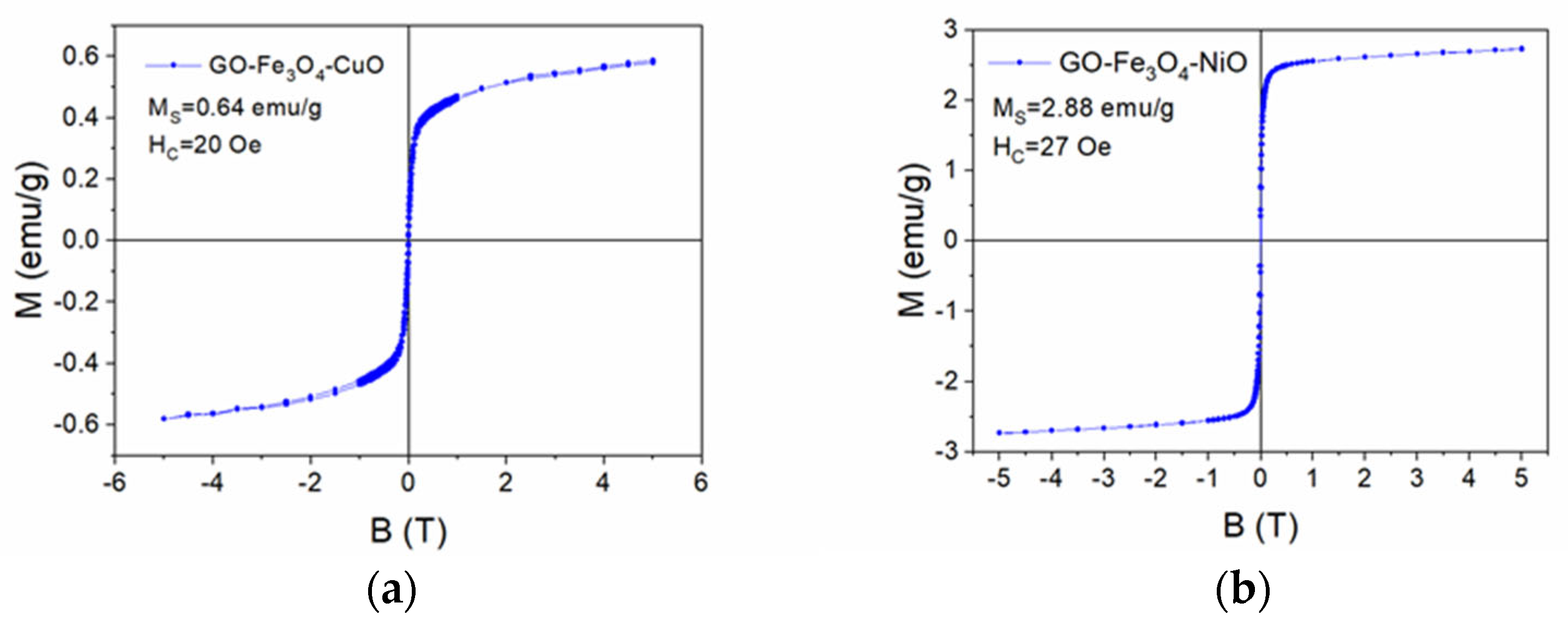
The magnetization curves vs. the applied magnetic field of the GO-Fe3O4-CuO and GO-Fe3O4-NiO samples are presented in Figure 9. The shape of the magnetization curves is typical of superparamagnetic materials. In the case of the GO-Fe3O4-CuO sample, the saturation magnetization was 0.64 emu/g, and the coercive field was 20 Oe (Figure 9a), while in the case of the GO-Fe3O4-NiO sample, the saturation magnetization was 2.88 emu/g, and the coercive field was 27 Oe (Figure 9b).

3.6. Removal of Pesticides and Drugs from Aqueous Solutions

The results of preliminary adsorption tests are promising (Figure 10). The obtained materials presented a better affinity for adsorbing the pesticides rather than the selected drugs. For the pesticides, the removal efficiencies were over 64%. For cymoxanil, the best removal efficiency was obtained using the material GO-MnO2 (98.59%), followed by GO (88.77%), GO-Fe3O4 (74.99%), GO-Fe3O4-NiO (67.74%), and GO-Fe3O4-CuO (64.74%) (Figure 10a). The removal efficiencies of triadimefon were 97.93% using the GO material, 81.52% with GO-Fe3O4-NiO, 76.31% with GO-Fe3O4-CuO, 74.84% with GO-Mn, and 70.58% with GO-Fe3O4.
For the selected drugs, the removal efficiencies were lower, between 20.32% and 63.33%. An exception from this trend occurred in the case of diclofenac, with a removal efficiency of 99.59% obtained with the GO-Fe3O4-NiO material (Figure 10b). Sulfamethoxazole was better adsorbed by the GO materials (63.33%) rather than the other tested materials. After GO, the best materials for sulfamethoxazole removal were GO-Fe3O4-CuO (51.41%), G-MnO2 (37.92%), GO-Fe3O4-NiO (40.56%), and GO-Fe3O4 (20.32%). Diclofenac was removed from the aqueous solution in proportions of 26.85% and 99.59% (Figure 10b).

4. Conclusions

This research presents the synthesis of graphene oxide (GO)-based materials functionalized with metal oxides (MOx: MnO2, Fe3O4, CuO, NiO), demonstrating their potential applications in water decontamination. The comprehensive characterization using XRD, TEM, FTIR, XPS, and VSM confirmed that the incorporation of the selected MOx significantly modified the morphological, structural, and chemical properties of the GO matrix. The XRD and TEM analyses revealed the successful formation of MOx phases, with noticeable shifts in diffraction patterns and material embeddings. FTIR and XPS spectra highlighted the interaction between GO and the MOx, suggesting possible changes in the electronic structure. The VSM analysis showed that the synthesized materials exhibited superparamagnetic behavior, which may be beneficial for magnetic separation processes in water purification.
The preliminary adsorption tests demonstrated that the synthesized materials showed a higher affinity toward pesticides than the selected drugs. Cymoxanil and triadimefon exhibited high removal efficiencies with G-MnO2 (98.59%) and GO (97.93%) materials, which were the most effective materials. Contrarily, the removal of drugs was generally lower, ranging from 20.32% to 63.33%, except for diclofenac, which was removed with high efficiency (99.59%) using the GO-Fe3O4-NiO material. Sulfamethoxazole showed the best adsorption with GO (63.33%). Overall, these functionalized GO-based materials showed promising prospects for efficient and sustainable water decontamination applications, offering an effective solution to environmental pollution. Therefore, future studies will be focused on the optimization of the adsorption processes for the materials that showed the best affinity for the selected pollutants.

Author Contributions

Conceptualization, data curation, writing—original draft, writing—review and editing, project administration: O.O.; conceptualization, methodology, data curation, investigation: A.S.; investigation: I.L.; investigation, writing—original draft: A.S.P.; investigation, writing—original draft: I.K.; investigation, G.B.; investigation, writing—original draft: C.L.; investigation, supervision: O.P.; writing—review and editing, supervision: M.-L.S. All authors have read and agreed to the published version of the manuscript.

Funding

This work was supported by a grant from the Ministry of Research, Innovation and Digitization, CNCS-UEFISCDI, project number PN-IV-P2-2.1-TE-2023-0522, within PNCDI IV.

Institutional Review Board Statement

Not applicable.

Informed Consent Statement

Not applicable.

Data Availability Statement

The original contributions presented in this study are included in the article. Further inquiries can be directed to the corresponding author.

Acknowledgments

This work was supported by a grant from the Ministry of Research, Innovation and Digitization, CNCS-UEFISCDI, project number PN-IV-P2-2.1-TE-2023-0522, within PNCDI IV.

Conflicts of Interest

The authors declare no conflicts of interest.

References

  1. Zhang, S.; Ding, J.; Tian, D. Incorporation of MIL-101 (Fe or Al) into chitosan hydrogel adsorbent for phosphate removal: Performance and mechanism. J. Solid State Chem. 2022, 306, 122709. [Google Scholar] [CrossRef]
  2. Chowdury, M.S.K.; Park, Y.J.; Park, S.B.; Park, Y.-I. Two-dimensional nanostructured pristine graphene and heteroatom-doped graphene-based materials for energy conversion and storage devices. Sustain. Mater. Technol. 2024, 42, e01124. [Google Scholar] [CrossRef]
  3. Ramezani, M.; Rahmani, O. A review of recent progress in the graphene syntheses and its applications. Mech. Adv. Mater. Struct. 2024, 1–33. [Google Scholar] [CrossRef]
  4. Hu, H.; Liu, J.; Zheng, X.; Zhao, K.; Lin, Y.; Xu, X.; Long, H.; Zhang, Y.; Wang, X.; Chen, D.; et al. Recent advances in iron-based catalyst-driven persulfate activation for organic pollutant degradation. J. Water Process Eng. 2025, 71, 107423. [Google Scholar] [CrossRef]
  5. Shahbazi, M.; Jäger, H.; Ettelaie, R.; Chen, J.; Kashi, P.A.; Mohammadi, A. Dispersion strategies of nanomaterials in polymeric inks for efficient 3D printing of soft and smart 3D structures: A systematic review. Adv. Colloid Interface Sci. 2024, 333, 103285. [Google Scholar] [CrossRef] [PubMed]
  6. Perala, R.S.; Chandrasekar, N.; Balaji, R.; Alexander, P.S.; Humaidi, N.Z.N.; Hwang, M.T. A comprehensive review on graphene-based materials: From synthesis to contemporary sensor applications. Mater. Sci. Eng. R. Rep. 2024, 159, 100805. [Google Scholar] [CrossRef]
  7. Altalbawy, F.M.A.; Zwamel, A.H.; Rachchh, N.; Ramachandran, T.; Shankhyan, A.; Karthikeyan, A.; Thatoi, D.N.; Gupta, D.; Formanova, S.; Alam, R.; et al. A review on graphene-based inorganic nanostructures: Synthesis, functionalization, and applications in photocatalytic degradation and electrochemical sensing of pollutants. Inorg. Chem. Comm. 2025, 177, 114398. [Google Scholar] [CrossRef]
  8. Badoni, A.; Thakur, S.; Vijayan, N.; Swart, H.C.; Bechelany, M.; Chen, Z.; Sun, S.; Cai, Q.; Chen, Y.; Prakash, J. Recent progress in understanding the role of graphene oxide, TiO2 and graphene oxide-TiO2 nanocomposites as multidisciplinary photocatalysts in energy and environmental applications. Cat. Sci. Technol. 2025, 15, 1702–1770. [Google Scholar] [CrossRef]
  9. Zafar, M.; Imran, S.M.; Iqbal, I.; Azeem, M.; Chaudhary, S.; Ahmad, S.; Kim, W.Y. Graphene-based polymer nanocomposites for energy applications: Recent advancements and future prospects. Results Phys. 2024, 60, 107655. [Google Scholar] [CrossRef]
  10. Khan, H. Graphene based semiconductor oxide photocatalysts for photocatalytic hydrogen (H2) production, a review. Int. J. Hydrog. Energy 2024, 84, 356–371. [Google Scholar] [CrossRef]
  11. Hu, H.; Ou, J.Z.; Xu, X.; Lin, Y.; Zhang, Y.; Zhao, H.; Chen, D.; He, M.; Huang, Y.; Deng, L. Graphene-assisted construction of electrocatalysts for carbon dioxide reduction. Chem. Eng. J. 2021, 425, 130587. [Google Scholar] [CrossRef]
  12. Pouthika, K.; Madhumitha, G. A mini review on recent advancements in metal oxide integrated nano-tubular inorganic clay mineral photocatalyst for organic pollutant degradation. Comments Inorg. Chem. 2025, 45, 30–62. [Google Scholar] [CrossRef]
  13. Abdel-Monem, R.A.; Khalil, A.M.; Darwesh, O.M.; Hashim, A.I.; Rabie, S.T. Antibacterial properties of carboxymethyl chitosan Schiff-base nanocomposites loaded with silver nanoparticles. J. Macromol. Sci. Part A 2020, 57, 145–155. [Google Scholar] [CrossRef]
  14. El-Sayed, A.A.; Khalil, A.M.; El-Shahat, M.; Khaireldin, N.Y.; Rabie, S.T. Antimicrobial activity of PVC-pyrazolone-silver nanocomposites. J. Macromol. Sci. Part A 2016, 53, 346–353. [Google Scholar] [CrossRef]
  15. Tiwari, G.; Devi, R.R.; Mahanta, S.P.; Raul, P.K.; Chatterjee, S.; Kamboj, D.V. Copper oxide nanoparticles modified activated carbon nanocomposite towards removal of tetracycline from waste water. Inorg. Chem. Commun. 2023, 152, 110687. [Google Scholar] [CrossRef]
  16. Su, P.; Chu, D.; Wang, L. Studies on catalytic activity of nanostructure Mn2O3, prepared by solvent-thermal method on degrading crystal violet. Mod. Appl. Sci. 2010, 4, 125–129. [Google Scholar] [CrossRef]
  17. Yang, M.; Bai, Q. Flower-like hierarchical Ni-Zn MOF microspheres: Efficient adsorbents for dye removal. Colloids Surf. A Physicochem. Eng. Asp. 2019, 582, 123795. [Google Scholar] [CrossRef]
  18. Arshad, A.; Iqbal, J.; Mansoor, Q. NiO-nanoflakes grafted graphene: An excellent photocatalyst and a novel nanomaterial for achieving complete pathogen control. Nanoscale 2017, 9, 16321–16328. [Google Scholar] [CrossRef] [PubMed]
  19. Alicanoglu, P.; Sponza, D. Removal of ciprofloxacin antibiotic with nano graphene oxide magnetite: Comparison of adsorption and photooxidation processes. In Proceedings of the 14th International Conference on Environmental Science and Technology, Rhodes, Grece, 3–5 September 2015. [Google Scholar]
  20. Razaq, A.; Bibi, F.; Zheng, X.; Papadakis, R.; Jafri, S.H.M.; Li, H. Review on graphene-, graphene oxide-, reduced graphene oxide-based flexible composites: From fabrication to applications. Materials 2022, 15, 1012. [Google Scholar] [CrossRef]
  21. Suhaimin, N.S.; Hanifah, M.F.R.; Azhar, M.; Jaafar, J.; Aziz, M.; Ismail, A.; Othman, M.; Rahman, M.A.; Aziz, F.; Yusof, N. The evolution of oxygen-functional groups of graphene oxide as a function of oxidation degree. Mater. Chem. Phys. 2022, 278, 125629. [Google Scholar] [CrossRef]
  22. Khine, Y.Y.; Wen, X.; Jin, X.; Foller, T.; Joshi, R. Functional groups in graphene oxide. PCCP 2022, 24, 26337–26355. [Google Scholar] [CrossRef] [PubMed]
  23. Hsu, C.-Y.; Ajaj, Y.; Mahmoud, Z.H.; Ghadir, G.K.; Alani, Z.K.; Hussein, M.M.; Hussein, S.A.; Karim, M.M.; Al-khalidi, A.; Abbas, J.K. Adsorption of heavy metal ions use chitosan/graphene nanocomposites: A review study. Results Chem. 2024, 7, 101332. [Google Scholar] [CrossRef]
  24. Ahmad, I.; Athar, M.S.; Muneer, M.; Altass, H.M.; Felemban, R.F.; Ahmed, S.A. Graphene oxide decorated BiOI/CdS nanocomposite: An efficient ternary heterostructure for photodegradation and adsorption study of organic pollutants. Surf. Interfaces 2024, 45, 103819. [Google Scholar] [CrossRef]
  25. Liaqat, M.; Iqbal, T.; Maryam, I.; Riaz, K.N.; Afsheen, S.; Sohaib, M.; Al-Zaqri, N.; Warad, I.; Al-Fatesh, A.S. Enhancing photocatalytic activity: Investigating the synthesis and characterization of BiVO4/Cu2O/graphene ternary nanocomposites. J. Photochem. Photobiol. A Chem. 2024, 446, 115122. [Google Scholar] [CrossRef]
  26. Budiarso, I.J.; Dabur, V.A.; Rachmantyo, R.; Judawisastra, H.; Hu, C.; Wibowo, A. Carbon nitride-and graphene-based materials for the photocatalytic degradation of emerging water pollutants. Mater. Adv. 2024, 5, 2668–2688. [Google Scholar] [CrossRef]
  27. Das, A.; Adak, M.K. Kinetic and mechanistic way for photocatalytic degradation of pollutants from textile wastewater by graphene oxide supported nanocomposite. Next Mater 2024, 3, 100153. [Google Scholar] [CrossRef]
  28. Sereshti, H.; Amirafshar, A.; Kadi, A.; Nodeh, H.R.; Rezania, S.; Hoang, H.Y.; Barghi, A.; Vasseghian, Y. Isolation of organophosphate pesticides from water using gold nanoparticles doped magnetic three-dimensional graphene oxide. Chemosphere 2023, 320, 138065. [Google Scholar] [CrossRef]
  29. European Union. European Commission, EU Pesticides Database, Pesticide EU-MRLs Regulation (EC) No 396/2005 [WWW document]. Off. J. Eur. Union 2025. Available online: https://food.ec.europa.eu/plants/pesticides/eu-pesticides-database_en (accessed on 23 March 2025).
  30. Marsin, F.M.; Wan Ibrahim, W.A.; Nodeh, H.R.; Sanagi, M.M. New magnetic oil palm fiber activated carbon-reinforced polypyrrole solid phase extraction combined with gas chromatography-electron capture detection for determination of organochlorine pesticides in water samples. J. Chromatogr. A 2020, 1612, 460638. [Google Scholar] [CrossRef]
  31. Arulsoosairaj, D.A.; Muthu-Pandian, C.K.; Sengottayan, S.N. Phycogenic nanoparticles efficiently catalyse pesticide degradation through a novel metabolic pathway utilizing solar light. Chemosphere 2024, 369, 143877. [Google Scholar] [CrossRef]
  32. Rajak, P. Green nanomaterial-based sustainable analysis of contaminant remediation in wastewater: A bird’s-eye view on recent advances and limitations. Sustain. Chem. Environ. 2025, 100238. [Google Scholar] [CrossRef]
  33. Rajak, P. Metal-based nanoparticles and nanohybrids for sensing and remediation of environmental pesticides. Hybrid Adv. 2025, 10, 100485. [Google Scholar] [CrossRef]
  34. Chinnappa, K.; Ananthai, P.K.; Srinivasan, P.P.; Glorybai, D.C. Green synthesis of rGO-AgNP composite using Curcubita maxima extract for enhanced photocatalytic degradation of the organophosphate pesticide chlorpyrifos. Environ. Sci. Pollut. Res. 2022, 29, 58121–58132. [Google Scholar] [CrossRef]
  35. Dehghan, A.; Aliasghar, A.; Rahmati, R.; Delnavaz, M.; Khoshvaght, H. Green synthesis of ZnO/αFe2O3 nano-photocatalyst for efficient removal of carbamate pesticides in wastewater: Optimization, mineralization, and financial analysis. Korean J. Chem. Eng. 2024, 41, 249–269. [Google Scholar] [CrossRef]
  36. Kumar, R.; Mukherji, S. Assessment of photocatalytic efficiency of graphene oxide–TiO2 nanocomposite for removal of binary mixtures of organophosphorus pesticides from water. ACS EST Water 2024, 4, 4075–4082. [Google Scholar] [CrossRef]
  37. Xu, Y.; Ding, J.; Chen, H.; Zhao, Q.; Hou, J.; Yan, J.; Wang, H.; Ding, L.; Ren, N. Fast determination of sulfonamides from egg samples using magnetic multiwalled carbon nanotubes as adsorbents followed by liquid chromatography-tandem mass spectrometry. Food Chem. 2013, 140, 83–90. [Google Scholar] [CrossRef] [PubMed]
  38. Wu, L.; Song, Y.; Hu, M.; Xu, X.; Zhang, H.; Yu, A.; Ma, Q.; Wang, Z. Determination of sulfonamides in butter samples by ionic liquid magnetic bar liquid-phase microextraction high-performance liquid chromatography. Anal. Bioanal. Chem. 2015, 407, 569–580. [Google Scholar] [CrossRef]
  39. Herrera-Herrera, A.V.; Hernandez-Borges, J.; Afonso, M.M.; Palenzuela, J.A.; Rodriguez-Delgado, M.A. Comparison between magnetic and non magnetic multi-walled carbon nanotubes-dispersive solid-phase extraction combined with ultra-high performance liquid chromatography for the determination of sulfonamide antibiotics in water samples. Talanta 2013, 116, 695–703. [Google Scholar] [CrossRef] [PubMed]
  40. Braschi, I.; Blasioli, S.; Gigli, L.; Gessa, C.E.; Alberti, A.; Martucci, A. Removal of sulfonamide antibiotics from water: Evidence of adsorption into anorganophilic zeolite Y by its structural modifications. J. Hazard. Mater. 2010, 178, 218–225. [Google Scholar] [CrossRef]
  41. Wu, J.; Zhao, H.; Chen, R.; Pham-Huy, C.; Hui, X.; He, H. Adsorptive removal of trace sulfonamide antibiotics bywater-dispersible magnetic reduced graphene oxide-ferrite hybrids from wastewater. J. Chromatogr. B 2016, 1029–1030, 106–112. [Google Scholar] [CrossRef]
  42. Sengupta, J.; Hussain, C.M. Advanced graphene-based technologies for antibiotic removal from wastewater: A review (2016–2024). C-J. Carbon Res. 2024, 10, 92. [Google Scholar] [CrossRef]
  43. Rajapaksha, P.; Orrell-Trigg, R.; Truong, Y.B.; Cozzolino, D.; Truong, V.K.; Chapman, J. Wastewater depollution of textile dyes and antibiotics using unmodified and copper oxide/zinc oxide nanofunctionalised graphene oxide materials. Environ. Sci. Adv. 2022, 1, 456–469. [Google Scholar] [CrossRef]
  44. Sathishkumar, P.; Meena, R.A.A.; Palanisami, T.; Ashokkumar, V.; Palvannan, T.; Gu, F.L. Occurrence, interactive effects and ecological risk of diclofenac in environmental compartments and biota—A review. Sci. Total Environ. 2020, 698, 134057. [Google Scholar] [CrossRef] [PubMed]
  45. Lonappan, L.; Brar, S.K.; Das, R.K.; Verma, M.; Surampalli, R.Y. Diclofenac and its transformation products: Environmental occurrence and toxicity—A review. Environ. Int. 2016, 96, 127–138. [Google Scholar] [CrossRef]
  46. Garg, S.P.; Kumar, V.; Mishra, L.; Dumee, R.S. Sharma, Environmental Health and Society, 1st ed.; Academic Publication: Cambridge, MA, USA, 2021; Chapter 17; pp. 188–206. [Google Scholar]
  47. Jurado, A.; Vázquez-Suñé, E.; Pujades, E. Urban groundwater contamination by non-steroidal anti-inflammatory drugs. Water 2021, 13, 720. [Google Scholar] [CrossRef]
  48. Zhang, Y.; Geißen, S.-U.; Gal, C.-U. Carbamazepine and diclofenac: Removal in wastewater treatment plants and occurrence in water bodies. Chemosphere 2008, 73, 1151–1161. [Google Scholar] [CrossRef]
  49. da Rocha Medeiros, G.; da Silva Pereira Júnior, A.; Galvão, F.M.F.; do Nascimento, J.H.O.; Tinôco, J.D. Optimization of diclofenac sodium adsorption onto graphene nanosheets: Capacity, kinetics, isotherms and removal. Desalination Water Treat. 2022, 271, 176–191. [Google Scholar] [CrossRef]
  50. Lung, I.; Soran, M.L.; Stegarescu, A.; Opriș, O.; Gutoiu, S.; Leostean, C.; Lazar, M.D.; Kacso, I.; Silipas, T.D.; Porav, A.S. Evaluation of CNT-COOH/MnO2/Fe3O4 nanocomposite for ibuprofen and paracetamol removal from aqueous solutions. J. Hazard. Mater. 2021, 403, 123528. [Google Scholar] [CrossRef] [PubMed]
  51. Lung, I.; Soran, M.L.; Stegarescu, A.; Opriș, O. Application of CNT-COOH/MnO2/Fe3O4 nanocomposite for the removal of cymoxanil from aqueous solution: Isotherm and kinetic studies. Anal. Lett. 2023, 56, 216–230. [Google Scholar] [CrossRef]
  52. Lung, I.; Soran, M.L.; Stegarescu, A.; Opriș, O. Devrinol and triadimefon removal from aqueous solutions using CNT-COOH/MnO2/Fe3O4 nanocomposite. J. Iran. Chem. Soc. 2022, 19, 2031–2039. [Google Scholar] [CrossRef]
  53. Sharma, N.; Sharma, V.; Jain, Y.; Kumari, M.; Gupta, R.; Sharma, S.K.; Sachdev, K. Synthesis and characterization of graphene oxide (GO) and reduced graphene oxide (rGO) for gas sensing application. Macromol. Symp. 2017, 376, 1700006. [Google Scholar] [CrossRef]
  54. Tarekegne, A.H.; Worku, D.A. Synthesis and characterization of reduced graphene oxide(rGO) started from graphene oxide (GO) using the tour method with different parameters. Adv. Mater. Sci. Eng. 2019, 2019, 5058163. [Google Scholar]
  55. Young, G.S.; Tran, Q.T.; Ok-Ja, Y.; Il-Yung, S.; Nae-Eung, L. Nanocomposites of reduced graphene oxide nanosheets and conducting polymer for stretchable transparent conducting electrodes. J. Mater. Chem. 2012, 22, 23759–23766. [Google Scholar]
  56. Mylarappa, M.; Lakshmi, V.V.; Mahesh, K.R.V.; Nagaswarupa, H.P.; Raghavendra, N. A facile hydrothermal recovery of nano sealed MnO2 particle from waste batteries: An advanced material for electrochemical and environmental applications. IOP Conf. Ser. Mater. Sci. Eng. 2016, 149, 012178. [Google Scholar] [CrossRef]
  57. Mohapatra, J.; Mitra, A.; Tyagi, H.; Bahadur, D.; Aslam, M. Iron oxide nanorods as high-performance magnetic resonance imaging contrast agents. Nanoscale 2015, 7, 9174–9184. [Google Scholar] [CrossRef]
  58. Arun, K.J.; Batra, A.K.; Krishna, A.; Bhat, K.; Aggarwal, M.D.; Fran, P.J.J. Surfactant free hydrothermal synthesis of copper oxide nanoparticles. Am. J. Mater. Sci. 2015, 5, 36–38. [Google Scholar]
  59. Sharma, P.K.; Singh, M.K.; Sharma, G.D.; Agrawal, A. NiO nanoparticles: Facile route synthesis, characterization and potential towards third generation solar cell. Mater. Today Proc. 2021, 43, 3061–3065. [Google Scholar] [CrossRef]
  60. Deng, H.; Yao, L.; Huang, Q.A.; Su, Q.; Zhang, J.; Zhang, F.; Du, G. Facile assembly of a S@carbon nanotubes/polyaniline/graphene composite for lithium–sulfur batteries. RSC Adv. 2017, 7, 9819–9825. [Google Scholar] [CrossRef]
Figure 1. X-ray diffraction patterns of synthesized materials: GO, GO-MnO2, GO-Fe3O4, GO-Fe3O4-CuO, and GO-Fe3O4-NiO.
Materials 18 02735 g001
Figure 2. TEM images for GO-Fe3O4 (a), GO-MnO2 (b), GO-Fe3O4-NiO (c), and GO- Fe3O4-CuO (d).
Materials 18 02735 g002
Figure 3. EDX spectrum and elements maps registered for the materials: GO-Fe3O4 (a), GO-MnO2 (b), GO-Fe3O4-NiO (c), and GO-Fe3O4-CuO (d).
Materials 18 02735 g003
Figure 4. FTIR spectra of GO, GO-MnO2, GO-Fe3O4, GO-Fe3O4-CuO, and GO-Fe3O4-NiO in the 3750–2750 cm−1 spectral domain (a), and 1750–400 cm−1 spectral domain (b).
Materials 18 02735 g004
Figure 5. XPS survey spectrum of the GO-Fe3O4 sample (a), XPS spectrum deconvolution corresponding to the Fe 2p line (b), and XPS spectrum deconvolution corresponding to the C 1s line (c).
Materials 18 02735 g005
Figure 6. XPS survey spectrum of the GO-MnO2 sample (a), XPS spectrum deconvolution corresponding to the Mn 2p line (b), and XPS spectrum deconvolution corresponding to the C 1s line (c).
Materials 18 02735 g006
Figure 7. XPS survey spectrum of the GO-Fe3O4-CuO sample (a), XPS spectrum deconvolution corresponding to the Fe 2p line (b), XPS spectrum deconvolution corresponding to the C 1s line (c), and XPS spectrum deconvolution corresponding to the Cu 2p line at different depths up to 0.39 nm (d).
Materials 18 02735 g007
Figure 8. XPS survey spectrum of the GO-Fe3O4-NiO sample (a), XPS spectrum deconvolution corresponding to the Fe 2p line at different depths up to 0.39 nm (b), XPS spectrum deconvolution corresponding to the C 1s line (c), and XPS spectrum deconvolution corresponding to the Ni 2p line at different depths up to 0.39 nm (d).
Materials 18 02735 g008
Figure 9. Magnetization as a function of the applied magnetic field for the GO−Fe3O4−CuO (a) and GO−Fe3O4−NiO samples (b).
Materials 18 02735 g009
Figure 10. Removal efficiency (η, %) of (a) pesticides (cymoxanil and triadimefon) and (b) drugs (sulfamethoxazole and diclofenac) from aqueous solution using prepared materials based on graphene oxide (GO) and metal oxides (MOx: MnO2, Fe3O4, CuO, NiO). Tested materials: GO, GO-MnO2, GO-Fe3O4, GO-Fe3O4-CuO, and GO-Fe3O4-NiO.
Materials 18 02735 g010
Disclaimer/Publisher’s Note: The statements, opinions and data contained in all publications are solely those of the individual author(s) and contributor(s) and not of MDPI and/or the editor(s). MDPI and/or the editor(s) disclaim responsibility for any injury to people or property resulting from any ideas, methods, instructions or products referred to in the content.