Article

High-Temperature Mechanochemical Synthesis of Nano-ZrO2 for Enhanced Densification and Fracture Toughness in B4C Ceramics

1
School of Metallurgy, Northeastern University, Shenyang 110819, China
2
Inner Mongolia First Machinery Group Co., Ltd., China National Weapons Industry Corp., Ltd., Baotou 014030, China
3
School of Metallurgy and Materials Engineering, Liaoning Institute of Science and Technology, Benxi 117004, China
4
National Key Laboratory for Remanufacturing, Army Academy of Armored Forces, Beijing 100072, China
*
Authors to whom correspondence should be addressed.
Materials 2025, 18(10), 2332; https://doi.org/10.3390/ma18102332
Submission received: 23 March 2025 / Revised: 28 April 2025 / Accepted: 12 May 2025 / Published: 16 May 2025
(This article belongs to the Section Manufacturing Processes and Systems)

Abstract

:
In this investigation, a novel process for the synthesis of nano-ZrO2 powders based on high-temperature mechanochemical technology (HTMT) in a short process is proposed and HTMT nano-ZrO2 enhancement mechanism as an additive on the properties of B4C ceramics was systematically investigated. ZrO(OH)2 was used as a precursor, and ZrO2-B4C composites were prepared by optimizing the ball milling temperature and time in combination with the hot-press sintering technique. The results demonstrated that the high-temperature mechanical force causes the transition temperature of ZrO2 from monoclinic to tetragonal crystal system to be decreased to 500 °C. The ZrO2 treated by high-temperature ball milling at 600 °C/6 h exhibits lower microstress, higher crystallinity, and a particle size of only about 9.12 nm. HTMT nano-ZrO2 effectively controls the size of in situ generated ZrB2 particles in B4C ceramics, reduces interfacial porosity and grain coarsening, and promotes densification of B4C ceramics compared to commercially available nano-ZrO2. With the addition of 4 wt% HTMT nano-ZrO2, the composite showed optimal comprehensive properties: relative density of 99.75% (2.57 g/cm3), fracture toughness of 4.74 MPa/m1/2, flexural strength of 266.61 MPa, Vickers hardness of 31.14 GPa, and fracture mode with mixed mechanism of through-crystallization and along-crystallization.

Graphical Abstract

1. Introduction

ZrO2 represents an outstanding inorganic material that has drawn much attention due to its excellent physical and chemical properties [1]. Its hardness is second only to diamond and exhibits extremely high mechanical strength [2]; meanwhile, its low coefficient of thermal expansion allows it to remain dimensionally stable in high-temperature environments and possesses excellent thermal stability [3]. In addition, ZrO2 has excellent chemical stability under extreme conditions such as high temperatures, strong acids, and bases. These properties enable it to show promising applications in ceramics [4], catalysts [5], refractories [6], and biomedicine [7]. ZrO2 exists as three crystal types: monoclinic, tetragonal, and cubic, which can be transformed into each other under certain conditions [8]. The monoclinic crystal type is stable from room temperature to 1170 °C, the tetragonal crystal type exists from 1170 °C to 2370 °C, and the elevated temperature is transformed into the cubic crystal type. The unique structure of zirconium dioxide with three crystal types lays a solid foundation for its diversified applications.
Currently, the preparation methods of nano-ZrO2 powders mainly include solid–liquid, liquid-phase and vapor-phase methods [9]. Among them, the liquid-phase method is the broadest category of preparation techniques, which mainly covers the co-precipitation method [10], sol–gel method [11], microemulsion method [12], and hydrothermal method [13], etc. The liquid-phase method, which has the advantages of clear chemical reaction process, simple operation, and easy scale-up production, has been widely used in industrial production. However, the nano-ZrO2 powders prepared by this method generally suffer from broad particle size distribution, complicated preparation process, and high raw material and energy consumption. With the rapid development of high-performance nano-ZrO2 downstream products, the demand for raw materials with better performance continues to increase [14]. Therefore, a short process and low-cost preparation process is urgently needed to produce high-purity, narrow particle size distribution, and highly active zirconia nano-powders to meet the growing market demand.
Mechanochemical processing of zirconium-containing compounds, particularly zircon sand (ZrSiO4) and zirconium salts, has emerged as an effective pathway for synthesizing materials such as zirconia (ZrO2). Previous studies have demonstrated the versatility of this approach in enhancing reactivity and enabling low-temperature conversion. Puclin et al. [15] systematically investigated the mechanochemical activation of zircon with reductants (e.g., Ca, Mg) and hydroxides. Their work revealed that prolonged ball milling induces structural disordering in ZrSiO4, significantly reducing its decomposition temperature from >1750 °C to 840–920 °C during subsequent calcination. This mechanochemically activated route successfully yielded crystalline ZrO2 and CaZrO3 phases, highlighting the critical role of surface amorphization and nanoscale mixing in promoting solid-state exchange reactions. Complementary research by Ding et al. [16] extended this methodology to zirconium salts. By ball milling ZrCl4 with CaO, they developed a nanocomposite that transformed into cubic ZrO2 nanoparticles (5–10 nm) embedded in a CaCl2 matrix after heating at 300–400 °C. These results underscore that mechanochemical pretreatment enables energy-efficient synthesis compared to conventional high-temperature routes. Collectively, these foundational studies establish mechanochemistry as a robust tool for modifying the reactivity of zirconium compounds. However, the synergistic interplay between mechanochemical and thermal effects in zirconium compound reactions remains underexplored, necessitating further investigation to optimize reaction pathways and material properties.
High-temperature mechanochemical technology (HTMT) provides a novel and efficient way to prepare high-performance ZrO2 nano-powders on a large scale. The combination of mechanical energy and thermal energy was realized by low-speed ball milling under high temperature conditions. During the ball milling process, the collision between the grinding balls intensifies the extrusion and collision between the powder particles, which triggers the displacement and reorganization of atoms or molecules on the surface of the particles, destroying the original grain boundary structure and then forming new grain boundaries and defects [17]. This process provides important conditions for the preparation of highly defective and active nano-powders and multicomponent composites. Currently, materials with better wave-absorbing properties such as microcrystalline graphite ZnFe2O4 [18], core-double-shell structure α-Fe(Si)@Fe3O4@SiO2 composites [19], lithium titanate [20], and other materials such as diffusion reinforcement have been successfully prepared based on the high-temperature mechanically forceful chemical method. However, there is still a gap in the research on the preparation of ZrO2 nano-powders using high-temperature mechanochemical techniques, which needs to be further explored and developed.
In this study, ZrO(OH)2 was employed as the zirconium source to synthesize nano-ZrO2 using a self-designed high-temperature ball milling setup. This approach circumvents the conventional two-step process involving separate dehydration and crystallization steps. By leveraging high-temperature mechanochemical forces, the formation of t-ZrO2 was directly promoted without relying on traditional size effects for stabilization, thereby achieving one-step preparation of nano-ZrO2 powders. The effects of ball milling temperature and time on the crystalline phase, morphology, and particle size of the synthesized nano-ZrO2 were systematically investigated. For comparative analysis, both commercial and HTMT nano-ZrO2 powders were utilized as raw materials to fabricate B4C-ZrO2 composites via an in situ hot-pressing method, aiming to elucidate their distinct impacts on the microstructure and mechanical properties of the composites. This study aims to provide scientific insights and technical support for optimizing the solid-phase synthesis of high-performance nano-ZrO2 powders via short-flow processes and their functional applications.

2. Experiment

2.1. Reagents and Instruments

The raw materials used in the experiments included high-purity ZrO(OH)2 (99.9% purity) and nano-ZrO2 (99.9% purity), supplied by Ningde Sanxiang Advanced Materials Co., Ltd. (Ningde, China). B4C (99.9% purity) and SiO2 (99.9% purity) were purchased from Sinopharm Chemical Reagent Co., Ltd. (Shanghai, China). The B4C raw material contains B4C ≥ 99.9% and the residual carbon ≤ 0.1%. Deionized water was used throughout the experiments. The experimental equipment and instruments comprised a laboratory-built high-energy ball mill (custom-designed, as shown in Ref. [15]) and a ZT-60-22Y vacuum hot-pressing sintering furnace (Shanghai Chenhua Electric Furnace Co., Ltd., Shanghai, China).

2.2. Sample Preparation

A predetermined amount of ZrO(OH)2 was dried in an electric blast drying oven at 100 °C for 6 h and subsequently ground to obtain the precursor for high-energy ball milling. A total of 100 g of pretreated ZrO(OH)2 was combined with zirconia grinding balls (diameters: 6, 8, and 12 mm; mass ratio of 1:1:1; total mass of 2 kg) in a 310S stainless steel ball milling jar. The jar was then loaded into a custom-designed high temperature ball mill. The high temperature ball mill used in this study is the same as that used by Liu et al. [17]. The temperature was raised to the target ball milling temperature (400, 500, 600, or 700 °C) at a heating rate of 5 °C min−1. Ball milling was conducted at 90 r min−1 for predetermined times (4, 5, 6, or 7 h). After completion, the samples were naturally cooled to room temperature to yield nano-ZrO2 powders. The normal distribution function in Origin 2020 software was used to statistically analyze the average grain size of HTMT.
For composite fabrication, ZrO2 (commercial or HTBM-synthesized), B4C, and SiO2 powders were mixed according to the mass fractions listed in Table 1. The mixture was ball milled in a 304 stainless steel jar with zirconia balls and ethanol at 280 r·min−1 for 3.5 h to obtain a homogeneous ZrO2-B4C slurry (sample S2 was specifically selected for supplementary analysis to compare the performance of commercial and HTMT-derived ZrO2). The slurry was dried at 100 °C for 6 h, and the resulting powder was compacted in a cylindrical graphite mold (diameter: 40 mm) under a forming pressure of 4600 kN. The green body was vacuum sintered in a sealed graphite heating furnace using the following protocol: (1) heating to 1500 °C at 40 °C·min−1, holding for 30 min; (2) applying a uniaxial pressure of 36 MPa while heating to 2240 °C at 100 °C·min−1, followed by a 1 h isothermal hold. Finally, the load was removed, and the sample was cooled naturally to room temperature.

2.3. Sample Characterization

The density and relative density of the materials were measured using an electronic densitometer (ZMD-2, Shanghai Fangrui Instrument Co., Ltd., Shanghai, China) based on Archimedes’ principle. The sintered ceramic samples were sectioned into specific dimensions using a diamond wire saw, followed by grinding and polishing for subsequent testing. The flexural strength and fracture toughness were determined via three-point bending and single-edge notched beam (SENB) methods, respectively, using a universal testing machine (AG-Xplus 100 kN, Shimadzu Corporation, Kyoto, Japan). Specimens with dimensions of 3 mm (height) × 4 mm (width) × 30 mm (length) were tested under a span of 20 mm with a loading rate of 0.5 mm/min applied along the 3 mm direction. For fracture toughness measurements, a notch (width: ~0.2 mm; depth: 1.4–1.6 mm) was introduced at the center of the tensile surface using wire electrical discharge machining prior to testing. The flexural strength test refers to GBT 16534-2009, and the fracture toughness test refers to GBT 23806-2009. Vickers hardness was evaluated with a hardness tester (HVS-50Z, Laizhou Huayin Testing Instrument Co., Ltd., Laizhou, China). The loading load is 1 kgf, and the indenter is kept under load on the surface of the specimen for 15 s. The Vickers hardness test refers to GBT 16534-2009. For all tests, seven replicates were performed, and the average value was calculated after excluding the maximum and minimum measurements. Thermal analysis of the raw material ZrO(OH)2 was performed using a synchronous thermal analyzer (STA 8000, PerkinElmer, Waltham, MA, USA) under an air atmosphere. The temperature range was set from 30 °C to 800 °C with a heating rate of 10 °C min−1. The particle size and morphology of nano-ZrO2 were observed by TEM (JEM-2010, JEOL, Tokyo, Japan), and the particle size distribution was quantified using Nano-measurer 1.2 software. Phase composition analysis of the products was conducted via XRD (D8 Advance, Bruker, Munich, Germany) with Cu-Kα radiation, scanning from 10° to 80° at a rate of 0.02° s−1. The average crystallite size (D) and microstrain (ε) were calculated using the Debye–Scherrer equations [21]:
D   = K λ β cos θ
ε   = β 4 tan θ
where K is the Scherrer constant (0.89), β is the full width at half maximum (FWHM) of the diffraction peak, θ is the Bragg angle, and λ is the X-ray wavelength (0.15406 nm).
The bulk density of sintered ceramics was measured using a gas pycnometer (AccuPyc 1340, Micromeritics, Norcross, GA, USA). The relative density was determined as the ratio of experimental bulk density to the theoretical density calculated via the rule of mixtures based on the final composition. Phase identification was further verified by XRD with Cu-Kα radiation at 40 kV, scanning from 5° to 90° at 10° min−1. The microstructure of sample surfaces and fracture cross-sections was examined by SEM and EDS (Sigma 500, ZEISS, Oberkochen, Germany) in secondary electron mode. For grain size analysis, sintered ceramics were polished to a thickness of 0.5 mm using diamond abrasive paper and observed under an optical microscope (MDS400, Olympus, Tokyo, Japan). Before observing with an optical microscope, the boron carbide ceramics were electrolytically etched. The specific parameters are as follows: the electrolyte is a KOH solution with a concentration of 0.1 mol/L; and the boron carbide ceramics serve as the anode, the electrolytic current is 0.1 A, and the electrolysis time is 60 s. The software Nano Measurer was used to mark the grain boundaries of B4C in the optical micrographs and calculate its grain size by the method of the diameter of the equivalent circle. Vickers hardness of B4C ceramics was measured using a microhardness tester (Tukon 2500, Wilson Hardness, Binghamton, NY, USA) with a 9.8 N load applied for 15 s.

3. Results and Discussion

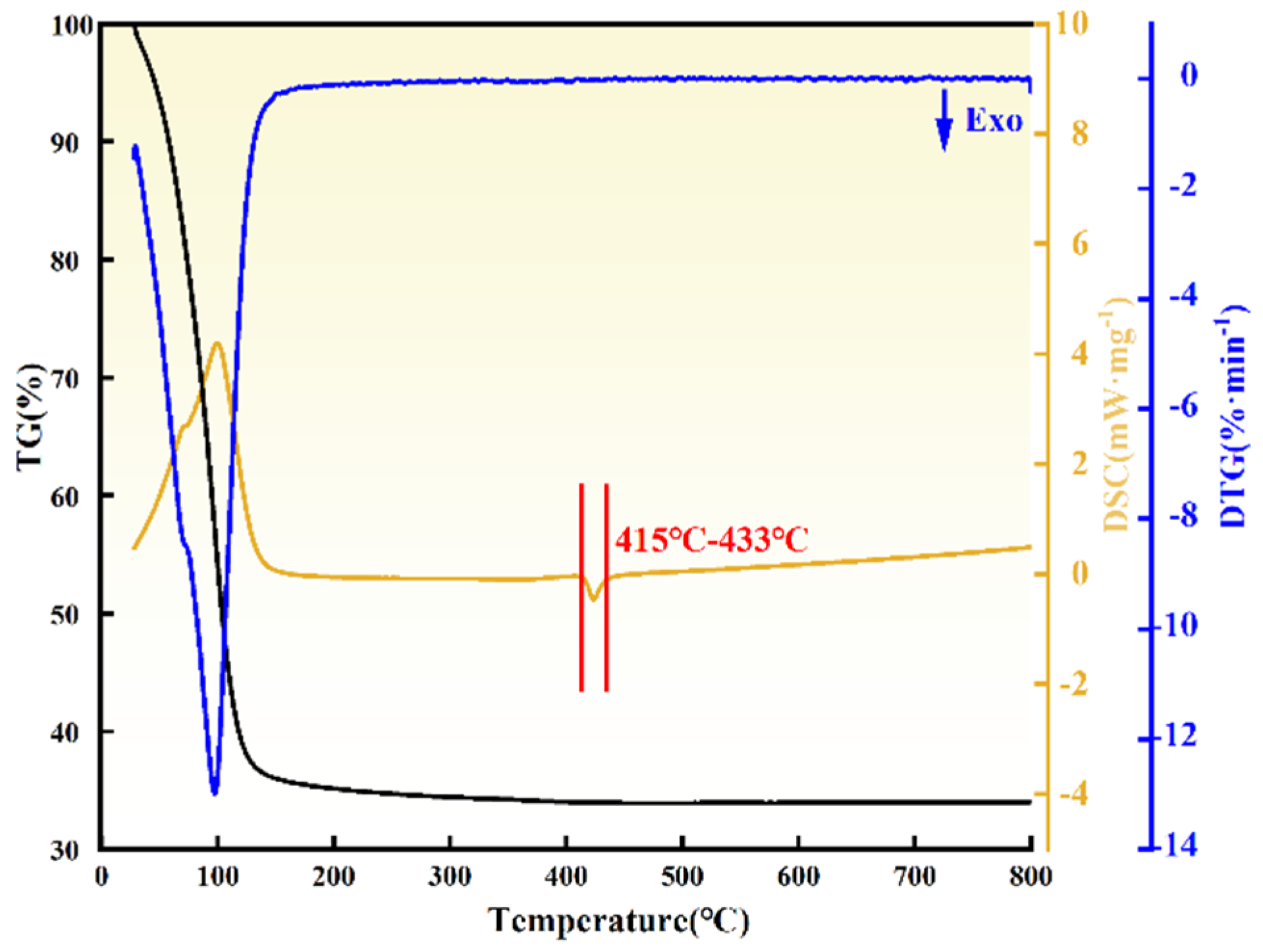
3.1. Thermogravimetric-Differential Thermal Analysis of ZrO(OH)2

The thermogravimetric-differential thermal analysis (TG-DTA) results of ZrO(OH)2 are shown in Figure 1. The thermal decomposition process of ZrO(OH)2 exhibited a single-stage weight loss behavior. As indicated by the TG curve, significant weight loss (43.3%) occurred between 30 °C and 130 °C, with the maximum weight loss rate observed at ~98 °C. This stage corresponds to the desorption of physically adsorbed water molecules from the surface of ZrO(OH)2, driven by overcoming van der Waals forces. From 130 °C to 415 °C, a gradual mass decline was attributed to the removal of internal hydroxyl groups, accompanied by the initial formation of the ZrO2 crystalline phase. A distinct exothermic peak appeared between 415 °C and 433 °C, corresponding to the cleavage of Zr–OH bonds and subsequent nucleation of ZrO2 crystals. Above 433 °C, the TG curve stabilized, indicating the completion of ZrO(OH)2 decomposition and the formation of thermally stable ZrO2 [22].

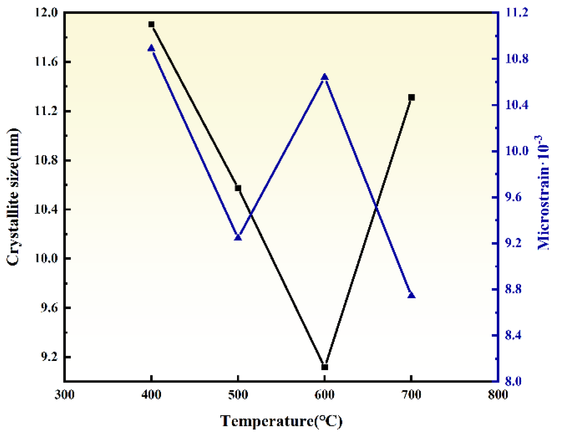
3.2. Effect of Ball Milling Temperature

The influence of ball milling temperature (400, 500, 600, and 700 °C) on the crystallinity, particle size, and microstrain of ZrO2 was investigated by ball milling dried ZrO(OH)2 for 6 h under each condition. The XRD patterns of ZrO2 (Figure 2) demonstrate that the sample ball milled at 400 °C for 6 h contains both m-ZrO2 and t-ZrO2 phases, with weak diffraction intensities indicative of low crystallinity. As the ball milling temperature increases to 500 °C, the monoclinic phase is transformed into the tetragonal phase, and further temperature elevation (500–700 °C) significantly enhances the crystallinity of t-ZrO2. The transition from monoclinic to tetragonal ZrO2 under HTMT conditions aligns with prior studies on mechanochemically driven phase stabilization. As demonstrated by Suryanarayana [23] the combination of shear stress and thermal activation reduces the energy barrier for t-ZrO2 formation, enabling its retention at room temperature despite subcritical crystallite sizes. The particle sizes and strain calculated via the Debye–Scherrer equations ((h k l) = (1 0 1), (1 1 2), and (2 1 1)) are summarized in Figure 3. Under 6 h of ball milling at 400–700 °C, the ZrO2 particle sizes are 11.91, 10.58, 9.12, and 11.31 nm, while the corresponding microstrain are 10.89, 9.25, 10.64, and 8.74 × 10⁻³, respectively. Notably, the particle size exhibits a non-monotonic trend, decreasing initially and then increasing with rising temperature. This behavior is attributed to the ~7% volume contraction during the m-ZrO2-to-t-ZrO2 phase transition, which generates additional internal stress within particles. At lower temperatures (400–500 °C), sluggish atomic diffusion and limited grain boundary migration allow the dominant impact and shear forces from milling media to refine ZrO2 particles effectively. Conversely, at elevated temperatures (600–700 °C), enhanced grain boundary diffusion facilitates particle coalescence, leading to increased particle sizes. Simultaneously, the reduced influence of milling-induced lattice distortion results in diminished microstrain. Considering the interplay between particle refinement and phase stability, a ball milling temperature of 600 °C is identified as the optimal condition.

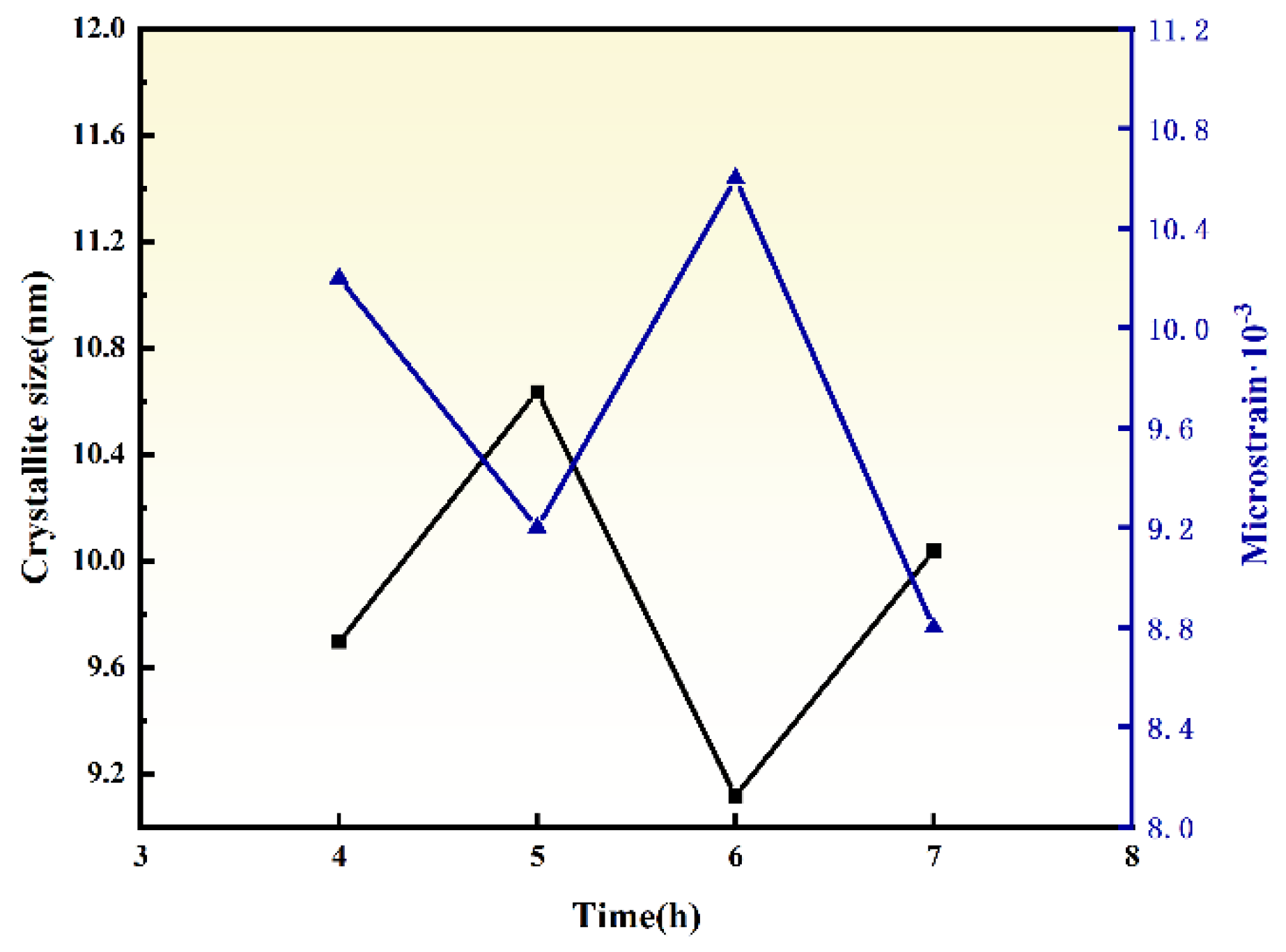
3.3. Effect of Ball Milling Time

The influence of ball milling time (4, 5, 6, and 7 h) on the phase composition, particle size, and microstrain of ZrO2 was investigated by ball milling dried ZrO(OH)2 at 600 °C. As shown in the XRD patterns of ZrO2 (Figure 4), the sample ball milled for 4 h at 600 °C contains both m-ZrO2 and t-ZrO2 phases, with weak diffraction peak intensities indicating low crystallinity. Prolonging the milling time to 5 h induces a transition from the monoclinic to the tetragonal phase, and further extension of milling time significantly enhances the crystallinity of t-ZrO2. The dominance of t-ZrO2 after prolonged milling (6 h) is consistent with the defect-mediated stabilization mechanism proposed by Garvie [24], where lattice strain and dislocation networks suppress the m-ZrO2 reconstruction during cooling.
The particle sizes and strain calculated via the Debye–Scherrer equations are summarized in Figure 5. Under 600 °C ball milling for 4–7 h, the ZrO2 particle sizes are 9.70, 10.62, 9.12, and 10.03 nm, while the corresponding microstrain are 10.2, 9.2, 10.6, and 8.8 × 10⁻³, respectively. Notably, after 6 h of milling, enhanced thermal vibration of Zr atoms facilitates grain boundary migration and rearrangement, leading to increased particle size (9.12 nm) and reduced microstrain (10.64 × 10⁻³). This phenomenon suggests that prolonged thermal–mechanical coupling promotes particle coalescence and stress relaxation. At 600 °C with 6 h of milling, ZrO2 exhibits the smallest particle size (9.12 nm) and highest microstrain (10.64 × 10⁻³). Considering the balance between phase stability and particle refinement, a ball milling time of 6 h is identified as optimal.
The TEM images, particle size distribution, and HRTEM analysis of the ZrO2 powders synthesized under these conditions are presented in Figure 6. As shown in Figure 6a, the ZrO2 particles exhibit irregular clusters with good dispersion and uniform spherical morphology. The particle size distribution histogram (Figure 6b) reveals a narrow size distribution centered at ~10 nm. The HRTEM image (Figure 6c) displays distinct lattice fringes, confirming high crystallinity and well-defined grain boundaries. The interplanar spacing measured in the orange-boxed region is 0.2986 nm, corresponding to the (101) plane of t-ZrO2, which aligns with the XRD analysis results.

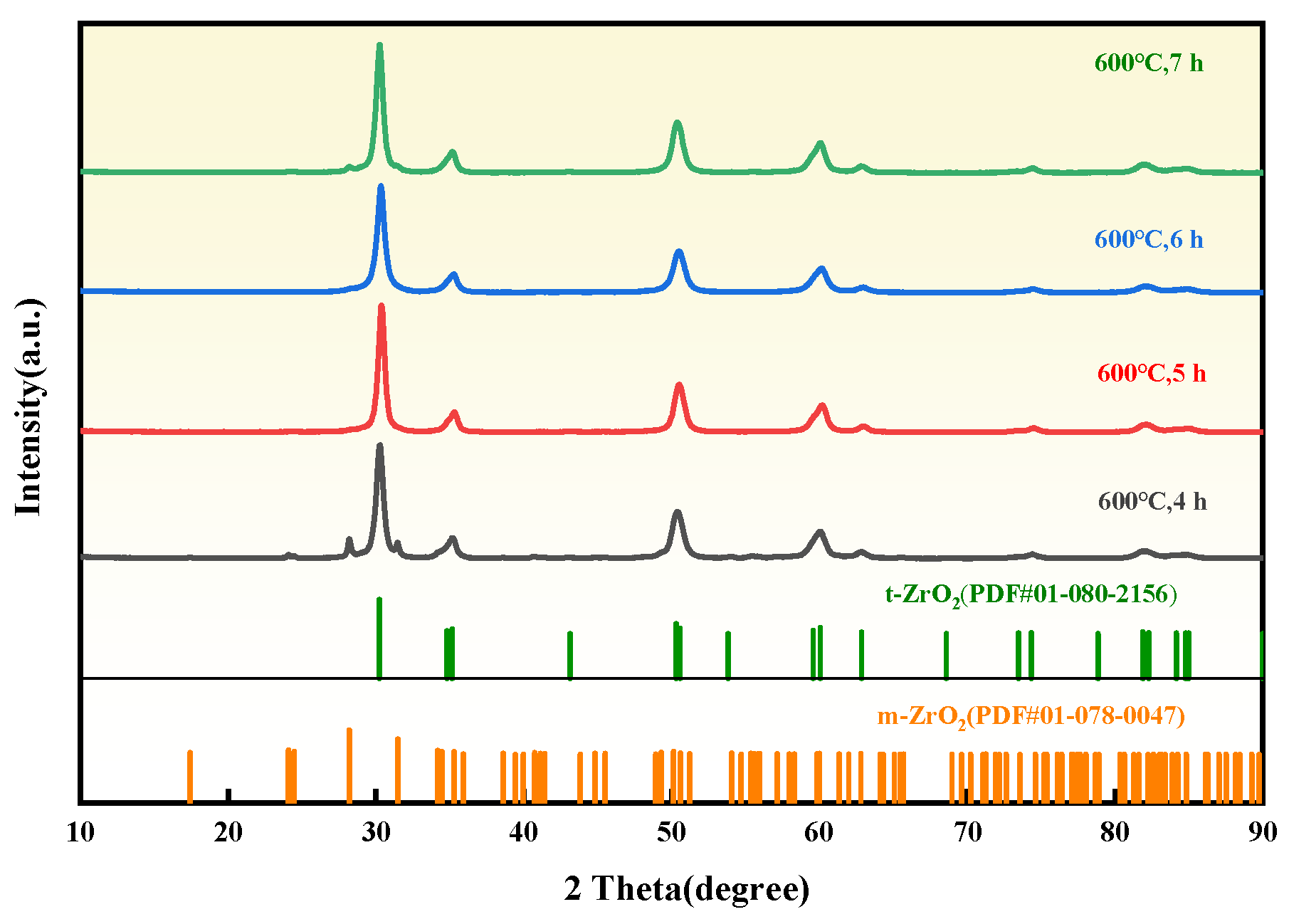
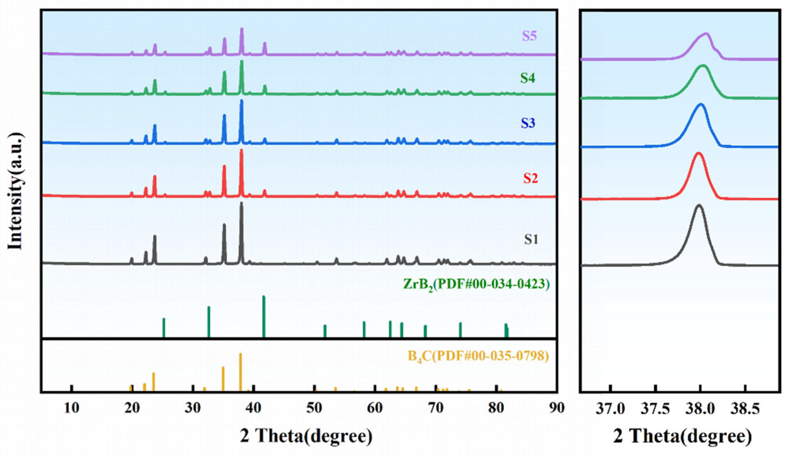
3.4. Phase Characterization of ZrO2-B4C Composites

The XRD patterns of B4C ceramics with varying HTMT-ZrO2 additions sintered at 2240 °C are shown in Figure 7. The rhombohedral B4C phase remains dominant in all samples, with B4C (Sample S1) exhibiting characteristic diffraction peaks at 2θ = 19.7° (101), 22.0° (003), 23.5° (012), 34.9° (104), and 37.8° (021). Upon introducing 2 wt% commercial nano-ZrO2 (Sample S2), weak ZrB2 peaks emerge at 2θ = 32.6° (100) and 41.6° (101). Above 2000 °C, the following interfacial reaction will occur between ZrO2 and B4C [25]:
4ZrO2(s) + 5B4C(s) → 5ZrB2(s) + 4CO(g) + 6BO(g)
Notably, the ZrB2 peak intensity in Sample S3 (2 wt% HTMT-ZrO2) increases by 23% compared to S2 (peak area ratio I Z r B 2 :   I Z r B 2 = 0.15   : 0.12 ), attributed to the enhanced reactivity of HTMT-ZrO2 due to optimized surface hydroxylation and reduced agglomeration. Increasing the HTMT-ZrO2 content from 2 to 4 wt% (Sample S4) significantly promotes ZrB2 formation [26], accompanied by a broadening of the B4C (021) peak FWHM from 0.26° to 0.28°, indicative of ZrO2-induced grain refinement. Further increasing HTMT-ZrO2 to 6 wt% (Sample S5) intensifies ZrB2 generation but reduces the B4C (021) FWHM to 0.25°, suggesting that ZrB2-mediated interfacial slip becomes the dominant stress relaxation mechanism.

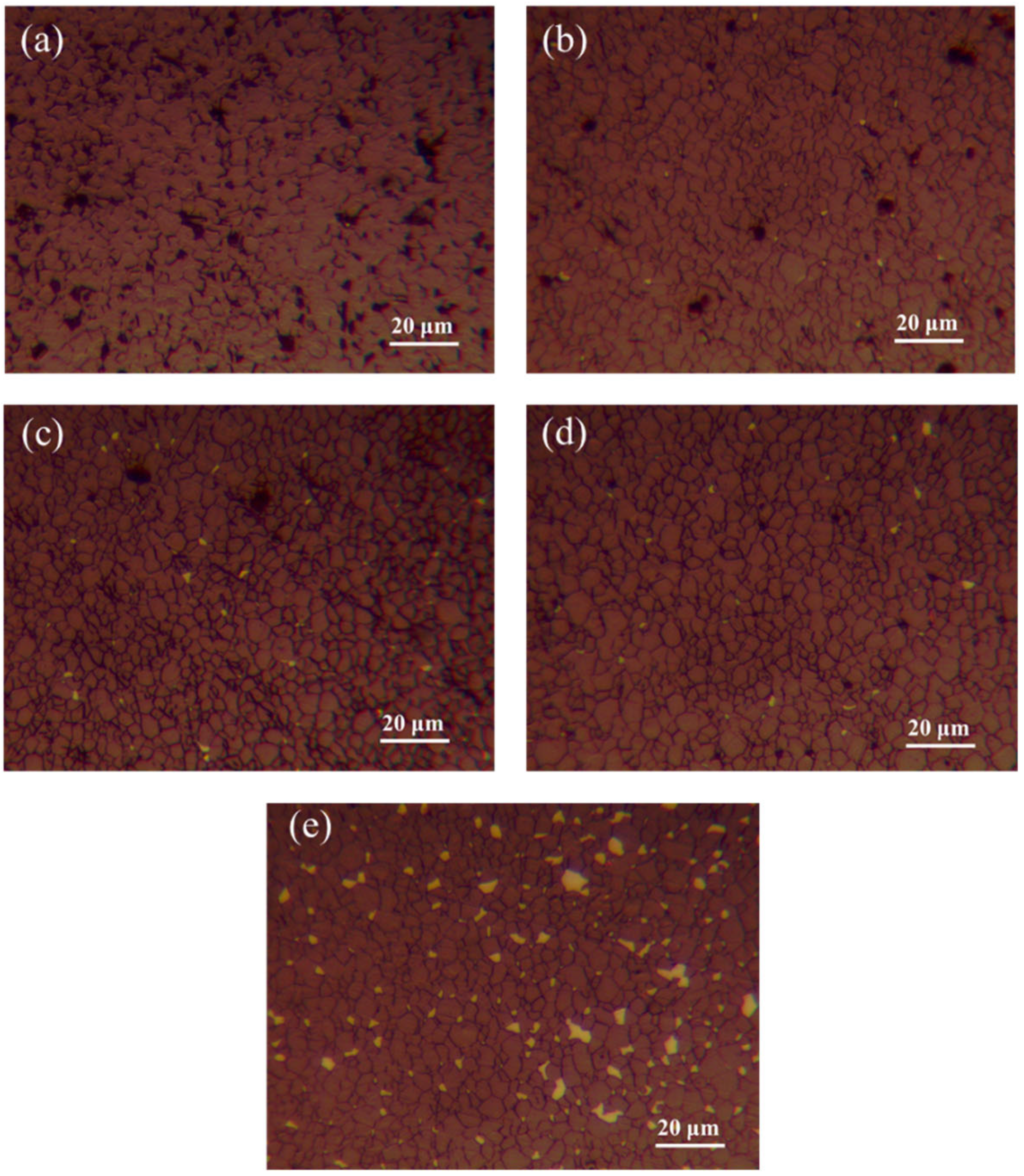
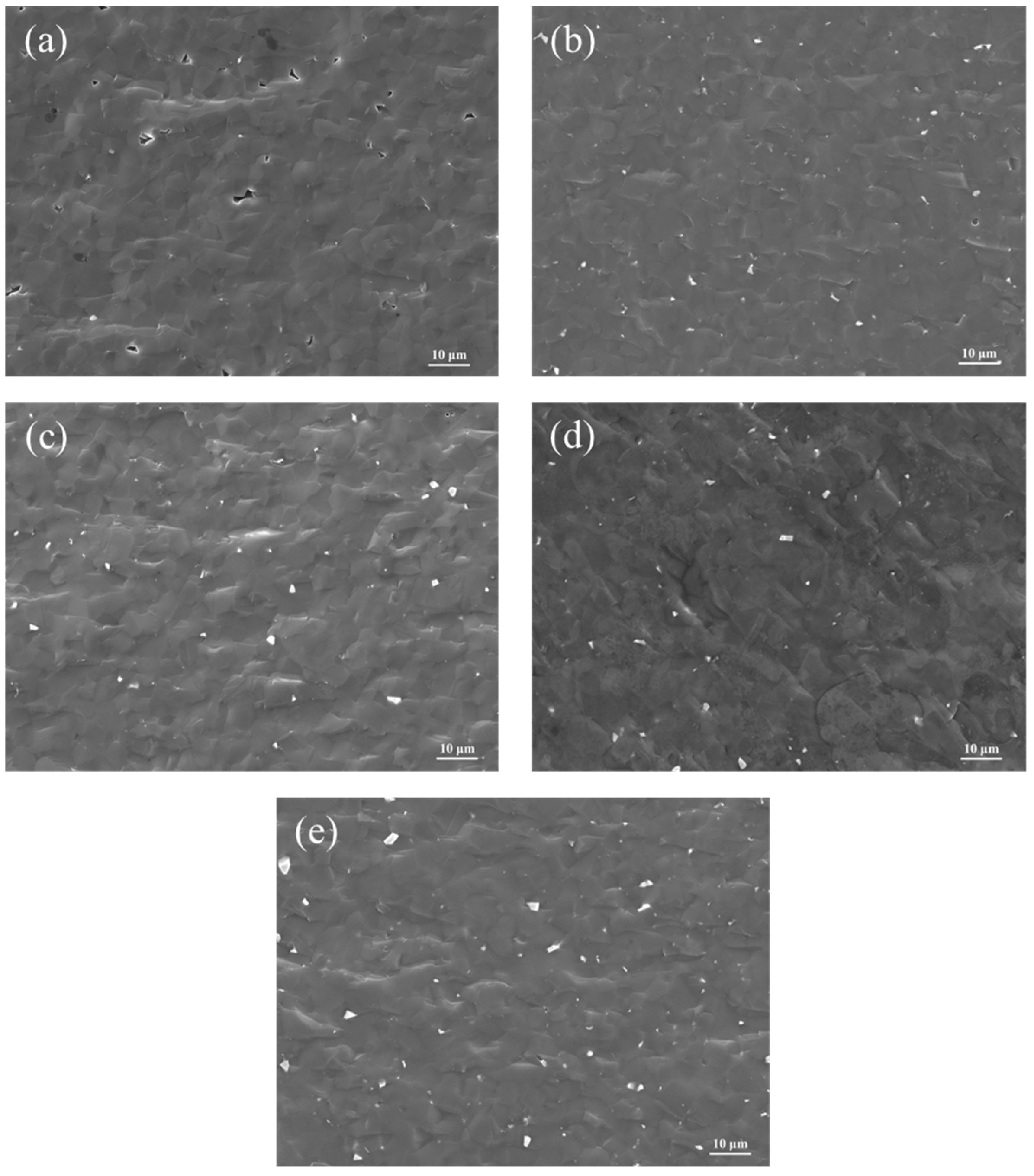
3.5. Effect of ZrO2 Content on the Microstructure of B4C Ceramics

The SEM images of B4C ceramics with varying ZrO2 additions sintered at 2240 °C are presented in Figure 8. As shown in Figure 8a, Sample S1 exhibits a porous structure with irregular pores (1–5 μm) due to incomplete densification caused by insufficient sintering activity. Sample S2 demonstrates improved densification compared to S1 but contains localized irregular platelet-like ZrB2 particles and non-uniform particle size distribution, attributed to the poor dispersion of commercial nano-ZrO2. In contrast, Sample S3 achieves a homogeneous microstructure with significantly reduced ZrB2 agglomerates of smaller dimensions (<500 nm), where the enhanced dispersion of HTMT-ZrO2 strengthens interfacial bonding [26]. Sample S4 displays a smooth surface with further reduced porosity, minimal ZrB2 agglomeration, and no visible microcracks. However, Sample S5 shows distinct platelet-like ZrB2 agglomerates, indicating that excessive ZrO2 addition triggers coarsening of the secondary ZrB2 phase, thereby degrading mechanical performance.
The etched metallographic images of B4C ceramics with varying ZrO2 additions sintered at 2240 °C are shown in Figure 9. As illustrated in Figure 9a, Sample S1 exhibits uniform grain distribution (average size: 9.7 ± 0.5 μm) but contains numerous intergranular pores (diameter: 5–8 μm, porosity: 8%), indicating poor densification. In Sample S2, the porosity decreases to 6.2%, with reduced grain size (7.4 ± 0.5 μm) and localized ZrB2 enrichment at B4C grain boundaries. In contrast, Sample S3 achieves finer grains (5.5 ± 0.3 μm) and lower porosity (3%), where in situ-formed ZrB2 particles at B4C grain boundaries inhibit grain boundary migration and mass transfer, thereby refining the grains [27]. Sample S4 demonstrates a reinforcing network of ZrB2 platelets, which enhances localized dislocation motion and significantly reduces pore size (diameter: 2–4 μm, porosity: 2.8%) through reactive liquid-phase sintering. For Sample S5, the porosity drops below 1%, with optimized densification due to second-phase strengthening by HTMT-ZrO2-derived products; however, localized Zr-rich zones are observed.
The fracture surface SEM images of B4C ceramics with varying ZrO2 additions sintered at 2240 °C are shown in Figure 10. As observed in Figure 10a, Sample S1 displays a relatively smooth fracture surface dominated by transgranular fracture, accompanied by sparse pores due to incomplete densification. In contrast, Samples S2–S5 exhibit a mixed fracture mode of transgranular and intergranular failure with minor porosity. This is attributed to CO gas generation during the in situ reaction between ZrO2 and B4C to form ZrB2. At elevated temperatures, the grain boundary migration rate of B4C exceeds the venting rate of CO gas, resulting in trapped pores. As the HTMT-ZrO2 content increases, its sintering-aid effect intensifies, leading to smoother fracture surfaces and a higher proportion of intergranular fracture [28], which correlates with enhanced interfacial reactions and improved densification.

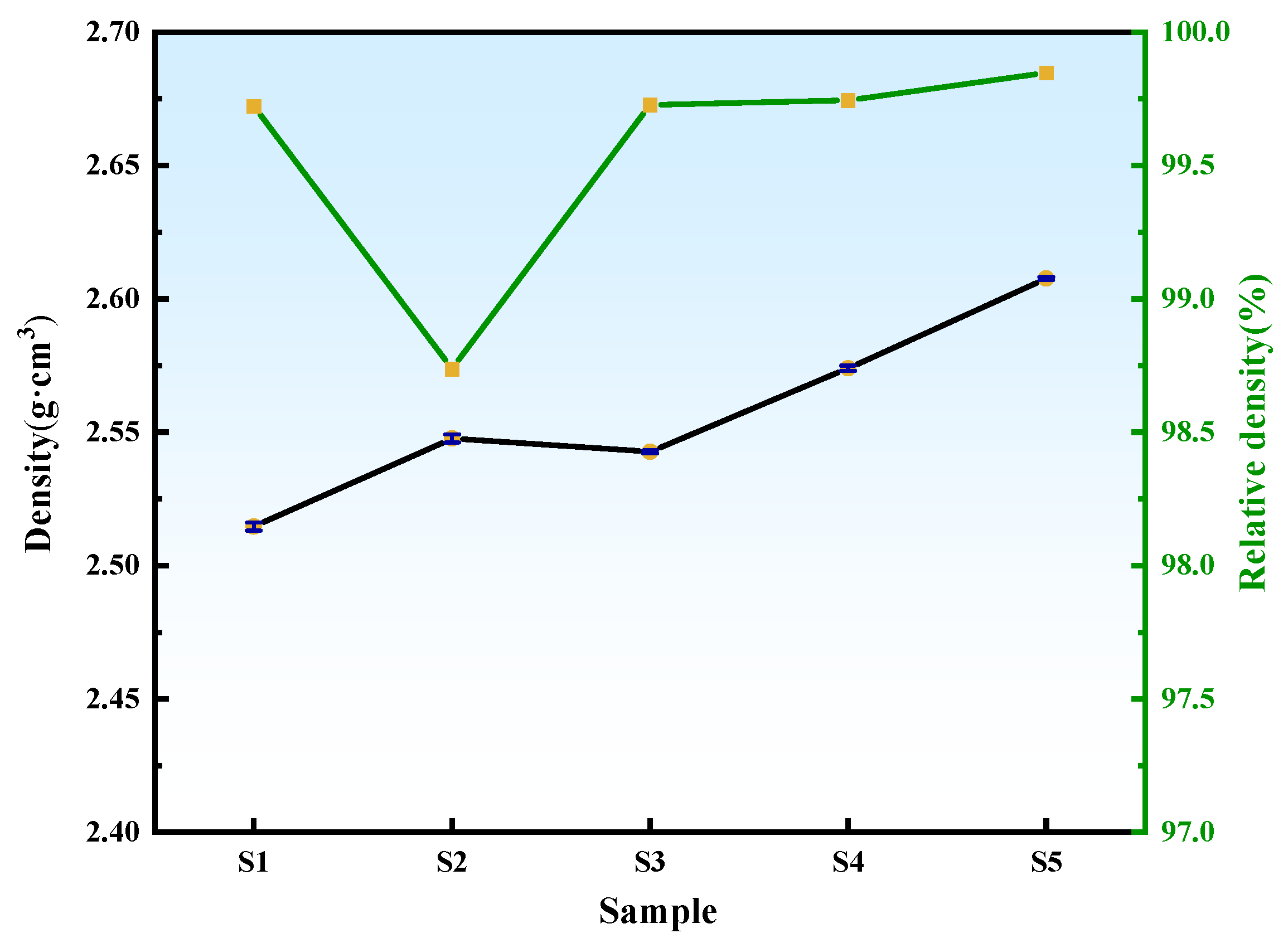
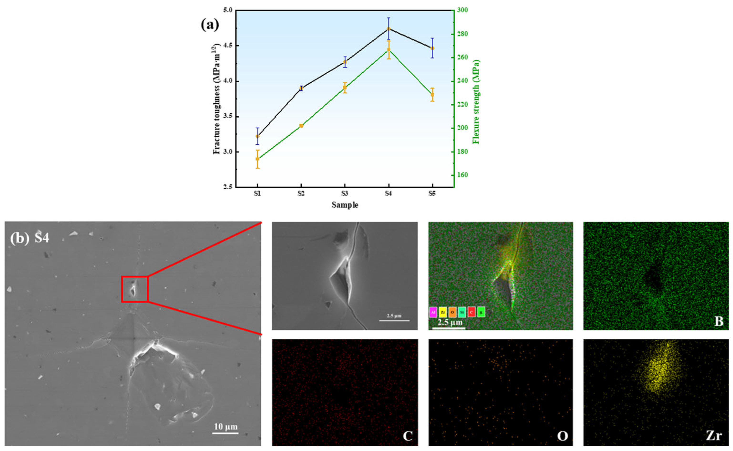
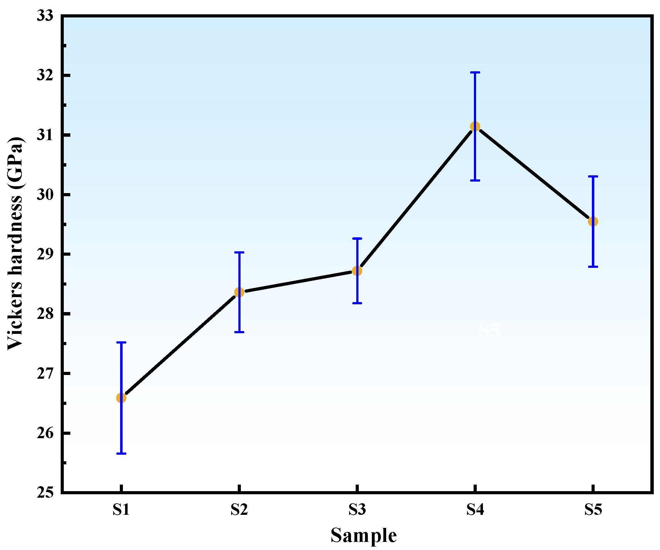
3.6. Effect of ZrO2 Content on the Mechanical Properties of B4C Ceramics

The density and relative density of B4C ceramics with varying ZrO2 additions sintered at 2240 °C are shown in Figure 11. Sample S1 exhibits a density of 2.51 g·cm−3 and a relative density of 99.72%, confirming the efficacy of SiO2-assisted liquid-phase sintering. For Sample S2, the density increases to 2.54 g·cm−3; however, agglomeration of commercial nano-ZrO2 particles generates large trapped pores, slightly reducing the relative density to 98.73% [29]. With increasing HTMT-ZrO2 content, the density of B4C ceramics progressively rises. The superior dispersion of HTMT-ZrO2 minimizes interfacial porosity caused by agglomeration, while the in situ formation of uniformly distributed ZrB2 platelets synergizes with the SiO2 liquid-phase sintering effect to enhance relative density.
The fracture toughness and flexural strength of B4C ceramics are summarized in Figure 12a. From Sample S1 to S4, the fracture toughness increases from 3.22 to 4.74 MPa·m1/2 (47.2% improvement), with Sample S2 showing a 9.6% enhancement over S1. Flexural strength similarly improves with higher HTMT-ZrO2 content, peaking at 266.62 MPa for Sample S4. However, Sample S5 exhibits reduced fracture toughness (4.32 MPa·m1/2) and flexural strength (228.43 MPa), even lower than that of S3 (234.40 MPa). This degradation is attributed to excessive in situ formation of micron-sized ZrB2, which acts as stress concentrators, significantly lowering fracture energy [25]. As evidenced by the dimple-crack morphology in Sample S4, cracks propagating to ZrB2/B4C interfaces are deflected along grain boundaries due to residual stress from thermal expansion mismatch between ZrB2 and B4C phases during cooling (as shown in Figure 12b). This prolongs the crack propagation path, requiring greater energy dissipation and thereby enhancing fracture toughness.
The Vickers hardness of B4C samples with varying ZrO2 additions sintered at 2240 °C is shown in Figure 13. The reference Sample S1 exhibits a hardness of 26.59 GPa. Upon adding 2 wt% commercial and HTMT-synthesized nano-ZrO2, Samples S2 and S3 achieve enhanced hardness values of 28.36 GPa and 28.72 GPa, respectively. This improvement confirms that the secondary ZrB2 phase refines B4C grains, reduces porosity, and optimizes densification, while also introducing residual stress due to the difference in coefficients of thermal expansion between B4C and ZrB2, thereby enhancing hardness. Sample S4 attains the maximum hardness of 31.14 GPa, while Sample S5 shows a decline to 29.55 GPa, attributed to the increased proportion of low-hardness ZrB2 phase.

4. Conclusions

In this study, nano-sized ZrO2 powders were successfully synthesized via a novel high-temperature mechanochemical technology (HTMT) using ZrO(OH)2 as the zirconium precursor. ZrB2-B4C composites were subsequently fabricated by hot-pressing mixtures of B4C powder with varying contents of HTMT-derived and commercial nano-ZrO2. Key findings include:
(1)
HTMT Process Optimization: The ZrO2 particle size exhibited a non-monotonic trend (initial decrease followed by increase) with rising ball milling temperature (400–700 °C) and prolonged duration (4–7 h). Optimal conditions were identified as 600 °C for 6 h, yielding ZrO2 powders with narrow size distribution (9.12 nm), low microstrain (10.64 × 10⁻³), high crystallinity, and excellent dispersion.
(2)
Composite Performance: Compared to commercial nano-ZrO2, HTMT-ZrO2 demonstrated superior uniformity and dispersion, enabling enhanced densification of ZrB2-B4C composites. With increasing in situ-generated ZrB2 content, B4C crystallite size initially decreased (5.5 ± 0.3 μm at 2 wt%) and then increased (7.2 ± 0.4 μm at 6 wt%), while mechanical properties (fracture toughness: 4.74 MPa·m1/2; flexural strength: 266.61 MPa; Vickers hardness: 31.14 GPa) peaked at 4 wt% HTMT-ZrO2.
(3)
This mechanism prolongs the crack extension path, resulting in a 47.2% increase in fracture toughness over B4C ceramics without ZrO2 addition.
The 4 wt% HTMT-ZrO2-B4C exhibited optimal comprehensive performance: density of 2.57 g·cm−3 (99.75% relative density), smooth microstructure, and balanced mechanical properties. This work establishes HTMT as a scalable, energy-efficient route for synthesizing high-performance ceramic composites, with implications for structural material design.

Author Contributions

Conceptualization, S.W., K.L. and Q.H.; methodology, J.X. and B.L.; formal analysis, J.X. and J.J.; resources, K.L. and C.W.; data curation, J.X. and J.J.; writing—original draft preparation, J.X.; writing—review and editing, J.X.; supervision, D.F. and Q.H.; funding acquisition, D.F. and C.W. All authors have read and agreed to the published version of the manuscript.

Funding

This research was supported by the Fundamental Research Funds for the Central Universities [N2425012, Daxue Fu] and a PhD start-up grant from Liaoning Institute of Science and Technology [No. 2407B10, Chunxin Wang].

Institutional Review Board Statement

Not applicable.

Informed Consent Statement

Not applicable.

Data Availability Statement

The original contributions presented in this study are included in this article, further inquiries can be directed to the corresponding authors.

Acknowledgments

All authors thank the editors and reviewers for their work on this manuscript.

Conflicts of Interest

Author Jingming Xu is employed by the Inner Mongolia First Machinery Group Co., Ltd., China National Weapons Industry Corp., Ltd. The remaining authors declare that the research was conducted in the absence of any commercial or financial relationships that could be construed as a potential conflict of interest.

References

  1. Xu, X.; Feng, Y.; Zhong, Z.; Guo, X.; Zhang, Z.; Li, J.; Zhao, S.; Wu, S.; Sun, H. Modified room temperature solid-state synthesis of yttria-stabilized zirconia (YSZ) nano-powders for solid oxide fuel cells. J. Rare Earths 2023, 41, 1385–1391. [Google Scholar] [CrossRef]
  2. Bugaeva, A.Y.; Nazarova, L.Y.; Tropnikov, E.M.; Shushkov, D.A.; Utkin, A.A.; Ryabkov, Y.I. Preparation, Microstructure, and Properties of a Ceramic Composite Based on Stabilized Zirconium Dioxide. Russ. J. Gen. Chem. 2023, 93, 2822–2830. [Google Scholar] [CrossRef]
  3. Ozsoy, M.; Tıkız, İ.; Pehlivan, H. Thermal analysis of a zirconium dioxide coated aluminum alloy piston. Int. J. Comput. Exp. Sci. Eng. Fail. Anal. 2018, 4, 43–50. [Google Scholar] [CrossRef]
  4. Ashok Kumaravel, V.K.; Elangovan, G. Influence of nano zirconia on the mechanical and durability properties of high-performance concrete containing nano-silica. Mater. Res. Express 2023, 10, 105012. [Google Scholar] [CrossRef]
  5. Yamagiwa, K.; Goudo, D. Synthesis of carbon nanotubes on carbon fiber substrates: Effects of nanozirconia dispersion on the growth of carbon nanotubes. Jpn. J. Appl. Phys. 2024, 63, 02SP04. [Google Scholar] [CrossRef]
  6. Song, Q.; Zha, X.; Gao, M.; Shi, J.; Ma, Y. Influence of ZrO2 on the phase composition and mechano-physical properties of MgO–ZrO2 refractories prepared by cold isostatic pressing. Ceram. Int. 2024, 50, 30474–30482. [Google Scholar] [CrossRef]
  7. Chakravarty, R.; Shukla, R.; Ram, R.; Tyagi, A.K.; Dash, A.; Venkatesh, M. Development of a nano-zirconia based 68Ge/68Ga generator for biomedical applications. Nucl. Med. Biol. 2011, 38, 575–583. [Google Scholar] [CrossRef]
  8. Bugaeva, A.Y.; Nazarova, L.Y.; Belyi, V.A.; Ryabkov, Y.I. Phase Transformations of Zirconium Dioxide and Crystal Growth During Heat Treatment of the ZrO2(CeO2,Y2O3)–La0.85Y0.15Al11O18–Al2O3 System. Russ. J. Gen. Chem. 2022, 92, 1488–1497. [Google Scholar] [CrossRef]
  9. Huang, W.; Qiu, H.; Zhang, Y.; Nan, L.; Gao, L.; Chen, J.; Omran, M.; Chen, G. Preparation of nano zirconia by binary doping: Effect of controlled sintering on structure and phase transformation. Ceram. Int. 2022, 48, 25374–25381. [Google Scholar] [CrossRef]
  10. Hsu, Y.-W.; Yang, K.-H.; Chang, K.-M.; Yeh, S.-W.; Wang, M.-C. Synthesis and crystallization behavior of 3mol% yttria stabilized tetragonal zirconia polycrystals (3Y-TZP) nanosized powders prepared using a simple co-precipitation process. J. Alloys Compd. 2011, 509, 6864–6870. [Google Scholar] [CrossRef]
  11. Sarkar, D.; Mohapatra, D.; Ray, S.; Bhattacharyya, S.; Adak, S.; Mitra, N. Synthesis and characterization of sol–gel derived ZrO2 doped Al2O3 nanopowder. Ceram. Int. 2007, 33, 1275–1282. [Google Scholar] [CrossRef]
  12. Li, N.; Dong, B.; Yuan, W.; Gao, Y.; Zheng, L.; Huang, Y.; Wang, S. ZrO2 Nanoparticles Synthesized using Ionic Liquid Microemulsion. J. Dispers. Sci. Technol. 2007, 28, 1030–1033. [Google Scholar] [CrossRef]
  13. Liu, L.; Wang, S.; Zhang, B.; Jiang, G.; Yang, J.; Li, Y.; Liu, W.; Wang, J.; Kong, W. From modification to mechanism: Supercritical hydrothermal synthesis of nano-zirconia. Ceram. Int. 2022, 48, 4401–4423. [Google Scholar] [CrossRef]
  14. Zhang, W.; Tan, Y.; Gao, Y.; Wu, J.; Tang, B. Ultrafine nano zirconia as electrochemical pseudocapacitor material. Ceram. Int. 2015, 41, 2626–2630. [Google Scholar] [CrossRef]
  15. Puclin, T.; Kaczmarek, W.A.; Ninham, B.W. Mechanochemical processing of ZrSiO4. Mater. Chem. Phys. 1995, 40, 73–81. [Google Scholar] [CrossRef]
  16. Ding, J.; Tsuzuki, T.; McCormick, P.G. Mechanochemical synthesis of ultrafine ZrO2 powder. Nanostruct. Mater. 1997, 8, 75–81. [Google Scholar] [CrossRef]
  17. Liu, M.; Wang, B.; Wang, Y.; Fu, D.; Chang, Y.; Li, B.; Liu, K.; He, X.; Chen, J.; Wei, S.; et al. Intercalation of AlCl3 in microcrystalline graphite via high-temperature mechanochemical method for electromagnetic wave absorption. Appl. Surf. Sci. 2024, 666, 160387. [Google Scholar] [CrossRef]
  18. Liu, M.; Chen, J.; Li, B.; Wang, B.; Wang, Y.; Han, Q.; Wei, S.; Liu, K.; He, X.; Sun, R.; et al. Electromagnetic wave absorption properties of Mn0.4Zn0.6Fe2O4 powders synthesized by high-temperature mechanochemical method. Mater. Sci. Eng. B 2024, 302, 117243. [Google Scholar] [CrossRef]
  19. Hou, Y.; Li, B.; Chen, J.; Shen, X.; Wang, B.; Liu, K.; Wei, S.; He, X.; Li, D.; Han, Q. Electromagnetic wave absorption properties of core double-shell structured α-Fe(Si)@Fe3O4@SiO2 composites. Appl. Surf. Sci. 2023, 615, 156345. [Google Scholar] [CrossRef]
  20. Jia, P.; Shao, Z.; Liu, K. Synthesis and electrochemical performance of Li4Ti5O12 by high temperature ball milling method. Mater. Lett. 2014, 125, 218–220. [Google Scholar] [CrossRef]
  21. Salim, E.T.; Ismail, R.A.; Halbos, H.T. Growth of Nb2O5 film using hydrothermal method: Effect of Nb concentration on physical properties. Mater. Res. Express 2019, 6, 116429. [Google Scholar] [CrossRef]
  22. Mondal, A.; Ram, S. Monolithic t-ZrO2 Nanopowder through a ZrO(OH)2·xH2O Polymer Precursor. J. Am. Ceram. Soc. 2004, 87, 2187–2194. [Google Scholar] [CrossRef]
  23. Suryanarayana, C. Mechanical alloying and milling. Prog. Mater. Sci. 2001, 46, 1–184. [Google Scholar] [CrossRef]
  24. Garvie, R.C. The Occurrence of Metastable Tetragonal Zirconia as a Crystallite Size Effect. J. Phys. Chem. 1965, 69, 1238–1243. [Google Scholar] [CrossRef]
  25. Kim, H.-W.; Koh, Y.-H.; Kim, H.-E. Reaction sintering and mechanical properties of B4C with addition of ZrO2. J. Mater. Res. 2000, 15, 2431–2436. [Google Scholar] [CrossRef]
  26. Chen, H.; Zeng, F.; Li, W.; Liu, J.A.; Gu, Y.; Zhang, F. Densification behavior and mechanical properties of spark plasma reaction sintered ZrB2–ZrC-B4C ceramics from B4C-Zr system. Ceram. Int. 2019, 45, 12122–12129. [Google Scholar] [CrossRef]
  27. Wang, X.-G.; Guo, W.-M.; Kan, Y.-M.; Zhang, G.-J. Hot-Pressed ZrB2 Ceramics With Composite Additives of Zr and B4C. Adv. Eng. Mater. 2010, 12, 893–898. [Google Scholar] [CrossRef]
  28. Xiong, Y.; Du, X.; Xiang, M.; Wang, H.; Wang, W.; Fu, Z. Densification mechanism during reactive hot pressing of B4C-ZrO2 mixtures. J. Eur. Ceram. Soc. 2018, 38, 4167–4172. [Google Scholar] [CrossRef]
  29. Sagar, J.S.; Kashyap, S.J.; Madhu, G.M. Investigation of mechanical, thermal and electrical parameters of gel combustion-derived cubic zirconia/epoxy resin composites for high-voltage insulation. Cerâmica 2020, 66, 186–196. [Google Scholar] [CrossRef]
Figure 1. TG-DSC-DTG plot of ZrO(OH)2.
Materials 18 02332 g001
Figure 2. XRD patterns of ZrO2 prepared by different ball milling temperatures.
Materials 18 02332 g002
Figure 3. Crystallite size and microstrains plot of ZrO2 prepared by different ball milling temperatures.
Materials 18 02332 g003
Figure 4. XRD patterns of ZrO2 prepared by different ball milling time.
Materials 18 02332 g004
Figure 5. Crystallite size and microstrains diagram of ZrO2 prepared by different ball milling time.
Materials 18 02332 g005
Figure 6. Ball milling at 600 °C for 6 h: (a) ZrO2 TEM image, (b) ZrO2 particle size distribution, and (c) HRTEM image.
Materials 18 02332 g006
Figure 7. XRD patterns of B4C samples with different ZrO2 additions.
Materials 18 02332 g007
Figure 8. Surface SEM images of B4C samples with different ZrO2 additions, (ae) Samples S1–S5.
Materials 18 02332 g008
Figure 9. Etched metallographs of B4C samples with different ZrO2 additions, (ae) Samples S1–S5.
Materials 18 02332 g009
Figure 10. SEM images of fracture surfaces of B4C samples with different ZrO2 additions, (ae) Samples S1–S5.
Materials 18 02332 g010
Figure 11. Density and relative density plots of B4C samples with different ZrO2 additions.
Materials 18 02332 g011
Figure 12. (a) Fracture toughness and flexural strength plots of B4C samples with different ZrO2 additions and (b) SEM/EDS mapping of cracked S4 samples.
Materials 18 02332 g012
Figure 13. Vickers hardness of B4C samples with different ZrO2 additions.
Materials 18 02332 g013
Table 1. Sample composition.
SampleCharacteristics (wt%)
B4CCommercially Available Nano-ZrO2HTMT Nano-ZrO2SiO2
S199001
S297201
S397021
S495041
S593061
Disclaimer/Publisher’s Note: The statements, opinions and data contained in all publications are solely those of the individual author(s) and contributor(s) and not of MDPI and/or the editor(s). MDPI and/or the editor(s) disclaim responsibility for any injury to people or property resulting from any ideas, methods, instructions or products referred to in the content.

