Article

Stress Behaviour of an Immature Maxillary Central Incisor: A 3D Finite Element Analysis

by
Petra Bučević Sojčić
1,*,
Jasna Leder Horina
2,
Nina Bočkaj
3,
Tea Borojević Renić
4,
Dubravka Turjanski
5,
Kristina Goršeta
1,6,
Tanja Jurčević Lulić
7 and
Hrvoje Jurić
1,6
1
Department of Paediatric and Preventive Dentistry, School of Dental Medicine, University of Zagreb, Gundulićeva 5, 10 000 Zagreb, Croatia
2
Independent Chairs, Chair of General Program Contents, Faculty of Transport and Traffic Sciences, University of Zagreb, Vukelićeva 4, 10 000 Zagreb, Croatia
3
Independent Researcher, 10 000 Zagreb, Croatia
4
Department of Prosthodontics, Dental Polyclinic Zagreb, Perkovčeva 3, 10 000 Zagreb, Croatia
5
Department of Pediatric and Preventive Dentistry, Dental Polyclinic Zagreb, Perkovčeva 3, 10 000 Zagreb, Croatia
6
University Dental Clinic, University Hospital Centre Zagreb, Kišpatićeva 12, 10 000 Zagreb, Croatia
7
Department of Applied Mechanics, Faculty of Mechanical Engineering and Naval Architecture, University of Zagreb, Ivana Lučića 5, 10 000 Zagreb, Croatia
*
Author to whom correspondence should be addressed.
Materials 2025, 18(10), 2305; https://doi.org/10.3390/ma18102305
Submission received: 5 April 2025 / Revised: 12 May 2025 / Accepted: 13 May 2025 / Published: 15 May 2025
(This article belongs to the Special Issue Biocompatibility of Restorative Dental Materials)

Abstract

:
Background and Objective: Immature maxillary incisors (IMIs) are especially susceptible to failure due to their thin dentinal walls and compromised structural integrity following endodontic treatment. This study aims to evaluate the stress distribution within the root dentin after various post-endodontic treatments. Materials and methods: A personalized finite element analysis model of IMI was created using cone beam computed tomography (CBCT) data. Based on data from the literature, five stages of root development were reconstructed: half root development (S1), three-quarter development (S2), more than three-quarter development (S3), fully developed root with open apex (S4), and fully developed root with closed apex (S5). Six experimental groups were analyzed: GC Fiber Post (PS1); RelyX Post (PS2); metal post Unimetric 1.0 (PS3); everStick Post (PS4); positive control group with only the gutta-percha filling (PC), and intact maxillary incisor as negative control group (NC). The resulting equivalent stresses were evaluated using the Hencky–von Mises (HMH) strength theory. Results: The mean HMH stress within the root dentin was statistically significantly higher at the cervical level in all stages, except in stage S1 and models PS2 and PS3 in stage S2, where it was significantly higher at the apical level (p < 0.001 for all models, except stage S3 [PC model p < 0.005; NC model p < 0.008]). The PS4 model showed the lowest stress values at the cervical level in stages S1, S2, and S3 (55.19 MPa, 58.78 MPa, 58.84 MPa) and the PS1 model in stages S4 and S5 (57.48 MPa, 58.81 MPa). At the apical level, model PS3 showed the lowest stress values in stage S1 (69.60 MPa), model PS1 in stages S2, S3, and S5 (35.99 MPa, 44.30 MPa, 12.51 MPa) and model PC in stage S4 (17.85 MPa). Conclusions: The results showed that the greatest stress in an immature maxillary central incisor occurred at the cervical level, except during the early stage of root development. Post placement did not reduce root dentin stress.

1. Introduction

The management of non-vital immature maxillary incisors poses a major clinical challenge due to their underdeveloped root structures and compromised mechanical properties [1] requiring careful selection of treatment modalities to optimize the long-term outcomes [2]. The amount of remaining coronal structure and the residual thickness of the root dentin are critical factors in determining the appropriate type of final restoration [3,4,5,6]. In cases of severe coronal damage, the use of a post is often recommended to ensure adequate retention for the core and enhance the overall stability of the coronal restoration [3].
The development of esthetic, non-metallic post systems has influenced a shift from mechanical to adhesive post concepts [7]. Fiber-reinforced composite (FRC) posts are widely used in contemporary clinical practice due to their biomechanical compatibility with dentin [8]. Their modulus of elasticity is very close to that of natural dentin, promoting more favorable stress distribution and reducing the incidence of catastrophic failures such as post debonding and root fractures that extend beyond the alveolar bone [9]. Prefabricated glass fiber-reinforced composite posts are most commonly used due to their excellent esthetic properties and the formation of a monobloc with the tooth structure [10,11]. To minimize the need for extensive dentin removal in roots with larger canal diameters, individually shaped glass fiber-reinforced composite posts have been introduced. This approach reduces stress concentrations in the apical region while improving core support and overall structural integrity [12,13]. The direct application of composite restorations following post-endodontic treatment in the anterior region is increasingly favored as it preserves more healthy tooth structure and reduces the risk of root fracture [14], especially in pediatric patients where prosthetic rehabilitation remains controversial [15].
Clinical studies have shown that more than 50% of young permanent teeth with incomplete root development and necrotic pulp are lost in the first 10 years following endodontic treatment [16,17,18], which can also have a profoundly negative impact on a child’s psychosocial development [19]. Finite element analysis is widely recognized as a valuable tool for complex biomechanical investigations [20,21], which are crucial for understanding treatment failures and evaluating the performance of restorative materials [22,23]. Most finite element method (FEM) studies on maxillary incisors have reported that stress concentrations are usually located in the cervical region of the root [23,24,25,26,27,28,29,30,31]. In studies by Okamoto et al. [28] and Jain et al. [31], the highest stress values were recorded on the inner cervical side in models with FRC posts, while Nokar et al. [27] reported the highest stress concentrations between the middle and cervical third, however in mature incisors. Madfa et al. [32] reported that the cervical and apical region is where failure begins due to the high stresses between the post and the surrounding materials. Root immaturity has usually been defined in general terms (e.g., open apex, short root, weakened dentin wall) or related to a specific stage of development, usually the third stage of the Cvek classification [16]. However, no study has yet investigated the distribution of stress across all stages of root development.
The aim of this study was to assess stress distribution in immature maxillary incisor roots treated with various post systems and in relation to the developmental stage of the root using the finite element method.
The research hypotheses were:
1. The highest stress occurs in the cervical part of the root of an endodontically treated tooth with intracanal retention.
2. The intracanal retention and its stability depend on the thickness of the remaining root dentin.

2. Materials and Methods

2.1. Study Design

This study is an experimental in silico (computer-aided) investigation designed to evaluate the biomechanical performance of various post-endodontic restorative materials using the finite element method. Stress distribution within the root dentin was analyzed across several stages of development applying a comparative approach to evaluate six experimental groups. This study was conducted according to the guidelines of the Declaration of Helsinki, and approved by the Ethics Committee of the School of Dentistry, University of Zagreb, Croatia (approval number: 05-PA-27-5/2018). Written informed consent was obtained from the parents or legal guardians for the use of existing cone beam computed tomography (CBCT) images acquired primarily for diagnostic purposes related to orthodontic treatment prior to the start of this study. The consent form included detailed information about the objectives of this study, the methodology, and the significance of the expected results.

2.2. Model Generation

The geometry of an immature maxillary incisor (IMI) was isolated from a CBCT image of a school-aged child with an intact maxillary central incisor exhibiting completed root development. A three-dimensional tooth model was generated using the Mimics software package (version 16.0, Materialize, Leuven, Belgium). The finite element mesh was subsequently created using the Abaqus finite element analysis (FEA) software package (version 2020, Dassault Systemes, Simulia Corp., Johnston, RI, USA). Each part of the model was assigned its own finite element mesh, namely second-order tetrahedral finite elements consisting of ten nodes. The final output was a personalized 3D tooth model. The material properties, including the modulus of elasticity and Poisson’s ratio, are listed in Table 1. All model components were assumed to be linearly elastic and isotropic.
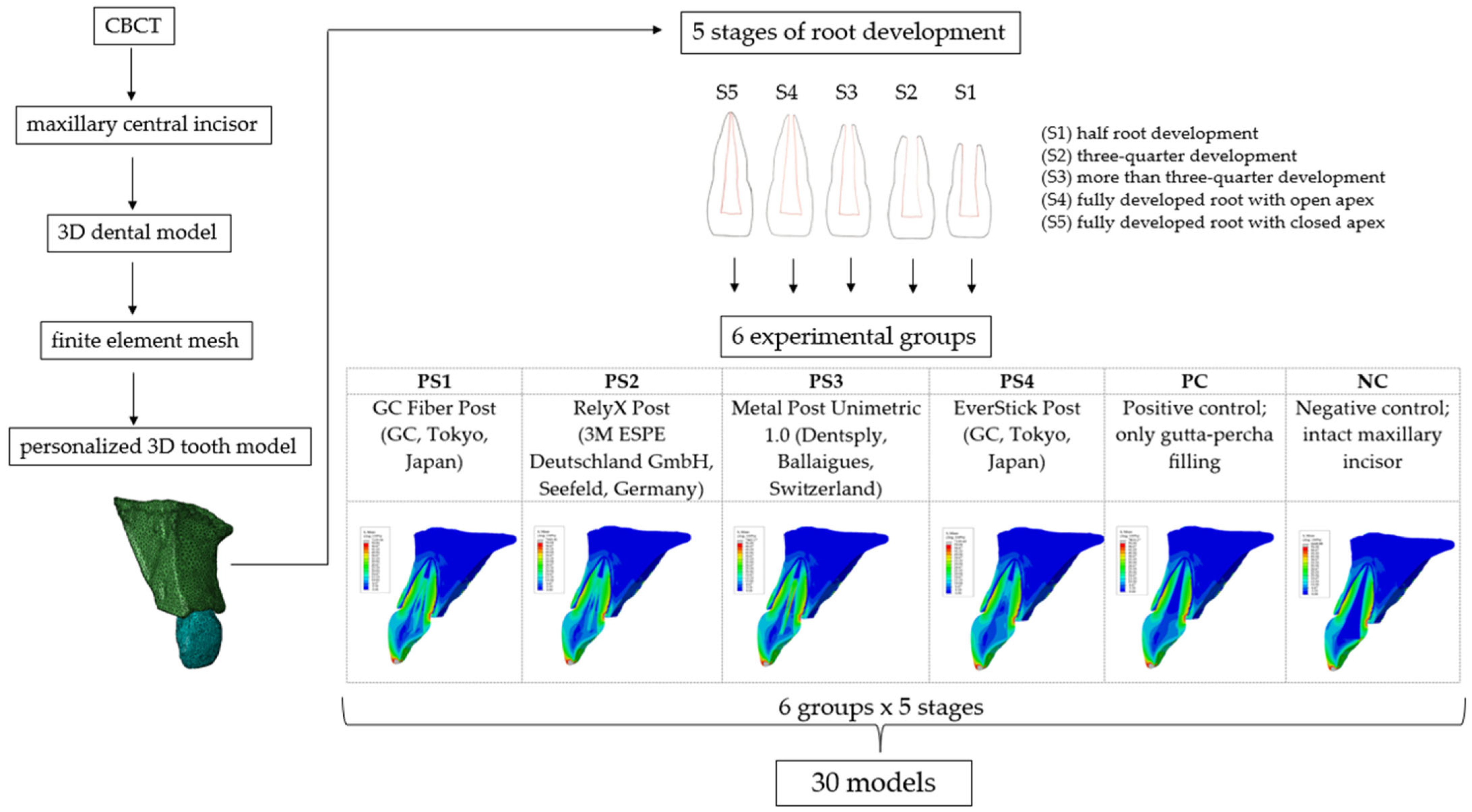
Based on data from a previous pilot study [42], dentin thickness measurements from intraoral images of IMIs in children aged from 7 to 11 years were used to reconstruct the five stages of root development: half root development (S1), three-quarter development (S2), more than three-quarter development (S3), fully developed root with open apex (S4), and fully developed root with closed apex (S5). In the next step, a complicated oblique crown fracture involving the loss of more than half of the crown was simulated, where intracanal retention can be considered. The overall therapeutic approach simulated a post-endodontic treatment with intracanal retention and a minimally invasive composite restoration of the crown (Figure 1). The depth of post space preparation varied according to the stage of root development, with the main requirement being to leave 4 mm of gutta-percha in the apical portion of the root. The models were divided into experimental groups according to the type of post system used: GC Fiber Post (GC, Tokyo, Japan) (PS1); RelyX Post (3M ESPE Deutschland GmbH, Seefeld, Germany) (PS2); Metal Post Unimetric 1.0 (Dentsply, Ballaigues, Switzerland) (PS3); everStick Post (GC, Tokyo, Japan) (PS4); positive control group with only the gutta-percha filling (PC), and intact maxillary incisor as negative control group (NC) (Figure 2).
The reconstruction involved modifying the mechanical properties within the root canal, which now included the properties of the gutta-percha, the post material, the cement, and the fractured crown segment restored with composite resin. The post was precisely positioned in the center of the root canal, maintaining a 4 mm distance between the apical foramen and the pulp chamber roof. The geometric specifications of each post were based on the manufacturer’s official documentation [43,44,45]. The remaining space surrounding the post and within the root canal was assigned the material properties of composite cement. In the case of the EverStick post, which is a customized post, it occupied almost the entire volume of the root canal, leaving 0.1 mm thick layer of composite cement surrounding the post. The simulation of the endodontic treatment included a gutta-percha filling up to 1 mm below the cervical margin, a 2 mm thick base layer of glass ionomer cement, and a composite restoration for the remaining portion of the crown filling. A total of 30 models were created.

2.3. Loading and Boundary Conditions

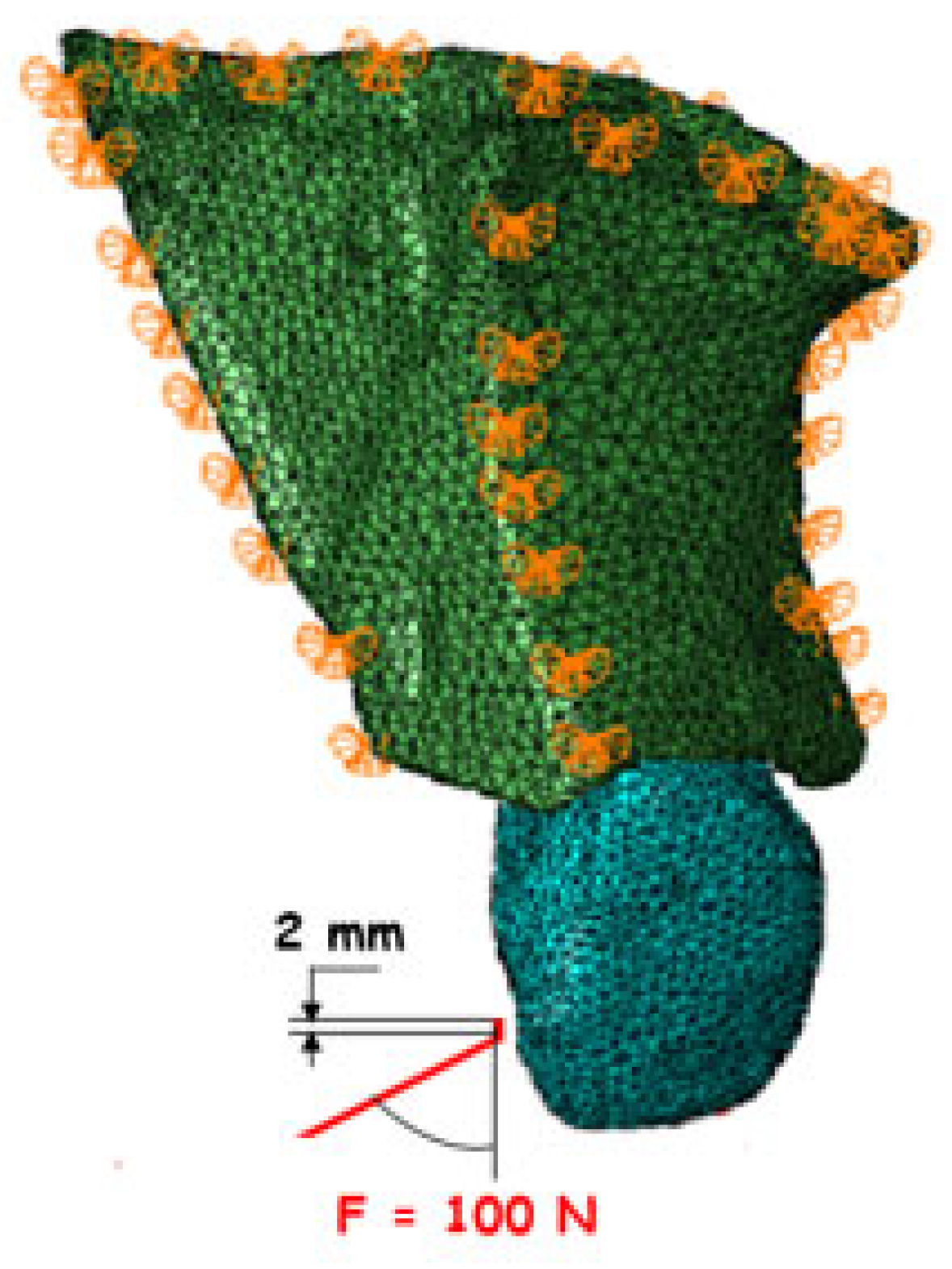
All directional movements were constrained to accurately simulate the anatomical connection with the rest of the maxilla. A static load of 100 N [46,47,48] was applied as a proportional continuous load to a small palatal surface of the simulated tooth at a 45-degree angle (Figure 3). The resulting equivalent stresses were evaluated using the Hencky–von Mises (HMH) stress theory.

2.4. Statistical Analysis

In all models, the highest stress concentrations were observed at the cervical and apical levels. Therefore, the 80 highest stress values from both levels were extracted for statistical analysis [49]. The middle level (between the apical and cervical levels) was not included as it did not show statistical significance in any of the groups. The normality of the data distribution within the individual groups of n = 80 was tested using the Shapiro–Wilk test and normal Q–Q plots. The analysis was performed using a three-way analysis of variance (ANOVA) with the factors “material”, “stage of development” and “root level (apical/cervical)”. For multiple comparisons, a one-way ANOVA with Tukey adjustment was used. The significance level was set at 0.05. Statistical analysis was performed with the SPSS software package, version 25.0 (IBM, Armonk, NY, USA).

3. Results

Descriptive Statistics

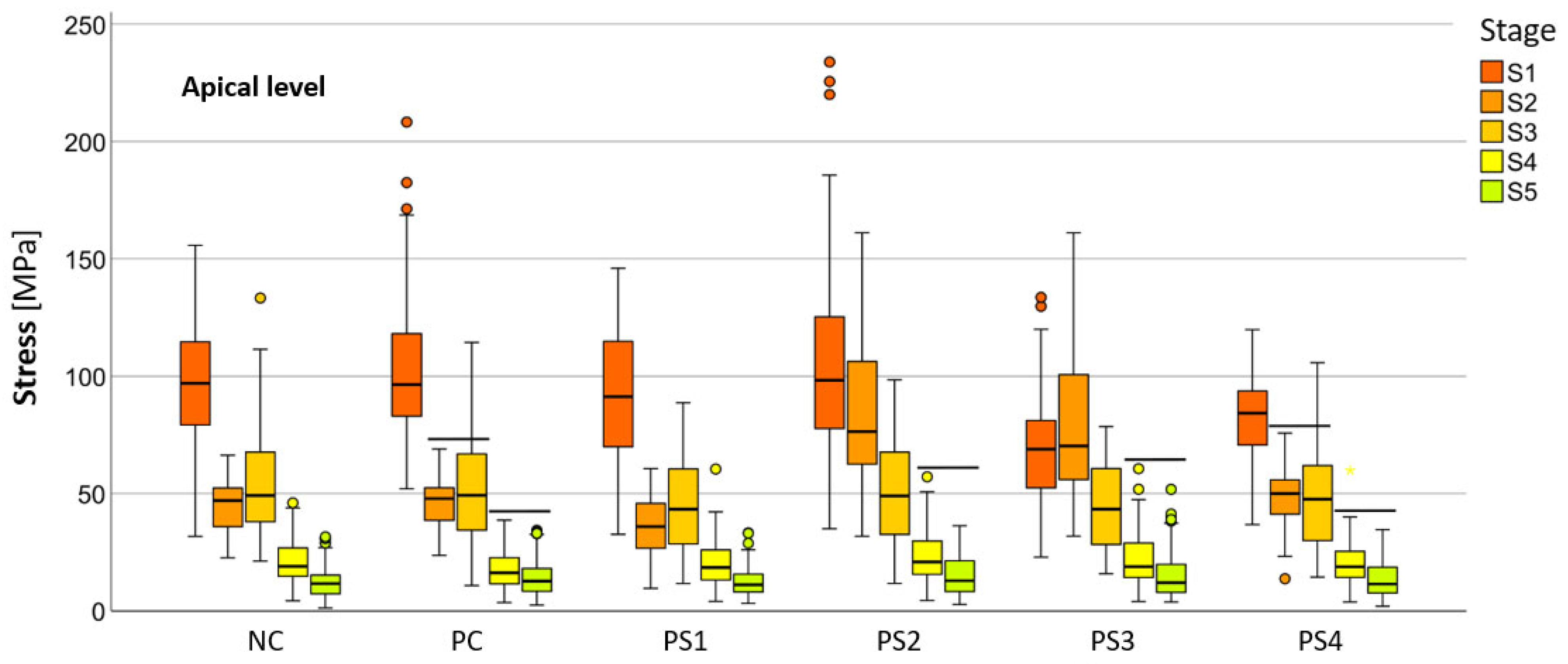
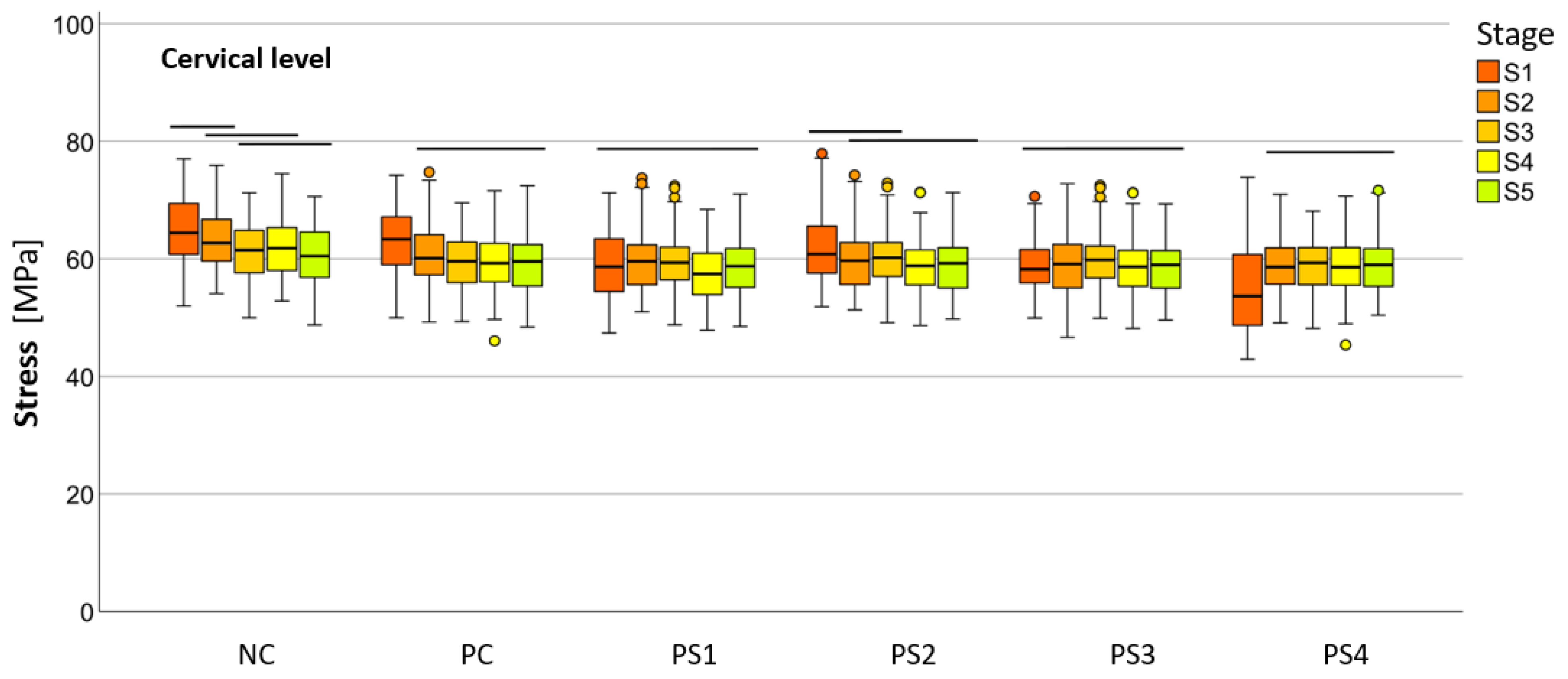
The mean HMH stress within the root dentin was statistically significantly higher at the cervical level in all stages, except in stage S1 and in models PS2 and PS3 in stage S2, where it was significantly higher at the apical level (p < 0.001 for all models, except stage S3 [PC model p < 0.005; NC model p < 0.008]) (Table 2). The PS4 model showed the lowest stress values at the cervical level in stages S1 (55.19 ± 1.70 MPa), S2 (58.78 ± 0.93 MPa), and S3 (58.84 ± 1.04 MPa), while the PS1 model showed the lowest values in stages S4 (57.48 ± −1.03 MPa) and S5 (58.81 ± 1.08 MPa). At the apical level, the lowest stress values were observed in the PS3 model at stage S1 (69.60 ± 5.03 MPa), in the PS1 model at stages S2 (35.99 ± 2.63 MPa), S3 (44.30 ± 4.07 MPa), and S5 (12.51 ± 1.42 MPa) and in the PC model at stage S4 (17.85 ± 1.88 MPa). Stress within the root dentin showed a decreasing trend from stage S1 to stage S5 in most models at both levels. However, notable deviations were observed in the PS1 and NC models, where stage S2 at the apical level exhibited significantly lower stress than stage S3, in the PS3 model, where stage S1 at the apical level exhibited significantly lower stress than stage S2, and in the PS4 model, where stage S1 at the cervical level exhibited significantly lower stress than stage S2 and all subsequent stages. In general, no statistically significant differences were observed between the post systems at both levels for all stages, especially for the stages with the highest clinical relevance (S4 and S5) (Table 3).
The numerical values of the equivalent stresses according to the HMH theory within the root dentin, categorized into five stages of root development at two levels for all materials, are shown in Figure 4 and Figure 5.
Figure 6, Figure 7, Figure 8, Figure 9, Figure 10 and Figure 11 show sagittal cross-sections of the individual finite element analysis models with respect to the different stages of root development.

4. Discussion

There are many FEM studies that have evaluated endodontically weakened or immature maxillary incisors, but given the different protocols and treatments, the results of this study could only be compared with a few.
HMH stress was highest at the cervical level in most models, which is consistent with findings in the literature [23,26,29,30,31]. Jain et al. [31] reported maximum stress concentration in the cervical third of the root when simulating two no-post systems with a decrease in stress intensity from the cervical to the apical third. Eram et al. [26] observed higher stresses in the cervical portion of the materials used for the apical stop simulating the third stage of Cvek on an extracted adult incisor before CBCT examination. Similarly, Gurbuz et al. [29] reported the highest stresses in the cervical region of the root dentin, but in primary central incisors when different composite materials were used to form a short post and core. Their maximum stress values (83.0–85.8 MPa) were higher than those observed in our study (59.44–63.00 MPa) at the same level. This discrepancy may be attributed to differences in anatomical properties, particularly the much higher crown-to-root ratio in primary incisors and the shorter length of the composite material used for the post function, as well as poorer mechanical properties compared to FRC posts. In the study by Kumar et al. [30], the highest stress areas in FRC posts were at the inner proximal wall at the cervical level (15.33–15.37 MPa) and at the inner apical side of the metal posts (14.92–15.01 MPa) in FEA models of IMI with completed root development, and therefore we could only compare their values with our S5 stage (58.81 MPa at the cervical level and 12.51 MPa at the apical level for the PS1 model and 58.66 MPa at the cervical level and 15.17 MPa at the apical level for the PS3 model). It is noteworthy that their values at the apical level are the same as ours, while those at the cervical level are 3 to 4 times higher. This could be due to differences in the approach to modeling the immature maxillary incisor. In their study, the FEA model was created by scanning the extracted maxillary incisor of an adult, whereas in our study, the FEA model was developed from CBCT data of an 11-year-old child. This highlights the methodological challenges of modeling the immature maxillary central incisor and the ethical considerations of evaluating therapeutic procedures in an in vivo model.
Manaktala et al. [50] investigated the stress distribution in endodontically treated maxillary incisors with external cervical resorptions restored with mineral trioxide aggregate (MTA) and Biodentine. The maximum stresses were found in the cervical region of the dentin and were also similar between intact and restored incisors, as observed in our study. This suggests that the cervical region remains a critical point for stress concentration, regardless of whether the tooth is intact or restored, highlighting the need for careful evaluation of the long-term durability of restorative materials in this area. Hassouneh et al. [23] assessed different therapeutic approaches in non-vital IMI at the third stage of Cvek and also reported the highest stress concentration in the cervical area. Their values for stress concentration in the glass fiber post (GFP) model (64.1 MPa) were comparable to those in our PS1 model in S3 stage (59.3 MPa). In addition, their intact model exhibited the highest stress values (90.3 MPa) similar to our NC model in S3 stage (61.4 MPa). However, in contrast to our findings, they reported that the use of GFP improved biomechanical performance. The larger deviations in stress values could be attributed to the higher load (240 N) used to simulate occlusal forces in their study.
Based on the above-mentioned studies on maxillary central incisors following post-endodontic treatment, it can be concluded that the highest stress concentrations typically occur in the cervical region, regardless of different modeling techniques or the stage of root development. However, there is lack of data in the literature regarding the very early stages of root development. In our study, the stress concentrations in these early stages were highest in the apical region, likely due to more pronounced change in the thickness of the root dentin. We believe that future studies will further validate the observed pattern of stress distribution. In addition, differences in geometric modeling between studies resulted in stress values that were not always directly comparable, emphasizing the need for a standardized protocol.
Comparison of stress concentrations in root dentin between developmental stages within each group in our study showed a decrease from S1 to S5, as expected given the progressive thickening of the dentin wall at each subsequent stage. The deviations observed in stages S2 and S3 can be attributed to differences in the modeling of these stages based on our pilot study [42]. In particular, a value corresponding to two-thirds of root development was used for stage S2, whereas a range between S2 and S4 (more than two-thirds of development) was applied for stage S3, which may have contributed to greater variability in the modeling.
A comparison between the model with intact tooth and the model with gutta-percha filling revealed no significant differences in the stress distribution within the root dentin. This finding supports the assumption that endodontic treatment alone has no significant influence on the stability of the tooth, consistent with previous reports in the literature [51,52]. Furthermore, the comparison between the gutta-percha model and the models with post retention in stages S4 and S5, where such restorations are most commonly used clinically, showed no significant differences in the stress concentrations at the either cervical or apical level of the root dentin. These results support a minimally invasive post-endodontic approach and emphasize the need to carefully evaluate the clinical benefits of intracanal retention. Alshabib et al. [7] observed an increasing trend towards the use of endocrown restorations and extended onlays that do not rely on a post-core system, instead utilizing a box-shaped preparation to enhance retention of the crowns or onlays. The long-standing debate among clinicians about the necessity of posts, often formulated as “To post or not to post?” [14,53,54,55], remains unresolved, especially in young permanent teeth, where clinical research is inherently challenging. In this context, finite element analysis can be a valuable tool to support the selection of appropriate therapeutic strategies [56,57].
All root development models restored with the GC Fiber Post system showed lower stress concentrations within the root dentin at the apical level compared to those restored with RelyX Post, suggesting that the geometry of the GC Fiber Post, which has a cylindrical base coronally and a tapered end apically, more closely follows the natural anatomy of the tooth [12,58,59]. Nevertheless, post placement did not reduce root dentin stress. Anthrayose et al. [60], using a similar protocol that simulated only Cvek stage 3, also reported no significant differences between the immature intact model (106.18 MPa) and the immature models subjected to apexification or revascularization (114.23 MPa), findings that are consistent with the results of our models.
No major deviations were observed in metal posts compared to esthetic post systems, which is in contrast to reports in the literature [61,62,63]. Interestingly, some FEM studies have shown that higher stresses occur in glass fiber-reinforced composite post than in a metal post [27,30]. Furthermore, it should be considered that cyclic mechanical loading would provide a more appropriate approach for evaluating the long-term performance of metal posts, as it would likely result in significantly higher stress concentrations. This consideration is particularly relevant given the weaker bond strength between metal posts and composite cement, which increases the risk of irreparable root fractures extending below the bone level, caused by the high modulus of elasticity [53,64]. Notably, in all models with metal posts, stress accumulation was consistently observed in the middle third of the post on the oral side, a result that was not seen in any of the groups with esthetic post systems.
The comparison between the customized (PS4 model) and the prefabricated aesthetic post system (PS1 model) showed insignificant differences in the stress concentration within the root dentin, except in the S1 stage at the apical level, which showed significantly lower values in the PS4 model. The Everstick Post does not require additional removal of tooth structure and, unlike other aesthetic posts, features individualized geometry that adapts to the shape of the canal. Furthermore, as a relatively new system, the mechanical properties required for an accurate FEM analysis have not yet been thoroughly investigated in the literature. Gomes et al. [65] pointed out the possible unreliability of FEM results if inappropriate values for the material properties are used. We believe that further research is needed to obtain more accurate values for mechanical properties of the Everstick post system and to update the data for other materials as well, recognizing that this process requires complex and time-consuming laboratory techniques.
In summary, both hypotheses were confirmed, except at the earliest stage of root development, where the higher HMH stress was at the apical level.
This study has some limitations. A single CBCT was used for this study to obtain a personalized model of a tooth with completed root development, while the stages of root development and therapeutic procedures were modeled using the literature data [66] and dentin wall measurements on intraoral radiographs [42]. Better model properties would be achieved with a CBCT that follows the growth and development of teeth from the same patient, which would be highly ethically questionable in children. Since all model components were defined as linearly elastic and isotropic, the numerical simulation did not fully replicate the real in vivo conditions of structures and dental materials. A perfect adhesive bond between the materials was assumed. The mechanical properties of the structures and materials used were taken from a large number of published studies. Additional tests are required to obtain more accurate values. Finally, there were no data in the literature with which our results could be fully compared, as no single study used the same protocol for modeling root immaturity, nor were all stages of root development of the maxillary central incisor evaluated. Therefore, our results represent a special contribution to this field of science.
Future research in this area should focus on the clinical validation of the proposed modeling protocol, alongside the development of more advanced and physiologically accurate material models. In addition, ongoing efforts to reach a standardized consensus on modeling protocols are essential. Furthermore, a more in-depth investigation into the mechanical properties of the materials is required.
It is also important to emphasize that CBCT scans should not be performed solely for research purposes unless they are part of a clinically justified and previously indicated diagnostic or therapeutic procedure. In summary, strict adherence to ethical and legal standards is essential when using CBCT imaging in dental studies involving pediatric patients.

5. Conclusions

Despite the limitations of this study, it can be concluded that the greatest stress in an immature maxillary central incisor occurred at the cervical level, except during the early stage of root development. Post placement did not reduce root dentin stress.
The Everstick Post was found to be the most suitable material in the early stages of root development, while the GC Fiber Post was found in the later stages, at the cervical level. The post design with a cylindrical coronal base and a tapered apical end demonstrated better performance when selecting an aesthetic prefabricated root canal post compared to a fully conical post design. In addition, the metal post did not result in a significantly higher development of HMH stresses within the root dentin.

Author Contributions

Conceptualization, P.B.S. and H.J.; methodology, P.B.S., J.L.H. and N.B.; resources, T.J.L. and J.L.H.; formal analysis, N.B., D.T. and T.B.R.; investigation, P.B.S.; data curation, P.B.S., T.B.R. and D.T.; writing—original draft preparation, P.B.S.; writing—review and editing, H.J., K.G., J.L.H. and N.B.; supervision, H.J., K.G. and T.J.L. All authors have read and agreed to the published version of the manuscript.

Funding

This research received no external funding.

Institutional Review Board Statement

This study was conducted in accordance with the Declaration of Helsinki. Ethical approval for the conduct of this study in the preparation of the dissertation was obtained from the Ethics Committee of University of Zagreb, School of Dental Medicine (No. 03-3160-1122).

Informed Consent Statement

We confirm that written informed consent was obtained for the use of CBCT data in this study.

Data Availability Statement

All the data used in this study are available on request from the corresponding author.

Conflicts of Interest

The authors declare no conflicts of interest.

Abbreviations

The following abbreviations are used in this manuscript:
ANOVAAnalysis of variance
CBCTCone beam computed tomography
FEAFinite element analysis
FEMFinite element method
FRCFiber-reinforced composite
GFPGlass fiber post
HMNHencky–von Mises
IMIImmature maxillary incisor
NCNegative control group—intact maxillary incisor
PCPositive control group with only the gutta-percha filling
PS1Post system with GC Fiber Post
PS2Post system with RelyX Post
PS3Post system with metal post Unimetric
PS4Post system with GC everStick Post
S1Stage with half of the root development
S2Stage with three quarters of root development
S3Stage with more than three quarters of root development
S4Stage with fully developed root with open apex
S5Stage with fully developed root with closed apex

References

  1. Mayya, A.; Bhandary, S.; Kolakemar, A.; George, A.M. Management of necrotic immature permanent maxillary incisors using a modified technique of revascularisation. BMJ Case Rep. 2021, 17, e240203. [Google Scholar] [CrossRef] [PubMed]
  2. Jadhav, K.; Vaidya, M.J.; Hegde, V.; Kawle, S. Management of non-vital immature teeth: A review. IOSR J. Dent. Med. Sci. 2021, 20, 35–40. [Google Scholar] [CrossRef]
  3. Cadenaro, M.; Josic, U.; Maravić, T.; Mazzitelli, C.; Marchesi, G.; Mancuso, E.; Breschi, L.; Mazzoni, A. Progress in dental adhesive materials. J. Dent. Res. 2023, 102, 254–262. [Google Scholar] [CrossRef] [PubMed]
  4. Mannocci, F.; Bitter, K.; Sauro, S.; Ferrari, P.; Austin, R.; Bhuva, B. Present status and future directions: The restoration of root filled teeth. Int. Endod. J. 2022, 55, 1059–1084. [Google Scholar] [CrossRef]
  5. de Andrade, G.; Saavedra, G.; Augusto, M.; Alfonzo, G.; Brandão, H.; Tribst, J.; Dal Piva, A. Post-endodontic restorative treatments and their mechanical behavior: A narrative review. Dent. Rev. 2023, 3, 100067. [Google Scholar] [CrossRef]
  6. Yang, A.; Lamichhane, A.; Xu, C. Remaining coronal dentin and risk of fiber-reinforced composite post-core restoration failure: A meta-analysis. Int. J. Prosthodont. 2015, 28, 258–264. [Google Scholar] [CrossRef]
  7. Alshabib, A.; Althaqafi, K.A.; AlMoharib, H.S.; Mirah, M.; AlFawaz, Y.F.; Algamaiah, H. Dental fiber-post systems: An in-depth review of their evolution, current practice, and future directions. Bioengineering 2023, 10, 551. [Google Scholar] [CrossRef]
  8. Suni, A.O.; Lassila, L.V.J.; Tuokko, J.K.; Garoushi, S.; Vallittu, P.K. Adhesion of individually formed fiber post adhesively luted with flowable short fiber composite. Biomater. Investig. Dent. 2023, 10, 2209593. [Google Scholar] [CrossRef]
  9. Dikbas, I.; Tanalp, J. An overview of clinical studies on fiber post systems. Sci. World J. 2013, 2013, 171380. [Google Scholar] [CrossRef]
  10. Parčina, I.; Baraba, A. Esthetic intracanal posts. Acta Stomatol. Croat. 2016, 50, 143–150. [Google Scholar] [CrossRef]
  11. de Morais, D.C.; Butler, S.; Santos, M.J.M.C. Current insights on fiber posts: A narrative review of laboratory and clinical studies. Dent. J. 2023, 11, 236. [Google Scholar] [CrossRef] [PubMed]
  12. Bonchev, A.; Radeva, E.; Tsvetanova, N. Fiber reinforced composite posts-A review of literature. Int. J. Sci. Res. 2017, 6, 1887–1893. [Google Scholar] [CrossRef]
  13. Amizic, I.P.; Baraba, A.; Ionescu, A.C.; Brambilla, E.; Van Ende, A.; Miletic, I. Bond strength of individually formed and prefabricated fiber-reinforced composite posts. J. Adhes. Dent. 2019, 21, 557–565. [Google Scholar] [CrossRef]
  14. de Carvalho, M.A.; Lazari, P.C.; Gresnigt, M.; Del Bel Cury, A.A.; Magne, P. Current options concerning the endodontically-treated teeth restoration with the adhesive approach. Braz. Oral. Res. 2018, 32, e74. [Google Scholar] [CrossRef]
  15. Nair, K.R.; Geetha, P.; Veedu, R.; George, E.; Joseph, M. Management of an oblique crown fracture of an anterior tooth with a missed coronal segment: A case report. Conserv. Dent. Endod. J. 2019, 4, 21–24. [Google Scholar] [CrossRef]
  16. Cvek, M. Prognosis of luxated non-vital maxillary incisors treated with calcium hydroxide and filled with gutta-percha. A retrospective clinical study. Endod. Dent. Traumatol. 1992, 8, 45–55. [Google Scholar] [CrossRef]
  17. Andreasen, J.O.; Farik, B.; Munksgaard, E.C. Long-term calcium hydroxide as a root canal dressing may increase risk of root fracture. Dent. Traumatol. 2002, 18, 134–137. [Google Scholar] [CrossRef]
  18. Al-Jundi, S.H. Type of treatment, prognosis, and estimation of time spent to manage dental trauma in late presentation cases at a dental teaching hospital: A longitudinal and retrospective study. Dent. Traumatol. 2004, 20, 1–5. [Google Scholar] [CrossRef]
  19. Rodd, H.; Noble, F. Psychosocial impacts relating to dental injuries in childhood: The bigger picture. Dent. J. 2019, 7, 23. [Google Scholar] [CrossRef]
  20. Shivakumar, S.; Kudagi, V.S.; Talwade, P. Applications of finite element analysis in dentistry: A review. J. Int. Oral. Health 2021, 13, 415–422. [Google Scholar] [CrossRef]
  21. Trivedi, S. Finite element analysis: A boon to dentistry. J. Oral Biol. Craniofacial Res. 2014, 4, 200–203. [Google Scholar] [CrossRef] [PubMed]
  22. Soares, C.J.; Rodrigues, M.P.; Faria-E-Silva, A.L.; Santos-Filho, P.C.F.; Veríssimo, C.; Kim, H.C.; Versluis, A. How biomechanics can affect the endodontic treated teeth and their restorative procedures? Braz. Oral. Res. 2018, 32, e76. [Google Scholar] [CrossRef] [PubMed]
  23. Hassouneh, L.; Matoug-Elwerfelli, M.; Al-Omari, T.; Setzer, F.C.; Nagendrababu, V. Assessment of biomechanical behavior of immature non-vital incisors with various treatment modalities by means of three-dimensional quasi–static finite element analysis. Sci. Rep. 2023, 13, 17491. [Google Scholar] [CrossRef]
  24. Santos Filho, P.C.F.; Soares, P.V.; Reis, B.R.; Veríssimo, C.; Soares, C.J. Effects of threaded post placement on strain and stress distribution of endodontically treated teeth. Braz. Oral. Res. 2013, 27, 305–310. [Google Scholar] [CrossRef]
  25. Ferrari, M.; Sorrentino, R.; Zarone, F.; Apicella, D.; Aversa, R.; Apicella, A. Non-linear viscoelastic finite element analysis of the effect of the length of glass fiber posts on the biomechanical behaviour of directly restored incisors and surrounding alveolar bone. Dent. Mater. J. 2008, 27, 485–498. [Google Scholar] [CrossRef]
  26. Eram, A.; Zuber, M.; Keni, L.G.; Kalburgi, S.; Naik, R.; Bhandary, S.; Amin, S.; Badruddin, I.A. Finite element analysis of immature teeth filled with MTA, Biodentine and Bioaggregate. Comput. Methods Programs Biomed. 2020, 190, 105356. [Google Scholar] [CrossRef]
  27. Nokar, S.; Bahrami, M.; Mostafavi, A.S. Comparative evaluation of the effect of different post and core materials on stress distribution in radicular dentin by three-dimensional finite element analysis. J. Dent. 2018, 15, 69–78. [Google Scholar] [PubMed Central]
  28. Okamoto, K.; Ino, T.; Iwase, N.; Shimizu, E.; Suzuki, M.; Satoh, G.; Ohkawa, S.; Fujisawa, M. Three-dimensional finite element analysis of stress distribution in composite resin cores with fiber posts of varying diameters. Dent. Mater. J. 2008, 27, 49–55. [Google Scholar] [CrossRef]
  29. Gurbuz, T.; Sengul, F.; Altun, C. Finite element stress analysis of short-post core and over restorations prepared with different restorative materials. Dent. Mater. J. 2008, 27, 499–507. [Google Scholar] [CrossRef]
  30. Kumar, P.; Rao, R.N. Three-dimensional finite element analysis of stress distribution in a tooth restored with metal and fiber posts of varying diameters: An in-vitro study. J. Conserv. Dent. 2015, 18, 100–104. [Google Scholar] [CrossRef]
  31. Jain, P.; Lakshmi, S.; Vigneshwaran, S. Endodontically treated teeth with custom cast post and core and metal ceramic crowns restoration: A case report. Int. J. Appl. Dent. Sci. 2021, 7, 16–19. [Google Scholar] [CrossRef]
  32. Madfa, A.A.; Al-Hamzi, M.A.; Al-Sanabani, F.A.; Al-Qudaimi, N.H.; Yue, X.G. 3D FEA of cemented glass fiber and cast posts with various dental cements in a maxillary central incisor. Springerplus 2015, 4, 598. [Google Scholar] [CrossRef] [PubMed]
  33. Oskui, I.Z.; Hashemi, A.; Jafarzadeh, H.; Kato, A. Finite element investigation of human maxillary incisor under traumatic loading: Static vs dynamic analysis. Comput. Methods Programs Biomed. 2018, 155, 121–125. [Google Scholar] [CrossRef] [PubMed]
  34. Jayasudha, K.; Hemanth, M.; Baswa, R.; Raghuveer, H.P.; Vedavathi, B.; Hegde, C. Traumatic impact loading on human maxillary incisor: A Dynamic finite element analysis. J. Indian Soc. Pedod. Prev. Dent. 2015, 33, 302–306. [Google Scholar] [CrossRef]
  35. Ruse, N.D. Propagation of erroneous data for the modulus of elasticity of periodontal ligament and gutta percha in FEM/FEA papers: A story of broken links. Dent. Mater. 2008, 24, 1717–1719. [Google Scholar] [CrossRef]
  36. Ausiello, P.; Dal Piva, A.; Borges, A.; Lanzotti, A.; Zamparini, F.; Epifania, E.; Tribst, J.P.M. Effect of shrinking and no shrinking dentine and enamel replacing materials in posterior restoration: A 3D-FEA study. Appl. Sci. 2021, 11, 11052215. [Google Scholar] [CrossRef]
  37. Matuda, A.G.N.; Silveira, M.P.M.; de Andrade, G.S.; Piva, A.M.O.D.; Tribst, J.P.M.; Borges, A.L.S.; Testarelli, L.; Mosca, G.; Ausiello, P. Computer aided design modelling and finite element analysis of premolar proximal cavities restored with resin composites. Materials 2021, 14, 2366. [Google Scholar] [CrossRef]
  38. Richert, R.; Robinson, P.; Viguie, G.; Farges, J.C.; Ducret, M. Multi-fiber-reinforced composites for the coronoradicular reconstruction of premolar teeth: A finite element analysis. BioMed Res. Int. 2018, 2018, 4302607. [Google Scholar] [CrossRef]
  39. Kedia, S.; Narayana, I.; Shetty, R.V.; Shukla, E.; Kalasaiah, D. Stress distribution in teeth restored with different posts using single or multi-post technique: A three-dimensional finite element analysis. Cons. Dent. Endod. J. 2022, 7, 1–6. [Google Scholar] [CrossRef]
  40. Aggarwal, S.; Garg, V. Finite element analysis of stress concentration in three popular brands of fiber posts systems used for maxillary central incisor teeth. J. Conserv. Dent. 2011, 14, 293–296. [Google Scholar] [CrossRef]
  41. Diana, H.H.; Oliveira, J.S.; Ferro, M.C.L.; Silva-Sousa, Y.T.C.; Gomes, É.A. Stress distribution in roots restored with fiber posts and an experimental dentin post: 3D-FEA. Braz. Dent. J. 2016, 27, 223–227. [Google Scholar] [CrossRef] [PubMed]
  42. Sojčić, P.B.; Horina, J.L.; Lulić, T.J.; Bočkaj, N.; Jurić, H. Measurement of the dentin wall thickness of the maxillary central incisor in relation to the stage of root development: A pilot study. Acta Stomatol. Croat. 2023, 57, 206–215. [Google Scholar] [CrossRef] [PubMed]
  43. 3M RelyX Fiber Post 3D Glass Fiber Post: Technical Data Sheet. Available online: https://multimedia.3m.com/mws/media/1146520O/3m-relyx-fiber-post-3d-glass-fiber-post-technical-data-sheet.pdf (accessed on 28 February 2024).
  44. GC Fiber Post—IFU. Available online: https://www.gc.dental/america/sites/america.gc.dental/files/products/downloads/gcfiberpost/ifu/gc-fiber-post-ifu.pdf (accessed on 28 February 2024).
  45. Dentsply Maillefer Product Catalogue. Available online: https://domdent.hu/wp-content/uploads/2023/10/DENTSPLY_MAILLEFER_2014_Product_Catalogue_Single_Page_HR_0714_EN.pdf (accessed on 28 February 2024).
  46. Agrawal, R.; Narang, S.; Ahmed, H.; Prasad, S.; Reddy, S.; Aila, S. Influence of occlusal bite forces on teeth with altered periodontal support: A three-dimensional finite element stress analysis. J. Pharm. Bioallied Sci. 2021, 13, S688–S691. [Google Scholar] [CrossRef] [PubMed]
  47. Nikam, D.; Milani, A.S. Physiological response of a natural central incisor tooth to various loading conditions: A 3D finite element study. Recent Prog. Mater. 2023, 5, 017. [Google Scholar] [CrossRef]
  48. Dorado, S.; Arias, A.; Jimenez-Octavio, J.R. Biomechanical modelling for tooth survival studies: Mechanical properties, loads and boundary conditions—A narrative review. Materials 2022, 15, 7852. [Google Scholar] [CrossRef]
  49. Verri, F.R.; Okumura, M.H.T.; Lemos, C.A.A.; Almeida, D.A.F.; de Souza Batista, V.E.; Cruz, R.S.; Oliveira, H.F.F.; Pellizzer, E.P. Three-dimensional finite element analysis of glass fiber and cast metal posts with different alloys for reconstruction of teeth without ferrule. J. Med. Eng. Technol. 2017, 41, 644–651. [Google Scholar] [CrossRef]
  50. Manaktala, M.; Taneja, S.; Bhalla, V.K. Stress distribution in endodontically treated external cervical resorption lesions restored with MTA and biodentine—A finite element analysis. J. Oral Biol. Craniofacial Res. 2024, 14, 415–422. [Google Scholar] [CrossRef]
  51. Peroz, I.; Blankenstein, F.; Lange, K.P.; Naumann, M. Restoring endodontically treated teeth with posts and cores-A review. Quintessence Int. 2005, 36, 737–746. [Google Scholar] [PubMed]
  52. Kalra, D.; Sukhija, D.; Rassawet, R.R.; Rani, D. A review on post and core. Sch. J. Dent. Sci. 2020, 07, 51–56. [Google Scholar] [CrossRef]
  53. Aurélio, I.L.; Fraga, S.; Rippe, M.P.; Valandro, L.F. Are posts necessary for the restoration of root filled teeth with limited tissue loss? A structured review of laboratory and clinical studies. Int. Endod. J. 2016, 49, 827–835. [Google Scholar] [CrossRef]
  54. Josic, U.; Radovic, I.; Juloski, J.; Beloica, M.; Popovic, M.; Alil, A.; Mandic, J.C. Can fiber-post placement reinforce structurally compromised roots? J. Adhes. Dent. 2020, 22, 409–414. [Google Scholar] [CrossRef] [PubMed]
  55. Fehrenbach, J.; de Soares, J.L.S.; do Nascimento Foly, J.C.S.; Miotti, L.L.; Münchow, E.A. Mechanical performance of endocrown restorations in anterior teeth: A systematic review and network meta-analysis. Dent. Mater. 2025, 41, 28–41. [Google Scholar] [CrossRef] [PubMed]
  56. Shetty, P.P.; Meshramkar, R.; Patil, K.N.; Nadiger, R.K. A finite element analysis for a comparative evaluation of stress with two commonly used esthetic posts. Eur. J. Dent. 2013, 7, 419–422. [Google Scholar] [CrossRef] [PubMed]
  57. Vikhe, D.M. Restoration of Endodontically Treated Teeth. In Clinical Concepts and Practical Management Techniques in Dentistry; Moolla, I., Ed.; IntechOpen: London, UK, 2021; Available online: https://www.intechopen.com/chapters/76852 (accessed on 21 February 2025).
  58. Goracci, C.; Ferrari, M. Current perspectives on post systems: A literature review. Aust. Dent. J. 2011, 56, 77–83. [Google Scholar] [CrossRef]
  59. Machado, J.; Almeida, P.; Fernandes, S.; Marques, A.; Vaz, M. Currently used systems of dental posts for endodontic treatment. Procedia Struct. Integr. 2017, 5, 27–33. [Google Scholar] [CrossRef]
  60. Anthrayose, P.; Nawal, R.R.; Yadav, S.; Talwar, S.; Yadav, S. Effect of revascularisation and apexification procedures on biomechanical behaviour of immature maxillary central incisor teeth: A three-dimensional finite element analysis study. Clin. Oral. Investig. 2021, 25, 6671–6679. [Google Scholar] [CrossRef]
  61. Almaroof, A.G.; Thyab, S.A.; Ali, A.H. Bond strength of a new Kevlar fiber-reinforced composite post with semi-interpenetrating polymer network (IPN) matrix. J. Clin. Exp. Dent. 2019, 11, e695–e700. [Google Scholar] [CrossRef]
  62. Li, J.; Parada, C.; Chai, Y. Cellular and molecular mechanisms of tooth root development. Development 2017, 144, 374–384. [Google Scholar] [CrossRef]
  63. Badami, V.; Ketineni, H.; Pb, S.; Akarapu, S.; Mittapalli, S.P.; Khan, A. Comparative evaluation of different post materials on stress distribution in endodontically treated teeth using the finite element analysis method: A systematic review. Cureus 2022, 14, e29753. [Google Scholar] [CrossRef]
  64. Jitumori, R.T.; Bittencourt, B.F.; Reis, A.; Gomes, J.C.; Gomes, G.M. Effect of root canal irrigants on fiber post bonding using self-adhesive composite cements. J. Adhes. Dent. 2019, 21, 537–544. [Google Scholar] [CrossRef]
  65. Gomes, E.A.; Diana, H.H.; Oliveira, J.S.; Silva-Sousa, Y.T.C.; Faria, A.C.L.; Ribeiro, R.F. Reliability of FEA on the results of mechanical properties of materials. Braz. Dent. J. 2015, 26, 667–670. [Google Scholar] [CrossRef] [PubMed]
  66. Brkić, H.; Dumančić, J.; Vodanović, M. Biologija i Morfologija Ljudskih Zuba; Naklada Slap: Zagreb, Croatia, 2016; p. 61. [Google Scholar]
Figure 1. Shematic illustration of post-endodontic treatment.
Materials 18 02305 g001
Figure 2. Shematic diagram of experimental groups.
Materials 18 02305 g002
Figure 3. Loading and boundary conditions.
Materials 18 02305 g003
Figure 4. Distribution of HMH stress between models divided into five stages of root development at the apical level (SPSS software package, version 25.0, IBM, Armonk, NY, USA).
Materials 18 02305 g004
Figure 5. Distribution of HMH stress with a smaller range on the Y-axis (0-100 MPa) between models divided into the five stages of root development at the cervical level (SPSS software package, version 25.0, IBM, Armonk, NY, USA).
Materials 18 02305 g005
Figure 6. PS1 model according to the root development stage—sagittal section.
Materials 18 02305 g006
Figure 7. PS2 model according to the root development stage—sagittal section.
Materials 18 02305 g007
Figure 8. PS3 model according to the root development stage—sagittal section.
Materials 18 02305 g008
Figure 9. PS4 model according to the root development stage—sagittal section.
Materials 18 02305 g009
Figure 10. PC model according to the developmental stage of the root—sagittal section.
Materials 18 02305 g010
Figure 11. NC model according to the developmental stage of the root—sagittal section.
Materials 18 02305 g011
Table 1. Mechanical properties.
Structure/MaterialE, GPaν, -References
Enamel41.00.30[33]
Dentin18.60.31[33]
Periodontal ligament (PDL)0.000670.45[34]
Pulp0.002070.45[33]
Cortical bone13.40.26[33]
Trabecular bone0.3450.36[33]
Gutapercha0.140.45[35]
Glass ionomer cement8.00.25[36]
Composite filling13.450.17[37]
Composite cement8.30.28[38]
PS1 (GC Fiberpost)40.00.25[39]
PS2 (RelyX post 3M ESPE)37.50.26[40,41]
PS3 (Metal post Unimetric 1.0 Dentsply)110.00.32[38]
PS4 (EverStick Post GC)15.00.18[39]
Table 2. Mean value of Hencky–von Mises (HMH) stress [MPa] ± 95% confidence interval and standard deviation (SD) at each stage of root development at the apical and cervical levels.
APICAL LEVEL
ModelS1S2S3S4S5
PS1Mean91.78 ± 6.1735.99 ± 2.6344.30 ± 4.0720.12 ± 2.1412.51 ± 1.42
SD 27.7511.8218.339.646.42
PS2Mean105.59 ± 8.7285.50 ± 6.8451.11 ± 4.5523.29 ± 2.4715.19 ± 1.95
SD 39.1930.7420.4711.138.78
PS3Mean69.60 ± 5.0380.84 ± 6.8744.53 ± 4.0821.73 ± 2.5315.17 ± 2.33
SD 22.6130.8718.3711.4010.50
PS4Mean82.83 ± 3.9548.43 ± 2.7347.95 ± 4.7120.24 ± 2.1414.08 ± 1.81
SD 17.7712.2921.199.638.17
PCMean103.44 ± 6.6846.10 ± 2.3252.49 ± 4.8917.85 ± 1.8814.40 ± 1.84
SD 30.0510.4522.018.468.28
NCMean97.95 ± 5.9544.96 ± 2.3954.28 ± 5.2021.25 ± 1.9612.67 ± 1.63
SD 26.7610.7723.368.827.35
CERVICAL LEVEL
ModelS1S2S3S4S5
PS1Mean59.06 ± 1.2859.70 ± 1.1159.35 ± 0.7957.48 ± 1.0358.81 ± 1.08
SD 5.775.014.974.654.88
PS2Mean61.60 ± 1.2560.12 ± 1.1459.89 ± 1.1458.79 ± 1.0258.98 ± 1.09
SD 5.635.165.144.594.91
PS3Mean58.77 ± 0.9659.29 ± 1.1359.64 ± 1.1158.62 ± 1.0858.66 ± 0.98
SD 4.345.115.024.874.42
PS4Mean55.19 ± 1.7058.78 ± 0.9358.84 ± 1.0458.74 ± 1.1158.87 ± 1.02
SD 7.674.194.685.034.58
PCMean63.00 ± 1.2460.51 ± 1.2059.64 ± 1.0859.44 ± 1.1459.50 ± 1.14
SD 5.585.424.855.125.14
NCMean64.88 ± 1.3363.12 ± 1.0661.42 ± 1.1362.07 ± 1.1360.68 ± 1.10
SD 6.014.775.125.114.96
Table 3. Results of univariate ANOVA comparing different types of materials between developmental stages at the apical and cervical level (Tukey HSD).
p-VALUE
APICAL LEVELCERVICAL LEVEL
ModelS1S2S3S4S5S1S2S3S4S5
PS1PS20.0250.0000.2990.3300.3250.0750.9950.9830.5431.000
PS30.0000.0001.0000.9090.3331.0000.9950.9990.6871.000
PS40.3370.0010.8741.0000.8420.0010.8480.9860.5861.000
PC0.0940.0190.1250.6970.7060.0000.9060.9990.1200.94
NC0.7350.0550.0290.9791.0000.0000.0000.0910.0000.141
PS2PS10.0250.0000.2990.3300.3250.0750.9950.9830.5431.000
PS30.0000.6830.3390.9191.0000.0310.8970.9991.0000.998
PS40.0000.0000.9290.3720.9590.0000.5240.7591.0001.000
PC0.9970.0000.9980.0070.9910.6660.9961.0000.9600.983
NC0.5200.0000.9270.7830.3970.0070.0020.3760.0000.224
PS3PS10.0000.0001.0000.9090.3331.0000.9950.9990.6871.000
PS20.0000.6830.3390.9191.0000.0310.8970.9991.0000.998
PS40.0360.0000.9030.9310.9620.0020.9870.9121.0001.000
PC0.0000.0000.1480.1330.9920.0000.6281.0000.8980.879
NC0.0000.0000.0361.0000.4060.0000.0000.2070.0000.087
PS4PS10.3370.0010.8741.0000.8420.0010.8480.9860.5861.000
PS20.0000.0000.9290.3720.9590.0000.5240.7591.0001.000
PS30.0360.0000.9030.9310.9620.0020.9870.9121.0001.000
PC0.0000.9770.7360.6511.0000.0000.2340.9110.9460.962
NC0.0100.8840.3830.9870.8940.0000.0000.0140.0000.169
PCPS10.0940.0190.1250.6970.7060.0000.9060.9990.1200.944
PS20.9970.0000.9980.0070.9910.6660.9961.0000.9600.983
PS30.0000.0000.1480.1330.9920.0000.6281.0000.8980.879
PS40.0000.9770.7360.6511.0000.0000.2340.9110.9460.962
NC0.8200.9990.9940.2540.7790.3380.0120.2080.0090.636
NCPS10.7350.0550.0290.9791.0000.0000.0000.0910.0000.141
PS20.5200.0000.9270.7830.3970.0070.0020.3760.0000.224
PS30.0000.0000.0361.0000.4060.0000.0000.2070.0000.087
PS40.0100.8840.3830.9870.8940.0000.0000.0140.0000.169
PC0.8200.9990.9940.2540.7790.3380.0120.2080.0090.636
Disclaimer/Publisher’s Note: The statements, opinions and data contained in all publications are solely those of the individual author(s) and contributor(s) and not of MDPI and/or the editor(s). MDPI and/or the editor(s) disclaim responsibility for any injury to people or property resulting from any ideas, methods, instructions or products referred to in the content.