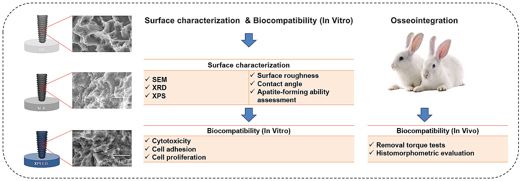
Improved Biocompatibility and Osseointegration of Nanostructured Calcium-Incorporated Titanium Implant Surface Treatment (XPEED®)

Abstract
1. Introduction
2. Materials and Methods
2.1. Surface Treatment of Samples
2.2. Surface Characterization
2.3. Cell Viability
2.3.1. Cell Cytotoxicity
2.3.2. Cell Proliferation
2.3.3. Cell Adhesion
2.4. Apatite-Forming Ability Assessment
2.5. Animal Tests and Surgical Procedures [31,41]
2.6. Removal Torque Tests [41,42]
2.7. Histomorphometric Evaluation: BIC and Bone Area (BA) Tests [31,41]
2.8. SPSS One-Way Analysis Test
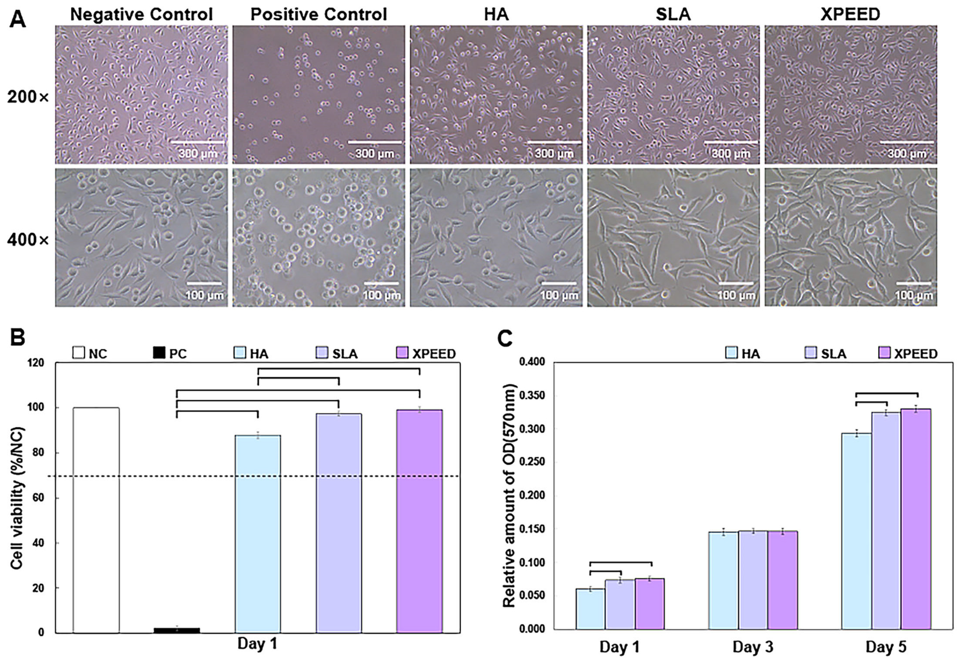
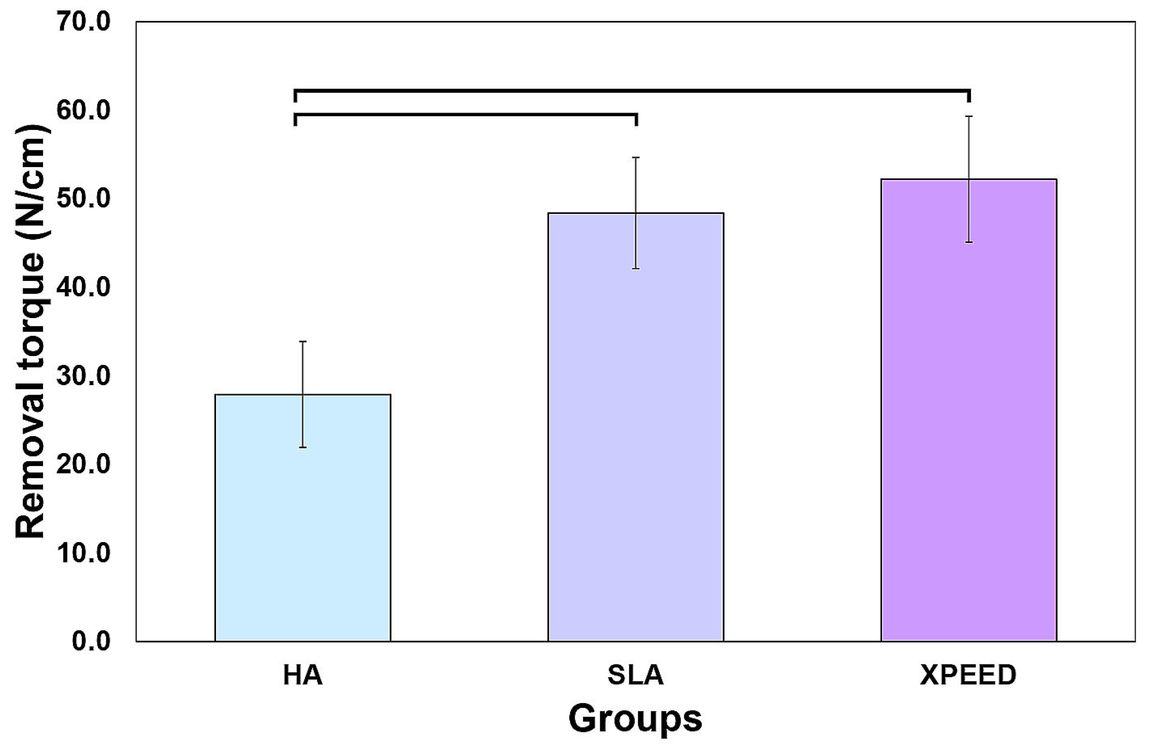
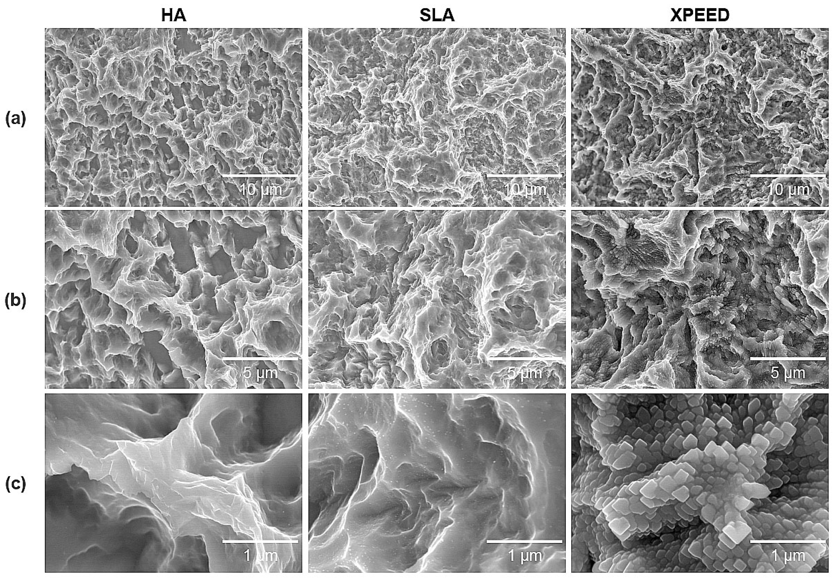
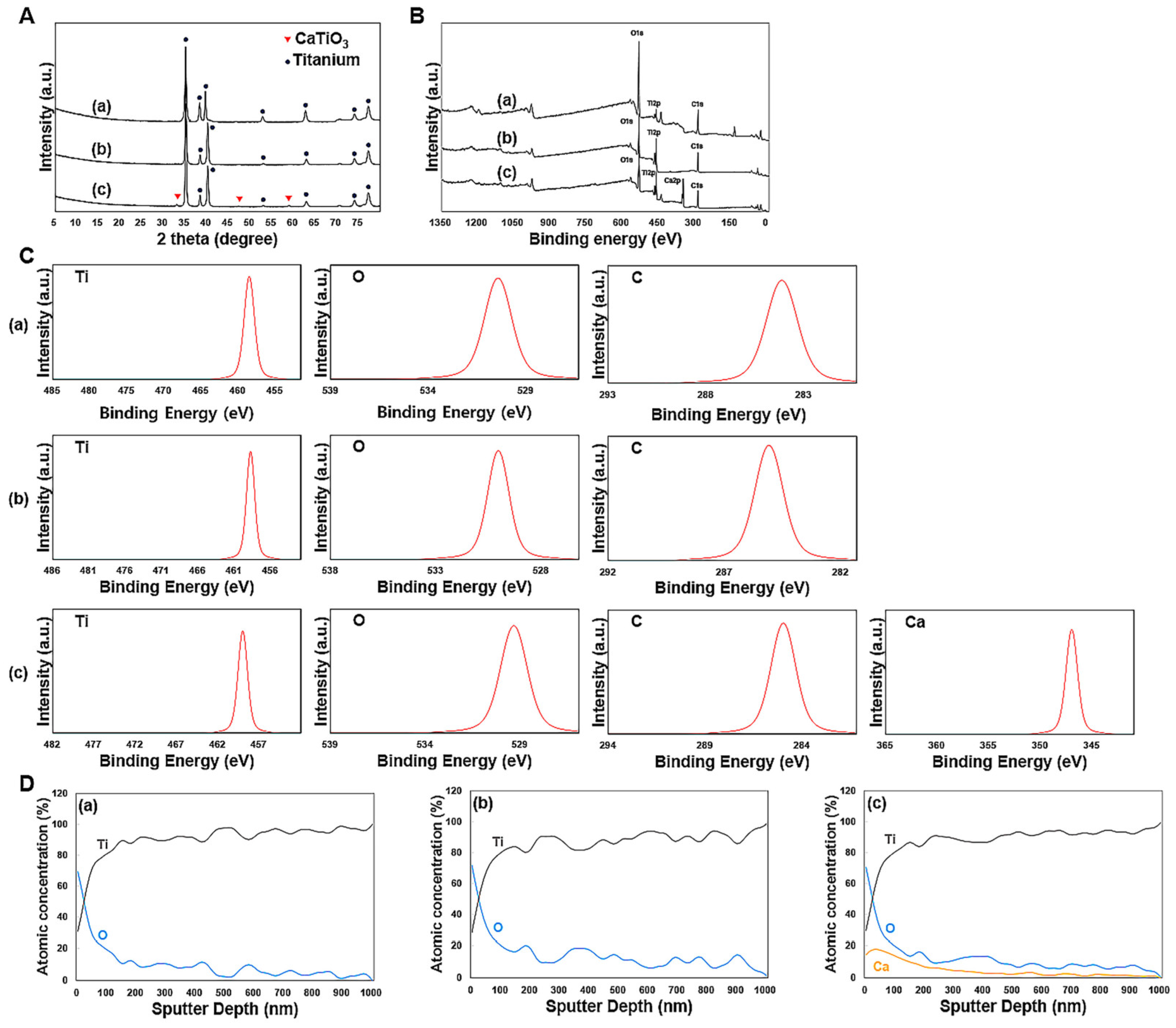
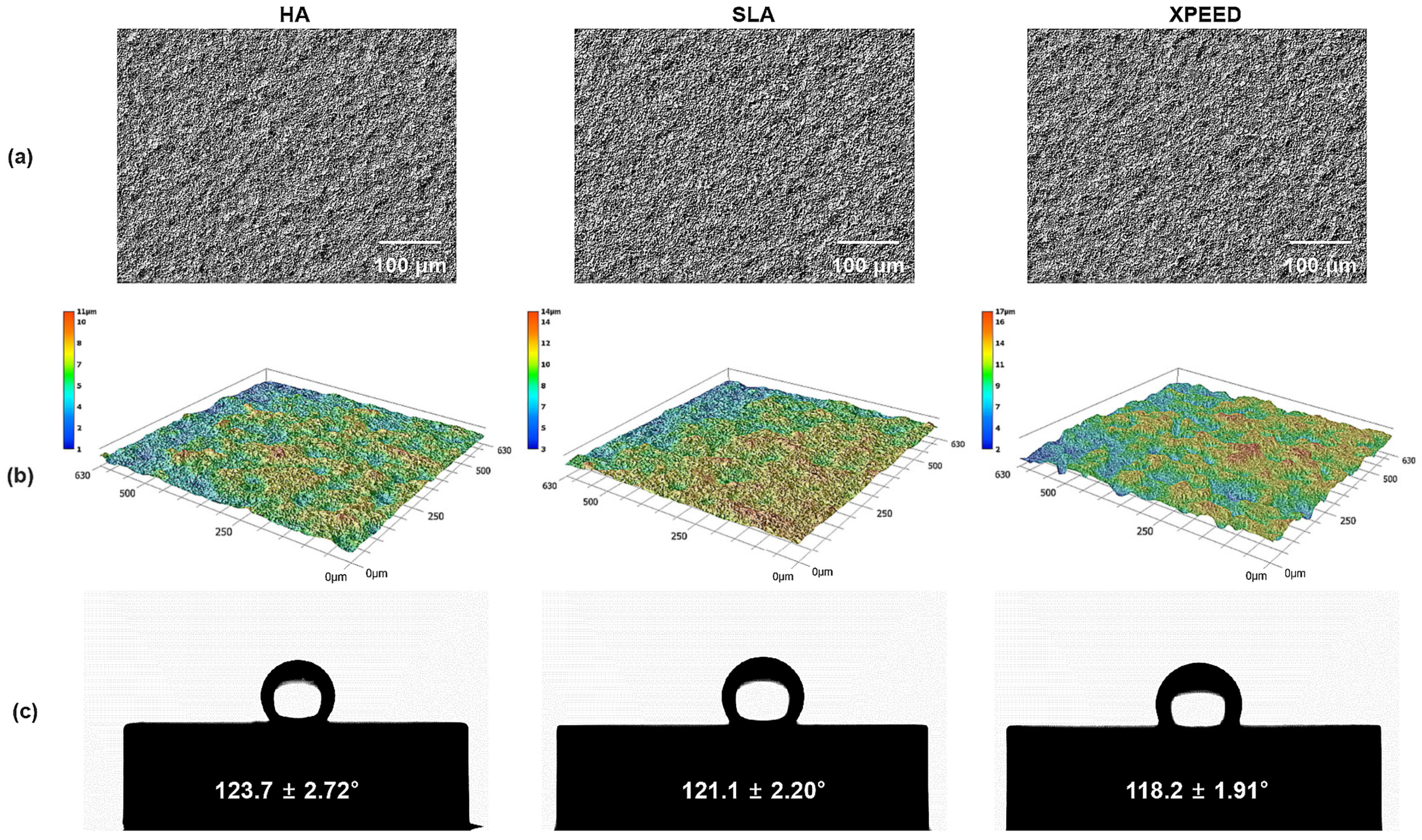
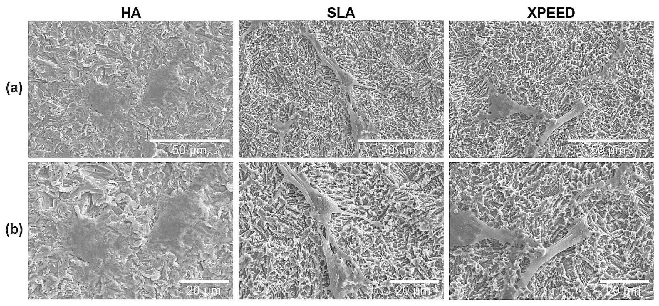
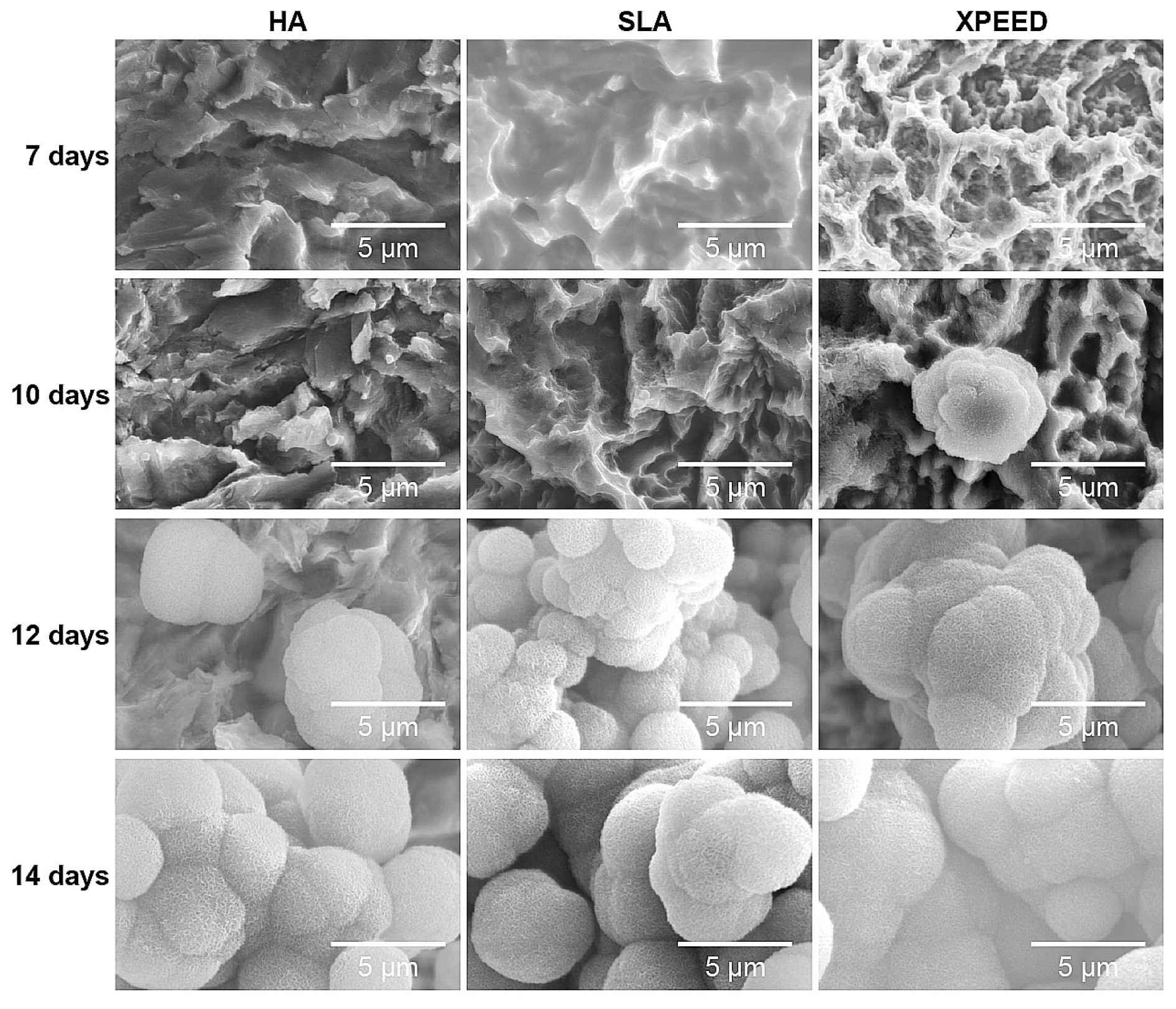
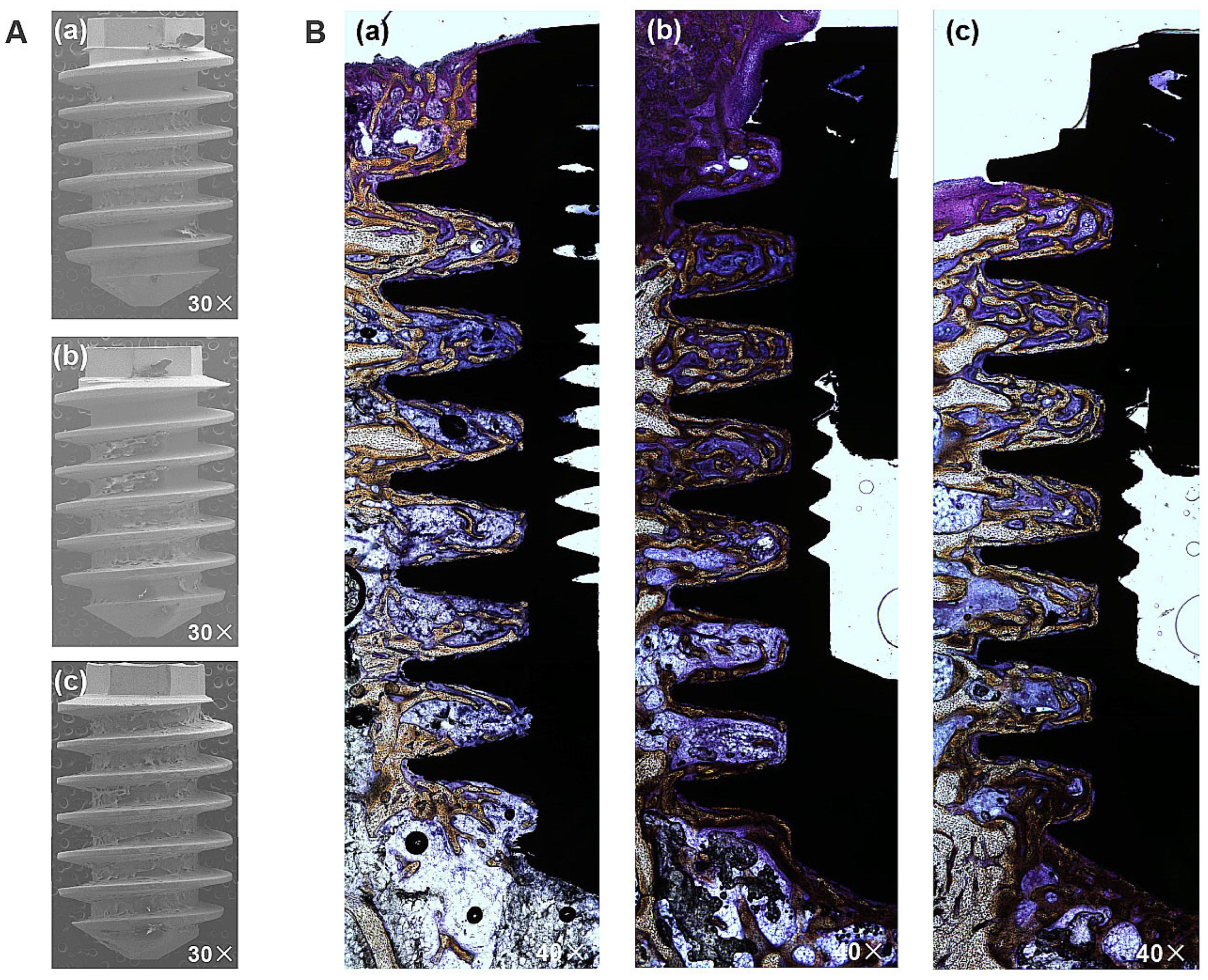
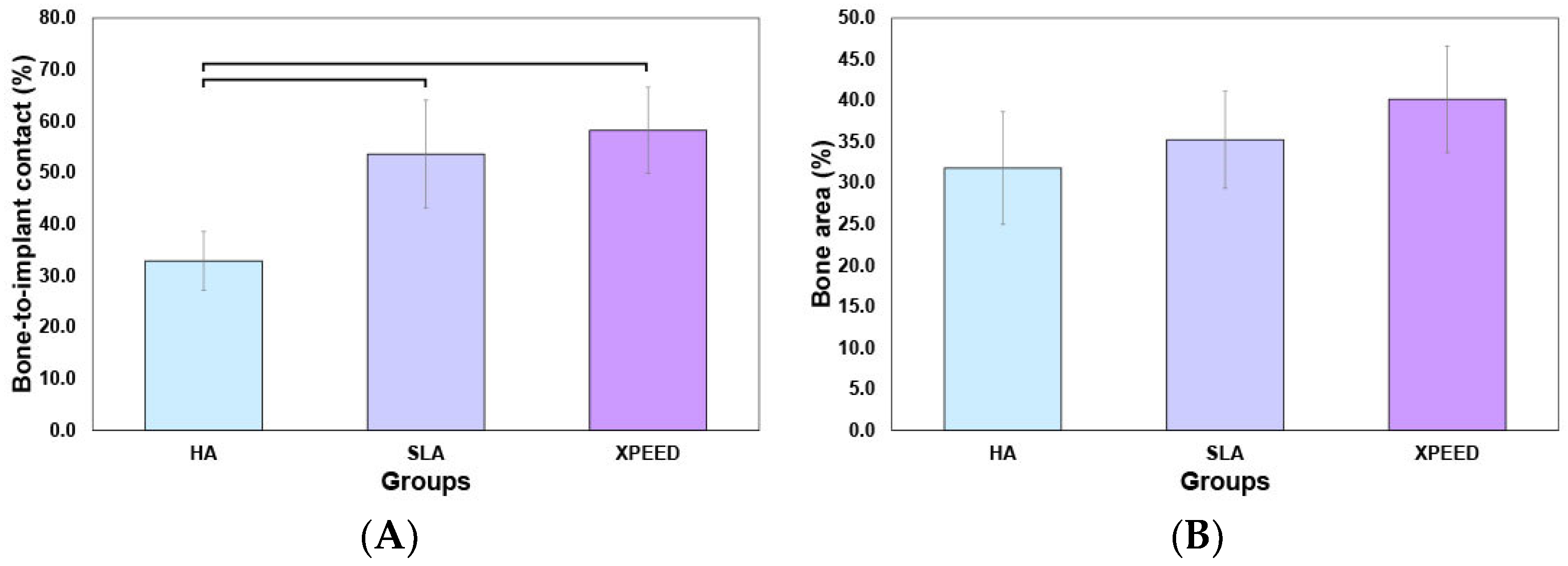
3. Results and Discussion
4. Conclusions
Author Contributions
Funding
Institutional Review Board Statement
Informed Consent Statement
Data Availability Statement
Acknowledgments
Conflicts of Interest
References
- Aziz-Kerrzo, M.; Conroy, K.G.; Fenelon, A.M.; Farrell, S.T. Electrochemical studies on the stability and corrosion resistance of titanium-based implant materials. Biomaterials 2001, 22, 1531–1539. [Google Scholar] [CrossRef] [Scilit]
- Choi, M.J.; Min, B.K.; Hong, M.H.; Lee, H.J.; Son, J.S.; Kwon, T.Y. Influence of oxidative etching solution temperatures on the surface roughness and wettability of a titanium alloy. J. Nanosci. Nanotechnol. 2019, 19, 1044–1047. [Google Scholar] [CrossRef] [Scilit] [PubMed]
- Schuler, M.; Trentin, D.; Textor, M.; Tosatti, S.G. Biomedical interfaces: Titanium surface technology for implants and cell carriers. Nanomedicine 2006, 1, 449–463. [Google Scholar] [CrossRef] [Scilit] [PubMed]
- Adell, R. Tissue integrated prostheses in clinical dentistry. Int. Dent. J. 1985, 35, 259–265. [Google Scholar] [PubMed]
- Albrektsson, T.; Brånemark, P.I.; Hansson, H.A.; Lindström, J. Osseointegrated titanium implants. Requirements for ensuring a long-lasting, direct bone-to-implant anchorage in man. Acta Orthop. Scand. 1981, 52, 155–170. [Google Scholar] [CrossRef] [Scilit] [PubMed]
- Esposito, M.; Hirsch, J.M.; Lekholm, U.; Thomsen, P. Biological factors contributing to failures of osseointegrated oral implants. (I). Success criteria and epidemiology. Eur. J. Oral Sci. 1998, 106, 527–551. [Google Scholar] [CrossRef] [Scilit] [PubMed]
- Jemat, A.; Ghazali, M.J.; Razali, M.; Otsuka, Y. Surface modifications and their effects on titanium dental implants. BioMed Res. Int. 2015, 2015, 791725. [Google Scholar] [CrossRef] [Scilit] [PubMed]
- Le Guehennec, L.; Lopez-Heredia, M.-A.; Enkel, B.; Weiss, P.; Amouriq, Y.; Layrolle, P. Osteoblastic cell behaviour on different titanium implant surfaces. Acta Biomater. 2008, 4, 535–543. [Google Scholar] [CrossRef] [Scilit] [PubMed]
- Stoilov, M.; Stoilov, L.; Enkling, N.; Stark, H.; Winter, J.; Marder, M.; Kraus, D. Effects of different titanium surface treatments on adhesion, proliferation and differentiation of bone cells: An in vitro study. J. Funct. Biomater. 2022, 13, 143. [Google Scholar] [CrossRef] [Scilit] [PubMed]
- Wennerberg, A.; Albrektsson, T.; Andersson, B.; Krol, J.J. A histomorphometric and removal torque study of screw-shaped titanium implants with three different surface topographies. Clin. Oral Implants Res. 1995, 6, 24–30. [Google Scholar] [CrossRef] [Scilit] [PubMed]
- Im, J.S.; Choi, H.; An, H.W.; Kwon, T.Y.; Hong, M.H. Effects of surface treatment method forming new nano/micro hierarchical structures on attachment and proliferation of osteoblast-like cells. Materials 2023, 16, 5717. [Google Scholar] [CrossRef] [Scilit]
- Albrektsson, T. Hydroxyapatite-coated implants: A case against their use. J. Oral Maxillofac. Surg. 1998, 56, 1312–1326. [Google Scholar] [CrossRef] [Scilit] [PubMed]
- Grassi, S.; Piattelli, A.; de Figueiredo, L.C.; Feres, M.; de Melo, L.; Iezzi, G.; Alba, R.C., Jr.; Shibli, J.A. Histologic evaluation of early human bone response to different implant surfaces. J. Periodontol. 2006, 77, 1736–1743. [Google Scholar] [CrossRef] [Scilit] [PubMed]
- Shibli, J.A.; Grassi, S.; de Figueiredo, L.C.; Feres, M.; Marcantonio, E., Jr.; Iezzi, G.; Piattelli, A. Influence of implant surface topography on early osseointegration: A histological study in human jaws. J. Biomed. Mater. Res. B Appl. Biomater. 2007, 80, 377–385. [Google Scholar] [CrossRef] [Scilit] [PubMed]
- Peta, K.; Love, G.; Brown, C.A. Comparing repeatability and reproducibility of topographic measurement types directly using linear regression analyses of measured heights. Precis. Eng. 2024, 88, 192–203. [Google Scholar] [CrossRef] [Scilit]
- Monjo, M.; Lamolle, S.F.; Lyngstadaas, S.P.; Ronold, H.J.; Ellingsen, J.E. In vivo expression of osteogenic markers and bone mineral density at the surface of fluoride-modified titanium implants. Biomaterials 2008, 29, 3771–3780. [Google Scholar] [CrossRef] [Scilit]
- Zhang, J.; Xie, Y.; Zuo, J.; Li, J.; Wei, Q.; Yu, Z.; Tang, Z. Cell responses to titanium treated by a sandblast-free method for implant applications. Mater. Sci. Eng. C Mater. Biol. Appl. 2017, 78, 1187–1194. [Google Scholar] [CrossRef] [Scilit]
- Peta, K.; Bartkowiak, T.; Rybicki, M.; Galek, P.; Mendak, M.; Wieczorowski, M.; Brown, C.A. Scale-dependent wetting behavior of bioinspired lubricants on electrical discharge machined Ti6Al4V surfaces. Tribol. Int. 2024, 194, 109562. [Google Scholar] [CrossRef] [Scilit]
- Elinor, Z.N.; Svetlana, L.; Alex, L.; Barbara, K.; Alexander, S. The study of hydroxyapatite growth kinetics on CP-Ti and Ti65Zr treated by plasma electrolytic oxidation process. J. Mater. Res. Technol. 2023, 24, 2169–2186. [Google Scholar]
- Barrere, F.; van der Valk, C.M.; Meijer, G.; Dalmei-jer, R.A.; de Groot, K.; Layrolle, P. Osteointegration of biomimetic apatite coating applied onto dense and porous metal implants in femurs of goats. J. Biomed. Mater. Res. B Appl. Biomater. 2003, 67, 655. [Google Scholar] [CrossRef] [Scilit]
- Lee, J.J.; Rouhfar, L.; Beirne, O.R. Survival of hydroxyapatite-coated implants: A meta-analytic review. J. Oral. Maxillofac. Surg. 2000, 58, 1372–1380. [Google Scholar] [CrossRef] [Scilit] [PubMed]
- Hanisch, O.; Cortella, C.A.; Boskovic, M.M.; James, R.A.; Slots, J.; Wikesjö, U.M. Experimental peri-implant tissue breakdown around hydroxyapatite-coated implants. J. Periodontol. 1997, 68, 59–66. [Google Scholar] [CrossRef] [Scilit] [PubMed]
- Verardi, S.; Swoboda, J.; Rebaudi, F.; Rebaudi, A. Osteointegration of tissue-level implants with very low insertion torque in soft bone: A clinical study on SLA surface treatment. Implant Dent. 2018, 27, 5–9. [Google Scholar] [CrossRef] [Scilit] [PubMed]
- Buser, D.; Broggini, N.; Wieland, M.; Schenk, R.K.; Denzer, A.J.; Cochran, D.L.; Hoffmann, B.; Lussi, A.; Steinemann, S.G. Enhanced bone apposition to a chemically modified SLA titanium surface. J. Dent. Res. 2004, 83, 529–533. [Google Scholar] [CrossRef] [Scilit]
- Smeets, R.; Stadlinger, B.; Schwarz, F.; Beck-Broichsitter, B.; Jung, O.; Precht, C.; Kloss, F.; Grobe, A.; Heiland, M.; Ebker, T. Impact of dental implant surface modifications on osseointegration. Biomed. Res. Int. 2016, 2016, 6285620. [Google Scholar] [CrossRef] [Scilit] [PubMed]
- Coelho, P.G.; Jimbo, R.; Tovar, N.; Bonfante, E.A. Osseointegration: Hierarchical designing encompassing the macrometer, micrometer, and nanometer length scales. Dent. Mater. 2015, 31, 37–52. [Google Scholar] [CrossRef] [Scilit] [PubMed]
- Makary, C.; Menhall, A.; Lahoud, P.; An, H.W.; Park, K.B.; Traini, T. Nanostructured calcium-incorporated surface compared to machined and SLA dental implants-a split-mouth randomized case/double-control histological human study. Nanomaterials 2023, 13, 357. [Google Scholar] [CrossRef] [Scilit]
- Park, J.W.; Suh, J.Y.; Chung, H.J. Effects of calcium ion incorporation on osteoblast gene expression in MC3T3-E1 cells cultured on microstructured titanium surfaces. J. Biomed. Mater. Res. A 2008, 86, 117–126. [Google Scholar] [CrossRef] [Scilit] [PubMed]
- Nayab, S.N.; Jones, F.H.; Olsen, I. Effects of calcium ion implantation on human bone cell inter-action with titanium. Biomaterials 2005, 26, 4717–4727. [Google Scholar] [CrossRef] [Scilit]
- Park, J.W.; Park, K.B.; Suh, J.Y. Effects of calcium ion incorporation on bone healing of TiłAl4V alloy implants in rabbit tibiae. Biomaterials 2007, 28, 3301–3313. [Google Scholar] [CrossRef] [Scilit]
- Park, J.W.; Kim, H.K.; Kim, Y.J.; An, C.H.; Hanawa, T. Enhanced osteoconductivity of micro-structured titanium implants (XiVE S CELLplus™) by addition of surface calcium chemistry: A histomorphometric study in the rabbit femur. Clin. Oral Implants Res. 2009, 20, 684–690. [Google Scholar] [CrossRef] [Scilit] [PubMed]
- McCullough, J.J.; Klokkevold, P.R. The effect of implant macro-thread design on implant stability in the early post-operative period: A randomized, controlled pilot study. Clin. Oral Implants Res. 2017, 28, 1218–1226. [Google Scholar] [CrossRef] [Scilit] [PubMed]
- Bernhardt, R.; Kuhlisch, E.; Schulz, M.C.; Eckelt, U.; Stadlinger, B. Comparison of bone-implant contact and bone-implant volume between 2D-histological sections and 3D-SRmicroCT slices. Eur. Cell Mater. 2012, 23, 237–247. [Google Scholar] [CrossRef] [Scilit] [PubMed]
- Park, Y.S.; Yi, K.Y.; Lee, I.S.; Jung, Y.C. Correlation between microtomography and histomorphometry for assessment of implant osseointegration. Clin. Oral Implant. Res. 2005, 16, 156–160. [Google Scholar] [CrossRef] [Scilit]
- Gasik, M.; Braem, A.; Chaudhari, A.; Duyck, J.; Vleugels, J. Titanium implants with modified surfaces: Meta-analysis of in vivo osteointegration. Mater. Sci. Eng. C Mater. Biol. Appl. 2015, 49, 152–158. [Google Scholar] [CrossRef] [Scilit]
- Von Wilmowsky, C.; Moest, T.; Nkenke, E.; Stelzle, F.; Schlegel, K.A. Implants in bone: Part II. Research on implant osseointegration: Material testing, mechanical testing, imaging and histoanalytical methods. Oral Maxillofac. Surg. 2014, 18, 355–372. [Google Scholar] [CrossRef] [Scilit]
- Lian, Z.; Guan, H.; Ivanovski, S.; Loo, Y.C.; Johnson, N.W.; Zhang, H. Effect of bone to implant contact percentage on bone remodelling surrounding a dental implant. Int. J. Oral Maxillofac. Surg. 2010, 39, 690–698. [Google Scholar] [CrossRef] [Scilit]
- World Medical Association. World Medical Association Declaration of Helsinki: Ethical principles for medical research involving human subjects. JAMA 2013, 310, 2191–2194. [Google Scholar] [CrossRef] [Scilit]
- Standard, British, and BSEN ISO. Biological evaluation of medical devices. Biol. Eval. Med. Devices 2003, 1, 10993. [Google Scholar]
- Oyane, A.; Onuma, K.; Ito, A.; Kim, H.M.; Kokubo, T.; Nakamura, T. Formation and growth of clusters in conventional and new kinds of simulated body fluids. J. Biomed. Mater. Res. A 2003, 64, 339–348. [Google Scholar] [CrossRef] [Scilit]
- Lee, S.Y.; Yang, D.J.; Yeo, S.; An, H.W.; Ryoo, K.H.; Park, K.B. The cytocompatibility and osseointegration of the Ti implants with XPEED® surfaces. Clin. Oral Implants Res. 2012, 23, 1283–1289. [Google Scholar] [CrossRef] [Scilit] [PubMed]
- Johansson, C.B.; Han, C.H.; Wennerberg, A.; Albrektsson, T. A quantitative comparison of machined commercially pure titanium and titanium-aluminum-vanadium implants in rabbit bone. Int. J. Oral Maxillofac. Implant. 1998, 13, 315–321. [Google Scholar]
- Webster, T.J.; Ergun, C.; Doremus, R.H.; Lanford, W.A. Increased osteoblast adhesion on titanium-coated hydroxyapatite that forms CaTiO3. J. Biomed. Mater. Res. A 2003, 67, 975–980. [Google Scholar] [CrossRef] [Scilit] [PubMed]
- Junker, R.; Dimakis, A.; Thoneick, M.; Jansen, J.A. Effects of implant surface coatings and composition on bone integration: A systematic review. Clin. Oral Implant. Res. 2009, 20, 185–206. [Google Scholar] [CrossRef] [Scilit] [PubMed]
- Brett, P.M.; Harle, J.; Salih, V.; Mihoc, R.; Olsen, I.; Jones, F.H.; Tonetti, M. Roughness response genes in osteoblasts. Bone 2004, 35, 124–133. [Google Scholar] [CrossRef] [Scilit] [PubMed]
- Serro, A.P.; Saramago, B. Influence of sterilization on the mineralization of titanium implants induced by incubation in various biological model fluids. Biomaterials 2003, 24, 4749–4760. [Google Scholar] [CrossRef] [Scilit] [PubMed]
- ISO 10993-5:2009; Biological Evaluation of Medical Devices-Part5: Tests for In Vitro Cytotoxicity. International Organization for Standardization (ISO): Geneva, Switzerland, 2009.
- Suh, J.Y.; Jeung, O.C.; Choi, B.J.; Park, J.W. Effects of a novel calcium titanate coating on the osseointegration of blasted endosseous implants in rabbit tibiae. Clin. Oral Implant. Res. 2007, 18, 362–369. [Google Scholar] [CrossRef] [Scilit] [PubMed]
- Borghi, F.F.; Bean, P.A.; Evans, M.D.M.; van der Laan, T.; Kumar, S.; Ostrikov, K. Nanostructured graphene surfaces promote different stages of bone cell differentiation. Nanomicro Lett. 2018, 10, 47. [Google Scholar] [CrossRef] [Scilit] [PubMed]
- Ryoo, S.R.; Kim, Y.K.; Kim, M.H.; Min, D.H. Behaviors of NIH-3T3 fibroblasts on graphene/carbon nanotubes: Proliferation, focal adhesion, and gene transfection studies. ACS Nano 2010, 4, 6587–6598. [Google Scholar] [CrossRef] [Scilit] [PubMed]
- Rasouli, R.; Barhoum, A.; Uludag, H. A review of nanostructured surfaces and materials for dental implants: Surface coating, patterning and functionalization for improved performance. Biomater. Sci. 2018, 6, 1312–1338. [Google Scholar] [CrossRef] [Scilit]
- Matos, G.R.M. Surface roughness of dental implant and osseointegration. J. Maxillofac. Oral Surg. 2021, 20, 1–4. [Google Scholar] [CrossRef] [Scilit] [PubMed]
- Ogura, A.; Yamaguchi, S.; Le, P.T.M.; Yamamoto, K.; Omori, M.; Inoue, K.; Kato-Kogoe, N.; Nakajima, Y.; Nakano, H.; Ueno, T.; et al. The effect of simple heat treatment on apatite formation on grit-blasted/acid-etched dental Ti implants already in clinical use. J. Biomed. Mater. Res. B Appl. Biomater. 2022, 110, 392–402. [Google Scholar] [CrossRef] [Scilit] [PubMed]
- Dridi, A.; Riahi, K.Z.; Somrani, S. Mechanism of apatite formation on a poorly crystallized calcium phosphate in a simulated body fluid (SBF) at 37 °C. J. Phys. Chem. Solids 2021, 156, 110122. [Google Scholar] [CrossRef] [Scilit]
- Morra, M.; Cassinelli, C.; Cascardo, G.; Mazzucco, L.; Borzini, P.; Fini, M.; Giavaresi, G.; Giardino, R. Collagen I-coated titanium surfaces: Mesenchymal cell adhesion and in vivo evaluation in trabecular bone implants. J. Biomed. Mater. Res. A 2006, 78, 449–458. [Google Scholar] [CrossRef] [Scilit] [PubMed]
- Buser, D.; Schenk, R.K.; Steinemann, S.; Fiorellini, J.P.; Fox, C.H.; Stich, H. Influence of surface characteristics on bone integration of titanium implants. A histomorphometric study in miniature pigs. J. Biomed. Mater. Res. 1991, 25, 889–902. [Google Scholar] [CrossRef] [Scilit] [PubMed]
- Bowers, K.T.; Keller, J.C.; Randolph, B.A.; Wick, D.G.; Michaels, C.M. Optimization of surface micromorphology for enhanced osteoblast responses in vitro. Int. J. Oral Maxillofac. Implant. 1992, 7, 302–310. [Google Scholar]
- Hong, W.S.; Kim, T.H.; Ryu, S.H.; Kook, M.S.; Park, H.J.; Oh, H.K. Comparative study of osseointegration of 4 different surfaced implants in the tibia of dogs. J. Korean Assoc. Oral Maxillofac. Surg. 2005, 31, 46–54. [Google Scholar]



| Group | Surface Treatment | Sample Shape |
|---|---|---|
| HA | Hydroxyapatite sandblasted | Disk shape (n = 25) |
| Fixture (n = 16) | ||
| SLA | Alumina sandblasted + acid-etched | Disk shape (n = 25) |
| Fixture (n = 16) | ||
| XPEED | Alumina sandblasted + acid-etched + calcium-coated | Disk shape (n = 25) |
| Fixture (n = 16) |
| Order | Reagent | Purity (%) | Amount (g) |
|---|---|---|---|
| 1 | NaCl | >99.5 | 5.403 |
| 2 | NaHCO3 | >99.5 | 0.504 |
| 3 | Na2CO3 | >99.5 | 0.426 |
| 4 | KCl | >99.5 | 0.225 |
| 5 | K2HPO4·3H2O | >99.0 | 0.230 |
| 6 | MgCl2·6H2O | >98.0 | 0.311 |
| 7 | HEPES (a) | >99.9 | 17.892 |
| 8 | CaCl2 | >95.0 | 0.293 |
| 9 | Na2SO4 | >99.0 | 0.072 |
| 10 | 1.0 M NaOH | - | ≒15 mL |
| Group | Average Surface Roughness (μm) | Value of Contact Angle (°) | |
|---|---|---|---|
| Ra (a) | Sa (b) | ||
| HA | 1.92 ± 0.08 | 2.05 ± 0.14 | 123.7 ± 2.72 |
| SLA | 1.87 ± 0.11 | 1.92 ± 0.06 | 121.1 ± 2.20 |
| XPEED | 2.01 ± 0.13 | 2.12 ± 0.07 | 118.2 ± 1.91 |
Disclaimer/Publisher’s Note: The statements, opinions and data contained in all publications are solely those of the individual author(s) and contributor(s) and not of MDPI and/or the editor(s). MDPI and/or the editor(s) disclaim responsibility for any injury to people or property resulting from any ideas, methods, instructions or products referred to in the content. |
© 2024 by the authors. Licensee MDPI, Basel, Switzerland. This article is an open access article distributed under the terms and conditions of the Creative Commons Attribution (CC BY) license (https://creativecommons.org/licenses/by/4.0/).
Share and Cite
Yang, K.R.; Hong, M.-H. Improved Biocompatibility and Osseointegration of Nanostructured Calcium-Incorporated Titanium Implant Surface Treatment (XPEED®). Materials 2024, 17, 2707. https://doi.org/10.3390/ma17112707
Yang KR, Hong M-H. Improved Biocompatibility and Osseointegration of Nanostructured Calcium-Incorporated Titanium Implant Surface Treatment (XPEED®). Materials. 2024; 17(11):2707. https://doi.org/10.3390/ma17112707
Chicago/Turabian StyleYang, Kyung Ran, and Min-Ho Hong. 2024. "Improved Biocompatibility and Osseointegration of Nanostructured Calcium-Incorporated Titanium Implant Surface Treatment (XPEED®)" Materials 17, no. 11: 2707. https://doi.org/10.3390/ma17112707
APA StyleYang, K. R., & Hong, M.-H. (2024). Improved Biocompatibility and Osseointegration of Nanostructured Calcium-Incorporated Titanium Implant Surface Treatment (XPEED®). Materials, 17(11), 2707. https://doi.org/10.3390/ma17112707