Article

(Na, Zr) and (Ca, Zr) Phosphate-Molybdates and Phosphate-Tungstates: II–Radiation Test and Hydrolytic Stability

1
Physical and Technical Research Institute, Lobachevsky State University of Nizhny Novgorod, Nizhny Novgorod 603022, Russia
2
G.N. Flerov Laboratory of Nuclear Reactions, Joint Institute of Nuclear Research, Dubna 141980, Russia
3
Institute of Nuclear Physics and Engineering, National Research Nuclear University MEPhI (Moscow Engineering Physics Institute), Moscow 115409, Russia
4
Department of Nuclear Physics, Dubna State University, Dubna 181982, Russia
5
International Department of Nuclear Physics, New Materials and Technologies, The Faculty of Physics and Technology, Gumilov Eurasian National University, Nur-Sultan 010000, Kazakhstan
6
Laboratory of Nuclear Processes, Nuclear Physics Department, The Institute of Nuclear Physics, Almaty 050032, Kazakhstan
7
Laboratory of Diagnostics of Radiation Defects in Solid State Nanostructure, Institute for Physics of Microstructure, Russian Academy of Science, Nizhniy Novgorod 603950, Russia
8
Center Collective Use “Materials Science and Metallurgy”, National University of Science and Technology “MISIS”, Moscow 119991, Russia
9
Laboratory “FIANIT”, Laser Materials and Technology Research Center, A.M. Prokhorov General Physics Institute of the Russian Academy of Sciences, Moscow 119991, Russia
*
Authors to whom correspondence should be addressed.
Materials 2023, 16(3), 965; https://doi.org/10.3390/ma16030965
Submission received: 28 November 2022 / Revised: 27 December 2022 / Accepted: 17 January 2023 / Published: 20 January 2023

Abstract

:
This paper introduces the results of hydrolytic stability tests and radiation resistance tests of phosphate molybdates and phosphate tungstates Na1−xZr2(PO4)3−x(XO4)x, X = Mo, W (0 ≤ x ≤ 0.5). The ceramics characterized by relatively high density (more than 97.5%) were produced by spark plasma sintering (SPS) of submicron powders obtained by sol–gel synthesis. The study focused on hydrolytic resistance of the ceramics in static mode at room temperature. After 28 days of testing in distilled water, the normalized leaching rate was determined. It was found that the ceramics demonstrated high hydrolytic resistance in static mode: the normalized leaching rates for Mo- and W-containing ceramics were 31·10−6 and 3.36·10−6 g·cm−2·day−1, respectively. The ceramics demonstrated high resistance to irradiation with 167 MeV Xe+26 multiple-charged ions at fluences ranging from 1·1012 to 6·1013 cm−2. The Mo-containing Na0.5Zr2(PO4)2.5(XO4)0.5 ceramics were shown to have higher radiation resistance than phosphate tungstates. Radiation was shown to trigger an increase in leaching rates for W and Mo in the crystal structure of NZP ceramics.

1. Introduction

The NaZr2(PO4)3 compounds (NZP type) are among the most promising materials that can be used as matrices to immobilize highly active components of high-level radioactive waste (HLW). As noted in Part I hereof, such compounds meet requirements as to radiation resistance and hydrolytic stability [1,2,3,4,5,6,7,8,9,10,11,12,13,14,15]. Ceramics with an NZP structure can be quite effective at binding W and Mo into stable crystalline compounds where W and Mo can partially replace P. NZP ceramics may be used to immobilize Mo- and W-containing fractions of HLW [1,16,17,18,19,20,21,22,23,24].
One of the most promising methods for obtaining specimens of mineral-like ceramics is Spark Plasma Sintering (SPS), a new method of rapid hot pressing [25,26,27,28,29,30,31,32,33,34,35,36,37]. Ceramics are sintered in graphite dyes and heated by passing a high-powered millisecond pulsed current through them [25]. During sintering, specimens are subjected to uniaxial pressure, which allows for the high relative density of ceramics [4,25,26,27,28,29,30,31,32,33,34,35,36,37], without any fusible additives that are often added to powders to accelerate sintering (see [38]). A literature review shows that ceramics obtained by SPS are characterized by higher relative density and a fine-grained microstructure compared to ceramics obtained by conventional sintering of pre-pressed powders [4,28]. High heating rates, low sintering temperatures, and a short process time help minimize, if necessary, the dissociation of hazardous elements from the ceramic surface. Ceramics obtained by SPS have high radiation resistance and hydrolytic stability [4,39,40,41,42,43,44,45,46,47,48,49,50,51,52]. The efficiency of using SPS to obtain promising materials for nuclear power engineering was described in many key papers (see for example [46,53,54,55,56,57,58,59,60,61,62,63,64,65,66,67,68,69,70,71,72,73,74]). Currently, there are research papers on the process of obtaining NZP ceramics by SPS [4,49,75,76,77]. This allows for SPS to be considered a promising method of obtaining ceramic matrices to immobilize HLW [4,39,40,41,42,43,44,45,46,47,48,49,50,51,52,76,77].
Part I herein describes the crystal structure, microstructure, phase composition, and properties of phosphate molybdates and phosphate tungstates Na1−xZr2(PO4)3−x(XO4)x (NZP type) and Ca1−xZr2(PO4)3−x(XO4)x (CZP type). Part II herein studies the hydrolytic and radiation resistances of NZP ceramics containing various concentrations of Mo and W in their crystal structures. Particular attention is paid to compounds with a high content of Mo and W (x = 0.4, 0.5).

2. Materials and Methods

The Na1−xZr2(PO4)3−x(XO4)x solid solutions, having X = Mo, W, and x = 0.1, 0.2, 0.3, 0.4, 0.5 were the targets of this research. The compounds were synthesized using the sol–gel method. The ceramics were sintered from powders obtained by SPS using Dr. Sinter™ SPS-625 (SPS SYNTEX®, Kanagawa, Japan). A detailed description of the synthesis and sintering modes can be found in Part I.
The surface of the specimens, after sintering, contained residual carbon (graphite), which formed as a result of interaction between the ceramic specimens and a graphite dye wall and graphite foil. The ceramic specimens had very low crack resistance (see Part 1), which often led to micro-cracks during mechanical grinding of the specimens. To avoid cracks and remove carbon, the specimens were annealed in air at 700 °C for 2 h. After annealing, no residual carbon was detected on the surface of the specimens.
The XRD analysis of the irradiated ceramics was performed using a Bruker® D8 Discover™ X-ray diffractometer in the symmetric Bragg–Brentano geometry. The microstructure of powders and ceramics was analyzed using a Jeol® JSM-6490 scanning electron microscope (SEM) (Jeol Ltd., Tokyo, Japan) with an Oxford Instruments® INCA 350 EDS microanalyzer (Oxford Instruments pls., Abingdon, UK). The methods used are described in Part I hereof.
The hydrolytic stability of the ceramic specimens was studied under static conditions, according to Russian National Standard GOST R 52126-2003 “Radioactive waste. Determination of chemical resistance”. Tests were performed in distilled water at room temperature (25–28 °C). Samples of the contact solution were taken 1, 3, 7, 10, 14, 21, and 28 days after the tests started. When testing irradiated ceramics, the non-irradiated sides of the specimens were covered with a waterproof varnish. Solution samples were analyzed for Mo and W content with inductively coupled plasma mass spectrometry using an ELEMENT™ 2 high resolution mass spectrometer (Thermo Scintific®, Bremen, Germany) with external calibration. Calibration was performed with ICP-MS-68A-B solution (High Purify Standards®, Charleston, SC, USA) using a Thermo Scientific® ELEMENTTM 2 high-resolution mass spectrometer (Thermo Scientific, Bremen, Germany).
In order to analyze the near-surface amorphous layer, a number of grazing incidence geometry (GIXRD) experiments were arranged. The GIXRD setup was equipped with a. parabolic Göbel mirror. With this geometric setup, the α angle between the specimen plane and the primary beam remained constant, while 2θ varied in the selected range of angles. In a series of experiments, α varied from 1° to 8° with an increment of 1°. Scanning in each experiment was carried out for the 2θ angle in the range from 22° to 24° using a point detector with an equatorial Soller slit. The depth of X-ray radiation penetration into the materials under study was calculated using a material X-ray properties database [78] and is shown in Figure 1. The α angle ranging from 1° to 8° corresponded to the penetration depth of 4–5 µm for the materials under study. The experiment focused on the dependence of integral intensity of diffraction peaks (211) and (031) for the Na0.5Zr2(PO4)2.5(WO4)0.5 phase and (113) for the Na0.5Zr2(PO4)2.5(MoO4)0.5 phase. The results were analyzed using the approach described earlier in [45,79].
The elemental composition of the ceramic surface layer was studied with secondary ion mass spectrometry (SIMS). Measurements were taken with the TOF.SIMS-5 setup, equipped with a time-of-flight mass analyzer with separate functions of probing and sputtering ion guns, operating in pulsed mode and not intersecting in time. A layer-by-layer analysis of the near-surface layer was carried out to a depth of about 500 nm with 25 keV Bi3+ cluster ions. Sputtering was carried out with 1 keV Cs+ ions. Measurements were taken in two modes of detecting secondary ions of both polarities (+ and −). Elementary and cluster secondary ions were detected in both modes.
The radiation stability of ceramics was assessed with high energy 167 MeV Xe+26 ion irradiation using an IC-100 FLNR JINR cyclotron (Joint Institute for Nuclear Research, Dubna, Russia). The specimens were irradiated at room temperature (23–27 °C) at fluences ranging from 1·1012 to 6·1013 cm−2. The average ion flux was about 2·109 cm−2⋅s−1 to avoid any significant heating of targets. The temperature of targets during irradiation did not exceed 30 °C. Uniform distribution of the ion beam over the irradiated target surface was achieved with ion beam scanning. The accuracy of ion flux and fluence measurements reached 15%.

3. Results and Discussion

The ceramic specimens with high relative densities were obtained from Na-containing compounds by means of SPS. For research purposes, 10 ceramic specimens, with varying W content, and 10 specimens with varying Mo content were prepared. These specimens had no visible macro- and micro-cracks. They were produced in line with the modes specified in Part I. The average SPS time was 13 min for the Na1−xZr2(PO4)3−x(MoO4)x phosphate molybdates and 16 min for the Na1−xZr2(PO4)3−x(WO4)x phosphate tungstates. The ceramic specimen density was consistent with the data presented in Part 1. Densities close to the theoretical ones were ensured for almost all ceramics. Relative density of the ceramics with 0.4 and 0.5% Mo was 100.2–100.9% of theoretical density, while relative density of the ceramics with 0.4 and 0.5% W was 100.1–100.6% of theoretical value. We reckoned that the increased relative density of the ceramics stemmed from secondary phase impurities found in them. The results of XRD analysis presented in Part I indicated the presence of secondary phases in the ceramics. In W-containing ceramics, the Zr2(WO4)(PO4)2 secondary phase was identified, and in Mo-containing ceramics the CaCO3 and Al3O0.34Zr5 secondary phases were found.
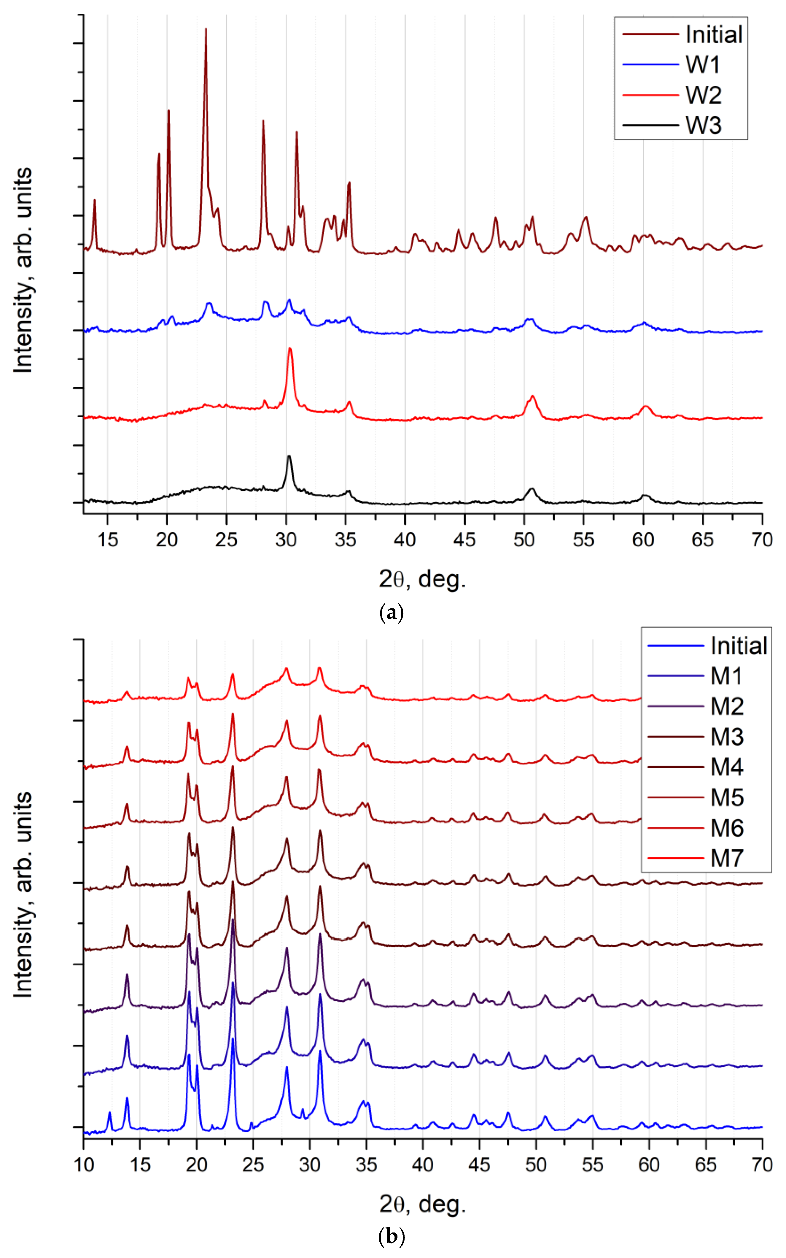
Radiation stabilities of phosphate molybdates and phosphate tungstates were compared with the help of 167 MeV Xe ion irradiation at various fluences, ranging from 1·1012 to 6·1013 cm–2. The dose dependence of XRD curves registered in the Na0.5Zr2(PO4)2.5(XO4)0.5 ceramics is shown in Figure 2. The results of XRD analysis proved that the initial Na0.5Zr2(PO4)2.5(WO4)0.5 ceramics (Figure 2a) were amorphized when exposed to ion irradiation at a minimum dose of 3·1012 cm−2. Dose increase resulted in further amorphization and phase decomposition in Na0.5Zr2(PO4)2.5(WO4)0.5 accompanied by the ZrO2 phase formation. When exposed to 3·1013 cm−2 irradiation, no XRD peaks were observed in the Na0.5Zr2(PO4)2.5(WO4)0.5 on an XRD curve, only peaks in the crystalline ZrO2 phase and a wide halo of an amorphous component in the specimen remained.
As for the Na0.5Zr2(PO4)2.5(MoO4)0.5 ceramics (Figure 2b), XRD results showed a weak impact of ion irradiation on crystallinity of these ceramics. Diffraction peaks were seen clearly, even at irradiation doses of up to 6·1013 cm−2. Changes caused by ion irradiation in this series concerned the coherent scattering region sizes of the Na0.5Zr2(PO4)2.5(MoO4)0.5 phase slightly. However, ion irradiation did not result in critical degradation of crystallinity, as in the case of the Na0.5Zr2(PO4)2.5(WO4)0.5 ceramics. Intensity of XRD peaks reduced 4 times with an increase in the irradiation dose of the Na0.5Zr2(PO4)2.5(MoO4)0.5 ceramics (Figure 2b).
Figure 3 shows the dependence of the intensity of an XRD peak (113) on X-ray incidence angle in Na0.5Zr2(PO4)2.5(MoO4)0.5 ceramics in the initial state and after irradiation at a dose of 6·1013 cm−2. The intensity for all experimental points of the irradiated specimen was multiplied by a factor of 4 for easier data comparison. Figure 3 shows a calculated curve plotted with due regard for material constants, and the geometry of the experiment as if crystalline quality and phase composition of the material were uniform over the entire depth of analysis. It was apparent that dependences were the same for the initial and irradiated specimens. It could be assumed that, within the depth of analysis of 5 µm, the degree of amorphization of the near-surface layer was the same and approximated 75% for M7 ceramics (irradiation dose of 6·1013 cm−2).
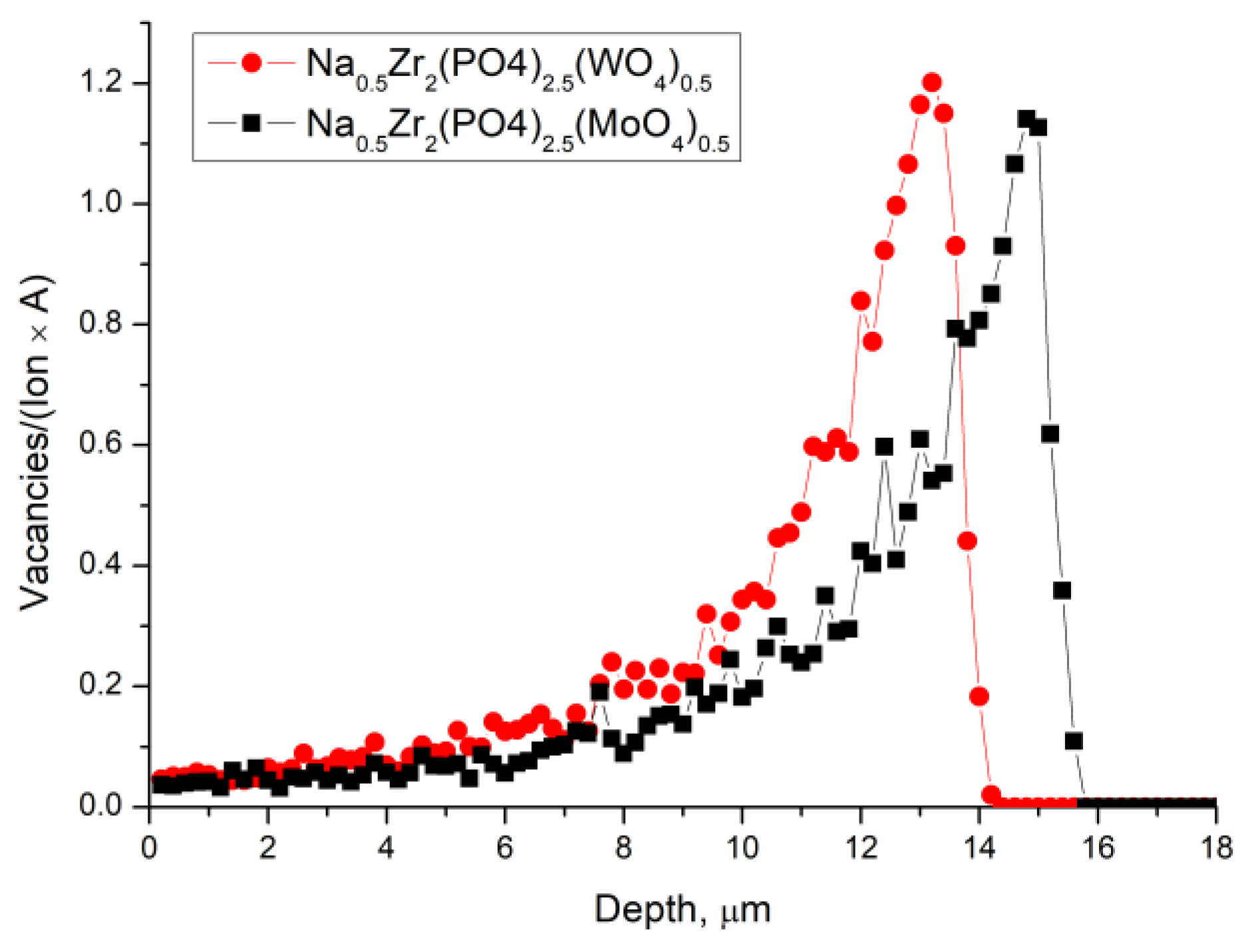
To assess how thick the damaged layer was, the depth of Xe ions penetration into the surface layers of the materials was simulated using SRIM-2013 software [80], in accordance with the method proposed in [81]. The results of simulating the distribution of vacancies depth for both materials under study are shown in Figure 4. It was apparent that the depth of the damaged layer significantly exceeded the depth of analysis in the GIXRD method for these materials and ion beam parameters. The assumption about uniform amorphization of the near-surface layer, about 5 μm thick, was confirmed by the simulation results. It could be seen that, in W-containing ceramics, the depth of defect formation was somewhat less, and the concentration of defects in the near-surface layer was slightly greater. We could assume that the energy of the ions was more efficiently transferred to the W-containing material, which led to its amorphization at lower doses. Probable explanations might involve differences in W and Mo chemical bonds with the environment and also in the atomic mass of W and Mo. This assumption agreed with the results of XRD analysis in the symmetric Bragg–Brentano geometry and was previously observed in other W- and Mo-containing ceramics [79].
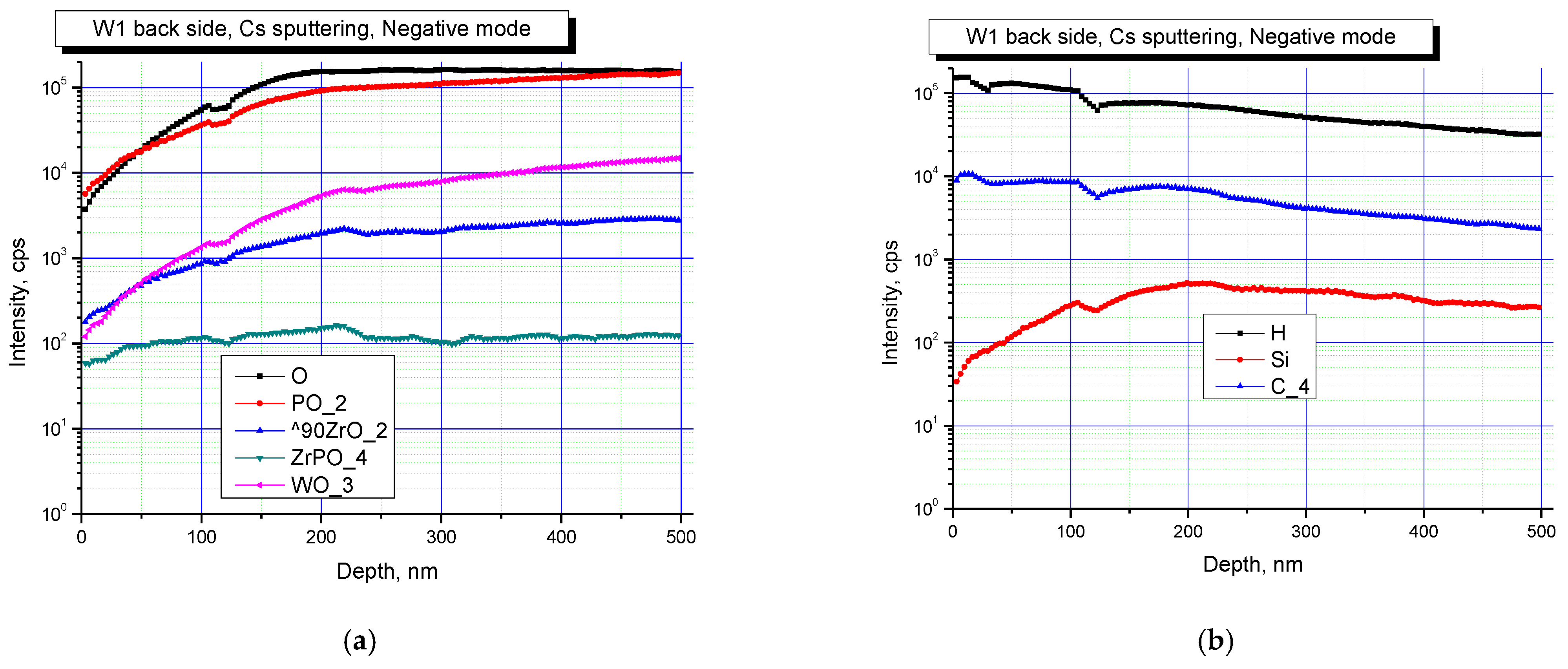
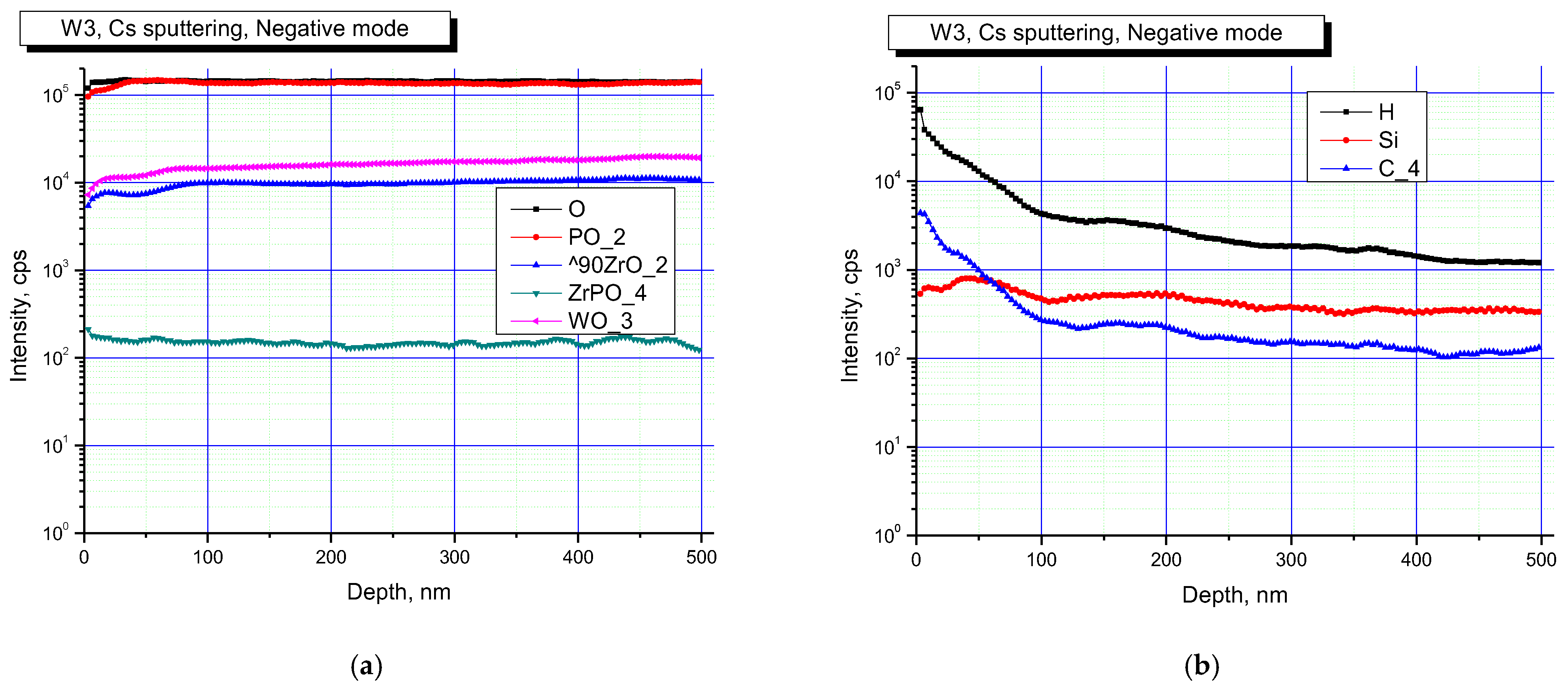
Figure 5 and Figure 6 show the results of SIMS studies of the surface layers of Na0.5Zr2(PO4)2.5(WO4)0.5 ceramics. Concentration depth profiles for the specimens in the initial state, and when exposed to irradiation at a dose of 3·1013 cm−2, are shown in negative secondary ion detection mode.
The analysis of results presented in Figure 5 and Figure 6 allows for a conclusion that the surface of all the specimens contained P, Zr, W oxides. The studies also indicated that the surface of the specimens was partially contaminated with Si. Irradiation led to increased contributions of P and Zr oxides. The changes observed might stem from changes in the phase composition of the specimens after irradiation, which led to a change in the probabilities of formation and the release of various cluster secondary ions.
It is noteworthy that the surface layer contained P, Zr, and W oxides, as well as a high concentration of C. Contamination of ceramics surface layers during SPS is one of the known drawbacks of this method, which has been described in a variety of research articles [45,82,83,84,85,86,87,88,89,90,91]. Carburization of surface layers during SPS occurs because the sintered material interacts with a graphite dye or graphite foil used to improve contact between the specimen surface and the inner wall of a graphite dye.
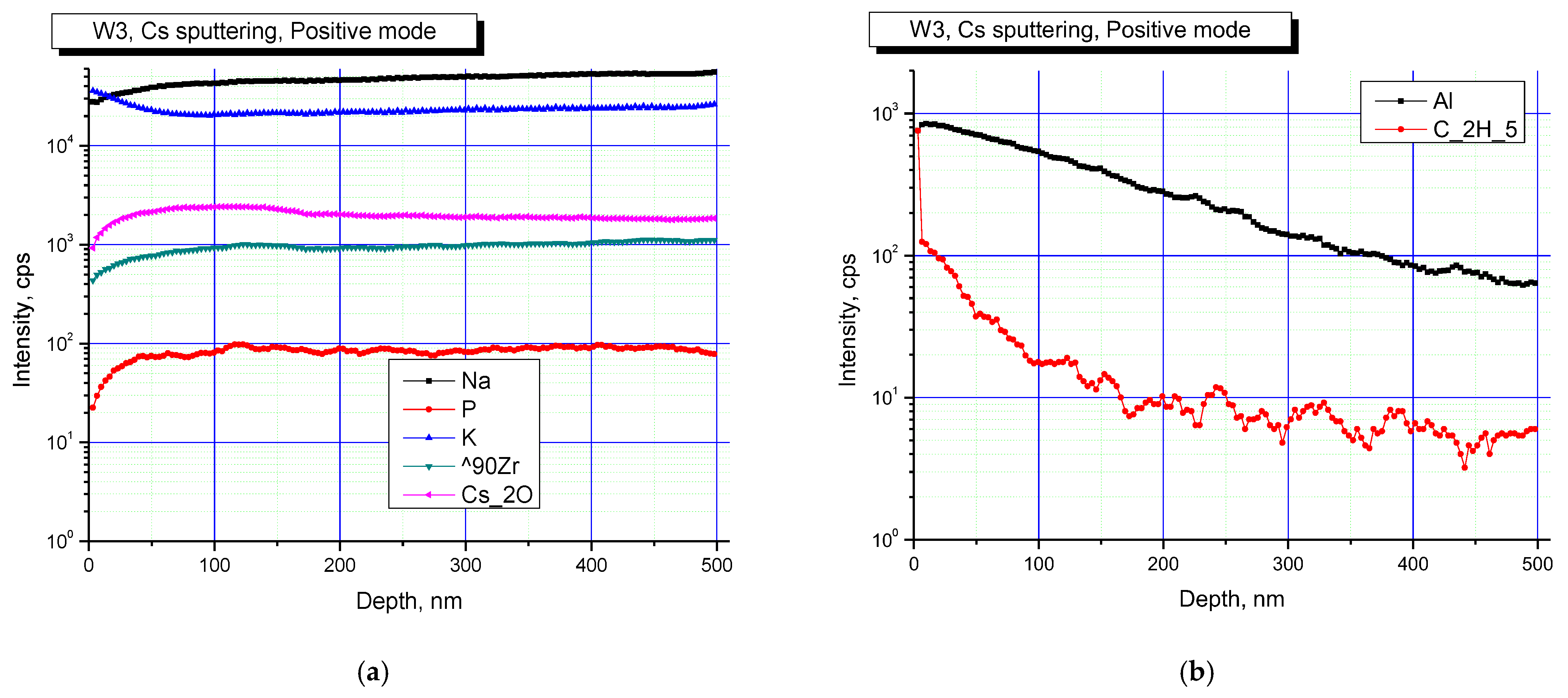
Figure 7 and Figure 8 show elemental profiles of the initial and irradiated Na0.5Zr2(PO4)2.5(WO4)0.5 ceramic specimens in positive secondary ion detection mode.
Figure 7 and Figure 8 show the results of studies in positive secondary ion detection mode. These results suggested that Na, K, Zr were present in all the specimens, and after irradiation, Na and K contributions increased. Doping elements or impurities in the specimens were mostly Al and hydrocarbon contaminants (C2H5 cluster line). After irradiation, the contribution of hydrocarbons decreased.
The hydrolytic stabilities of the specimens with high Mo and W contents (x = 0.4 and 0.5) were studied. According to XRD data, no crystal structure damage was observed during the hydrolytic tests. Unit cell parameters of Na1–xZr2(PO4)3–x(XO4)x were identical before and after the hydrolytic tests. Normalized release rates per unit surface area (R) for particular components were determined according to the formulae:
R = NL/t,
NL = m/(ω·S),
where m [g] is the mass of a component leached for a given time, t [days] is the test duration, S [cm2] is the open surface area, and ω is the mass fraction of the component in the initial specimen.
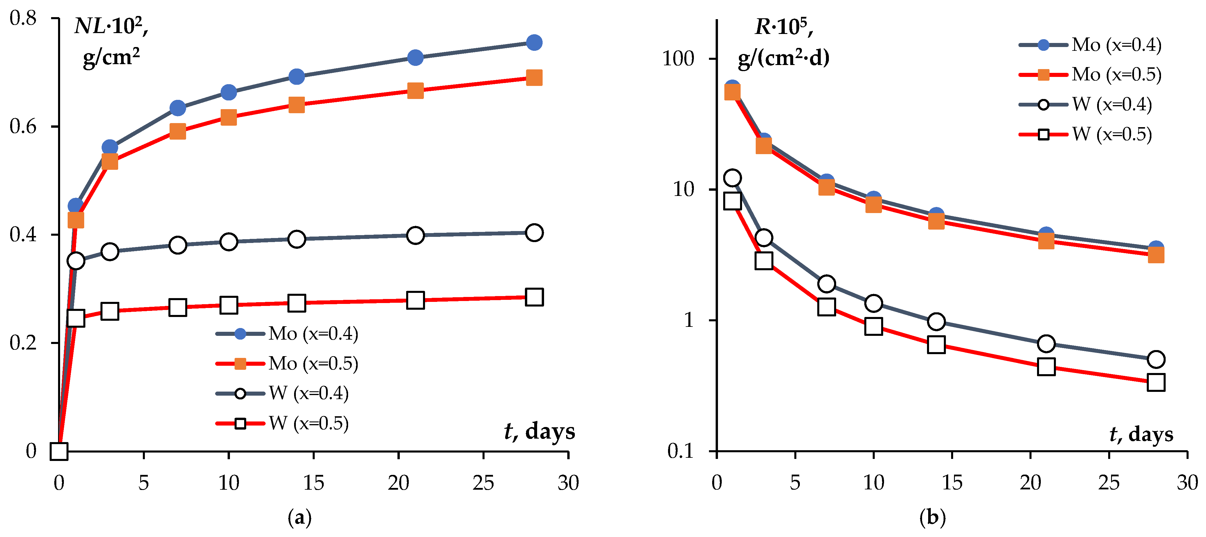
The normalized weight loss values NL and the normalized leaching rates R after 28 days of testing are presented in Table 1. Time dependencies of the above values are shown in Figure 9. The normalized leaching rates after 28 days of testing (Rmin) were 31.6·10−6 g·cm−2·day−1 for Mo-containing compounds (x = 0.5) and 3.36·10−6 g·cm−2·day−1 for W-containing ones (x = 0.5). After 28 days of testing, the normalized leaching rate for Mo from the Na1−xZr2(PO4)3−x(XO4)x ceramics at x = 0.4 and 0.5 was by an order of magnitude greater than that of W. This was a very low normalized leaching rate after 28 days of testing of NZP ceramics suggesting their having high chemical resistance. We reckoned that this result indicated that inorganic compounds of the NZP family could have advanced applications as binders for W- and Mo-containing fractions of HLW.
As follows from Table 1 and Figure 9, the normalized leaching rates R decreased while W and Mo contents increased. At the moment, we have no clear explanation of this effect. In our opinion, it might be specific to a stationary mode of testing phosphate tungstates and phosphate molybdates i.e., metal atoms that are leached react with oxygen diluted in water, which results in thin oxide films that form on ceramics surfaces and prevent further leaching of heavy metals. The surface area covered with an oxide film grows along with an increase in W and/or Mo contents in ceramics.
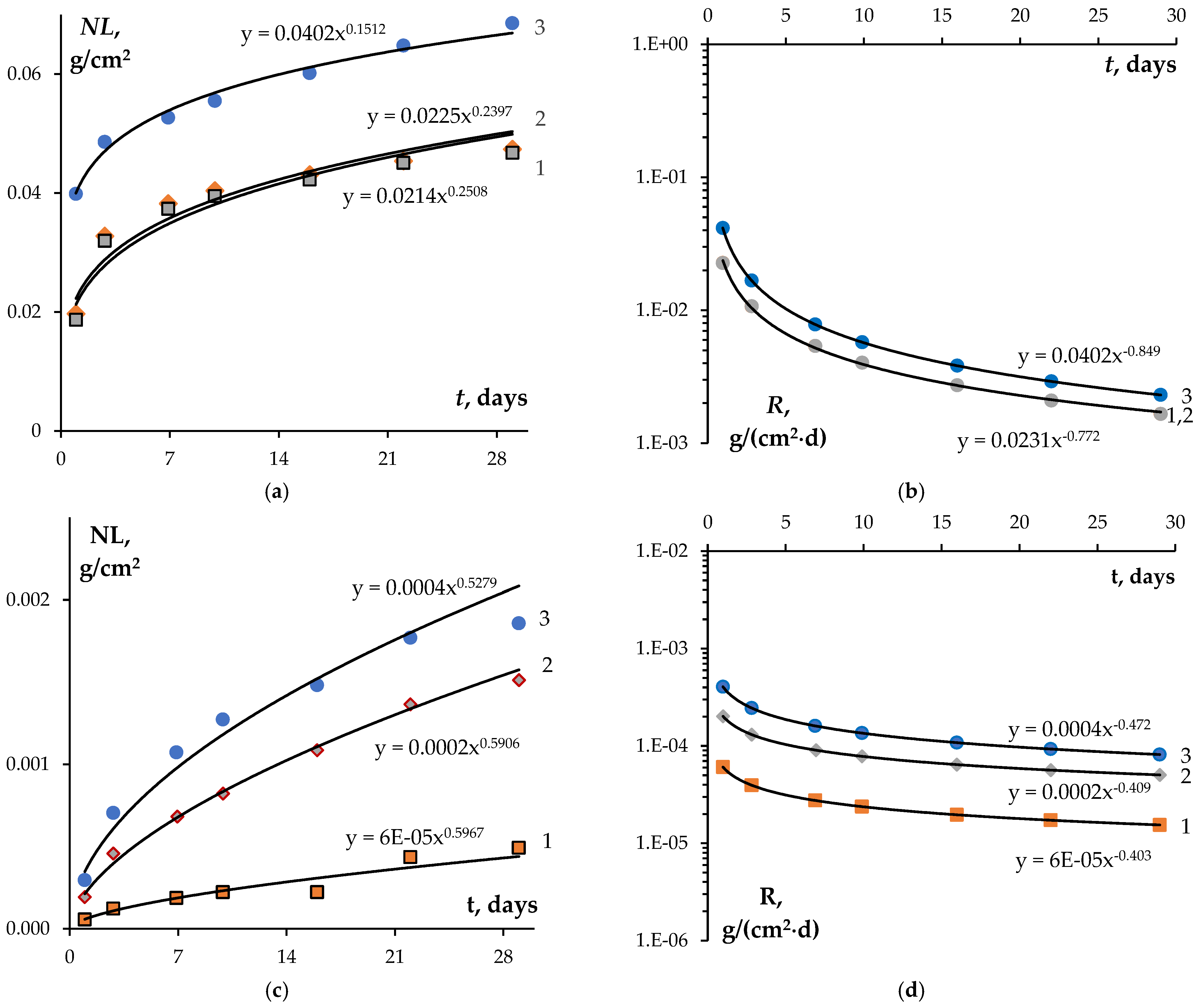
Figure 10 shows the results of hydrolytic testing of the Na0.5Zr2(PO4)2.5(MoO4)0.5 (a, b) and Na0.5Zr2(PO4)2.5(WO4)0.5 ceramics after irradiation at different doses. NL(t) and R(t) data were interpolated with a power function. The analysis of Ri(t) and NLi(t) dependencies showed that higher doses increased W and Mo leaching rates. The Mo leaching rate turned out to be significantly higher than that of W. After irradiation at a fluence of 3·1012 cm−2, the Mo leaching rate after 28 days of testing was RMo = 1.6·10−3 g⋅cm−2⋅d−1, while the W leaching rate was RW = 8.2·10−5 g⋅cm−2⋅d−1. After irradiation at a dose of 3·1013 cm−2, the Mo leaching rate increased to 2.3·10−3 g⋅cm−2⋅d−1, while the W leaching rate reached 5.0·10−5 g⋅cm−2⋅d−1. Comparison of these results with the data presented in Table 1 shows that the hydrolytic stability of the irradiated ceramics decreased, but remained high for the W-containing ceramics.
Let us compare the leaching rate R estimates with literature data. It was noted that key data on resistance of NZP ceramics obtained by conventional methods are presented in many works (see, for example, [2,6,92,93,94,95,96,97,98], etc.). Here we shall point out only some data that is crucial in order to analyze the results obtained. NZP ceramics demonstrated high chemical resistance, including after irradiation. It is known that NZP ceramics do not decompose even after 2 years of exposure to hydrothermal conditions at 400 °C, including after irradiation with 60Co [99,100]. According to [101], the Ca leaching rate R for Ca0.75Zr2(PO4)2.5(SiO4)0.5, obtained by cold pressing (P = 200 MPa) followed by sintering (900 °C, 10 h), was ~1·10-8 g⋅cm−2⋅day−1. Tests were carried out under static conditions at room temperature for 21 days. As shown in [92], the Zr leaching rate for La1/3Zr2(PO4)3 under static conditions was less than 10−5 g⋅m−2⋅day−1, while the La leaching rate depended on the ratio of the ceramic surface area to the solution volume and was ~10−6 g⋅m−2⋅day−1 after 14 days of testing. According to [102], the Pu leaching rate in Pu1/3Zr2(PO4)3 after testing for 14 days at room temperature under static conditions was ~9.9·10−6 g⋅cm−2⋅day−1. Pu1/3Zr2(PO4)3 ceramics was produced by cold pressing of powders (P = 200 MPa) and sintering at 950 °C (7 h). Low Sr leaching rates (less than 10−6 g⋅m−2⋅day−1) at room temperature in deionized water were measured for ceramics obtained by thermal treatment of HZr2(PO4)3 + Sr(NO3)2 [103]. The same high chemical stability of NZP ceramics was found during tests based on the Soxhlet method [104], as well as during tests of multicomponent compounds with an NZP structure that simulated the composition of various RAW fractions [105]. The Cs leaching rate in CsMgPO4, CsZr2(PO4)3, and Cs2Mg0.5Zr1.5(PO4)3 specimens obtained by SPS varied between 3·10−4 and 4·10−6 g⋅m−2⋅day−1 [106,107]. As shown in [44], in NaRe2(PO4)3 ceramics with relative density of 85% obtained by SPS under static conditions at room temperature, the Re leaching rate was 1.3·10−5 g⋅cm−1⋅day−1. Higher Re leaching rates were explained in [44] by the low density of ceramics and, as a result, the large specific surface area. Given high leaching rates of Mo and W from the structure of phosphates (see, for example, [108]), it could be assumed that the ceramics obtained had high chemical stability.
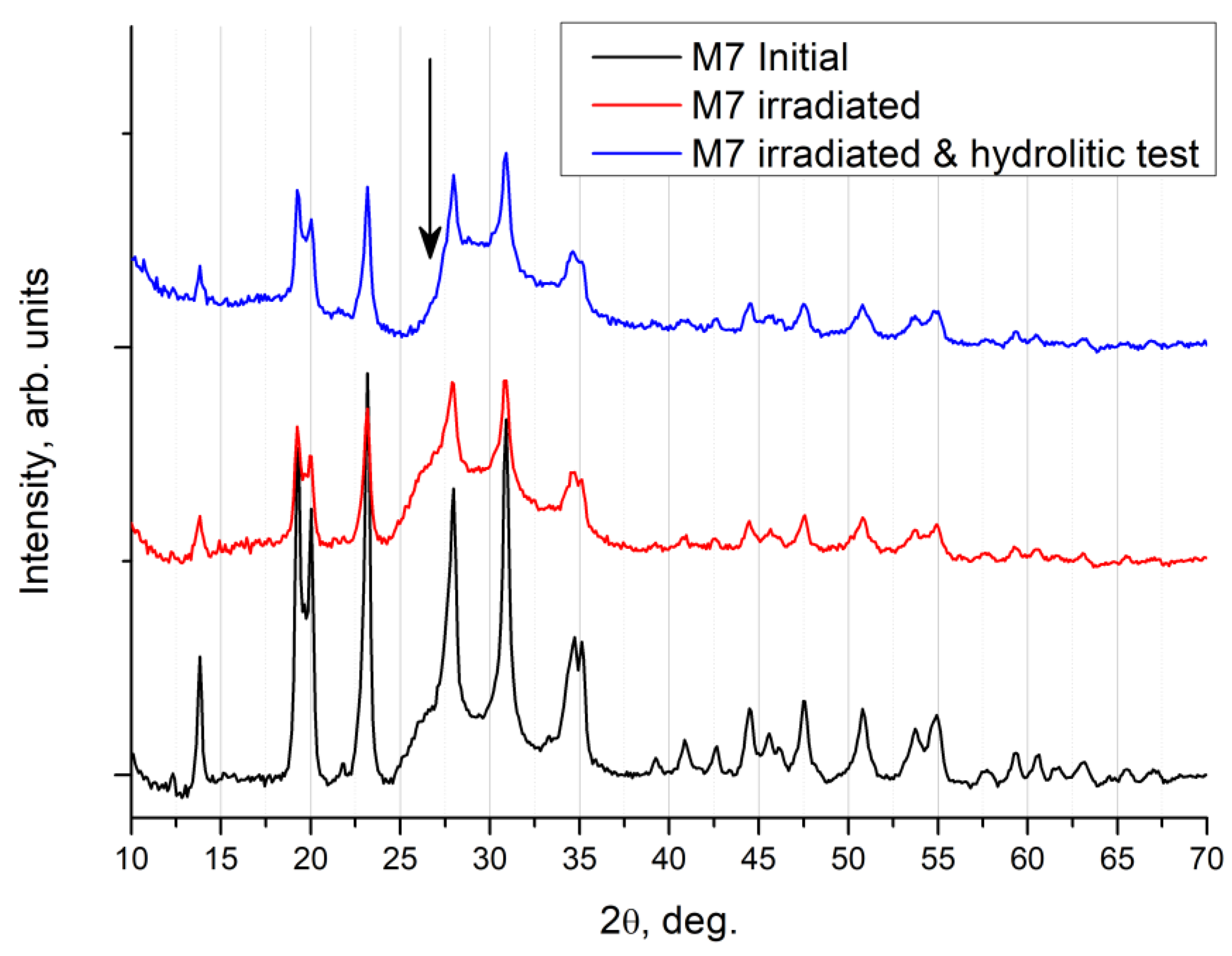
Figure 11 shows the XRD results in the symmetric Bragg–Brentano geometry of the surface of the irradiated Na0.5Zr2(PO4)2.5(MoO4)0.5 ceramics after the hydrolytic tests.
The Na0.5Zr2(PO4)2.5(MoO4)0.5 ceramics after hydrolytic tests showed no change in peak intensity of the main phase, and no peaks of auxiliary phases could be observed. A broad peak of a microcrystalline phase in graphite (shown by an arrow in the figure near 26° in 2θ) disappeared, which was the only change. Apparently, a side phase of graphite was washed out from the near-surface layer and from the pores of the specimen as a result of the hydrolytic tests.
Representative data of an XRD experiment in the symmetric Bragg–Brentano geometry for the Na0.5Zr2(PO4)2.5(WO4)0.5 ceramics is shown in Figure 12.
With the W-containing ceramics, it was apparent that the hydrolytic tests did not lead to a change in peak intensity of the main phase in Na0.5Zr2(PO4)2.5(WO4)0.5, and no peaks of auxiliary phases could be observed. There are no intensity changes near 26° associated with the graphite phase. Apparently, a graphite phase was initially absent in the W-series specimens since they were less porous or had other preparation-induced features. Carbon contamination, detected with SIMS, stemmed from an increase in the content of carbon ions in the Na0.5Zr2(PO4)2.5(WO4)0.5 crystal lattice. As noted earlier, this might be due to the intense diffusion of carbon from the graphite mold or graphite paper, with which the specimen surface interacted during SPS.

4. Conclusions

XRD analysis showed that the structure of the compounds Na1−xZr2(PO4)3−x(XO4)x remained unchanged during sintering and hydrolytic stability tests. The normalized leaching rates after 28 days of testing were 31·10−6 g·cm−2·d−1 for compounds Na0.5Zr2(PO4)2.5(MoO4)0.5 and 3.36·10−6 g·cm−2·d−1 for Na0.5Zr2(PO4)2.5(WO4)0.5 ones.
Irradiation tests proved that the destruction of the NZP crystal lattice was less expressed in the Mo-containing specimens, as compared to phosphate tungstates irradiated under similar conditions. The crystal lattice of W-containing ceramic specimens broke down as a result of irradiation at a fluence of 3·1013 cm−2.
Irradiation led to an increase in the leaching rate of W and Mo from the crystal structure of the ceramics. The irradiated W-containing ceramics had higher hydrolytic resistance, compared to the Mo-containing NZP ceramics. The leaching rates observed on the 28th day of testing for the irradiated Na0.5Zr2(PO4)2.5(MoO4)0.5 specimens were 1.6⋅10−3 g⋅cm−2⋅d−1 at a fluence of 3·1012 cm−2 and 2.3·10−3 g⋅cm−2⋅d−1 at a fluence of 3·1013 cm−2. The Na0.5Zr2(PO4)2.5(WO4)0.5 ceramics after irradiation at similar fluences had the leaching rates of 8.2·10−5 and 5.0·10−5 g⋅cm−2⋅d−1, respectively.

Author Contributions

Conceptualization, A.I.O. and A.V.N.; methodology, A.I.O.; validation, A.I.O., A.V.N. and V.N.C.; formal analysis, A.I.O. and A.V.N.; investigation, M.E.K., D.O.S., M.S.B., A.A.M., P.A.Y., A.A.N., N.Y.T., V.A.S., A.T.I., E.A.P. and M.N.D.; resources, A.I.O. and V.N.C.; data curation, A.I.O. and A.V.N.; writing—original draft preparation, A.I.O., A.V.N. and V.N.C.; writing—review and editing, A.I.O., A.V.N. and V.N.C.; visualization, A.V.N.; supervision, A.I.O.; project administration, A.I.O.; funding acquisition, A.I.O. All authors have read and agreed to the published version of the manuscript.

Funding

This research was funded by the Russian Science Foundation (Grant No. 21-13-00308). The TEM study of the powders was carried out on the equipment of the Center Collective Use “Materials Science and Metallurgy” (National University of Science and Technology “MISIS”, Moscow, Russia) with the financial support of the Ministry of Science and Higher Education of the Russian Federation (Grant No. 075-15-2021-696). XRD and SIMS analyses of the specimens exposed to ion radiation were carried out at the Laboratory of Diagnostics of Radiation Defects in Solid State Nanostructure, Institute for Physics of Microstructure, Russian Academy of Sciences (IPM RAS) with financial support from the Ministry of Science and Higher Education of Russian Federation (Grant No. 0030-2021-0030). This research was partially supported by the Ministry of Science and Higher Education of the Russian Federation (contract 075-15-2021-709, unique identifier of the project RF-2296.61321X0037 (radiation test equipment maintenance)).

Institutional Review Board Statement

Not applicable.

Informed Consent Statement

Not applicable.

Data Availability Statement

Not applicable.

Conflicts of Interest

The authors declare no conflict of interest.

References

  1. Orlova, A.I.; Ojovan, M.I. Ceramic mineral waste-forms for nuclear waste immobilization. Materials 2019, 12, 2638. [Google Scholar] [CrossRef] [PubMed] [Green Version]
  2. Bohre, A.; Avasthi, K.; Pet’kov, V.I. Vitreous and crystalline phosphate high level waste matrices: Present status and future challenges. J. Ind. Eng. Chem. 2017, 50, 1–14. [Google Scholar] [CrossRef]
  3. Ewing, R.; Wang, L. Phosphates as Nuclear Waste forms. Review in Mineralogy and Geochemistry. Phosphates Geochem. Geobiol. Mater. Importance 2002, 48, 673–689. [Google Scholar] [CrossRef]
  4. Orlova, A.I. Crystalline phosphates for HLW immobilization—Composition, structure, properties and production of ceramics. Spark Plasma Sintering as a promising sintering technology. J. Nucl. Mater. 2022, 559, 153407. [Google Scholar] [CrossRef]
  5. Savinykh, D.O.; Khainakov, S.A.; Orlova, A.I.; Garcia-Granda, S. New phosphate-sulfates with NZP Structure. Russ. J. Inorg. Chem. 2018, 63, 685–694. [Google Scholar] [CrossRef]
  6. Pet’kov, V.; Asabina, E.; Loshkarev, V.; Sukhanov, M. Systematic investigation of the strontium zirconium phosphate ceramic form for nuclear waste immobilization. J. Nucl. Mater. 2016, 471, 122–128. [Google Scholar] [CrossRef]
  7. Scheetz, B.E.; Agrawal, D.K.; Breval, E.; Roy, R. Sodium zirconium phosphate (NZP) as a host structure for nuclear waste immobilization: A review. Waste Manag. 1994, 14, 489–505. [Google Scholar] [CrossRef]
  8. Scales, N.; Dayal, P.; Aughterson, R.D.; Zhang, Y.; Gregg, D.I. Sodium zirconium phosphate-based glass-ceramics as potential wasteforms for the immobilization of nuclear wastes. J. Am. Cer. Soc. 2022, 105, 901–912. [Google Scholar] [CrossRef]
  9. Ojovan, M.I.; Lee, W.E.; Kalmykov, S.N. An Introduction to Nuclear Waste Immobilisation, 3rd ed.; Elsevier: Amsterdam, The Netherlands, 2019; 497p. [Google Scholar] [CrossRef]
  10. Weber, W.J.; Navrotsky, A.; Stefanovsky, S.; Vance, E.R.; Vernaz, E. Materials Science of High-Level Nuclear Waste Immobilization. MRS Bull. 2009, 34, 46–53. [Google Scholar] [CrossRef]
  11. Ewing, R.C. The design and evaluation of nuclear-waste forms: Clues from mineralogy. Can. Mineral. 2001, 39, 697–715. [Google Scholar] [CrossRef] [Green Version]
  12. Lumpkin, G.R. Ceramic waste forms for actinides. Elements 2006, 2, 365–372. [Google Scholar] [CrossRef]
  13. Terra, O.; Dacheux, N.; Audubert, F.; Podor, R. Immobilization of tetravalent actinides in phosphate ceramics. J Nucl. Mater. 2006, 352, 224–232. [Google Scholar] [CrossRef]
  14. Dacheux, N.; Clavier, N.; Podor, R. Versatile Monazite: Resolving geological records and solving challenges in materials science. Monazite as a promising long-term radioactive waste matrix: Benefits of high-structural flexibility and chemical durability. Am. Mineral. 2013, 98, 833–847. [Google Scholar] [CrossRef]
  15. Ananthanarayanan, A.; Ambashta, R.D.; Sudarsan, V.; Ajithkumar, T.; Sen, D.; Mazumber, S.; Wattal, P.K. Structure and short time degradation studies of sodium zirconium phosphate ceramics loaded with simulated fast breeder (FBR) waste. J. Nucl. Mater. 2017, 487, 5–12. [Google Scholar] [CrossRef]
  16. Pet’kov, V.I.; Sukhanov, M.V.; Kurazhkovskaya, V.S. Molybdenum fixation in crystalline NZP matrices. Radiochemistry 2003, 45, 620–625. [Google Scholar] [CrossRef]
  17. Chourasiaa, R.; Shrivastavaa, O.P.; Wattal, P.K. Synthesis, characterization and structure refinement of sodium zirconium molibdato-phosphate: Na0.9Zr2Mo0.1P2.9O12 (MoNZP). J. Alloys Compd. 2009, 473, 579–583. [Google Scholar] [CrossRef]
  18. Bohre, A.; Shrivastava, O.P.; Avasthi, K. Solid state synthesis and structural refinement of polycrystalline phases: Ca1−2xZr4M2xP6−2xO24 (M = Mo, x = 0.1 and 0.3). Arab. J. Chem. 2016, 9, 736–744. [Google Scholar] [CrossRef] [Green Version]
  19. Kumar, S.P.; Gopal, B. Immobilization of “Mo6+” in monazite lattice: Synthesis and characterization of new phosphomolybdates, La1−xCaxP1−yMoyO4, where x = y = 0.1–0.9. J. Am. Cer. Soc. 2011, 94, 1008–1013. [Google Scholar] [CrossRef]
  20. Daub, M.; Lehner, A.J.; Höppe, H.A. Synthesis, crystal structure and optical properties of Na2RE(PO4)(WO)4 (RE = Y,Tb-Lu). Dalton Trans. 2012, 41, 12121–12128. [Google Scholar] [CrossRef]
  21. Sukhanov, M.V.; Pet’kov, V.I.; Kurazhkovskaya, V.S.; Eremin, N.N.; Urusov, V.S. Computer-assisted structure simulation, synthesis, and phase formation of molybdophosphates A1−xZr2(PO4)3−x(MoO4)x (A is an alkali metal). Russ. J. Inorg. Chem. 2006, 51, 706–711. [Google Scholar] [CrossRef]
  22. Bennouna, L.; Arsalane, S.; Brochu, R.; Lee, M.R.; Chassaing, J.; Quarton, M. Spécificités des ions NbIV and MoIV dans les monophosphates de type Nasicon. J. Solid State Chem. 1995, 114, 224–229. [Google Scholar] [CrossRef]
  23. Buzlukov, A.L.; Fedorov, D.S.; Serdtsev, A.V.; Kotova, I.Y.; Tyutyunnik, A.P.; Korona, D.V.; Baklanova, Y.V.; Ogloblichev, V.V.; Kozhevnikova, N.M.; Denisova, T.A.; et al. Ion mobility in triple sodium molybdates and tungstates with a NASICON structure. J. Exp. Theor. Phys. 2022, 134, 42–50. [Google Scholar] [CrossRef]
  24. Vereshchagina, T.A.; Fomenko, E.V.; Vasilieva, N.G.; Solovyov, L.A.; Vereshchagin, S.N.; Bazarova, Z.G.; Anshits, A.G. A novel layered zirconium molybdate as a precursor to a ceramic zirconomolybdate host for lanthanide bearing radioactive waste. J. Mater. Chem. 2011, 21, 12001–12007. [Google Scholar] [CrossRef]
  25. Tokita, M. Progress of Spark Plasma Sintering (SPS): Method, Systems, Ceramics Applications and Industrialization. Ceramics 2021, 4, 160–198. [Google Scholar] [CrossRef]
  26. Olevsky, E.; Dudina, D. Field-Assisted Sintering: Science and Applications; Springer: Cham, Switzerland, 2018; 425p. [Google Scholar] [CrossRef]
  27. Dudina, D.V.; Vidyuk, T.M.; Korchagin, M.A. Synthesis of ceramic reinforcements in metallic matrices during Spark Plasma Sintering: Consideration of Reactant/Matrix Mutual Chemistry. Ceramics 2021, 4, 592–599. [Google Scholar] [CrossRef]
  28. Hu, Z.-Z.; Zhang, Z.-H.; Cheng, X.-W.; Wang, F.-C.; Zhang, Y.-F.; Li, S.-L. A review of multi-physical fields induced phenomena and effects in spark plasma sintering: Fundamentals and applications. Mater. Des. 2020, 191, 108662. [Google Scholar] [CrossRef]
  29. Grasso, S.; Sakka, Y.; Maizza, G. Electric current activated/assisted sintering (ECAS): A review of patents 1906–2008. Sci. Technol. Adv. Mater. 2009, 10, 053001. [Google Scholar] [CrossRef]
  30. Wang, L.; Zhang, J.; Jiang, W. Recent development in reactive synthesis of nanostructured bulk materials by spark plasma sintering. Int. J. Refract. Met. Hard Mater. 2013, 39, 103–112. [Google Scholar] [CrossRef]
  31. Yamanoglu, R. Pressureless Spark Plasma Sintering: A perspective from conventional sintering to accelerated sintering without pressure. Powder Metall. Met. Ceram. 2019, 57, 513–525. [Google Scholar] [CrossRef]
  32. Kai, R.; Mukhopadhyay, A. Spark plasma sintered/synthesized dense and nanostructured materials for solid-state Li-ion batteries: Overview and perspective. J. Power Source 2014, 247, 920–931. [Google Scholar] [CrossRef]
  33. Chaim, R.; Chevallier, G.; Weibel, A.; Estournès, C. Grain growth during spark plasma and flash sintering of ceramic nanoparticles: A review. J. Mater. Sci. 2018, 53, 3087–3105. [Google Scholar] [CrossRef] [Green Version]
  34. Dudina, D.V.; Bokhonov, B.B.; Olevsky, E.A. Fabrication of porous materials by spark plasma sintering: A review. Materials 2019, 12, 541. [Google Scholar] [CrossRef] [PubMed] [Green Version]
  35. Hulbert, D.M.; Anders, A.; Dudina, D.V.; Andersson, J.; Jiang, D.; Unuvar, C.; Anselmi-Tamburini, U.; Lavernia, E.J.; Mukherjee, A.K. The absence of plasma in “spark plasma sintering”. J. Appl. Phys. 2008, 104, 033305. [Google Scholar] [CrossRef] [Green Version]
  36. Mukasyan, A.S.; Rogachev, A.S.; Moskovskikh, D.O.; Yermekova, Z.S. Reactive spark plasma sintering of exothermic systems: A critical review. Ceram. Int. 2022, 48, 2988–2998. [Google Scholar] [CrossRef]
  37. Munir, Z.A.; Anselmi-Tamburini, U.; Ohyanagi, M. The effect of electric field and pressure on the synthesis and consolidation of materials: A review of the spark plasma sintering method. J. Mater. Sci. 2006, 41, 763–777. [Google Scholar] [CrossRef]
  38. Sukhanov, M.V.; Pet’kov, V.I.; Firsov, D.V. Sintering mechanism for high-density NZP ceramics. Inorg. Mater. 2011, 47, 674–678. [Google Scholar] [CrossRef]
  39. Shichalin, O.O.; Papynov, E.K.; Maiorov, V.Y.; Belov, A.A.; Modin, E.B.; Buravlev, I.Y.; Azarova, Y.A.; Golub, A.V.; Gridasova, E.A.; Sukhorada, A.E.; et al. Spark Plasma Sintering of Aluminosilicate Ceramic Matrices for Immobilization of Cesium Radionuclides. Radiochemistry 2019, 61, 185–191. [Google Scholar] [CrossRef]
  40. Papynov, E.K.; Belov, A.A.; Shichalin, O.O.; Buravlev, I.Y.; Azon, S.A.; Gridasova, E.A.; Parotkina, Y.A.; Yagofarov, V.Y.; Drankov, A.N.; Golub, A.V.; et al. Synthesis of perovskite-like SrTiO3 ceramics for radioactive strontium immobilization by spark plasma sintering-reactive synthesis. Russ. J. Inorg. Chem. 2021, 66, 645–653. [Google Scholar] [CrossRef]
  41. Shichalin, O.O.; Papynov, E.K.; Nepomnyushchaya, V.A.; Ivanets, A.I.; Belov, A.A.; Dran’kov, A.N.; Yarusova, S.B.; Buravlev, I.Y.; Tarabanova, A.E.; Fedorets, A.N.; et al. Hydrothermal synthesis and spark plasma sintering of NaY zeolite as solid-state matrices for cesium-137 immobilization. J. Eur. Ceram. Soc. 2022, 42, 3004–3014. [Google Scholar] [CrossRef]
  42. Orlova, A.I.; Troshin, A.N.; Mikhailov, D.A.; Chuvil’deev, V.N.; Boldin, M.S.; Sakharov, N.V.; Nokhrin, A.V.; Skuratov, V.A.; Kirilkin, N.S. Phosphorous-containing cesium compounds of pollucite structure. Preparation of high-density ceramic and its radiation test. Radiochemistry 2014, 56, 98–104. [Google Scholar] [CrossRef]
  43. Potanina, E.; Golovkina, L.; Orlova, A.; Nokhrin, A.; Boldin, M.; Sakharov, N. Lanthanide (Nd, Gd) compounds with garnet and monazite structures. Powders synthesis by "wet" chemistry to sintering ceramics by Spark Plasma Sintering. J. Nucl. Mater. 2016, 473, 93–98. [Google Scholar] [CrossRef]
  44. Alekseeva, L.S.; Nokhrin, A.V.; Orlova, A.I.; Boldin, M.S.; Lantsev, E.A.; Murashov, A.A.; Korchenkin, K.K.; Ryabkov, D.V.; Chuvil’deev, V.N. Ceramics based on the NaRe2(PO4)3 phosphate with the kosnarite structure as waste forms for technetium immobilization. Inorg. Mater. 2022, 58, 325–332. [Google Scholar] [CrossRef]
  45. Mikhaylov, D.A.; Potanina, E.A.; Nokhrin, A.V.; Orlova, A.I.; Yunin, P.A.; Sakharov, N.V.; Boldin, M.S.; Belkin, O.A.; Skuratov, V.A.; Issatov, A.T.; et al. Investigation of the Microstructure of Fine-Grained YPO4:Gd Ceramics with Xenotime Structure after Xe Irradiation. Ceramics 2022, 5, 237–252. [Google Scholar] [CrossRef]
  46. Alekseeva, L.; Nokhrin, A.; Boldin, M.; Lantsev, E.; Murashov, A.; Orlova, A.; Chuvil’deev, V. Study of the hydrolytic stability of fine-grained ceramics based on Y2.5Nd0.5AlO12 oxide with a garnet structure under hydrothermal conditions. Materials 2021, 14, 2152. [Google Scholar] [CrossRef]
  47. Papynov, E.K.; Shichalin, O.O.; Mayorov, V.Y.; Kuryavyi, V.G.; Kaidalova, T.A.; Teplukhina, L.V.; Portnyagin, A.S.; Slobodyuk, A.B.; Belov, A.A.; Tananaev, I.G.; et al. SPS technique for ionizing radiation source fabrication based on dense cesium-containing core. J. Hazardous Mater. 2019, 369, 25–30. [Google Scholar] [CrossRef]
  48. Mikhailov, D.A.; Orlova, A.I.; Malanina, N.V.; Nokhrin, A.V.; Potanina, E.A.; Chuvil’deev, V.N.; Boldin, M.S.; Sakharov, N.V.; Belkin, O.A.; Kalenova, M.Y.; et al. A study of fine-grained ceramics based on complex oxides ZrO2-Ln2O3 (Ln = Sm, Yb) obtained by Spark Plasma Sintering for inert matrix fuel. Ceram. Int. 2018, 44, 18595–18608. [Google Scholar] [CrossRef]
  49. Orlova, A.I.; Volgutov, V.Y.; Mikhailov, D.A.; Bykov, D.M.; Skuratov, V.A.; Chuvil’Deev, V.N.; Nokhrin, A.V.; Boldin, M.S.; Sakharov, N.V. Phosphate Ca1/4Sr1/4Zr2(PO4)3 of the NaZr2(PO4)3 structure type: Synthesis of a dense ceramic material and its radiation testing. J. Nucl. Mater. 2014, 446, 232–239. [Google Scholar] [CrossRef]
  50. Shichalin, O.O.; Yarusova, S.B.; Ivanets, A.I.; Papynov, E.K.; Belov, A.A.; Azon, S.A.; Buravlev, I.Y.; Panasenko, A.E.; Zadorozhny, P.A.; Mayorov, V.Y.; et al. Synthesis and spark plasma sintering of solid-state matrices based on calcium silicate for 60Co immobilization. J. Alloys Compd. 2022, 912, 165233. [Google Scholar] [CrossRef]
  51. Shichalin, O.O.; Belov, A.A.; Zavyalov, A.P.; Papynov, E.K.; Azon, S.A.; Fedorets, A.N.; Buravlev, I.Y.; Balanov, M.I.; Tananaev, I.G.; Shi, Y.; et al. Reaction synthesis of SrTiO3 mineral-like ceramics for strontium-90 immobilization via additional in situ synchrotron studies. Ceram. Int. 2022, 48, 19597–19605. [Google Scholar] [CrossRef]
  52. Papynov, E.K.; Shichalin, O.O.; Buravlev, I.Y.; Belov, A.A.; Portnyagin, A.S.; Fedorets, A.N.; Azarova, Y.A.; Tananaev, I.G.; Sergienko, V.I. Spark plasma sintering-reactive synthesis of SrWO4 ceramic matrices for 90Sr immobilization. Vacuum 2020, 180, 109628. [Google Scholar] [CrossRef]
  53. Ge, L.; Subhash, G.; Baney, R.H.; Tulenko, J.S.; McKenna, E. Densification of uranium dioxide fuel pellets prepared by spark plasma sintering (SPS). J. Nucl. Mater. 2013, 435, 1–9. [Google Scholar] [CrossRef]
  54. Cologna, M.; Tyrpekl, V.; Ernstberger, M.; Stohr, S.; Somers, J. Sub-micrometre grained UO2 pellets consolidated from sol gel beads using spark plasma sintering (SPS). Ceram. Int. 2016, 42, 6619–6623. [Google Scholar] [CrossRef]
  55. Papynov, E.K.; Shichalin, O.O.; Mironenko, A.Y.; Ryakov, A.V.; Manakov, I.V.; Makhrov, P.V.; Buravlev, I.Y.; Tananaev, I.G.; Avramenko, V.A.; Sergienko, V.I. Synthesis of high-density pellets of uranium dioxide by spark plasma sintering in dies of different types. Radiochemistry 2018, 60, 362–370. [Google Scholar] [CrossRef]
  56. Malkki, P.; Jolkkonen, M.; Hollmer, T.; Wallenius, J. Manufacture of fully dense uranium nitride pellets using hydride derived powders with spark plasma sintering. J. Nucl. Mater. 2014, 452, 548–551. [Google Scholar] [CrossRef]
  57. Johnson, K.D.; Wallenius, J.; Jolkkonen, M.; Claisse, A. Spark plasma sintering and porosity studies of uranium nitride. J. Nucl. Mater. 2016, 473, 13–17. [Google Scholar] [CrossRef]
  58. Yang, K.; Kardoulaki, E.; Zhao, D.; Broussard, A.; Metzger, K.; White, J.T.; Sivack, M.R.; Mcclellan, K.J.; Lahoda, E.J.; Lian, J. Uranium nitride (UN) pellets with controllable microstructure and phase—Fabrication by spark plasma sintering and their thermal-mechanical and oxidation properties. J. Nucl. Mater. 2021, 557, 153272. [Google Scholar] [CrossRef]
  59. Salvato, D.; Vigier, J.F.; Cologna, M.; Luzzi, L.; Somers, J.; Tyrpekl, V. Spark Plasma Sintering of fine uranium carbide powders. Ceram. Int. 2017, 43, 866–869. [Google Scholar] [CrossRef]
  60. Alekseeva, L.S.; Orlova, A.I.; Nokhrin, A.V.; Boldin, M.S.; Lantsev, E.A.; Chuvil’deev, V.N.; Murashov, A.A.; Sakharov, N.V. Spark Plasma Sintering of fine-grained YAG:Nd+MgO composite ceramics based on garnet-type oxide Y2.5Nd0.5Al5O12 for inert fuel matrices. Mater. Chem. Phys. 2018, 226, 323–330. [Google Scholar] [CrossRef]
  61. Kim, G.; Ahn, J.; Ahn, S. Grain growth and densification of uranium mononitride during spark plasma sintering. Ceram. Int. 2021, 47, 7258–7262. [Google Scholar] [CrossRef]
  62. Yeo, S.; Mckenna, E.; Baney, R.; Subhash, G.; Tulenko, J. Enhanced thermal conductivity of uranium dioxide-silicon carbide composite fuel pellets prepared by Spark Plasma Sintering (SPS). J. Nucl. Mater. 2013, 433, 66–73. [Google Scholar] [CrossRef]
  63. Margueret, A.; Balice, L.; Popa, K.; Holzhäuser, M.; De Bona, E.; Bonani, W.; Bulgheroni, A.; Audubert, F.; Cologna, M. Spark plasma sintering of UO2 nanopowders: Pressure, heating rate and current effects. J. Eur. Ceram. Soc. 2022, 42, 6056–6066. [Google Scholar] [CrossRef]
  64. Golovkina, L.S.; Orlova, A.I.; Nokhrin, A.V.; Boldin, M.S.; Lantsev, E.A.; Chuvil’deev, V.N.; Sakharov, N.V.; Shotin, S.V.; Zelenov, A.Y. Spark Plasma Sintering of fine-grained ceramic-metal composites YAG:Nd-(W,Mo) based on garnet-type oxide Y2.5Nd0.5Al5O12 for inert matrix fuel. J. Nucl. Mater. 2018, 511, 109–121. [Google Scholar] [CrossRef]
  65. De Bona, E.; Balice, L.; Cognini, L.; Holzhäuser, M.; Popa, K.; Walter, O.; Cologna, M.; Prieur, D.; Wiss, T.; Baldinozzi, G. Single-step, high pressure, and two-step spark plasma sintering of UO2 nanopowders. J. Nucl. Mater. 2021, 41, 3655–3663. [Google Scholar] [CrossRef]
  66. Golovkina, L.S.; Orlova, A.I.; Chuvil’deev, V.N.; Boldin, M.S.; Lancev, E.A.; Nokhrin, A.V.; Sakharov, N.V.; Zelenov, A.Y. Spark Plasma Sintering of high-density fine-grained Y2.5Nd0.5Al5O12+SiC composite ceramics. Mater. Res. Bull. 2018, 103, 211–215. [Google Scholar] [CrossRef]
  67. Chen, Z.; Subhash, G.; Tulenko, J.S. Master sintering curves for UO2 and UO2-SiC composite processed by spark plasma sintering. J. Nucl. Mater. 2014, 454, 427–433. [Google Scholar] [CrossRef] [Green Version]
  68. Muta, H.; Murakami, Y.; Uno, M.; Kurosaki, K.; Yamanaka, S. Thermophysical properties of Th1−xUxO2 pellets prepared by spark plasma sintering technique. J. Nucl. Sci. Techn. 2013, 50, 181–187. [Google Scholar] [CrossRef]
  69. Golovkina, L.S.; Orlova, A.I.; Nokhrin, A.V.; Boldin, M.S.; Chuvil’deev, V.N.; Sakharov, N.V.; Belkin, O.A.; Shotin, S.V.; Zelenov, A.Y. Spark Plasma Sintering of fine-grain ceramic-metal composites based on garnet-structure oxide Y2.5Nd0.5Al5O12 for Inert Matrix Fuel. Mater. Chem. Phys. 2018, 214, 516–526. [Google Scholar] [CrossRef]
  70. O’Brien, R.C.; Jerred, N.D. Spark Plasma Sintering of W-UO2 cermets. J. Nucl. Mater. 2013, 433, 50–54. [Google Scholar] [CrossRef]
  71. Yao, T.; Scott, S.M.; Xin, G.; Lian, J. TiO2 doped UO2 fuels sintered by spark plasma sintering. J. Nucl. Mater. 2016, 469, 251–261. [Google Scholar] [CrossRef] [Green Version]
  72. Wangle, T.; Tyrpekl, V.; Cologna, M.; Somers, J. Simulated UO2 fuel containing CsI by spark plasma sintering. J. Nucl. Mater. 2015, 466, 150–153. [Google Scholar] [CrossRef]
  73. Alekseeva, L.S.; Nokhrin, A.V.; Boldin, M.S.; Lantsev, E.A.; Orlova, A.I.; Chuvil’deev, V.N.; Sakharov, N.V. Fabrication of fine-grained CeO2-SiC ceramics for inert fuel matrices by Spark Plasma Sintering. J. Nucl. Mater. 2020, 539, 152225. [Google Scholar] [CrossRef]
  74. Gong, B.; Kardoulaki, E.; Yang, K.; Broussard, A.; Zhao, D.; Metzger, K.; White, J.T.; Sivack, M.R.; Mcclellan, K.J.; Lahoda, E.J.; et al. UN and U3Si composites densified by spark plasma sintering for accident-tolerant fuels. Ceram. Int. 2022, 48, 10762–10769. [Google Scholar] [CrossRef]
  75. Savinykh, D.O.; Boldin, M.S.; Orlova, A.I.; Aleksandrov, A.A.; Popov, A.A.; Murashov, A.A.; Nokhrin, A.V.; Chuvil’deev, V.N.; Khainakov, S.A.; Garcia-Granda, S. Synthesis, thermal expansion behavior and sintering of sodium zirconium nickel and calcium zirconium nickel phosphates. Inorg. Mater. 2021, 57, 529–540. [Google Scholar] [CrossRef]
  76. Savinykh, D.O.; Khainakov, S.A.; Boldin, M.S.; Orlova, A.I.; Aleksandrov, A.A.; Lantsev, E.A.; Sakharov, N.V.; Murashov, A.A.; Garcia-Granda, S.; Nokhrin, A.V.; et al. Preparation of NZP-type Ca0.75+0.5xZr1.5Fe0.5(PO4)3-x(SiO4)x powders and ceramics, thermal expansion behavior. Inorg. Mater. 2018, 54, 1267–1273. [Google Scholar] [CrossRef]
  77. Orlova, A.I.; Koryttseva, A.K.; Kanunov, A.E.; Chuvil’deev, V.N.; Moskvicheva, A.V.; Sakharov, N.V.; Boldin, M.S.; Nokhrin, A.V. Fabrication of NaZr2(PO4)3-type ceramic materials by spark plasma sintering. Inorg. Mater. 2012, 48, 372–377. [Google Scholar] [CrossRef]
  78. X-ray Interactions with Matter. Available online: https://henke.lbl.gov/optical_constants/ (accessed on 27 November 2022).
  79. Yunin, P.A.; Nazarov, A.A.; Potanina, E.A. Application of the GIXRD technique to investigation of damaged layers in NaNd(WO4)2 and NaNd(MoO4)2 ceramics irradiated with high-energy ions. Tech. Phys. 2022, 92, 1137. (In Russian) [Google Scholar] [CrossRef]
  80. Ziegler, J.F.; Ziegler, M.D.; Biersack, J.P. SRIM—The stopping and range of ions in matter. Nucl. Instrum. Methods Phys. Res. Sect. B Beam Interact. Mater. At. 2010, 268, 1818–1823. [Google Scholar] [CrossRef] [Green Version]
  81. Stoller, R.E.; Toloczko, M.B.; Was, G.S.; Certain, A.G.; Dwaraknath, S.; Garner, F.A. On the use of SRIM for computing radiation damage exposure. Nucl. Instrum. Methods Phys. Res. Sect. B Beam Interact. Mater. At. 2013, 310, 75–80. [Google Scholar] [CrossRef]
  82. Bernard-Granger, G.; Benameur, N.; Guizard, C.; Nygren, M. Influence of graphite contamination on the optical properties of transparent spinel obtained by spark plasma sintering. Scr. Mater. 2009, 60, 164–167. [Google Scholar] [CrossRef]
  83. Dudina, D.V.; Bokhonov, B.B.; Ukhina, A.V.; Anisimov, A.G.; Mali, V.I.; Esikov, M.A.; Batraev, I.S.; Kuznechik, O.O.; Pilinevich, L.P. Reactivity of materials towards carbon of graphite foil during Spark Plasma Sintering: A case study using Ni-W powders. Mater. Lett. 2016, 168, 62–67. [Google Scholar] [CrossRef]
  84. Wang, P.; Yang, M.; Zhang, S.; Tu, R.; Goto, T.; Zhang, L. Suppression of carbon contamination in SPSed CaF2 transparent ceramics by Mo foil. J. Eur. Ceram. Soc. 2017, 37, 4103–4107. [Google Scholar] [CrossRef]
  85. Kosyanov, D.Y.; Vornovskikh, A.A.; Zakharenko, A.M.; Gridasova, E.A.; Yavetskiy, R.P.; Dobrotvorskaya, M.V.; Tolmacheva, A.V.; Shichalin, O.O.; Papynov, E.K.; Ustinov, A.Y.; et al. Influence of sintering parameters on transparency of reactive SPSed Nd3+:YAG ceramics. Opt. Mater. 2021, 112, 110760. [Google Scholar] [CrossRef]
  86. Wang, P.; Huang, Z.; Morita, K.; Li, Q.; Yang, M.; Zhang, S.; Goto, T.; Tu, R. Influence of spark plasma sintering conditions on microstructure, carbon contamination, and transmittance of CaF2 ceramics. J. Eur. Ceram. Soc. 2022, 42, 245–257. [Google Scholar] [CrossRef]
  87. Yong, S.-K.; Choi, D.H.; Lee, K.; Ko, S.-Y.; Cheong, D.-I.; Park, Y.-J.; Go, S.-I. Study of the carbon contamination and carboxylate group formation in Y2O3-MgO nanocomposites fabricated by spark plasma sintering. J. Eur. Ceram. Soc. 2020, 40, 847–851. [Google Scholar] [CrossRef]
  88. Hammoud, H.; Garnier, V.; Fantozzi, G.; Lachaud, E.; Taider, S. Mechanism of carbon contamination in transparent MgAl2O4 and Y3Al5O12 ceramics sintered by Spark Plasma Sintering. Ceramics 2019, 2, 612–619. [Google Scholar] [CrossRef] [Green Version]
  89. Nokhrin, A.; Andreev, P.; Boldin, M.; Chuvil’deev, V.; Chegurov, M.; Smetanina, K.; Gryaznov, M.; Shotin, S.; Nazarov, A.; Shcherbak, G.; et al. Investigation of microstructure and corrosion resistance of Ti-Al-V titanium alloys obtained by Spark Plasma Sintering. Metals 2021, 11, 945. [Google Scholar] [CrossRef]
  90. Morita, K.; Kim, B.-N.; Yoshida, H.; Higara, K.; Sakka, Y. Distribution of carbon contamination in oxide ceramics occurring during spark-plasma-sintering (SPS) processing: II—Effect of SPS and loading temperatures. J. Eur. Cer. Soc. 2018, 38, 2596–2604. [Google Scholar] [CrossRef]
  91. Nečina, V.; Pabst, W. Reduction of temperature gradient and carbon contamination in electric current assisted sintering (ECAS/SPS) using a “saw-tooth” heating schedule. Ceram. Int. 2019, 45, 22987–22990. [Google Scholar] [CrossRef]
  92. Bois, L.; Guittet, M.J.; Carrot, F.; Trocellier, P.; Gautier-Soyer, M. Preliminary results on the leaching process of phosphate ceramics, potential host for actinide immobilization. J. Nucl. Mater. 2001, 297, 129–137. [Google Scholar] [CrossRef]
  93. Nakata, H.; Kageyama, T.; Itoh, K.; Nakayama, S. Leaching properties of alkali and alkaline-earth metallic elements immobilized by HZr2(PO4)3. J. Cer. Soc. Jpn. 2003, 111, 366–368. [Google Scholar] [CrossRef] [Green Version]
  94. Nakayama, S. Simultaneous immobilization of cesium and strontium by crystalline zirconium phosphate. J. Cer. Soc. Jpn. 2022, 130, 731–734. [Google Scholar] [CrossRef]
  95. Hashimoto, C.; Nakayama, S. Effect of treatment temperature on the immobilization of Cs and Sr to HZr2(PO4)3 using a autoclave. J. Nucl. Mater. 2013, 440, 153–157. [Google Scholar] [CrossRef]
  96. Nakayama, S. Immobilization of alkali, alkaline-earth and rare-earth elements by crystalline zirconium phosphate HZr2(PO4)3. J. Cer. Soc. Jpn. 2012, 120, 334–337. [Google Scholar] [CrossRef] [Green Version]
  97. Wei, Y.; Luo, P.; Wang, J.; Wen, J.; Zhan, L.; Zhang, X.; Yang, S.; Wang, J. Microwave-sintering preparation, phase evolution and chemical stability of Na1-2xSrxZr2(PO4)3 ceramics for immobilizing simulated radionuclides. J. Nucl. Mater. 2020, 540, 152366. [Google Scholar] [CrossRef]
  98. Wang, J.; Wei, Y.; Wang, J.; Zhang, X.; Wang, Y.; Li, N. Simultaneous immobilization of radionuclides Sr and Cs by sodium zirconium phosphate type ceramics and its chemical durability. Cer. Int. 2022, 48, 12772–12778. [Google Scholar] [CrossRef]
  99. Orlova, A.I.; Zyryanov, V.N.; Egor’kova, O.V.; Demarin, V.T. Long-term hydrothermal testing of crystalline phosphates of the NZP family. Radichemistry 1996, 38, 22–26. (In Russian) [Google Scholar]
  100. Orlova, A.I.; Zyryanov, V.N.; Kotel’nikov, A.R. Ceramic phosphate matrices of high-level waste. Behavior under hydrothermal conditions. Radiochemistry 1993, 35, 120–126. (In Russian) [Google Scholar]
  101. Kanunov, A.E.; Orlova, A.I.; Demarin, V.T. Synthesis and investigation of calcium-containing phosphatosilicates with NaZr2(PO4)3 structure. Russ. J. Gen. Chem. 2013, 83, 1029–1034. [Google Scholar] [CrossRef]
  102. Bykov, D.M.; Orlova, A.I.; Tomilin, S.V.; Lizin, A.A.; Lukinykh, A.N. Americium and plutonium in trigonal phosphates (NZP type) Am1/3[Zr2(PO4)3] and Pu1/4[Zr2(PO4)3]. Radiochemistry 2006, 48, 234–239. [Google Scholar] [CrossRef]
  103. Nakayama, S.; Itoh, K. Immobilization of strontium by crystalline zirconium phosphate. J. Eur. Cer. Soc. 2003, 23, 1047–1052. [Google Scholar] [CrossRef]
  104. Sugantha, M.; Kumar, N.R.S.; Varadaraju, U.V. Synthesis and leachability studies of NZP and eulytine phases. Waste Manag. 1998, 18, 275–279. [Google Scholar] [CrossRef]
  105. Buvaneswari, G.; Varadaraju, U.V. Low leachability phosphate lattices for fixation of select metal ions. Mater. Res. Bull. 2000, 35, 1313–1323. [Google Scholar] [CrossRef]
  106. Pet’kov, V.I.; Asabina, E.A.; Lukuttsov, A.A.; Lorchemkin, I.V.; Alekseev, A.A.; Demarin, V.T. Immobilization of cesium into mineral-like matrices of tridymite, kosnarite, and langbeinite structure. Radiochemistry 2015, 57, 632–639. [Google Scholar] [CrossRef]
  107. Shrivastava, O.P.; Chourasia, R. Crystal chemistry of sodium zirconium phosphate based simulated ceramic waste forms of effluent cations (Ba2+, Sn4+, Fe3+, Cr3+, Ni2+ and Si4+) from light water reaction fuel reprocessing plants. J. Hazard. Mater. 2008, 153, 285–292. [Google Scholar] [CrossRef]
  108. Kumar, S.P.; Gopal, B. Simulated monazite crystalline wasteform La0.4Nd0.1Y0.1Gd0.1Sm0.1Ce0.1Ca0.1(P0.9Mo0.1O4): Synthesis, phase stability and chemical durability study. J. Nucl. Mater. 2015, 458, 224–232. [Google Scholar] [CrossRef]
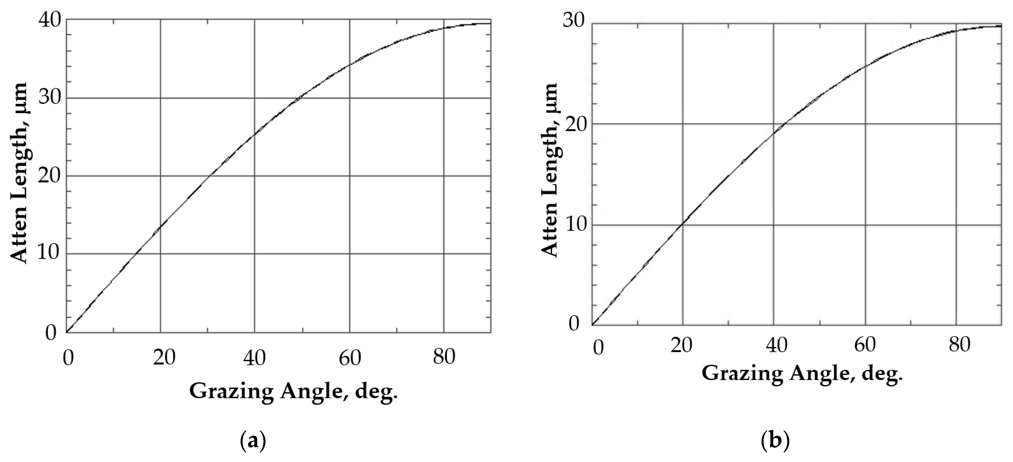
Figure 1. Calculating the depth of CuKα X-ray radiation penetration into the Na0.5Zr2(PO4)2.5(MoO4)0.5 (a) and Na0.5Zr2(PO4)2.5(WO4)0.5 (b) specimens depending on α incidence angle. Energy = 8000 eV.
Materials 16 00965 g001
Figure 2. XRD curves of the phosphate tungstates specimens (a) and the phosphate molybdates specimens (b) with x = 0.5 after irradiation at the following fluences (cm−2). Initial state and when exposed to different Xe ion doses (in cm−2): (a): W1—3·1012; W2—1013; W3—3·1013; (b): M1—1012; M2 –3·1012; M3—6·1012; M4—8·1012; M5—1013; M6—3·1013; M7—6·1013.
Materials 16 00965 g002
Figure 3. Dependence of integral intensity of a XRD peak (113) in Na0.5Zr2(PO4)2.5(MoO4)0.5 phase on α incidence angle for Na0.5Zr2(PO4)2.5(MoO4)0.5 ceramics before and after irradiation. Irradiation intensity is multiplied by a factor of 4. The calculated curve is plotted with due regard for material constants and the geometry of the experiment as if crystalline quality and phase composition of the material were uniform over the entire depth of analysis.
Materials 16 00965 g003
Figure 4. Results of SRIM simulation of vacancies depth distribution caused by 167 MeV Xe ions in M- and W-series specimens.
Materials 16 00965 g004
Figure 5. Profiles of main (a) and doping (b) elements on the surface of the Na0.5Zr2(PO4)2.5(WO4)0.5 ceramics in negative secondary ion detection mode.
Materials 16 00965 g005
Figure 6. Profiles of main (a) and doping (b) elements on the surface of the irradiated Na0.5Zr2(PO4)2.5(WO4)0.5 ceramics in negative secondary ion detection mode.
Materials 16 00965 g006
Figure 7. Profiles of main (a) and doping (b) elements on the surface of the Na0.5Zr2(PO4)2.5(WO4)0.5 ceramics in the initial state. Positive secondary ion detection mode. SIMS.
Materials 16 00965 g007
Figure 8. Profiles of main (a) and doping (b) elements on the surface of the irradiated Na0.5Zr2(PO4)2.5(WO4)0.5 ceramics. Positive secondary ion detection mode. SIMS.
Materials 16 00965 g008
Figure 9. Time dependences of normalized weight loss NL (a) and normalized leaching rates for certain components per unit surface area R (b) in the Na1−xZr2(PO4)3−x(XO4)x ceramics.
Materials 16 00965 g009
Figure 10. Dependence of normalized weight loss (NL) (a,c) and leaching rate (R) (b,d) on testing time t for the Na0.5Zr2(PO4)2.5(MoO4)0.5 (a,b) and Na0.5Zr2(PO4)2.5(WO4)0.5 (c,d) ceramic specimens. Fluence: 1–3·1012 cm−2, 2–1·1013 cm−2, 3–3·1013 cm−2.
Materials 16 00965 g010
Figure 11. XRD patterns for the Na0.5Zr2(PO4)2.5(MoO4)0.5 ceramic specimen: initial state, irradiated with Xe ions at a dose of 6·1013 cm−2, and after hydrolytic tests.
Materials 16 00965 g011
Figure 12. XRD patterns for the Na0.5Zr2(PO4)2.5(WO4)0.5 ceramic specimen: initial state, irradiated with Xe ions at a dose of 3·1012 cm−2, and after hydrolytic tests.
Materials 16 00965 g012
Table 1. Normalized weight loss (NL) and normalized leaching rates (R) after 28 days of testing for Mo and W in the Na1−xZr2(PO4)3−x(XO4)x ceramics.
xt, Daysm·104, gNL·102, g·cm−2R·105, g·cm−2·d−1
MoWMoWMoW
0.415.9178.3330.4530.35260.00012.300
31.4170.4170.5610.36923.5834.289
70.9580.2750.6340.38111.4771.903
100.3750.1330.6630.3878.4751.352
140.3750.1250.6920.3926.3670.979
210.4580.1670.7270.3994.5110.664
280.3750.1250.7550.4043.5320.504
0.517.2507.5000.4270.24655.6008.200
31.8330.3920.5350.25921.5912.859
70.9580.2080.5910.26610.4101.269
100.4330.1250.6170.2707.6570.901
140.4000.1080.6400.2745.7310.653
210.4420.1670.6660.2794.0420.442
280.4080.1670.6900.2853.1560.336
Disclaimer/Publisher’s Note: The statements, opinions and data contained in all publications are solely those of the individual author(s) and contributor(s) and not of MDPI and/or the editor(s). MDPI and/or the editor(s) disclaim responsibility for any injury to people or property resulting from any ideas, methods, instructions or products referred to in the content.