
Recent Trends in Hydroxyapatite Supplementation for Osteoregenerative Purposes


 ,
,
Abstract
1. Introduction
2. Materials and Methods
3. Hydroxyapatite
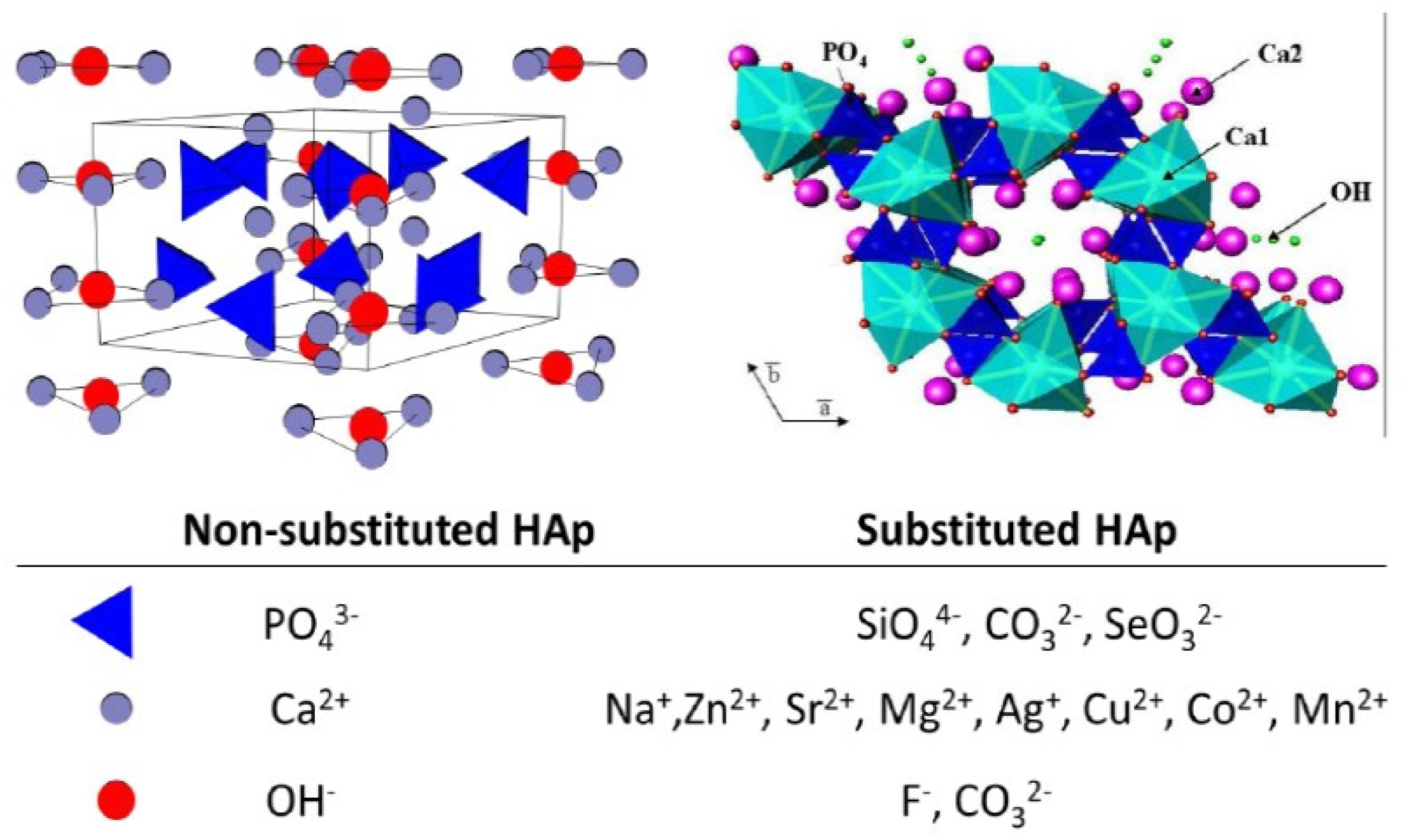
3.1. Components, Structure and Properties of Hydroxyapatite
3.2. Bone Cells and Their Interactions with Hydroxyapatite
3.2.1. Osteoblasts
3.2.2. Osteocytes
3.2.3. Osteoclasts

| Cell Type | Role | Reference |
|---|---|---|
| Osteoblasts | Bone formation Synthesis of extracellular proteins Synthesis of endogenous HAP | [17,18,19,20,23] [21,22] [24,25] |
| Osteocytes | Bone remodeling Influence on bone integrity | [37] [38] |
| Osteoclasts | Bone repair and remodeling Bone matrix resorption | [37] [42] |
3.3. Molecular Role of Hydroxyapatite and Signaling Pathways
4. Hydroxyapatite Nanoformulations
4.1. Nano-Hydroxyapatite Synthesis Methods
4.1.1. Wet Chemical Precipitation Synthesis
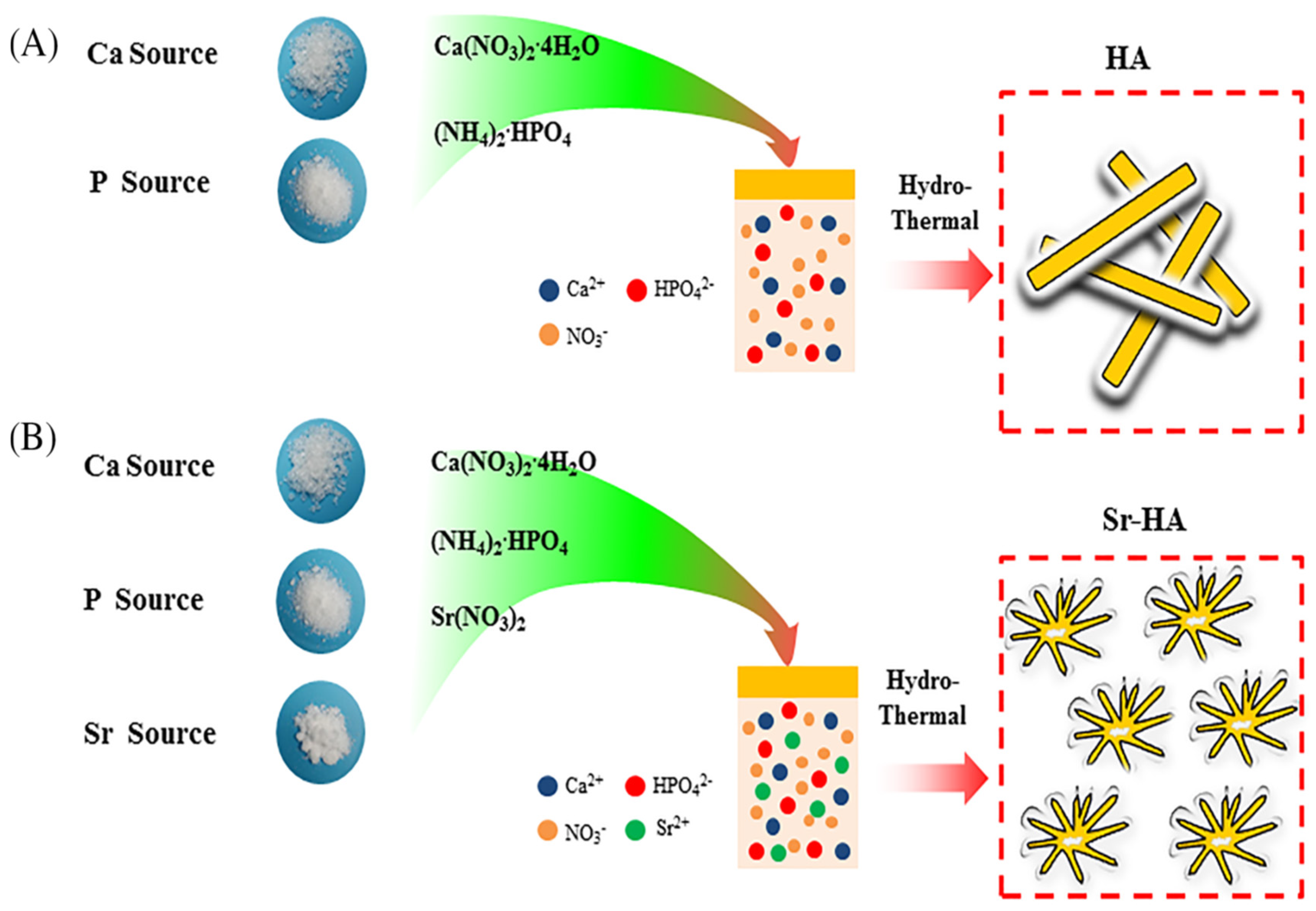
4.1.2. Hydrothermal Synthesis
4.1.3. Micro-Emulsion Synthesis
4.1.4. Sol–Gel Synthesis
| HA Synthesis Method | Advantages | Disadvantages | Reference |
|---|---|---|---|
| Wet chemical precipitation synthesis | Able to control nHAP particle size Versatile, reliable, feasible | Irregular shape Unsatisfactory surface morphology | [70,71,72,73,74,75,76,77,78,79,80] [9,79] |
| Hydrothermal synthesis | Rapid fabrication Technical simplicity Increased crystallinity Influences surface morphology independent of scaffold shape | Difficult to control agglomeration | [9,79] [82] [84,85] |
| Microemulsion synthesis | Creates nanometer-sized particles with minimal agglomeration | Unable to perform at low temperature | [86,87,88] |
| Sol-gel synthesis | Performs at low temperatures Cost-effective High-purity, homogenous coatings Uniform dispersation | [88,89,90,91,92,93] | |
| Microwave synthesis | Rapid, homogenous internal and volumetric heating High crystallinity | [64,65,66,67,68,69,70] |
4.2. Nano-Hydroxyapatite Mixed with Ions
4.2.1. Magnesium-Nano-Hydroxyapatite
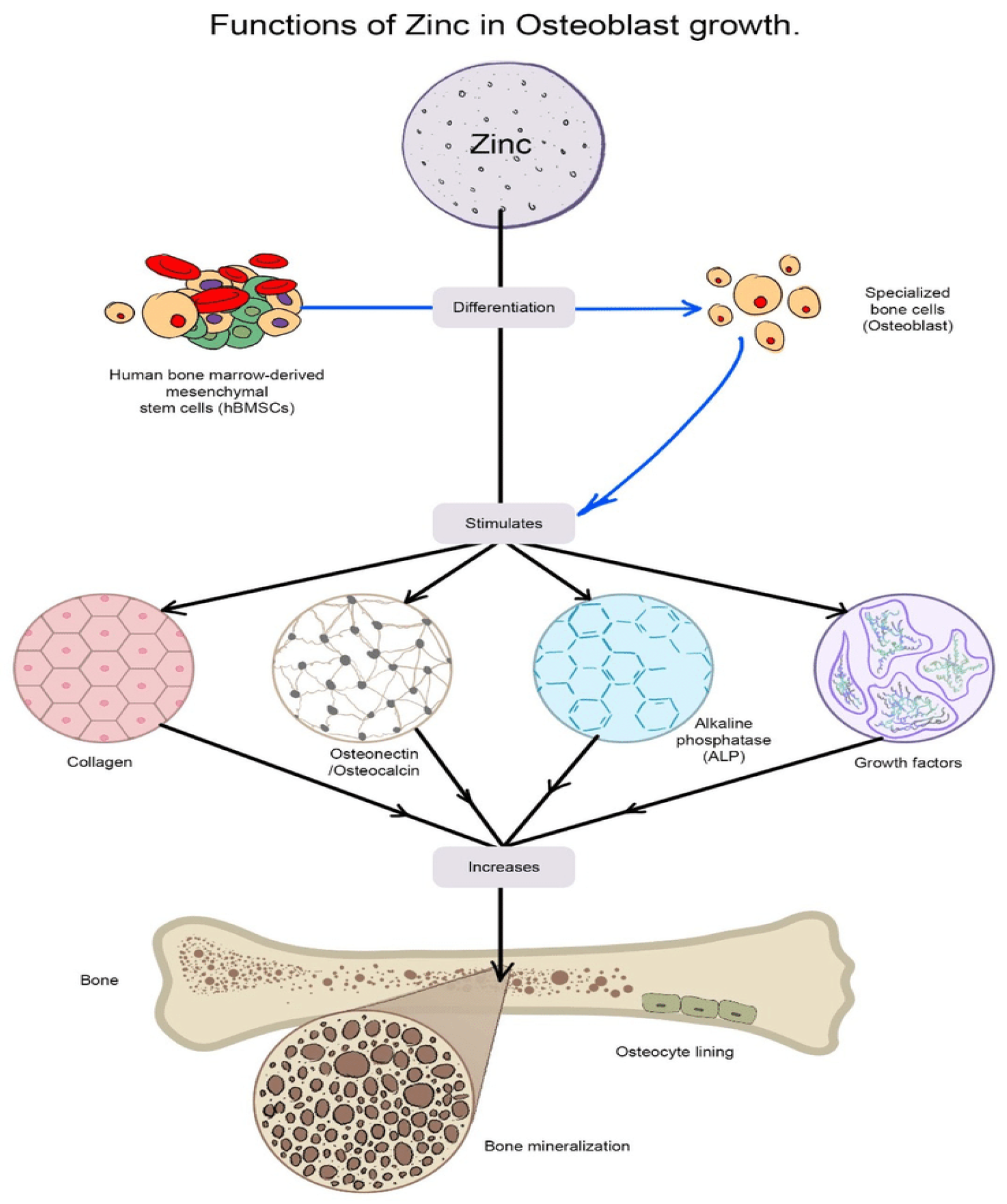
4.2.2. Zinc-Nano-Hydroxyapatite
4.2.3. Selenium-Nano-Hydroxyapatite
4.2.4. Strontium-Nano-Hydroxyapatite
4.2.5. Boron-Nano-Hydroxyapatite
4.2.6. Cobalt-Nano-Hydroxyapatite
4.2.7. Copper-Nano-Hydroxyapatite
4.2.8. Silicon Nano-Hydroxyapatite
4.2.9. Multi-Substituted Hydroxyapatite
4.2.10. Hydroxyapatite Combined with Other Ions
5. Hydroxyapatite Enriched with Curcumin
6. Conclusions and Future Perspectives
Author Contributions
Funding
Institutional Review Board Statement
Informed Consent Statement
Data Availability Statement
Conflicts of Interest
References
- Hellwinkel, J.E.; Working, Z.M.; Certain, L.; García, A.J.; Wenke, J.C.; Bahney, C.S. The intersection of fracture healing and infection: Orthopaedics research society workshop 2021. J. Orthop. Res. 2022, 40, 541–552. [Google Scholar] [CrossRef] [PubMed]
- Nauth, A.; Schemitsch, E.; Norris, B.; Nollin, Z.; Watson, J.T. Critical-Size Bone Defects: Is There a Consensus for Diagnosis and Treatment? J. Orthop. Trauma 2018, 32 (Suppl. 1), S7–S11. [Google Scholar] [CrossRef] [PubMed]
- Karpiński, R.; Szabelski, J.; Krakowski, P.; Jojczuk, M.; Jonak, J.; Nogalski, A. Evaluation of the Effect of Selected Physiological Fluid Contaminants on the Mechanical Properties of Selected Medium-Viscosity PMMA Bone Cements. Materials 2022, 15, 2197. [Google Scholar] [CrossRef]
- Szabelski, J.; Karpiński, R.; Krakowski, P.; Jojczuk, M.; Jonak, J.; Nogalski, A. Analysis of the Effect of Component Ratio Imbalances on Selected Mechanical Properties of Seasoned, Medium Viscosity Bone Cements. Materials 2022, 15, 5577. [Google Scholar] [CrossRef] [PubMed]
- Gisep, A.; Kugler, S.; Wahl, D.; Rahn, B. Mechanical characterisation of a bone defect model filled with ceramic cements. J. Mater. Sci. Mater. Med. 2004, 15, 1065. [Google Scholar] [CrossRef]
- Habibah, T.; Amlani, D.; Brizuela, M. Hydroxyapatite Dental Material. In StatPearls; StatPearls Publishing: Treasure Island, FL, USA, 2022. [Google Scholar]
- Ramesh, S.; Tan, C.Y.; Aw, K.L.; Yeo, W.H.; Hamdi, M.; Sopyan, I.; Teng, W.D. Sintering behaviour of hydroxyapatite bioceramics. Med. J. Malays. 2008, 63 (Suppl. A), 89–90. [Google Scholar]
- Barabas, R.; Rigo, M.; Sarkozi, M.; Hoaghia, M.; Cadar, O. Hydroxyapatite-carbon nanotube composites for drug delivery applications. Braz. J. Chem. Eng. 2019, 36, 913–922. [Google Scholar] [CrossRef]
- Loo, S.; Moore, T.; Banik, B.; Alexis, F. Biomedical applications of hydroxyapatite nanoparticles. Curr. Pharm. Biotechnol. 2010, 11, 333–342. [Google Scholar] [CrossRef]
- Kowal, T.J.; Hahn, N.C.; Eider, S.; Marzillier, J.Y.; Fodera, D.M.; Thamma, U.; Jain, H.; Falk, M.M. New bioactive glass scaffolds with exceptional qualities for bone tissue regeneration: Response of osteoblasts and osteoclasts. Biomed. Mater. 2018, 13, 025005. [Google Scholar] [CrossRef]
- Komur, B.; Lohse, T.; Can, H.M.; Khalilova, G.; Geçimli, Z.N.; Aydoğdu, M.O.; Kalkandelen, C.; Stan, G.E.; Sahin, Y.M.; Sengil, A.Z. Fabrication of naturel pumice/hydroxyapatite composite for biomedical engineering. BioMed. Eng. OnLine 2016, 15, 1–20. [Google Scholar] [CrossRef]
- Meleshko, A.A.; Tolstoy, V.P.; Afinogenov, G.E.; Levshakova, A.S.; Afinogenova, A.G.; Muldiyarov, V.P.; Vissarionov, S.V.; Linnik, S.A. Prospects of hydroxyapatite-based nanomaterials application synthesized by layer-by-layer method for pediatric traumatology and orthopedics. Pediatr. Traumatol. Orthop. Reconstr. Surg. 2020, 8, 217–230. [Google Scholar] [CrossRef]
- Sobti, M.M.; Shams, F.; Jawaheer, L.; Cauchi, P.; Chadha, V. Unwrapped hydroxyapatite orbital implants: Our experience in 347 cases. Eye 2020, 34, 675–682. [Google Scholar] [CrossRef] [PubMed]
- Compton, J.T.; Lee, F.Y. A review of osteocyte function and the emerging importance of sclerostin. J. Bone Jt. Surg Am 2014, 96, 1659–1668. [Google Scholar] [CrossRef] [PubMed]
- Knothe Tate, M.L.; Adamson, J.R.; Tami, A.E.; Bauer, T.W. The osteocyte. Int. J. Biochem. Cell Biol. 2004, 36, 1–8. [Google Scholar] [CrossRef]
- Creecy, A.; Damrath, J.G.; Wallace, J.M. Control of bone matrix properties by osteocytes. Front. Endocrinol. 2021, 11, 578477. [Google Scholar] [CrossRef] [PubMed]
- Liu, X.; Zhao, M.; Lu, J.; Ma, J.; Wei, J.; Wei, S. Cell responses to two kinds of nanohydroxyapatite with different sizes and crystallinities. Int. J. Nanomed. 2012, 7, 1239–1250. [Google Scholar] [CrossRef]
- Liu, F.; Fang, F.; Yuan, H.; Yang, D.; Chen, Y.; Williams, L.; Goldstein, S.A.; Krebsbach, P.H.; Guan, J. Suppression of autophagy by FIP200 deletion leads to osteopenia in mice through the inhibition of osteoblast terminal differentiation. J. Bone Miner. Res. 2013, 28, 2414–2430. [Google Scholar] [CrossRef]
- Nollet, M.; Santucci-Darmanin, S.; Breuil, V.; Al-Sahlanee, R.; Cros, C.; Topi, M.; Momier, D.; Samson, M.; Pagnotta, S.; Cailleteau, L. Autophagy in osteoblasts is involved in mineralization and bone homeostasis. Autophagy 2014, 10, 1965–1977. [Google Scholar] [CrossRef]
- Seleverstov, O.; Zabirnyk, O.; Zscharnack, M.; Bulavina, L.; Nowicki, M.; Heinrich, J.; Yezhelyev, M.; Emmrich, F.; O’Regan, R.; Bader, A. Quantum dots for human mesenchymal stem cells labeling. A size-dependent autophagy activation. Nano Lett. 2006, 6, 2826–2832. [Google Scholar] [CrossRef]
- Kim, J.; Lin, C.; Stavre, Z.; Greenblatt, M.B.; Shim, J. Osteoblast-osteoclast communication and bone homeostasis. Cells 2020, 9, 2073. [Google Scholar] [CrossRef]
- Long, F. Building strong bones: Molecular regulation of the osteoblast lineage. Nat. Rev. Mol. Cell Biol. 2012, 13, 27–38. [Google Scholar] [CrossRef] [PubMed]
- Ansari, N.; Sims, N.A. The cells of bone and their interactions. In Bone Regulators and Osteoporosis Therapy; Springer: Berlin/Heidelberg, Germany, 2019; pp. 1–25. [Google Scholar]
- Simonet, W.; Lacey, D.; Dunstan, C.; Kelley, M.; Chang, M.; Lüthy, R.; Nguyen, H.; Wooden, S.; Bennett, L.; Boone, T. Osteoprotegerin: A novel secreted protein involved in the regulation of bone density. Cell 1997, 89, 309–319. [Google Scholar] [CrossRef] [PubMed]
- Dallas, S.L.; Prideaux, M.; Bonewald, L.F. The osteocyte: An endocrine cell … and more. Endocr. Rev. 2013, 34, 658–690. [Google Scholar] [CrossRef]
- Asano, T.; Okamoto, K.; Nakai, Y.; Tsutsumi, M.; Muro, R.; Suematsu, A.; Hashimoto, K.; Okamura, T.; Ehata, S.; Nitta, T. Soluble RANKL is physiologically dispensable but accelerates tumour metastasis to bone. Nat. Metab. 2019, 1, 868–875. [Google Scholar] [CrossRef]
- Jimi, E.; Nakamura, I.; Amano, H.; Taguchi, Y.; Tsurukai, T.; Tamura, M.; Takahashi, N.; Suda, T. Osteoclast function is activated by osteoblastic cells through a mechanism involving cell-to-cell contact. Endocrinology 1996, 137, 2187–2190. [Google Scholar] [CrossRef]
- Tamura, T.; Udagawa, N.; Takahashi, N.; Miyaura, C.; Tanaka, S.; Yamada, Y.; Koishihara, Y.; Ohsugi, Y.; Kumaki, K.; Taga, T. Soluble interleukin-6 receptor triggers osteoclast formation by interleukin 6. Proc. Natl. Acad. Sci. USA 1993, 90, 11924–11928. [Google Scholar] [CrossRef] [PubMed]
- Nakashima, T.; Hayashi, M.; Fukunaga, T.; Kurata, K.; Oh-Hora, M.; Feng, J.Q.; Bonewald, L.F.; Kodama, T.; Wutz, A.; Wagner, E.F. Evidence for osteocyte regulation of bone homeostasis through RANKL expression. Nat. Med. 2011, 17, 1231–1234. [Google Scholar] [CrossRef]
- Xiong, J.; Piemontese, M.; Onal, M.; Campbell, J.; Goellner, J.J.; Dusevich, V.; Bonewald, L.; Manolagas, S.C.; O’Brien, C.A. Osteocytes, not osteoblasts or lining cells, are the main source of the RANKL required for osteoclast formation in remodeling bone. PloS ONE 2015, 10, e0138189. [Google Scholar] [CrossRef]
- Schaffler, M.B.; Cheung, W.; Majeska, R.; Kennedy, O. Osteocytes: Master orchestrators of bone. Calcif. Tissue Int. 2014, 94, 5–24. [Google Scholar] [CrossRef]
- Van Wesenbeeck, L.; Odgren, P.R.; MacKay, C.A.; D’Angelo, M.; Safadi, F.F.; Popoff, S.N.; Van Hul, W.; Marks, S.C.J. The osteopetrotic mutation toothless (tl) is a loss-of-function frameshift mutation in the rat Csf1 gene: Evidence of a crucial role for CSF-1 in osteoclastogenesis and endochondral ossification. Proc. Natl. Acad. Sci. USA 2002, 99, 14303–14308. [Google Scholar] [CrossRef]
- Wong, B.R.; Besser, D.; Kim, N.; Arron, J.R.; Vologodskaia, M.; Hanafusa, H.; Choi, Y. TRANCE, a TNF family member, activates Akt/PKB through a signaling complex involving TRAF6 and c-Src. Mol. Cell. 1999, 4, 1041–1049. [Google Scholar] [CrossRef]
- Buenzli, P.R.; Sims, N.A. Quantifying the osteocyte network in the human skeleton. Bone 2015, 75, 144–150. [Google Scholar] [CrossRef]
- Clarke, B. Normal bone anatomy and physiology. Clin. J. Am. Soc. Nephrol. 2008, 3 (Suppl. 3), S131–S139. [Google Scholar] [CrossRef] [PubMed]
- Blair, H.C.; Larrouture, Q.C.; Li, Y.; Lin, H.; Beer-Stoltz, D.; Liu, L.; Tuan, R.S.; Robinson, L.J.; Schlesinger, P.H.; Nelson, D.J. Osteoblast differentiation and bone matrix formation in vivo and in vitro. Tissue Eng. Part B Rev. 2017, 23, 268–280. [Google Scholar] [CrossRef] [PubMed]
- Crockett, J.C.; Rogers, M.J.; Coxon, F.P.; Hocking, L.J.; Helfrich, M.H. Bone remodelling at a glance. J. Cell Sci. 2011, 124, 991–998. [Google Scholar] [CrossRef] [PubMed]
- Hartgers, F.C.; Vissers, J.L.; Looman, M.W.; Zoelen, C.v.; Huffine, C.; Figdor, C.G.; Adema, G.J. DC-STAMP, a novel multimembrane-spanning molecule preferentially expressed by dendritic cells. Eur. J. Immunol. 2000, 30, 3585–3590. [Google Scholar] [CrossRef]
- Kukita, T.; Wada, N.; Kukita, A.; Kakimoto, T.; Sandra, F.; Toh, K.; Nagata, K.; Iijima, T.; Horiuchi, M.; Matsusaki, H. RANKL-induced DC-STAMP is essential for osteoclastogenesis. J. Exp. Med. 2004, 200, 941–946. [Google Scholar] [CrossRef]
- Miyamoto, H.; Suzuki, T.; Miyauchi, Y.; Iwasaki, R.; Kobayashi, T.; Sato, Y.; Miyamoto, K.; Hoshi, H.; Hashimoto, K.; Yoshida, S. Osteoclast stimulatory transmembrane protein and dendritic cell–specific transmembrane protein cooperatively modulate cell–cell fusion to form osteoclasts and foreign body giant cells. J. Bone Miner. Res. 2012, 27, 1289–1297. [Google Scholar]
- Jansen, I.D.; Vermeer, J.A.; Bloemen, V.; Stap, J.; Everts, V. Osteoclast fusion and fission. Calcif. Tissue Int. 2012, 90, 515–522. [Google Scholar]
- Martin, T.J.; Sims, N.A. RANKL/OPG. Critical role in bone physiology. Rev. Endocr. Metab. Disord. 2015, 16, 131–139. [Google Scholar] [CrossRef]
- Henriksen, K.; Karsdal, M.A.; John Martin, T. Osteoclast-derived coupling factors in bone remodeling. Calcif. Tissue Int. 2014, 94, 88–97. [Google Scholar] [CrossRef]
- Dougall, W.C.; Glaccum, M.; Charrier, K.; Rohrbach, K.; Brasel, K.; De Smedt, T.; Daro, E.; Smith, J.; Tometsko, M.E.; Maliszewski, C.R.; et al. RANK is essential for osteoclast and lymph node development. Genes Dev. 1999, 13, 2412–2424. [Google Scholar] [CrossRef]
- Kong, Y.; Yoshida, H.; Sarosi, I.; Tan, H.; Timms, E.; Capparelli, C.; Morony, S.; Oliveira-dos-Santos, A.J.; Van, G.; Itie, A. OPGL is a key regulator of osteoclastogenesis, lymphocyte development and lymph-node organogenesis. Nature 1999, 397, 315–323. [Google Scholar] [CrossRef] [PubMed]
- Blank, M.; Sims, N.A. Cellular processes by which osteoblasts and osteocytes control bone mineral deposition and maturation revealed by stage-specific EphrinB2 knockdown. Curr. Osteoporos. Rep. 2019, 17, 270–280. [Google Scholar] [CrossRef]
- Tsourdi, E.; Jahn, K.; Rauner, M.; Busse, B.; Bonewald, L.F. Physiological and pathological osteocytic osteolysis. J. Musculoskelet. Neuronal. Interact. 2018, 18, 292–303. [Google Scholar] [PubMed]
- Yasuda, H.; Shima, N.; Nakagawa, N.; Yamaguchi, K.; Kinosaki, M.; Mochizuki, S.; Tomoyasu, A.; Yano, K.; Goto, M.; Murakami, A.; et al. Osteoclast differentiation factor is a ligand for osteoprotegerin/osteoclastogenesis-inhibitory factor and is identical to TRANCE/RANKL. Proc. Natl. Acad. Sci. USA 1998, 95, 3597–3602. [Google Scholar] [CrossRef] [PubMed]
- Nakagawa, N.; Kinosaki, M.; Yamaguchi, K.; Shima, N.; Yasuda, H.; Yano, K.; Morinaga, T.; Higashio, K. RANK is the essential signaling receptor for osteoclast differentiation factor in osteoclastogenesis. Biochem. Biophys. Res. Commun. 1998, 253, 395–400. [Google Scholar] [CrossRef]
- Zhang, F.; Zhou, Z.; Yang, S.; Mao, L.; Chen, H.; Yu, X. Hydrothermal synthesis of hydroxyapatite nanorods in the presence of anionic starburst dendrimer. Mater. Lett. 2005, 59, 1422–1425. [Google Scholar] [CrossRef]
- Ioku, K.; Yamauchi, S.; Fujimori, H.; Goto, S.; Yoshimura, M. Hydrothermal preparation of fibrous apatite and apatite sheet. Solid State Ion. 2002, 151, 147–150. [Google Scholar] [CrossRef]
- Zhang, H.; Zhou, K.; Li, Z.; Huang, S. Plate-like hydroxyapatite nanoparticles synthesized by the hydrothermal method. J. Phys. Chem. Solids 2009, 70, 243–248. [Google Scholar] [CrossRef]
- Kaygili, O.; Keser, S.; Kom, M.; Eroksuz, Y.; Dorozhkin, S.V.; Ates, T.; Ozercan, I.H.; Tatar, C.; Yakuphanoglu, F. Strontium substituted hydroxyapatites: Synthesis and determination of their structural properties, in vitro and in vivo performance. Mater. Sci. Eng. C 2015, 55, 538–546. [Google Scholar] [CrossRef] [PubMed]
- Kaygili, O.; Tatar, C.; Yakuphanoglu, F.; Keser, S. Nano-crystalline aluminum-containing hydroxyapatite based bioceramics: Synthesis and characterization. J. Sol. Gel. Sci. Technol. 2013, 65, 105–111. [Google Scholar] [CrossRef]
- Eshtiagh-Hosseini, H.; Housaindokht, M.R.; Chahkandi, M. Effects of parameters of sol–gel process on the phase evolution of sol–gel-derived hydroxyapatite. Mater. Chem. Phys. 2007, 106, 310–316. [Google Scholar] [CrossRef]
- Pattanayak, D.K.; Dash, R.; Prasad, R.; Rao, B.; Mohan, T.R. Synthesis and sintered properties evaluation of calcium phosphate ceramics. Mater. Sci. Eng. C 2007, 27, 684–690. [Google Scholar] [CrossRef]
- Testinon, A.; Buscaglia, M.T.; Viviani, M.; Buscaglia, V.; Nanni, P. Synthesis of BaTiO3 particles with tailored size by precipitation from aqueous solutions. J. Am. Ceram. Soc. 2004, 87, 79–83. [Google Scholar] [CrossRef]
- Veljović, D.; Jokić, B.; Petrović, R.; Palcevskis, E.; Dindune, A.; Mihailescu, I.N.; Janaćković, D. Processing of dense nanostructured HAP ceramics by sintering and hot pressing. Ceram Int. 2009, 35, 1407–1413. [Google Scholar] [CrossRef]
- Liu, J.; Li, K.; Wang, H.; Zhu, M.; Yan, H. Rapid formation of hydroxyapatite nanostructures by microwave irradiation. Chem. Phys. Lett. 2004, 396, 429–432. [Google Scholar] [CrossRef]
- Coelho, J.; Moreira, J.A.; Almeida, A.; Monteiro, F. Synthesis and characterization of HAp nanorods from a cationic surfactant template method. J. Mater. Sci. Mater. Med. 2010, 21, 2543–2549. [Google Scholar] [CrossRef]
- Kalita, S.J.; Bhardwaj, A.; Bhatt, H.A. Nanocrystalline calcium phosphate ceramics in biomedical engineering. Mater. Sci. Eng. C 2007, 27, 441–449. [Google Scholar] [CrossRef]
- Simon, V.; Lazăr, D.; Turcu, R.; Mocuta, H.; Magyari, K.; Prinz, M.; Neumann, M.; Simon, S. Atomic environment in sol–gel derived nanocrystalline hydroxyapatite. Mater. Sci. Eng. B 2009, 165, 247–251. [Google Scholar] [CrossRef]
- Kim, I.; Kumta, P.N. Sol–gel synthesis and characterization of nanostructured hydroxyapatite powder. Mater. Mater. Mater. Sci. Eng. B 2004, 111, 232–236. [Google Scholar] [CrossRef]
- Shakir, M.; Kushwaha, S.; Maurya, K.; Bhagavannarayana, G.; Wahab, M. Characterization of ZnSe nanoparticles synthesized by microwave heating process. Solid State Commun. 2009, 149, 2047–2049. [Google Scholar] [CrossRef]
- Sutton, W.H. Microwave processing of ceramic materials. Am. Ceram. Soc. Bull. 1989, 68, 376–386. [Google Scholar]
- Shkir, M.; Yahia, I.; AlFaify, S.; Abutalib, M.; Muhammad, S. Facile synthesis of lead iodide nanostructures by microwave irradiation technique and their structural, morphological, photoluminescence and dielectric studies. J. Mol. Struct. 2016, 1110, 83–90. [Google Scholar] [CrossRef]
- Chanda, A.; Dasgupta, S.; Bose, S.; Bandyopadhyay, A. Microwave sintering of calcium phosphate ceramics. Mater. Sci. Eng. C 2009, 29, 1144–1149. [Google Scholar] [CrossRef]
- Silva, C.; Valente, M.; Graça, M.; Sombra, A. The modulus formalism used in the dielectric analysis of hydroxyapatite and calcium phosphate with titanium formed by dry ball milling. J. Non. Cryst. Solids 2005, 351, 2945–2950. [Google Scholar] [CrossRef]
- Escudero, A.; Calvo, M.E.; Rivera-Fernández, S.; De la Fuente, J.M.; Ocaña, M. Microwave-assisted synthesis of biocompatible europium-doped calcium hydroxyapatite and fluoroapatite luminescent nanospindles functionalized with poly (acrylic acid). Langmuir 2013, 29, 1985–1994. [Google Scholar] [CrossRef]
- Yahia, I.; Shkir, M.; AlFaify, S.; Ganesh, V.; Zahran, H.; Kilany, M. Facile microwave-assisted synthesis of Te-doped hydroxyapatite nanorods and nanosheets and their characterizations for bone cement applications. Mater. Sci. Eng. C 2017, 72, 472–480. [Google Scholar] [CrossRef]
- Afshar, A.; Ghorbani, M.; Ehsani, N.; Saeri, M.; Sorrell, C. Some important factors in the wet precipitation process of hydroxyapatite. Mater. Des. 2003, 24, 197–202. [Google Scholar] [CrossRef]
- Pang, Y.; Bao, X. Influence of temperature, ripening time and calcination on the morphology and crystallinity of hydroxyapatite nanoparticles. J. Eur. Ceram. Soc. 2003, 23, 1697–1704. [Google Scholar] [CrossRef]
- Welzel, T.; Meyer-Zaika, W.; Epple, M. Continuous preparation of functionalised calcium phosphate nanoparticles with adjustable crystallinity. Chem. Commun. 2004, 1204–1205. [Google Scholar] [CrossRef] [PubMed]
- Kumar, R.; Prakash, K.; Cheang, P.; Khor, K. Temperature driven morphological changes of chemically precipitated hydroxyapatite nanoparticles. Langmuir 2004, 20, 5196–5200. [Google Scholar] [CrossRef] [PubMed]
- Li-yun, C.; Chuan-bo, Z.; Jian-feng, H. Influence of temperature, [Ca2+], Ca/P ratio and ultrasonic power on the crystallinity and morphology of hydroxyapatite nanoparticles prepared with a novel ultrasonic precipitation method. Mater. Lett. 2005, 59, 1902–1906. [Google Scholar] [CrossRef]
- Bouyer, E.; Gitzhofer, F.; Boulos, M. Morphological study of hydroxyapatite nanocrystal suspension. J. Mater. Sci. Mater. Med. 2000, 11, 523–531. [Google Scholar] [CrossRef] [PubMed]
- Liu, Y.; Hou, D.; Wang, G. A simple wet chemical synthesis and characterization of hydroxyapatite nanorods. Mater. Chem. Phys. 2004, 86, 69–73. [Google Scholar] [CrossRef]
- Ślósarczyk, A.; Paszkiewicz, Z.; Paluszkiewicz, C. FTIR and XRD evaluation of carbonated hydroxyapatite powders synthesized by wet methods. J. Mol. Struct. 2005, 744, 657–661. [Google Scholar] [CrossRef]
- Loo, S.C.J.; Siew, Y.E.; Ho, S.; Boey, F.Y.C.; Ma, J. Synthesis and hydrothermal treatment of nanostructured hydroxyapatite of controllable sizes. J. Mater. Sci. Mater. Med. 2008, 19, 1389–1397. [Google Scholar] [CrossRef]
- Rahaman, M. Ceramic Processing Sintering; CRC Press: Boca Raton, FL, USA, 1995. [Google Scholar]
- Byrappa, K.; Yoshimura, M. Handbook of Hydrothermal Technology; Elsevier: Amsterdam, The Netherlands, 2012. [Google Scholar]
- López-Macipe, A.; Gómez-Morales, J.; Rodríguez-Clemente, R. Nanosized hydroxyapatite precipitation from homogeneous calcium/citrate/phosphate solutions using microwave and conventional heating. Adv. Mater. 1998, 10, 49–53. [Google Scholar] [CrossRef]
- Taheri, M.M.; Kadir, M.R.A.; Shokuhfar, T.; Hamlekhan, A.; Assadian, M.; Shirdar, M.R.; Mirjalili, A. Surfactant-assisted hydrothermal synthesis of fluoridated hydroxyapatite nanorods. Ceram Int. 2015, 41, 9867–9872. [Google Scholar] [CrossRef]
- Qin, J.; Zhong, Z.; Ma, J. Biomimetic synthesis of hybrid hydroxyapatite nanoparticles using nanogel template for controlled release of bovine serum albumin. Mater. Sci. Eng. C 2016, 62, 377–383. [Google Scholar] [CrossRef]
- Qi, Y.; Shen, J.; Jiang, Q.; Jin, B.; Chen, J.; Zhang, X. The morphology control of hydroxyapatite microsphere at high pH values by hydrothermal method. Adv. Powder Technol. 2015, 26, 1041–1046. [Google Scholar] [CrossRef]
- Elrayah, A.; Zhi, W.; Feng, S.; Al-Ezzi, S.; Lei, H.; Weng, J. Preparation of micro/nano-structure copper-substituted hydroxyapatite scaffolds with improved angiogenesis capacity for bone regeneration. Materials 2018, 11, 1516. [Google Scholar] [CrossRef] [PubMed]
- Bose, S.; Saha, S.K. Synthesis and characterization of hydroxyapatite nanopowders by emulsion technique. Chem. Mater. 2003, 15, 4464–4469. [Google Scholar] [CrossRef]
- Furuzono, T.; Sonoda, K.; Tanaka, J. A hydroxyapatite coating covalently linked onto a silicone implant material. J. Biomed. Mater. Res. 2001, 56, 9–16. [Google Scholar] [CrossRef]
- Jarudilokkul, S.; Tanthapanichakoon, W.; Boonamnuayvittaya, V. Synthesis of hydroxyapatite nanoparticles using an emulsion liquid membrane system. Colloids Surf Phys. Eng. Asp. 2007, 296, 149–153. [Google Scholar] [CrossRef]
- Pileni, M. The role of soft colloidal templates in controlling the size and shape of inorganic nanocrystals. Nat. Mater. 2003, 2, 145–150. [Google Scholar] [CrossRef]
- Arcos, D.; Vallet-Regí, M. Substituted hydroxyapatite coatings of bone implants. J. Mater. Chem. B 2020, 8, 1781–1800. [Google Scholar] [CrossRef]
- Danks, A.E.; Hall, S.R.; Schnepp, Z. The evolution of ‘sol–gel’chemistry as a technique for materials synthesis. Mater. Horiz. 2016, 3, 91–112. [Google Scholar] [CrossRef]
- Feinle, A.; Elsaesser, M.S.; Huesing, N. Sol–gel synthesis of monolithic materials with hierarchical porosity. Chem. Soc. Rev. 2016, 45, 3377–3399. [Google Scholar] [CrossRef]
- Dave, B.C. Sol-Gel Coating Methods in Biomedical Systems. In Medical Coatings and Deposition Technologies; Scrivener: Beverly, MA, USA, 2016; p. 373. [Google Scholar]
- Côté, A.S.; Cormack, A.N.; Tilocca, A. Reactive molecular dynamics: An effective tool for modelling the sol–gel synthesis of bioglasses. J. Mater. Sci. 2017, 52, 9006–9013. [Google Scholar] [CrossRef]
- Ishikawa, K.; Garskaite, E.; Kareiva, A. Sol–gel synthesis of calcium phosphate-based biomaterials—A review of environmentally benign, simple, and effective synthesis routes. J. Sol. Gel. Sci. Technol. 2020, 94, 551–572. [Google Scholar] [CrossRef]
- Wopenka, B.; Pasteris, J.D. A mineralogical perspective on the apatite in bone. Mater. Sci. Eng. C 2005, 25, 131–143. [Google Scholar] [CrossRef]
- Clara, M.; Magalhães, F.; Williams, P.A. Apatite group minerals: Solubility and environmental remediation. In Thermodynamics, Solubility and Environmental Issues; Elsevier: Amsterdam, The Netherlands, 2007; pp. 327–340. [Google Scholar]
- Hall, S.; Dimai, H.; Farley, J. Effects of zinc on human skeletal alkaline phosphatase activity in vitro. Calcif. Tissue Int. 1999, 64, 163–172. [Google Scholar] [CrossRef] [PubMed]
- Peretz, A.; Papadopoulos, T.; Willems, D.; Hotimsky, A.; Michiels, N.; Siderova, V.; Bergmann, P.; Neve, J. Zinc supplementation increases bone alkaline phosphatase in healthy men. J. Trace Elem. Med. Biol. 2001, 15, 175–178. [Google Scholar] [CrossRef] [PubMed]
- Kanchana, P.; Sekar, C. Influence of sodium fluoride on the synthesis of hydroxyapatite by gel method. J. Cryst. Growth 2010, 312, 808–816. [Google Scholar] [CrossRef]
- Chen, Y.; Miao, X. Thermal and chemical stability of fluorohydroxyapatite ceramics with different fluorine contents. Biomaterials 2005, 26, 1205–1210. [Google Scholar] [CrossRef]
- Eslami, H.; Solati-Hashjin, M.; Tahriri, M. Synthesis and characterization of nanocrystalline fluorinated hydroxyapatite powder by modified wet-chemical process. J. Ceram. Process. Res. 2008, 9, 224–229. [Google Scholar]
- Kaygili, O.; Keser, S.; Ates, T.; Al-Ghamdi, A.A.; Yakuphanoglu, F. Controlling of dielectrical and optical properties of hydroxyapatite based bioceramics by Cd content. Powder Technol. 2013, 245, 1–6. [Google Scholar] [CrossRef]
- Percival, M. Bone health & osteoporosis. Appl. Nutr. Sci. Rep. 1999, 5, 1–6. [Google Scholar]
- Cheng, P.T.; Grabher, J.J.; LeGeros, R.Z. Effects of magnesium on calcium phosphate formation. Magnesium 1988, 7, 123–132. [Google Scholar]
- Bigi, A.; Falini, G.; Foresti, E.; Ripamonti, A.; Gazzano, M.; Roveri, N. Magnesium influence on hydroxyapatite crystallization. J. Inorg. Biochem. 1993, 49, 69–78. [Google Scholar] [CrossRef]
- TenHuisen, K.S.; Brown, P.W. Effects of magnesium on the formation of calcium-deficient hydroxyapatite from CaHPO4·2H2O and Ca4 (PO4)2O. J. Biomed. Mater. Res. 1997, 36, 306–314. [Google Scholar] [CrossRef]
- Gibson, I.R.; Bonfield, W. Preparation and characterization of magnesium/carbonate co-substituted hydroxyapatites. J. Mater. Sci. Mater. Med. 2002, 13, 685–693. [Google Scholar] [CrossRef] [PubMed]
- Bigi, A.; Falini, G.; Foresti, E.; Gazzano, M.; Ripmonti, A.; Roveri, N. Rietveld structure refinements of calcium hydroxylapatite containing magnesium. Acta Crystallogr. Sect. B Struct. Sci. 1996, 52, 87–92. [Google Scholar] [CrossRef]
- Correia, R.; Magalhaes, M.; Marques, P.; Senos, A. Wet synthesis and characterization of modified hydroxyapatite powders. J. Mater. Sci. Mater. Med. 1996, 7, 501–505. [Google Scholar] [CrossRef]
- Ryu, H.; Hong, K.S.; Lee, J.; Kim, D.J.; Lee, J.H.; Chang, B.; Lee, D.; Lee, C.; Chung, S. Magnesia-doped HA/β-TCP ceramics and evaluation of their biocompatibility. Biomaterials 2004, 25, 393–401. [Google Scholar] [CrossRef]
- Landi, E.; Logroscino, G.; Proietti, L.; Tampieri, A.; Sandri, M.; Sprio, S. Biomimetic Mg-substituted hydroxyapatite: From synthesis to in vivo behaviour. J. Mater. Sci. Mater. Med. 2008, 19, 239–247. [Google Scholar] [CrossRef]
- Yajing, Y.; Qiongqiong, D.; Yong, H.; Han, S.; Pang, X. Magnesium substituted hydroxyapatite coating on titanium with nanotublar TiO2 intermediate layer via electrochemical deposition. Appl. Surf. Sci. 2014, 305, 77–85. [Google Scholar] [CrossRef]
- Zhao, S.; Dong, W.; Jiang, Q.; He, F.; Wang, X.; Yang, G. Effects of zinc-substituted nano-hydroxyapatite coatings on bone integration with implant surfaces. J. Zhejiang Univ. Sci. B 2013, 14, 518–525. [Google Scholar] [CrossRef]
- Bhardwaj, P.; Rai, D.; Garg, M. Zinc improves the bone mechanical strength in ovariectomized rat model by restoring bone composition and hydroxyapatite crystallite dimension. Vitam. Min. 2016, 5, 137. [Google Scholar] [CrossRef]
- Nasser, M.E.; Khaled, H.F.; Kaddah, E.A.; Elbadrawy, A.M.; Mahdi, S.M.; Sharobeem, M.A. Role of vascular endothelial growth factor expression in pathogenesis of postmenopausal osteoporosis. Egypt. Rheumatol. Rehabil. 2013, 40, 211–223. [Google Scholar] [CrossRef]
- Nava-Valdivia, C.; Ponce-Guarneros, J.; Saldaña-Cruz, A.; Corona-Sanchez, E.; Ramirez-Villafaña, M.; Perez-Guerrero, E.; Murillo-Saich, J.; Contreras-Haro, B.; Vazquez-Villegas, M.; Gonzalez-Ponce, F. Assessment of Serum sRANKL, sRANKL/OPG Ratio, and Other Bone Turnover Markers with the Estimated 10-Year Risk of Major and Hip Osteoporotic Fractures in Rheumatoid Arthritis: A Cross-Sectional Study. BioMed Res. Int. 2021, 2021, 5567666. [Google Scholar] [CrossRef]
- Novella, S.; Heras, M.; Hermenegildo, C.; Dantas, A.P. Effects of estrogen on vascular inflammation: A matter of timing. Arter. Thromb. Vasc. Biol. 2012, 32, 2035–2042. [Google Scholar] [CrossRef] [PubMed]
- Khajuria, D.K.; Zahra, S.F.; Razdan, R. Effect of locally administered novel biodegradable chitosan based risedronate/zinc-hydroxyapatite intra-pocket dental film on alveolar bone density in rat model of periodontitis. J. Biomater. Sci. Polym. Ed. 2018, 29, 74–91. [Google Scholar] [CrossRef] [PubMed]
- Tokudome, Y.; Ito, A.; Otsuka, M. Effect of zinc-containing β-tricalcium phosphate nano particles injection on jawbone mineral density and mechanical strength of osteoporosis model rats. Biol. Pharm. Bull. 2011, 34, 1215–1218. [Google Scholar] [CrossRef]
- Chou, J.; Hao, J.; Hatoyama, H.; Ben-Nissan, B.; Milthorpe, B.; Otsuka, M. The therapeutic effect on bone mineral formation from biomimetic zinc containing tricalcium phosphate (ZnTCP) in zinc-deficient osteoporotic mice. PLoS ONE 2013, 8, e71821. [Google Scholar] [CrossRef] [PubMed]
- Bhattacharjee, P.; Begam, H.; Chanda, A.; Nandi, S.K. Animal trial on zinc doped hydroxyapatite: A case study. J. Asian Ceram. Soc. 2014, 2, 44–51. [Google Scholar] [CrossRef]
- Elghareeb, M.M.; Elshopakey, G.E.; Elkhooly, T.A.; Salama, B.; Samy, A.; Bazer, F.W.; Elmetwally, M.A.; Almutairi, M.H.; Aleya, L.; Abdel-Daim, M.M. Estradiol and zinc-doped nano hydroxyapatite as therapeutic agents in the prevention of osteoporosis; oxidative stress status, inflammation, bone turnover, bone mineral density, and histological alterations in ovariectomized rats. Front. Physiol. 2022, 1900, 989487. [Google Scholar] [CrossRef] [PubMed]
- Barceloux, D.; Vanadium, J. Toxicol. Clin. Toxicol. 1999, 37, 231–237. [Google Scholar]
- Mandal, A.K.; Katuwal, S.; Tettey, F.; Gupta, A.; Bhattarai, S.; Jaisi, S.; Bhandari, D.P.; Shah, A.K.; Bhattarai, N.; Parajuli, N. Current research on zinc oxide nanoparticles: Synthesis, characterization, and biomedical applications. Nanomaterials 2022, 12, 3066. [Google Scholar] [CrossRef]
- Wu, Q.; Rayman, M.P.; Lv, H.; Schomburg, L.; Cui, B.; Gao, C.; Chen, P.; Zhuang, G.; Zhang, Z.; Peng, X. Low population selenium status is associated with increased prevalence of thyroid disease. J. Clin. Endocrinol. Metab. 2015, 100, 4037–4047. [Google Scholar] [CrossRef] [PubMed]
- Li, Y.; Li, X.; Wong, Y.; Chen, T.; Zhang, H.; Liu, C.; Zheng, W. The reversal of cisplatin-induced nephrotoxicity by selenium nanoparticles functionalized with 11-mercapto-1-undecanol by inhibition of ROS-mediated apoptosis. Biomaterials 2011, 32, 9068–9076. [Google Scholar] [CrossRef] [PubMed]
- Broome, C.S.; McArdle, F.; Kyle, J.A.; Andrews, F.; Lowe, N.M.; Hart, C.A.; Arthur, J.R.; Jackson, M.J. An increase in selenium intake improves immune function and poliovirus handling in adults with marginal selenium status. Am. J. Clin. Nutr. 2004, 80, 154–162. [Google Scholar] [CrossRef]
- Narayan, V.; Ravindra, K.C.; Liao, C.; Kaushal, N.; Carlson, B.A.; Prabhu, K.S. Epigenetic regulation of inflammatory gene expression in macrophages by selenium. J. Nutr. Biochem. 2015, 26, 138–145. [Google Scholar] [CrossRef] [PubMed]
- Wu, F.; Cao, W.; Xu, H.; Zhu, M.; Wang, J.; Ke, X. Treatment with a selenium-platinum compound induced T-cell acute lymphoblastic leukemia/lymphoma cells apoptosis through the mitochondrial signaling pathway. Oncol. Lett. 2017, 13, 1702–1710. [Google Scholar] [CrossRef] [PubMed]
- Kohler, L.N.; Florea, A.; Kelley, C.P.; Chow, S.; Hsu, P.; Batai, K.; Saboda, K.; Lance, P.; Jacobs, E.T. Higher plasma selenium concentrations are associated with increased odds of prevalent type 2 diabetes. J. Nutr. 2018, 148, 1333–1340. [Google Scholar] [CrossRef] [PubMed]
- Truta, Z.; Garlovanu, M.; Lerintiu, S.; Micu, R. A new method for human semen glucose concentration evaluation. Rom Biotech Lett 2010, 15, 5764–5772. [Google Scholar]
- Casaril, A.M.; Ignasiak, M.T.; Chuang, C.Y.; Vieira, B.; Padilha, N.B.; Carroll, L.; Lenardão, E.J.; Savegnago, L.; Davies, M.J. Selenium-containing indolyl compounds: Kinetics of reaction with inflammation-associated oxidants and protective effect against oxidation of extracellular matrix proteins. Free Radic. Biol. Med. 2017, 113, 395–405. [Google Scholar] [CrossRef]
- Mistry, H.D.; Pipkin, F.B.; Redman, C.W.; Poston, L. Selenium in reproductive health. Obs. Gynecol. 2012, 206, 21–30. [Google Scholar] [CrossRef]
- Jin, N.; Zhu, H.; Liang, X.; Huang, W.; Xie, Q.; Xiao, P.; Ni, J.; Liu, Q. Sodium selenate activated Wnt/β-catenin signaling and repressed amyloid-β formation in a triple transgenic mouse model of Alzheimer’s disease. Exp. Neurol. 2017, 297, 36–49. [Google Scholar] [CrossRef]
- Barbanente, A.; Palazzo, B.; Degli Esposti, L.; Adamiano, A.; Iafisco, M.; Ditaranto, N.; Migoni, D.; Gervaso, F.; Nadar, R.; Ivanchenko, P. Selenium-doped hydroxyapatite nanoparticles for potential application in bone tumor therapy. J. Inorg. Biochem. 2021, 215, 111334. [Google Scholar] [CrossRef] [PubMed]
- Behne, D.; Alber, D.; Kyriakopoulos, A. Long-term selenium supplementation of humans: Selenium status and relationships between selenium concentrations in skeletal muscle and indicator materials. J. Trace Elem. Med. Biol. 2010, 24, 99–105. [Google Scholar] [CrossRef] [PubMed]
- Kieliszek, M.; Błażejak, S. Selenium: Significance, and outlook for supplementation. Nutrition 2013, 29, 713–718. [Google Scholar] [CrossRef]
- Hu, M.; Fang, J.; Zhang, Y.; Wang, X.; Zhong, W.; Zhou, Z. Design and evaluation a kind of functional biomaterial for bone tissue engineering: Selenium/mesoporous bioactive glass nanospheres. J. Colloid Interface Sci. 2020, 579, 654–666. [Google Scholar] [CrossRef]
- Lu, Z.; Jin, M.; Huang, M.; Wang, Y.; Wang, Y. Bioactivity of selenium-enriched exopolysaccharides produced by Enterobacter cloacae Z0206 in broilers. Carbohydr. Polym. 2013, 96, 131–136. [Google Scholar] [CrossRef]
- Trandafir, D.; Ponta, O.; Ciceo-Lucacel, R.; Simon, V. Effects of sodium and potassium ions on a novel SeO2–B2O3–SiO2–P2O5–CaO bioactive system. J. Mol. Struct. 2015, 1080, 111–116. [Google Scholar] [CrossRef]
- Rodríguez-Valencia, C.; López-Álvarez, M.; Cochón-Cores, B.; Pereiro, I.; Serra, J.; González, P. Novel selenium-doped hydroxyapatite coatings for biomedical applications. J. Biomed. Mater. Res. Part A 2013, 101, 853–861. [Google Scholar] [CrossRef]
- Wallenberg, M.; Misra, S.; Björnstedt, M. Selenium cytotoxicity in cancer. Basic Clin. Pharmacol. Toxicol. 2014, 114, 377–386. [Google Scholar] [CrossRef] [PubMed]
- Zhao, L.; Li, J.; Li, Y.; Liu, J.; Wirth, T.; Li, Z. Selenium-containing naphthalimides as anticancer agents: Design, synthesis and bioactivity. Bioorg. Med. Chem. 2012, 20, 2558–2563. [Google Scholar] [CrossRef]
- Kolmas, J.; Oledzka, E.; Sobczak, M.; Nałęcz-Jawecki, G. Nanocrystalline hydroxyapatite doped with selenium oxyanions: A new material for potential biomedical applications. Mater. Sci. Eng. C 2014, 39, 134–142. [Google Scholar] [CrossRef]
- Black, J.; Hastings, G. Handbook of Biomaterial Properties; Springer Science & Business Media: Berlin/Heidelberg, Germany, 2013. [Google Scholar]
- Verberckmoes, S.; Behets, G.; Oste, L.; Bervoets, A.; Lamberts, L.; Drakopoulos, M.; Somogyi, A.; Cool, P.; Dorrine, W.; De Broe, M. Effects of strontium on the physicochemical characteristics of hydroxyapatite. Calcif. Tissue Int. 2004, 75, 405–415. [Google Scholar] [CrossRef] [PubMed]
- Li, Y.; Leong, J.; Lu, W.; Luk, K.; Cheung, K.; Chiu, K.; Chow, S. A novel injectable bioactive bone cement for spinal surgery: A developmental and preclinical study. J. Biomed. Mater. Res. 2000, 52, 164–170. [Google Scholar] [CrossRef] [PubMed]
- Ni, G.; Lu, W.; Xu, B.; Chiu, K.; Yang, C.; Li, Z.; Lam, W.; Luk, K. Interfacial behaviour of strontium-containing hydroxyapatite cement with cancellous and cortical bone. Biomaterials 2006, 27, 5127–5133. [Google Scholar] [CrossRef]
- Christoffersen, J.; Christoffersen, M.; Kolthoff, N.; Bärenholdt, O. Effects of strontium ions on growth and dissolution of hydroxyapatite and on bone mineral detection. Bone 1997, 20, 47–54. [Google Scholar] [CrossRef] [PubMed]
- Landi, E.; Tampieri, A.; Celotti, G.C.; Mattioli-Belmonte, M.; Logroscino, G. Synthetic biomimetic nanostructured hydroxyapatite. Key Eng. Mater. 2005, 284–286, 949–952. [Google Scholar] [CrossRef]
- Landi, E.; Tampieri, A.; Celotti, G.; Sprio, S.; Sandri, M.; Logroscino, G. Sr-substituted hydroxyapatites for osteoporotic bone replacement. Acta Biomater. 2007, 3, 961–969. [Google Scholar] [CrossRef] [PubMed]
- Rapuntean, S.; Frangopol, P.T.; Hodisan, I.; Tomoaia, G.; Oltean-Dan, D.; Mocanu, A.; Prejmerean, C.; Soritau, O.; Racz, L.Z.; Tomoaia-Cotisel, M. In vitro response of human osteoblasts cultured on strontium substituted hydroxyapatites. Rev. Chim. 2018, 69, 3537–3544. [Google Scholar] [CrossRef]
- Li, J.; Liu, X.; Park, S.; Miller, A.L.; Terzic, A.; Lu, L. Strontium-substituted hydroxyapatite stimulates osteogenesis on poly (propylene fumarate) nanocomposite scaffolds. J. Biomed. Mater. Res. Part A 2019, 107, 631–642. [Google Scholar] [CrossRef]
- Kumar, D.; Schooler, J.; Zuo, J.; McCulloch, C.E.; Nardo, L.; Link, T.M.; Li, X.; Majumdar, S. Trabecular bone structure and spatial differences in articular cartilage MR relaxation times in individuals with posterior horn medial meniscal tears. Osteoarthr. Cartil. 2013, 21, 86–93. [Google Scholar] [CrossRef]
- Gallardo-Williams, M.T.; Maronpot, R.R.; Turner, C.H.; Johnson, C.S.; Harris, M.W.; Jayo, M.J.; Chapin, R.E. Effects of boric acid supplementation on bone histomorphometry, metabolism, and biomechanical properties in aged female F-344 rats. Biol. Trace Elem. Res. 2003, 93, 155–169. [Google Scholar] [CrossRef]
- Calis, M.; Demirtas, T.T.; Vatansever, A.; Irmak, G.; Sakarya, A.H.; Atilla, P.; Ozgur, F.; Gumusderelioglu, M. A biomimetic alternative to synthetic hydroxyapatite: “boron-containing bone-like hydroxyapatite” precipitated from simulated body fluid. Ann. Plast. Surg. 2017, 79, 304–311. [Google Scholar] [CrossRef] [PubMed]
- Hakki, S.S.; Malkoc, S.; Dundar, N.; Kayis, S.A.; Hakki, E.E.; Hamurcu, M.; Baspinar, N.; Basoglu, A.; Nielsen, F.H.; Götz, W. Dietary boron does not affect tooth strength, micro-hardness, and density, but affects tooth mineral composition and alveolar bone mineral density in rabbits fed a high-energy diet. J. Trace Elem. Med. Biol. 2015, 29, 208–215. [Google Scholar] [CrossRef] [PubMed]
- Boyacioglu, O.; Orenay-Boyacioglu, S.; Yildirim, H.; Korkmaz, M. Boron intake, osteocalcin polymorphism and serum level in postmenopausal osteoporosis. J. Trace Elem. Med. Biol. 2018, 48, 52–56. [Google Scholar] [CrossRef] [PubMed]
- Gizer, M.; Köse, S.; Karaosmanoglu, B.; Taskiran, E.Z.; Berkkan, A.; Timuçin, M.; Korkusuz, F.; Korkusuz, P. The effect of boron-containing nano-hydroxyapatite on bone cells. Biol. Trace Elem. Res. 2020, 193, 364–376. [Google Scholar] [CrossRef] [PubMed]
- Hakki, S.S.; Bozkurt, B.S.; Hakki, E.E. Boron regulates mineralized tissue-associated proteins in osteoblasts (MC3T3-E1). J. Trace Elem. Med. Biol. 2010, 24, 243–250. [Google Scholar] [CrossRef]
- Gümüşderelioğlu, M.; Tunçay, E.Ö.; Kaynak, G.; Demirtaş, T.T.; Aydın, S.T.; Hakkı, S.S. Encapsulated boron as an osteoinductive agent for bone scaffolds. J. Trace Elem. Med. Biol. 2015, 31, 120–128. [Google Scholar] [CrossRef]
- Lin, W.; Chuang, C.; Yao, C.; Tang, C. Effect of cobalt precursors on cobalt-hydroxyapatite used in bone regeneration and MRI. J. Dent. Res. 2020, 99, 277–284. [Google Scholar] [CrossRef]
- Chim, S.M.; Tickner, J.; Chow, S.T.; Kuek, V.; Guo, B.; Zhang, G.; Rosen, V.; Erber, W.; Xu, J. Angiogenic factors in bone local environment. Cytokine Growth Factor Rev. 2013, 24, 297–310. [Google Scholar] [CrossRef]
- Ignjatović, N.; Ajduković, Z.; Savić, V.; Najman, S.; Mihailović, D.; Vasiljević, P.; Stojanović, Z.; Uskoković, V.; Uskoković, D. Nanoparticles of cobalt-substituted hydroxyapatite in regeneration of mandibular osteoporotic bones. J. Mater. Sci. Mater. Med. 2013, 24, 343–354. [Google Scholar] [CrossRef]
- Tank, K.P.; Chudasama, K.S.; Thaker, V.S.; Joshi, M.J. Cobalt-doped nanohydroxyapatite: Synthesis, characterization, antimicrobial and hemolytic studies. J. Nanoparticle Res. 2013, 15, 1–11. [Google Scholar] [CrossRef]
- Malhotra, A.; Habibovic, P. Calcium phosphates and angiogenesis: Implications and advances for bone regeneration. Trends Biotechnol. 2016, 34, 983–992. [Google Scholar] [CrossRef]
- Amini, A.R.; Laurencin, C.T.; Nukavarapu, S.P. Bone tissue engineering: Recent advances and challenges. Crit. Rev. Biomed. Eng. 2012, 40, 363–408. [Google Scholar] [CrossRef] [PubMed]
- Levingstone, T.J.; Barron, N.; Ardhaoui, M.; Benyounis, K.; Looney, L.; Stokes, J. Application of response surface methodology in the design of functionally graded plasma sprayed hydroxyapatite coatings. Surf. Coat. Technol. 2017, 313, 307–318. [Google Scholar] [CrossRef]
- Komur, B.; Ozturk, E.; Ekren, N.; Inan, A.; Gunduz, O.; Andronescu, E.; Ficai, A.; Oktar, F. Characterization of Cu/Ag/Eu/hydroxyapatite composites produced by wet chemical precipitation. Acta Phys. Pol. A 2017, 131, 392–396. [Google Scholar] [CrossRef]
- Tomoaia, G.; Mocanu, A.; Vida-Simiti, I.; Jumate, N.; Bobos, L.; Soritau, O.; Tomoaia-Cotisel, M. Silicon effect on the composition and structure of nanocalcium phosphates: In vitro biocompatibility to human osteoblasts. Mater. Sci. Eng. C 2014, 37, 37–47. [Google Scholar] [CrossRef]
- Oltean-Dan, D.; Dogaru, G.; Jianu, E.; Riga, S.; Tomoaia-Cotisel, M.; Mocanu, A.; Barbu-Tudoran, L.; Tomoaia, G. Biomimetic composite coatings for activation of titanium implant surfaces: Methodological approach and in vivo enhanced osseointegration. Micromachines 2021, 12, 1352. [Google Scholar] [CrossRef]
- Mocanu, A.; Cadar, O.; Frangopol, P.T.; Petean, I.; Tomoaia, G.; Paltinean, G.; Racz, C.P.; Horovitz, O.; Tomoaia-Cotisel, M. Ion release from hydroxyapatite and substituted hydroxyapatites in different immersion liquids: In vitro experiments and theoretical modelling study. R. Soc. Open Sci. 2021, 8, 201785. [Google Scholar] [CrossRef]
- Garbo, C.; Locs, J.; D’Este, M.; Demazeau, G.; Mocanu, A.; Roman, C.; Horovitz, O.; Tomoaia-Cotisel, M. Advanced Mg, Zn, Sr, Si Multi-Substituted Hydroxyapatites for Bone Regeneration. Int. J. Nanomed. 2020, 15, 1037–1058. [Google Scholar] [CrossRef]
- Oltean-Dan, D.; Dogaru, G.; Tomoaia-Cotisel, M.; Apostu, D.; Mester, A.; Benea, H.; Paiusan, M.; Jianu, E.; Mocanu, A.; Balint, R.; et al. Enhancement of bone consolidation using high-frequency pulsed electromagnetic short-waves and titanium implants coated with biomimetic composite embedded into PLA matrix: In vivo evaluation. Int. J. Nanomed. 2019, 14, 5799–5816. [Google Scholar] [CrossRef]
- Mocanu, A.; Furtos, G.; Rapuntean, S.; Horovitz, O.; Flore, C.; Garbo, C.; Danisteanu, A.; Rapuntean, G.; Prejmerean, C.; Tomoaia-Cotisel, M. Synthesis; characterization and antimicrobial effects of composites based on multi-substituted hydroxyapatite and silver nanoparticles. Appl. Surf. Sci. 2014, 298, 225–235. [Google Scholar] [CrossRef]
- Xiao, D.; Guo, T.; Yang, F.; Feng, G.; Shi, F.; Li, J.; Wang, D.; Duan, K.; Weng, J. In situ formation of nanostructured calcium phosphate coatings on porous hydroxyapatite scaffolds using a hydrothermal method and the effect on mesenchymal stem cell behavior. Ceram Int. 2017, 43, 1588–1596. [Google Scholar] [CrossRef]
- Saghiri, M.A.; Asatourian, A.; Orangi, J.; Sorenson, C.M.; Sheibani, N. Functional role of inorganic trace elements in angiogenesis—Part II: Cr, Si, Zn, Cu, and S. Crit. Rev. Oncol. 2015, 96, 143–155. [Google Scholar] [CrossRef] [PubMed]
- Imrie, F.; Skakle, J.; Gibson, I. Preparation of copper-doped hydroxyapatite with varying x in the composition Ca10 (PO4) 6CuxOyHz. Bioceram. Dev. Appl. 2013, 1, 2013. [Google Scholar]
- Ai, F.; Chen, L.; Yan, J.; Yang, K.; Li, S.; Duan, H.; Cao, C.; Li, W.; Zhou, K. Hydroxyapatite scaffolds containing copper for bone tissue engineering. J. Sol. Gel. Sci. Technol. 2020, 95, 168–179. [Google Scholar] [CrossRef]
- Guo, C.; Li, L.; Li, S.; Wang, Y.; Yu, X. Preparation, characterization, bioactivity and degradation behavior in vitro of copper-doped calcium polyphosphate as a candidate material for bone tissue engineering. RSC Adv. 2017, 7, 42614–42626. [Google Scholar]
- Rodriguez-Vazquez, M.; Vega-Ruiz, B.; Ramos-Zuniga, R.; Saldana-Koppel, D.; Quinones-Olvera, L. Chitosan and Its Potential Use as a Scaffold for Tissue Engineering in Regenerative Medicine. Biomed Res. Int. 2015, 2015, 821279. [Google Scholar] [CrossRef]
- Hung, Y.H.; Bush, A.I.; Cherny, R.A. Copper in the brain and Alzheimer’s disease. JBIC J. Biol. Inorg. Chem. 2010, 15, 61–76. [Google Scholar] [CrossRef] [PubMed]
- Abdul Halim, N.A.; Hussein, M.Z.; Kandar, M.K. Nanomaterials-Upconverted Hydroxyapatite for Bone Tissue Engineering and a Platform for Drug Delivery. Int. J. Nanomed. 2021, 16, 6477–6496. [Google Scholar] [CrossRef]
- Boanini, E.; Gazzano, M.; Bigi, A. Ionic substitutions in calcium phosphates synthesized at low temperature. Acta Biomater. 2010, 6, 1882–1894. [Google Scholar] [CrossRef]
- Kaygili, O.; Dorozhkin, S.V.; Ates, T.; Al-Ghamdi, A.A.; Yakuphanoglu, F. Dielectric properties of Fe doped hydroxyapatite prepared by sol–gel method. Ceram Int. 2014, 40, 9395–9402. [Google Scholar] [CrossRef]
- Gloria, A.; Russo, T.; d’Amora, U.; Zeppetelli, S.; d’Alessandro, T.; Sandri, M.; Bañobre-López, M.; Piñeiro-Redondo, Y.; Uhlarz, M.; Tampieri, A. Magnetic poly (ε-caprolactone)/iron-doped hydroxyapatite nanocomposite substrates for advanced bone tissue engineering. J. R. Soc. Interface 2013, 10, 20120833. [Google Scholar] [CrossRef] [PubMed]
- Jiang, H.; Li, Y.; Zuo, Y.; Yang, W.; Zhang, L.; Li, J.; Wang, L.; Zou, Q.; Cheng, L.; Li, J. Physical and chemical properties of superparamagnetic Fe-incorporated nano hydroxyapatite. J. Nanosci. Nanotechnol. 2009, 9, 6844–6850. [Google Scholar] [CrossRef] [PubMed]
- Kurtoğlu, F.; Kurtoğlu, V.; Celik, I.; Kececi, T.; Nizamlioğlu, M. Effects of dietary boron supplementation on some biochemical parameters, peripheral blood lymphocytes, splenic plasma cells and bone characteristics of broiler chicks given diets with adequate or inadequate cholecalciferol (vitamin D3) content. Br. Poult. Sci. 2005, 46, 87–96. [Google Scholar] [CrossRef]
- Nakahira, A.; Nakamura, S.; Horimoto, M. Synthesis of modified hydroxyapatite (HAP) substituted with Fe ion for DDS application. IEEE Trans. Magn. 2007, 43, 2465–2467. [Google Scholar] [CrossRef]
- Patel, N.; Best, S.; Bonfield, W.; Gibson, I.R.; Hing, K.; Damien, E.; Revell, P. A comparative study on the in vivo behavior of hydroxyapatite and silicon substituted hydroxyapatite granules. J. Mater. Sci. Mater. Med. 2002, 13, 1199–1206. [Google Scholar] [CrossRef]
- Lim, P.N.; Chang, L.; San Thian, E. Development of nanosized silver-substituted apatite for biomedical applications: A review. Nanomed. Nanotechnol. Biol. Med. 2015, 11, 1331–1344. [Google Scholar] [CrossRef] [PubMed]
- Lin, Y.; Yang, Z.; Cheng, J. Preparation, characterization and antibacterial property of cerium substituted hydroxyapatite nanoparticles. J. Rare Earths 2007, 25, 452–456. [Google Scholar] [CrossRef]
- Iconaru, S.; Motelica-Heino, M.; Predoi, D. Study on europium-doped hydroxyapatite nanoparticles by fourier transform infrared spectroscopy and their antimicrobial properties. J. Spectrosc. 2013, 2013, 284285. [Google Scholar] [CrossRef]
- Landi, E.; Celotti, G.; Logroscino, G.; Tampieri, A. Carbonated hydroxyapatite as bone substitute. J. Eur. Ceram. Soc. 2003, 23, 2931–2937. [Google Scholar] [CrossRef]
- Chen, M.; Hanagata, N.; Ikoma, T.; Huang, J.; Li, K.; Lin, C.; Lin, F. Hafnium-doped hydroxyapatite nanoparticles with ionizing radiation for lung cancer treatment. Acta Biomater. 2016, 37, 165–173. [Google Scholar] [CrossRef]
- Sarkar, N.; Bose, S. Controlled delivery of curcumin and vitamin K2 from hydroxyapatite-coated titanium implant for enhanced in vitro chemoprevention, osteogenesis, and in vivo osseointegration. ACS Appl. Mater. Interfaces 2020, 12, 13644–13656. [Google Scholar] [CrossRef] [PubMed]
- Liang, Z.; Xue, Y.; Wang, T.; Xie, Q.; Lin, J.; Wang, Y. Curcumin inhibits the migration of osteoclast precursors and osteoclastogenesis by repressing CCL3 production. BMC Complement. Med. Ther. 2020, 20, 234. [Google Scholar] [CrossRef] [PubMed]
- Yang, M.; Wang, T.; Yan, P.; Chu, L.; Yu, J.; Gao, Z.; Li, Y.; Guo, B. Curcumin improves bone microarchitecture and enhances mineral density in APP/PS1 transgenic mice. Phytomedicine 2011, 18, 205–213. [Google Scholar] [CrossRef] [PubMed]
- French, D.; Muir, J.; Webber, C. The ovariectomized, mature rat model of postmenopausal osteoporosis: An assessment of the bone sparing effects of curcumin. Phytomedicine 2008, 15, 1069–1078. [Google Scholar] [CrossRef] [PubMed]
- Folwarczna, J.; Zych, M.; Trzeciak, H.I. Effects of curcumin on the skeletal system in rats. Pharmacol. Rep. 2010, 62, 900–909. [Google Scholar] [CrossRef]
- Hussan, F.; Ibraheem, N.; Kamarudin, T.; Shuid, A.; Soelaiman, I.; Othman, F. Evid Curcumin protects against ovariectomy-induced bone changes in rat model. Based Complement Altern. Med. 2012, 2012, 174916. [Google Scholar]
- Kim, W.; Ke, K.; Sul, O.; Kim, H.; Kim, S.; Lee, M.; Kim, H.; Kim, S.; Chung, H.; Choi, H. Curcumin protects against ovariectomy-induced bone loss and decreases osteoclastogenesis. J. Cell. Biochem. 2011, 112, 3159–3166. [Google Scholar] [CrossRef]
- Heo, D.N.; Ko, W.; Moon, H.; Kim, H.; Lee, S.J.; Lee, J.B.; Bae, M.S.; Yi, J.; Hwang, Y.; Bang, J.B. Inhibition of osteoclast differentiation by gold nanoparticles functionalized with cyclodextrin curcumin complexes. ACS Nano 2014, 8, 12049–12062. [Google Scholar] [CrossRef]
- Li, X.; Chen, Y.; Mao, Y.; Dai, P.; Sun, X.; Zhang, X.; Cheng, H.; Wang, Y.; Banda, I.; Wu, G. Curcumin protects osteoblasts from oxidative stress-induced dysfunction via GSK3β-Nrf2 signaling pathway. Front. Bioeng. Biotechnol. 2020, 8, 625. [Google Scholar] [CrossRef]
- Dai, P.; Mao, Y.; Sun, X.; Li, X.; Muhammad, I.; Gu, W.; Zhang, D.; Zhou, Y.; Ni, Z.; Ma, J.; et al. Attenuation of Oxidative Stress-Induced Osteoblast Apoptosis by Curcumin is Associated with Preservation of Mitochondrial Functions and Increased Akt-GSK3beta Signaling. Cell Physiol. Biochem. 2017, 41, 661–677. [Google Scholar] [CrossRef]
- Chen, S.; Liang, H.; Ji, Y.; Kou, H.; Zhang, C.; Shang, G.; Shang, C.; Song, Z.; Yang, L.; Liu, L. Curcumin modulates the crosstalk between macrophages and bone mesenchymal stem cells to ameliorate osteogenesis. Front. Cell Dev. Biol. 2021, 9, 634650. [Google Scholar] [CrossRef] [PubMed]
- Racz, L.Z.; Racz, C.P.; Pop, L.; Tomoaia, G.; Mocanu, A.; Barbu, I.; Sárközi, M.; Roman, I.; Avram, A.; Tomoaia-Cotisel, M. Strategies for Improving Bioavailability, Bioactivity, and Physical-Chemical Behavior of Curcumin. Molecules 2022, 27, 6854. [Google Scholar] [CrossRef] [PubMed]
- He, J.; Yang, X.; Liu, F.; Li, D.; Zheng, B.; Abdullah, A.O.; Liu, Y. The impact of curcumin on bone osteogenic promotion of MC3T3 cells under high glucose conditions and enhanced bone formation in diabetic mice. Coatings 2020, 10, 258. [Google Scholar] [CrossRef]
- Fan, D.; Lu, J.; Yu, N.; Xie, Y.; Zhen, L. Curcumin Prevents Diabetic Osteoporosis through Promoting Osteogenesis and Angiogenesis Coupling via NF-κB Signaling. Evid.-Based Complement. Altern. Med. 2022, 2022, 4974343. [Google Scholar] [CrossRef]
- Sarkar, N.; Bose, S. Liposome-encapsulated curcumin-loaded 3D printed scaffold for bone tissue engineering. ACS Appl. Mater. Interfaces 2019, 11, 17184–17192. [Google Scholar] [CrossRef]
- Banerjee, S.; Ji, C.; Mayfield, J.E.; Goel, A.; Xiao, J.; Dixon, J.E.; Guo, X. Ancient drug curcumin impedes 26S proteasome activity by direct inhibition of dual-specificity tyrosine-regulated kinase 2. Proc. Natl. Acad. Sci. USA 2018, 115, 8155–8160. [Google Scholar] [CrossRef]
- Datta, S.; Misra, S.K.; Saha, M.L.; Lahiri, N.; Louie, J.; Pan, D.; Stang, P.J. Orthogonal self-assembly of an organoplatinum (II) metallacycle and cucurbit[8]uril that delivers curcumin to cancer cells. Proc. Natl. Acad. Sci. USA 2018, 115, 8087–8092. [Google Scholar] [CrossRef]
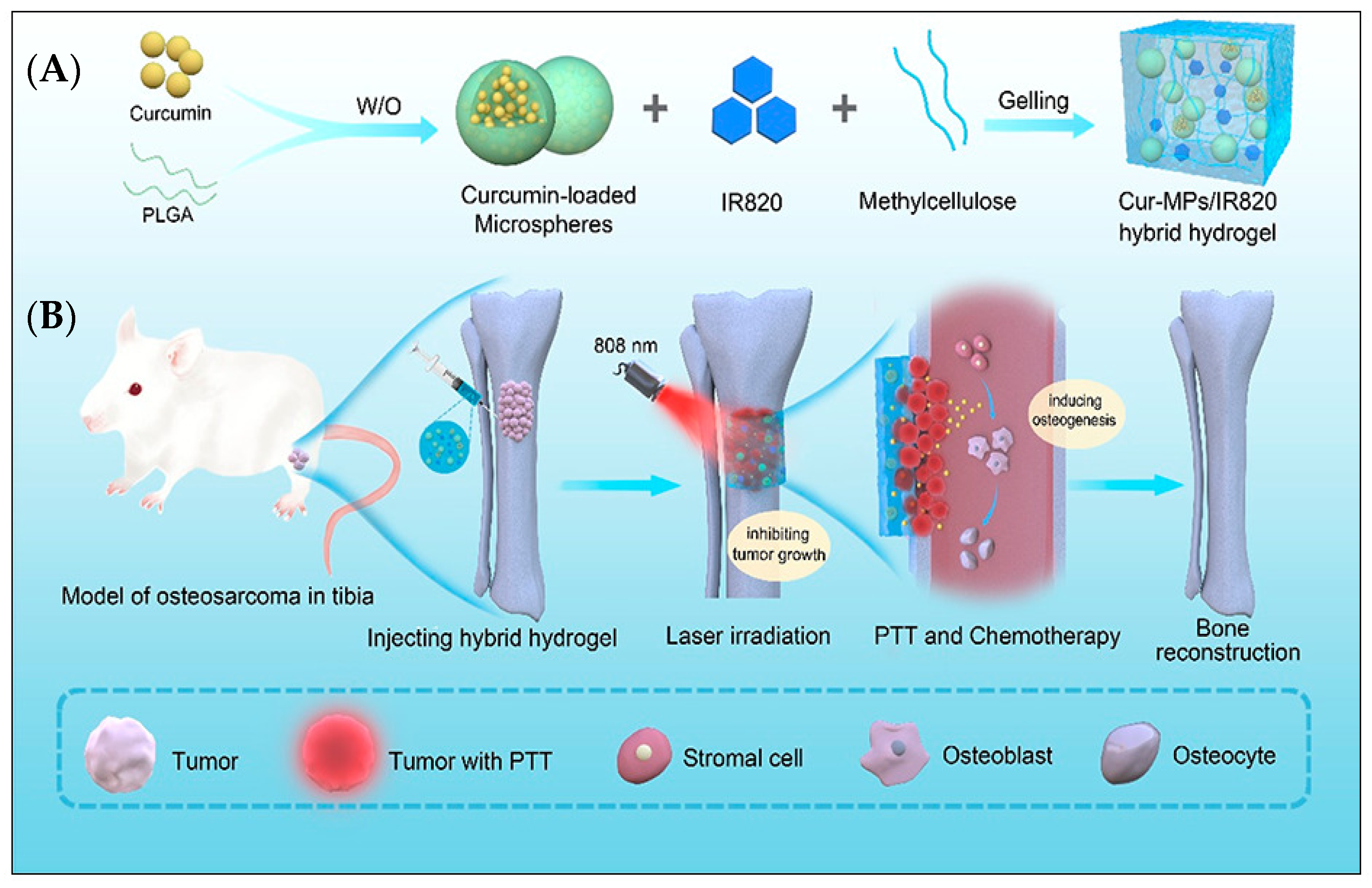
- Tan, B.; Wu, Y.; Wu, Y.; Shi, K.; Han, R.; Li, Y.; Qian, Z.; Liao, J. Curcumin-microsphere/IR820 hybrid bifunctional hydrogels for in situ osteosarcoma chemo-co-thermal therapy and bone reconstruction. ACS Appl. Mater. Interfaces 2021, 13, 31542–31553. [Google Scholar] [CrossRef]


| Ions Added to nHAP | Ion Roles | Advantages of HAP Ion Supplementation | Reference |
|---|---|---|---|
| Magnesium | Cartilage and bone structure Skeletal metabolism Osteoblast and osteoclast activity | Superior mineralization Improved osseointegration Superior morphology, granulation, composition, solubility and crystallinity Biocompatibility | [105,114] [70] [113] |
| Zinc | Bone metabolism stimulation Positive impact on osteoblast and osteoclast activity Decrease of early bone resorption and deterioration | Promotes osteogenesis in osteoporotic bones Osteoregeneration Anti-inflammatory ability | [114] [120,121,122] [115,123] |
| Selenium | Regulation of thyroid hormone levels Redox homeostasis Inflammatory and immunological reaction response Carbohydrate metabolism Cardiovascular health Reproductive health Physiological brain function Enzyme cofactor and protection against tissue deterioration | Regeneration Aging delay Free radical inhibition Endemic diseases prevention and treatment Selective toxicity and apoptosis of cancer cells Bone tumor and metastasis prevention and treatment Preosteoblast differentiation No toxicity | [127,128,129,130,131,132,133,134,135,136,137,138,139] |
| Strontium | Bone synthesis Inhibition of bone resorption | Increased osteoblast function Impeded osteoclast proliferation Osteoporosis treatment | [149,150,151,152,153,154] |
| Boron | Bone metabolism molecular control Synthesis of steroid hormone Bone strength and biomineral density (BMD) increase | Vitamin D metabolism optimization Treatment of calvarial defects in rats Osteoporosis, osteoarthritis and rheumatoid arthritis treatment Osteogenic differentiation of cell lines | [156,157,158,190] [158,161,162,163] |
| Cobalt | Bone tissue regeneration Antibacterial and antiviral action | Stimulation of in vivo osteogenesis | [160] [164,165] |
| Copper | Angiogenesis Endothelial cell migration | Synthesis of micro-vessels Positive impact on chemical and physical properties of HAP Improvement of HAP bioactivity Growth and proliferation of bone mesenchymal cells Osteoblast proliferation (reduced Copper ion concentrations) | [163,164,165,166,167,168] [83,171] |
| Silicon | Bone growth and development | Promotion of in vitro osteoblast adhesion and proliferation | [172,184] |
| Manganese | Bone tissue regeneration | Osteoblast proliferation promotion and metabolism activation | [178] |
| Iron | Magnetic properties Drug delivery | Stimulation of bone tissue remodeling and regeneration Enhancement of osteoblastic activity | [179,180] |
| Silver Caesium Europium | Antibacterial properties | Use in dental and orthopedic implants | [171] [185,186,187] |
| Carbonate | Superior bio-integration of HAP implants | [188] | |
| Hafnium | Creation of reactive oxygen species | Photodynamic anti-tumor treatment | [189] |
Disclaimer/Publisher’s Note: The statements, opinions and data contained in all publications are solely those of the individual author(s) and contributor(s) and not of MDPI and/or the editor(s). MDPI and/or the editor(s) disclaim responsibility for any injury to people or property resulting from any ideas, methods, instructions or products referred to in the content. |
© 2023 by the authors. Licensee MDPI, Basel, Switzerland. This article is an open access article distributed under the terms and conditions of the Creative Commons Attribution (CC BY) license (https://creativecommons.org/licenses/by/4.0/).
Share and Cite
Zastulka, A.; Clichici, S.; Tomoaia-Cotisel, M.; Mocanu, A.; Roman, C.; Olteanu, C.-D.; Culic, B.; Mocan, T. Recent Trends in Hydroxyapatite Supplementation for Osteoregenerative Purposes. Materials 2023, 16, 1303. https://doi.org/10.3390/ma16031303
Zastulka A, Clichici S, Tomoaia-Cotisel M, Mocanu A, Roman C, Olteanu C-D, Culic B, Mocan T. Recent Trends in Hydroxyapatite Supplementation for Osteoregenerative Purposes. Materials. 2023; 16(3):1303. https://doi.org/10.3390/ma16031303
Chicago/Turabian StyleZastulka, Ana, Simona Clichici, Maria Tomoaia-Cotisel, Aurora Mocanu, Cecilia Roman, Cristian-Doru Olteanu, Bogdan Culic, and Teodora Mocan. 2023. "Recent Trends in Hydroxyapatite Supplementation for Osteoregenerative Purposes" Materials 16, no. 3: 1303. https://doi.org/10.3390/ma16031303
APA StyleZastulka, A., Clichici, S., Tomoaia-Cotisel, M., Mocanu, A., Roman, C., Olteanu, C.-D., Culic, B., & Mocan, T. (2023). Recent Trends in Hydroxyapatite Supplementation for Osteoregenerative Purposes. Materials, 16(3), 1303. https://doi.org/10.3390/ma16031303