Article

Histological Comparison of Collagenated Cancellous Equine Bone Blocks Used as Inlay or Onlay for Lateral Bone Augmentation in Rabbits

by
Ryuichi Sakaguchi
1,
Samuel Porfirio Xavier
2,
Kenzo Morinaga
1,
Daniele Botticelli
1,3,*,
Erick Ricardo Silva
2,
Yasushi Nakajima
1,2 and
Shunsuke Baba
1
1
Department of Oral Implantology, Osaka Dental University, 8-1 Kuzuhahanazonocho, Hirakata 573-1121, Japan
2
Department of Oral and Maxillofacial Surgery and Periodontology, Faculty of Dentistry of Ribeirão Preto, University of São Paulo, Av. do Café, Subsetor Oeste, 11 (N-11), Ribeirao Preto 14040-904, SP, Brazil
3
ARDEC Academy, 47923 Rimini, Italy
*
Author to whom correspondence should be addressed.
Materials 2023, 16(20), 6742; https://doi.org/10.3390/ma16206742
Submission received: 9 September 2023 / Revised: 12 October 2023 / Accepted: 17 October 2023 / Published: 18 October 2023
(This article belongs to the Special Issue Effects of Bone Substitute Materials in Bone Defect Regeneration)

Abstract

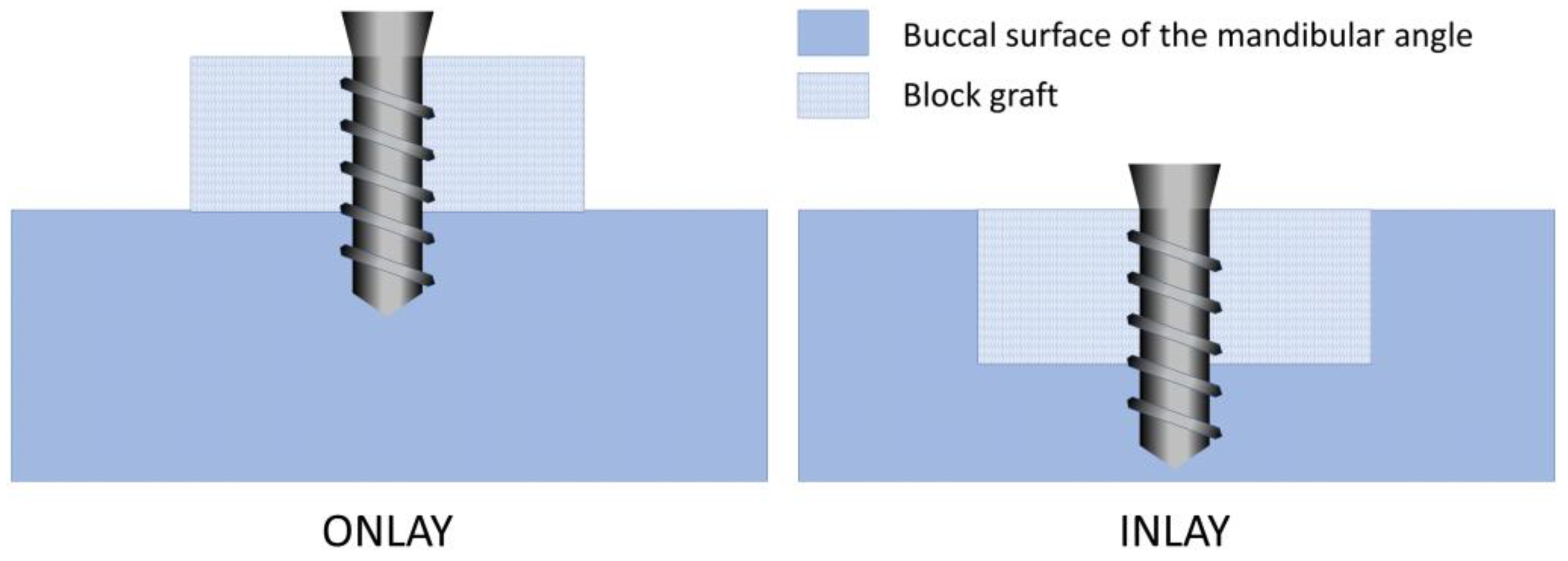
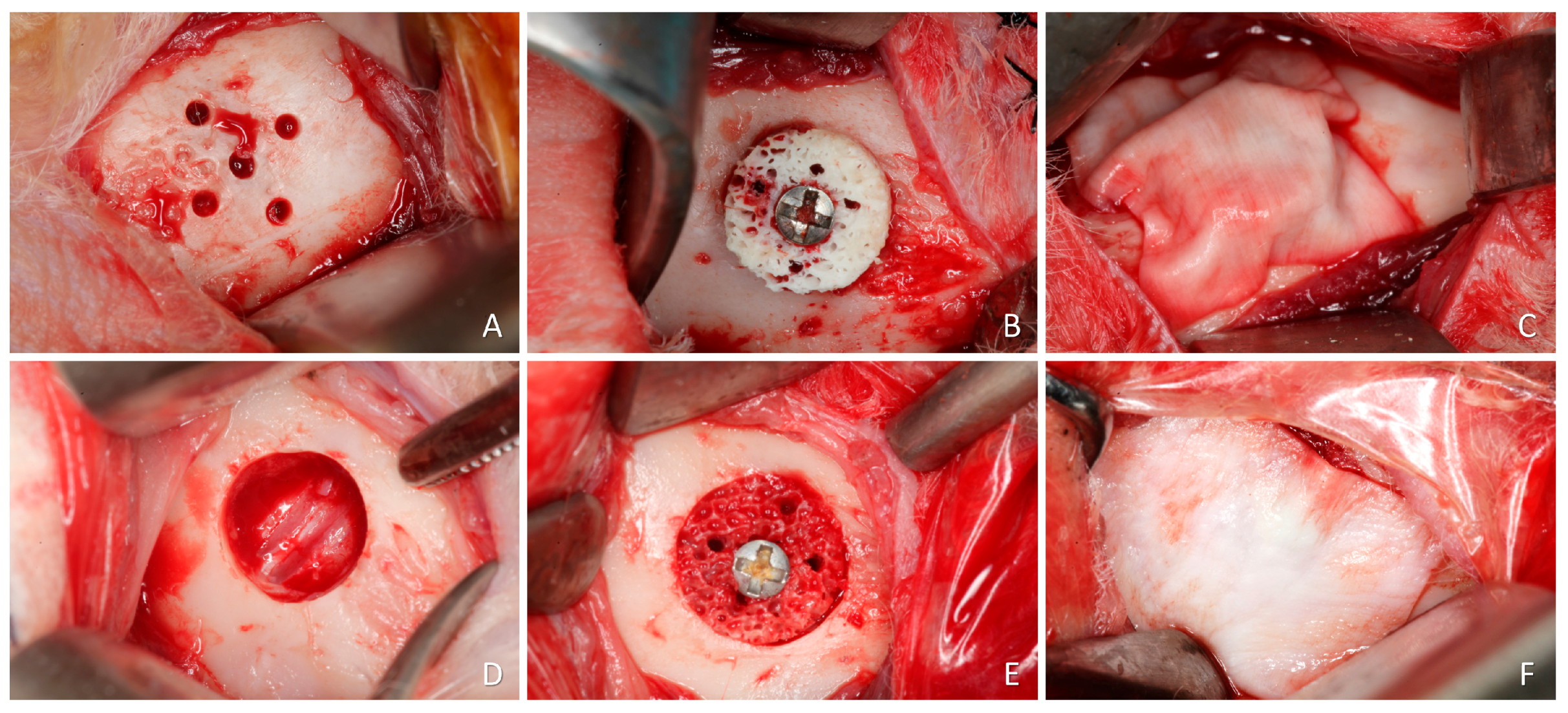
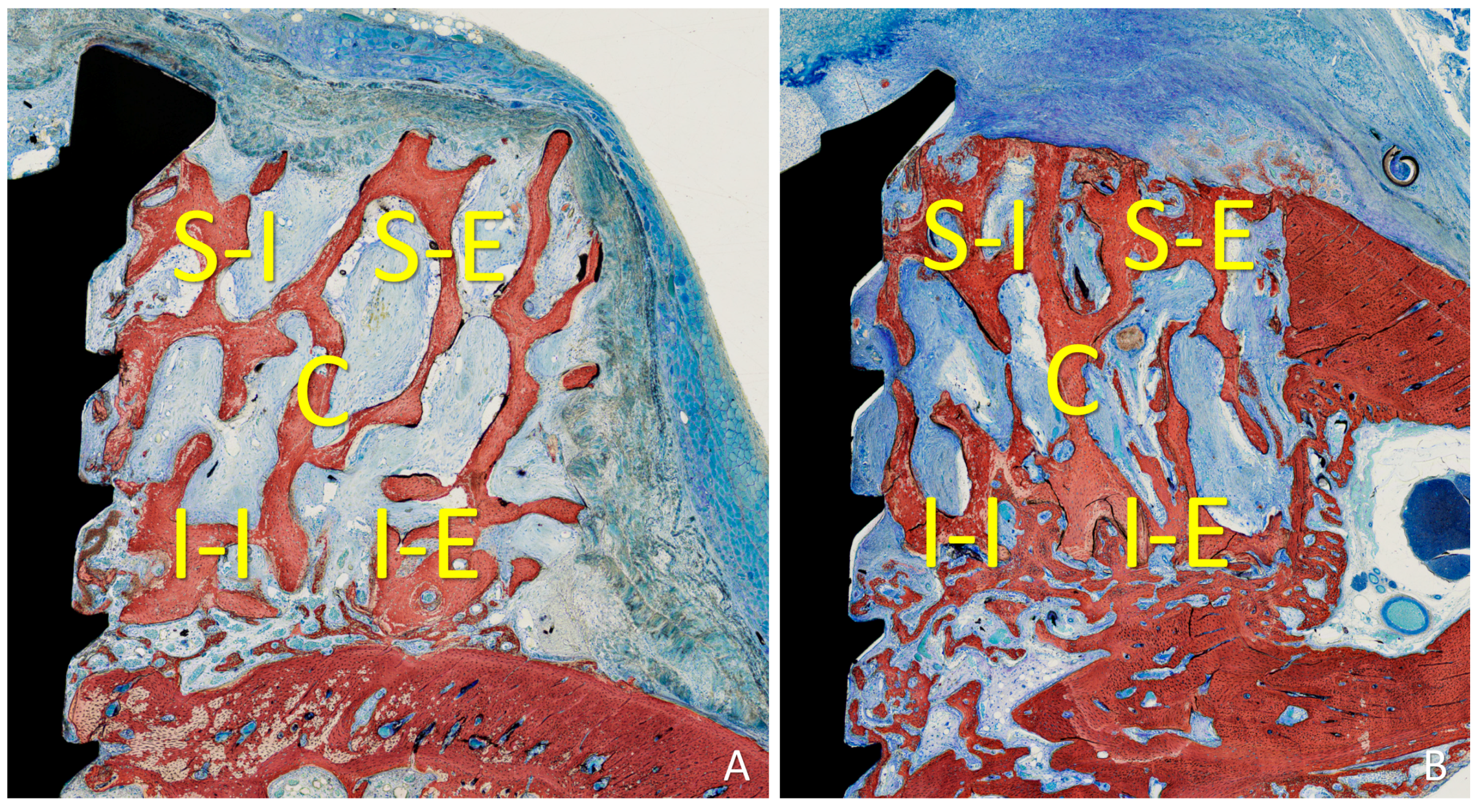
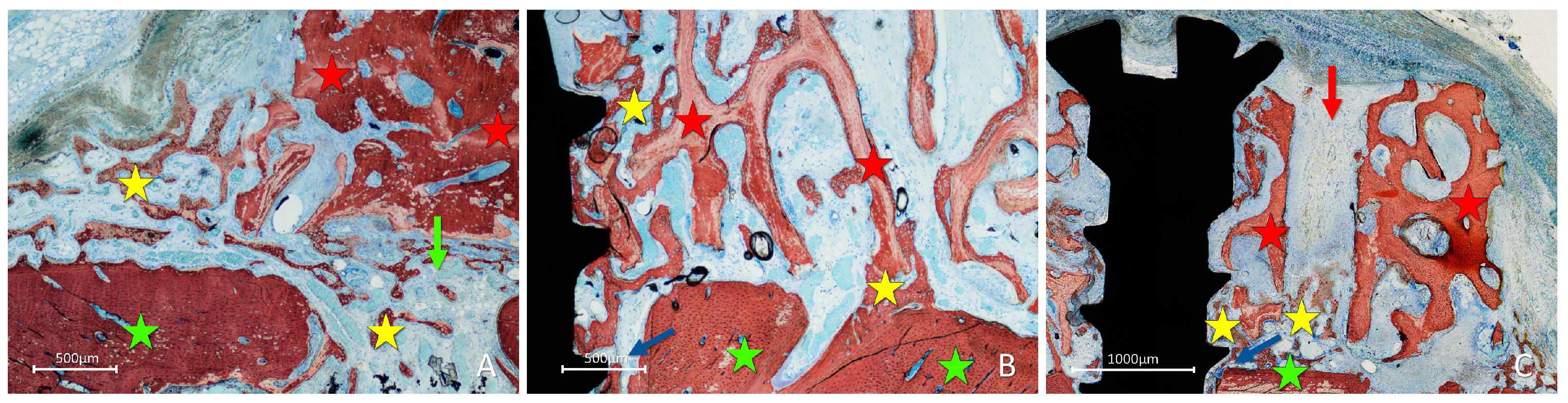
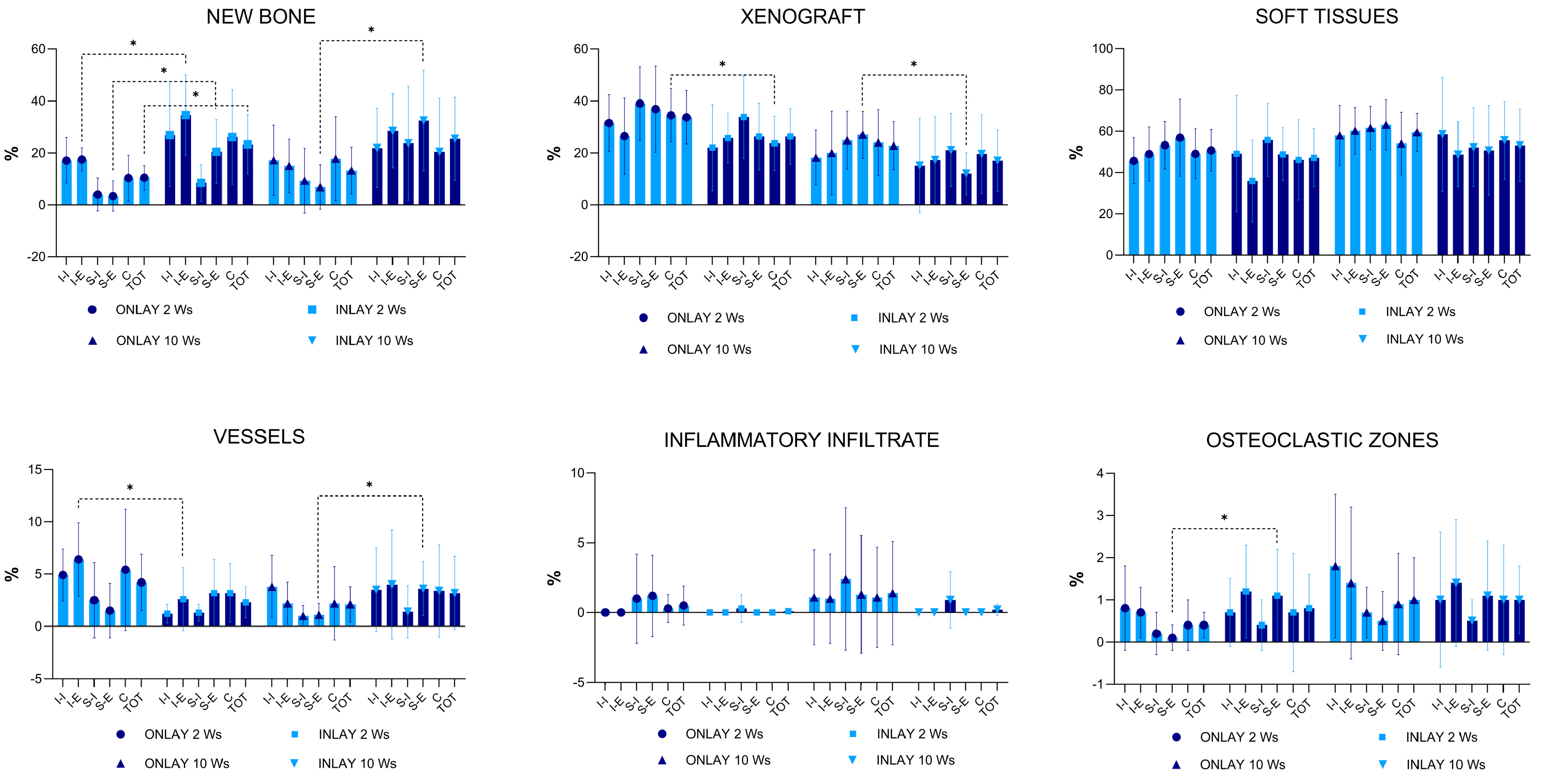
Background: The conformation of the recipient site for an inlay graft presents an increased contact with the parent bone compared to an onlay graft. This might favor bone growth within the inlay compared to onlay grafts. Hence, the objective of this study was to compare the bone incorporation and remodeling processes of xenogeneic en bloc grafts placed using two bone grafting techniques, i.e., onlay vs. inlay. Methods: In this prospective, randomized, split-mouth study (test and control sides in the same animal), two bone grafting techniques were comparatively evaluated. The lateral aspect of the rabbit mandible was used as the recipient site, bilaterally. On one side of the mandible, the cortical bone was perforated with drills to allow a better bone formation from the bone wound and the marrow spaces. A xenogeneic bone block was fixed in the center of the prepared region, representing the onlay site. On the other side of the mandible, a 7 mm wide and 3 mm deep circumferential defect was prepared using trephines and drills. A xenogeneic bone block was fixed in the center of the defect, representing the inlay site. Two healing periods were applied in the study: 2 and 10 weeks, each represented by 10 rabbits (n = 10 for each period). Results: After 2 weeks of healing, the mean percentage of new bone was 10.4% and 23.3% at the onlay and inlay grafts, respectively (p = 0.022). After 10 weeks of healing, new bone increased to 13.2% at the onlay sites and 25.4% at the inlay sites (p = 0.080). In the 10-week period, the inlay grafts presented a homogeneous growth of new bone in all regions, while in the onlay grafts, low percentages of new bone were observed in the external regions. Conclusion: The percentage of new bone increased faster and was higher in the inlay grafts than in the onlay grafts. This outcome might be related to the self-contained conformation of the recipient site in the inlay group, which offered more sources for new bone formation compared to the one-wall conformation of the recipient sites in the onlay group. The osteoconductive properties of the biomaterial allowed the newly formed bone to reach the most peripheral regions in both groups. The osteoconductive properties of the biomaterial, together with the protection offered by the collagen membrane, allowed marginal closure of the defects by newly formed bone in the inlay group.
Keywords: animal study; bone healing; histology; lateral augmentation; bone transplantation; biomaterial; bone defect animal study; bone healing; histology; lateral augmentation; bone transplantation; biomaterial; bone defect