Article

Crystallography and Microstructure of 7M Martensite in Ni-Mn-Ga Thin Films Epitaxially Grown on (1 1 2¯ 0)-Oriented Al2O3 Substrate

1
Key Laboratory for Anisotropy and Texture of Materials (Ministry of Education), School of Material Science and Engineering, Northeastern University, Shenyang 110819, China
2
Laboratoire d’Étude des Microstructures et de Mécanique des Matériaux (LEM3), CNRS UMR 7239, Université de Lorraine, CEDEX, 57045 Metz, France
3
Laboratory of Excellence on Design of Alloy Metals for Low-mAss Structures (DAMAS), Université de Lorraine, CEDEX, 57045 Metz, France
*
Author to whom correspondence should be addressed.
Materials 2022, 15(5), 1916; https://doi.org/10.3390/ma15051916
Submission received: 31 December 2021 / Revised: 22 February 2022 / Accepted: 3 March 2022 / Published: 4 March 2022
(This article belongs to the Section Metals and Alloys)

Abstract

:
Epitaxial Ni-Mn-Ga thin films have been extensively investigated, due to their potential applications in magnetic micro-electro-mechanical systems. It has been proposed that the martensitic phase in the <1 1 0>A-oriented film is much more stable than that in the <1 0 0>A-oriented film. Nevertheless, the magnetic properties, microstructural features, and crystal structures of martensite in such films have not been fully revealed. In this work, the <1 1 0>A-oriented Ni51.0Mn27.5Ga21.5 films with different thicknesses were prepared by epitaxially growing on Al2O3(1 1 2 ¯ 0) substrate by magnetron sputtering. The characterization by X-ray diffraction technique and transmission electron microscopy revealed that all the Ni51.0Mn27.5Ga21.5 films are of 7M martensite at the ambient temperature, with their Type-I and Type-II twinning interfaces nearly parallel to the substrate surface.

1. Introduction

Ni-Mn-based ferromagnetic shape memory alloys have been considered as the promising materials for magnetic field-driven sensors and actuators, new type solid state refrigeration, and thermomagnetic generators [1,2,3,4,5,6,7,8,9,10,11,12]. Under the external magnetic-, thermal-, and force-field stimulus, they usually occur martensite variant reorientation and/or martensitic transformation [9,10,11,12], thus exhibiting multi-functionalities such as giant magnetic field-induced strain [1,2,3,4,5], excellent magnetocaloric effects [6,7,8], and colossal elastocaloric effects, etc. Among these functionalities, the giant magnetic field-induced strain has gained much attention, since both the single crystalline and polycrystalline bulk materials have demonstrated giant magnetic field-induced strain as much as 6–12% and 1% [1,2,3,4,5], respectively. Such giant magnetic field-induced strains also promote the research interest in epitaxial Ni-Mn-Ga thin films, as the films possess a cost-effective advantage in the application of magnetic micro-electro-mechanical systems [13,14,15,16,17,18,19,20,21,22,23]. So far, the magnetically induced reorientation of martensite variant has been reported in the <1 0 0>A-oriented Ni-Mn-Ga thin films epitaxially grown on the MgO(1 0 0) substrate and NaCl (1 0 0) substrate. Nevertheless, magnetic field-induced strains have not been achieved yet, since these kinds of thin films are of single crystalline state in the Austenite at evaluated temperatures and transform to self-accommodated martensite at ambient temperature. The self-accommodated martensite microstructure always contains two kinds of distinct microstructures: Type-X and Type-Y, which is composed of several different oriented martensite variants [17,24,25].
Recently, G. Jakob and his coworkers [26,27] proposed that the <1 1 0>A-oriented Ni2MnGa thin films can be achieved by epitaxially growing on the (1 1 2 ¯ 0) sapphire substrate (Al2O3). In addition, the martensitic phase in the <1 1 0>A-oriented film on Al2O3 substrate is much more stable than those in the <1 0 0>A-oriented film on MgO (1 0 0) substrate. However, the martensitic transformation temperatures of the <1 1 0>-oriented Ni2MnGa thin films are between 260 K and 280 K, suggesting that the Ni2MnGa thin films are of Austenite at ambient temperature. The magnetic properties, microstructure, and crystal structure of martensite in <1 1 0>A-oriented Ni2MnGa thin films have not been revealed yet. A. Sharma and his coworkers [28] also prepared Ni-Mn-Ga thin films on (1 1 2 ¯ 0) sapphire substrate (Al2O3), which is of martensite at ambient temperature. However, the twining interfaces of martensite are not parallel to the substrate surface [29], since the film thickness is of several micron meter.
In the present work, we successfully prepared the <1 1 0>A-oriented Ni51.0Mn27.5Ga21.5 films with different thicknesses from 200 nm to 600 nm on Al2O3 (1 1 2 ¯ 0) substrates by magnetron sputtering. The characterization by X-ray diffraction technique, transmission electron microscopy revealed that all the Ni51.0Mn27.5Ga21.5 films are of 7M martensite at the ambient temperature, and with their Type-I and Type-II twinning interfaces nearly parallel to the substrate surface.

2. Materials and Methods

2.1. Thin Films Preparation

Ni51.0Mn27.5Ga21.5 thin films with different thicknesses from 200 nm to 600 nm were grown on Al2O3 (1 1 2 ¯ 0) mono-crystalline substrates by DC magnetron sputtering and using polycrystalline Ni48Mn30Ga22 as the target materials. The target is of 50.8 mm in diameter and 1.5 mm in thickness. Before deposition, the base pressure of the sputtering equipment was vacuumed to below 9.0 × 10⁻⁵ Pa. In order to obtain high-quality films, the substrate was heated to 650 °C. The sputtering process was conducted under a constant Ar working pressure of 0.15 Pa with an applied power of 70 W. Under this condition, the sputtering rate is of 0.1 nm/s roughly estimated through film thicknesses divided by deposition time. Before the actual depositions, a pre-sputtering was performed for 15 min.

2.2. Structure and Microstructure Characterization

The crystal structure and macroscopic crystallographic features were analyzed by temperature-dependent X-ray diffractometer using Cu-Kα radiation (λ = 0.15406 nm) (Rigaku Smartlab 9 kW, Tokyo, Japan) and a four-circle X-ray diffractometer (Rigaku Smartlab 3 kW, Tokyo, Japan), respectively. A stylus profiler (Veeco DEKTAK 150, Plainview, NY, USA) was employed to measure the film thickness. The microstructures and chemical composition were examined by scanning electron microscopy (SEM, JEOL-JSM 7001F, Tokyo, Japan) and energy dispersive spectrometry (EDS, Bruker XFlash 4010, Berlin, Germany), respectively. The cross-sectional microstructures at nano and atomic scale were characterized by transmission electron microscopy (JEOL JEM 2100F, Tokyo, Japan) working at 200 kV. The cross-sectional sample for TEM characterization was prepared using the focused-ion beam (FIB, FEI Helios nanolab, Hillsboro, OR, USA) lift-out technique. Temperature-dependent magnetization curves and magnetic hysteresis loops were measured by Versalab (Quantum Design, San Diego, CA, USA).

3. Results and Discussion

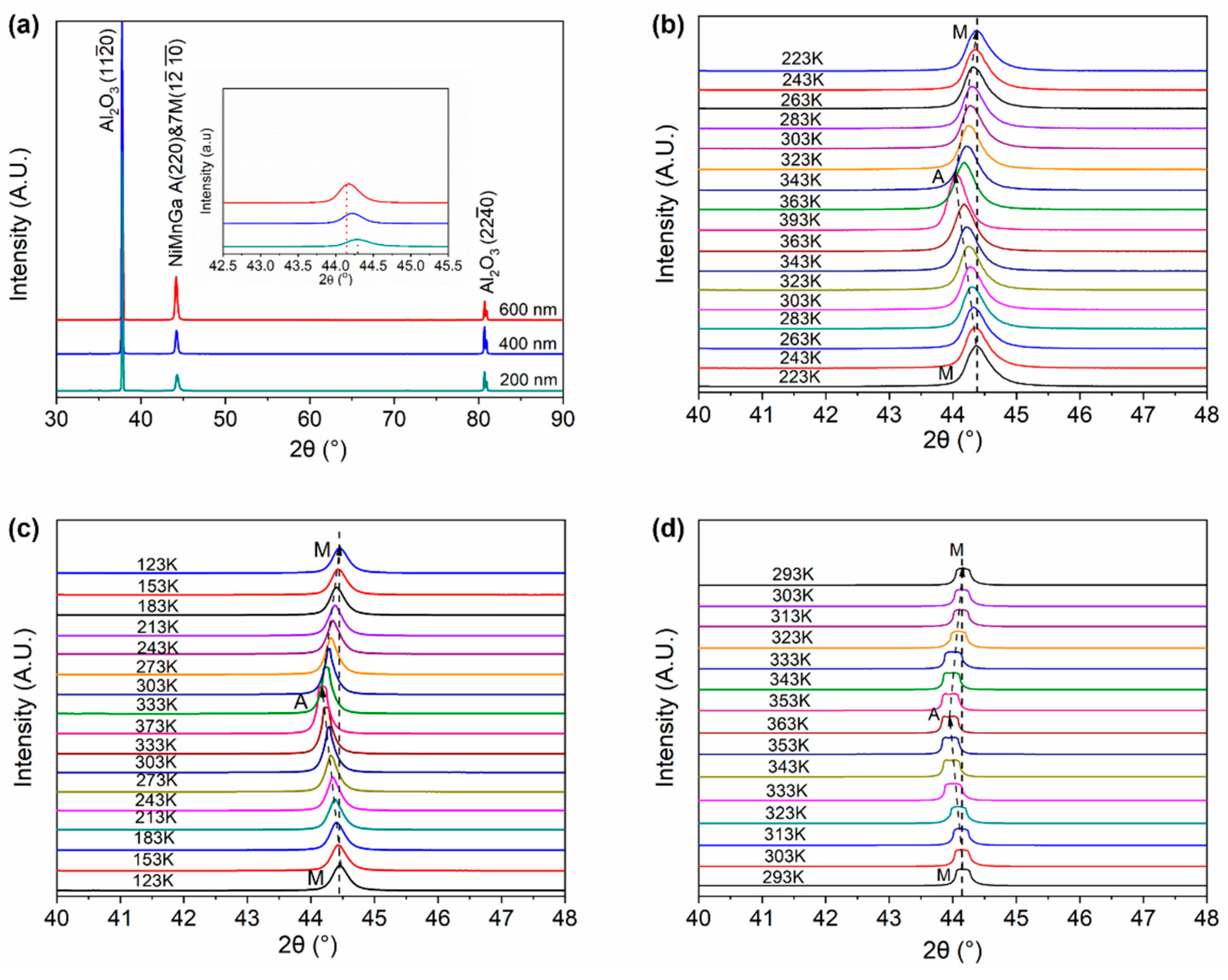
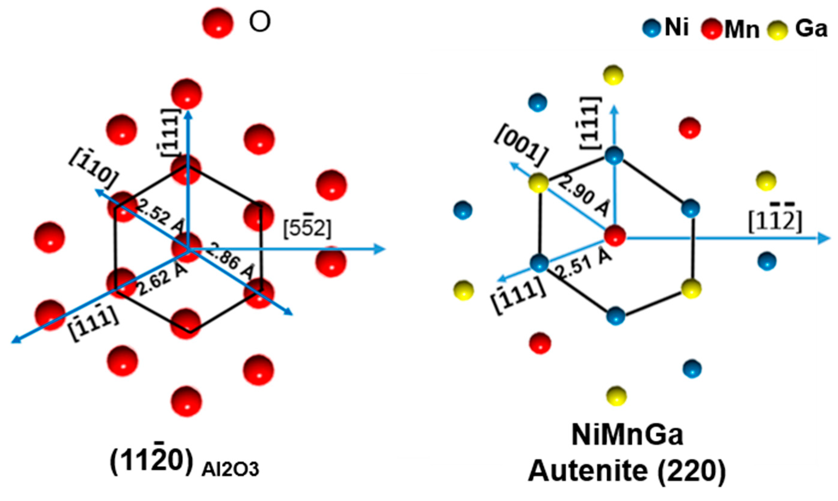
3.1. Crystal Structure and Microstructure

Figure 1 displays the conventional and temperature-dependent X-ray diffraction patterns of the Ni51.0Mn27.5Ga21.5 thin films with different thicknesses from 200 nm to 600 nm grown on Al2O3 (1 1 2 ¯ 0) mono-crystalline substrates. As shown in Figure 1a, it is seen that only one specific diffraction peak of Ni51.0Mn27.5Ga21.5 thin films is observed in the patterns, except the peaks from the Al2O3 substrate, indicating that the films possess a strong preferred orientation. However, since the planar distance of (2 2 0)A plane of Austenite and (1 2 ¯   10 ¯ )7M plane of 7M martensite are roughly equal, we cannot distinguish which specific diffraction peak belongs to the 7M martensite or the Austenite. We performed the temperature-dependent X-ray diffraction measurement, as shown in Figure 1b–d. As can be seen from Figure 1b–d, for all the films, with the increase in temperature, the specific diffraction peak of (1 2 ¯   10 ¯ )7M gradually shifts to the low angle side. When the temperature is higher than 350 K, there is a sharp shift to the low angle side in the XRD patterns, which indicated that the (1 2 ¯   10 ¯ )7M of 7M martensite transformed to the (2 2 0)A of Austenite. It should be noted that the coexistence of Austenite and martensite phases in the 600 nm-thick Ni51.0Mn27.5Ga21.5 film, which may be due to the restriction from the substrate and the temperature is not high or low enough for the full martensitic transformation.
In order to determine the lattice parameters of the martensite, a four-circle X-ray diffractometer was employed to measure more XRD patterns at various azimuth angles (Phi) and tilt angles (Psi), as shown in Figure 2. With the employment of a four-circle XRD diffractometer, the crystal structure of the Ni51.0Mn27.5Ga21.5 thin films is identified as 7M modulated martensite and of monoclinic. The lattice parameters are shown in Table 1.
Figure 3a–c shows the microstructure of Ni51.0Mn27.5Ga21.5 thin films with different thicknesses taken by FE-SEM. From these FE-SEM images, no twining microstructure of the martensite variants can be observed in the top-view SEM images. All the films can be identified as continuously grown and the surface of the film becomes coarse with increasing thickness of the films. It is worth noting that white areas on the surface of the films can be identified as precipitates and the pit on the films might be defect pores among the column grains.
To further analyze the microstructure and the martensitic configuration of the Ni51.0Mn27.5Ga21.5 films on the Al2O3 substrate, the cross-section TEM characterization was conducted. As displayed in Figure 4a–c, the martensite is visible in all films. The twin interfaces of martensite are parallel to the substrate surface. A selected area electron diffraction confirmed that the martensite is of 7M martensite (insets in Figure 4a). In addition, with the increase in film thickness, the number of both martensite variants and the twin interfaces increased, but the thickness of each martensite variant remains unchanged. Even though the microstructure of all the films can be identified as column grains, a detailed observation in Figure 4d shows that the twin can pass the grain boundaries, which indicated that the column grain boundaries are small-angle grain boundaries. From Figure 4d–f, martensite can be classified as the hierarchical structure in which martensite lath incorporates martensitic variants. The further crystallographic analysis demonstrated that each plate corresponds to one crystallographic variant, there are a maximum of four-oriented variants in each group and Type-I twin, Type-II twin, and Compound twin are present in the film, which is marked as the dotted line.

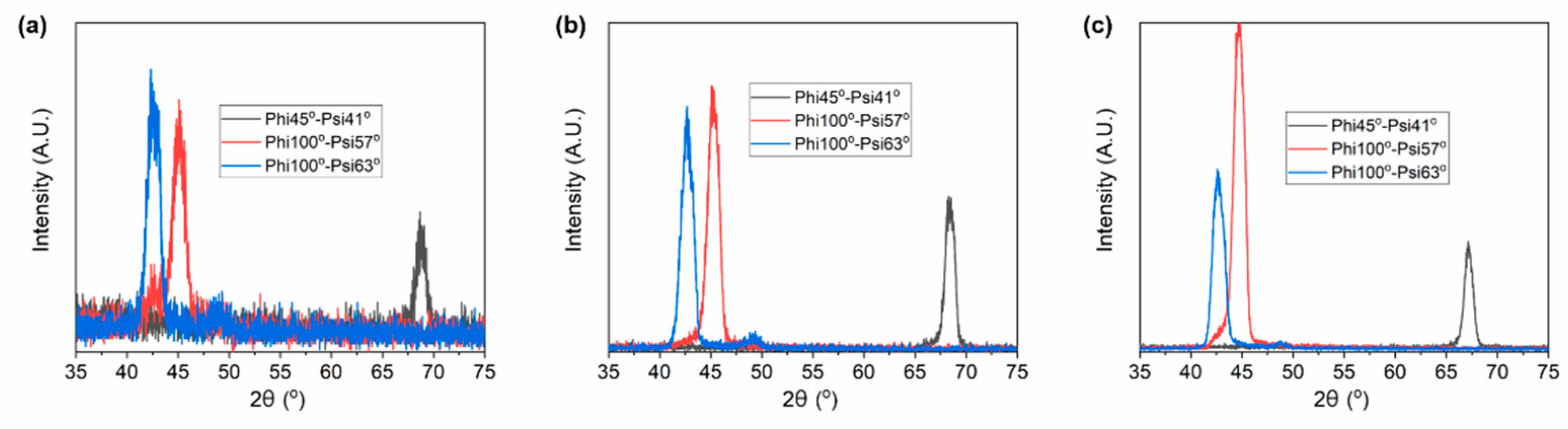
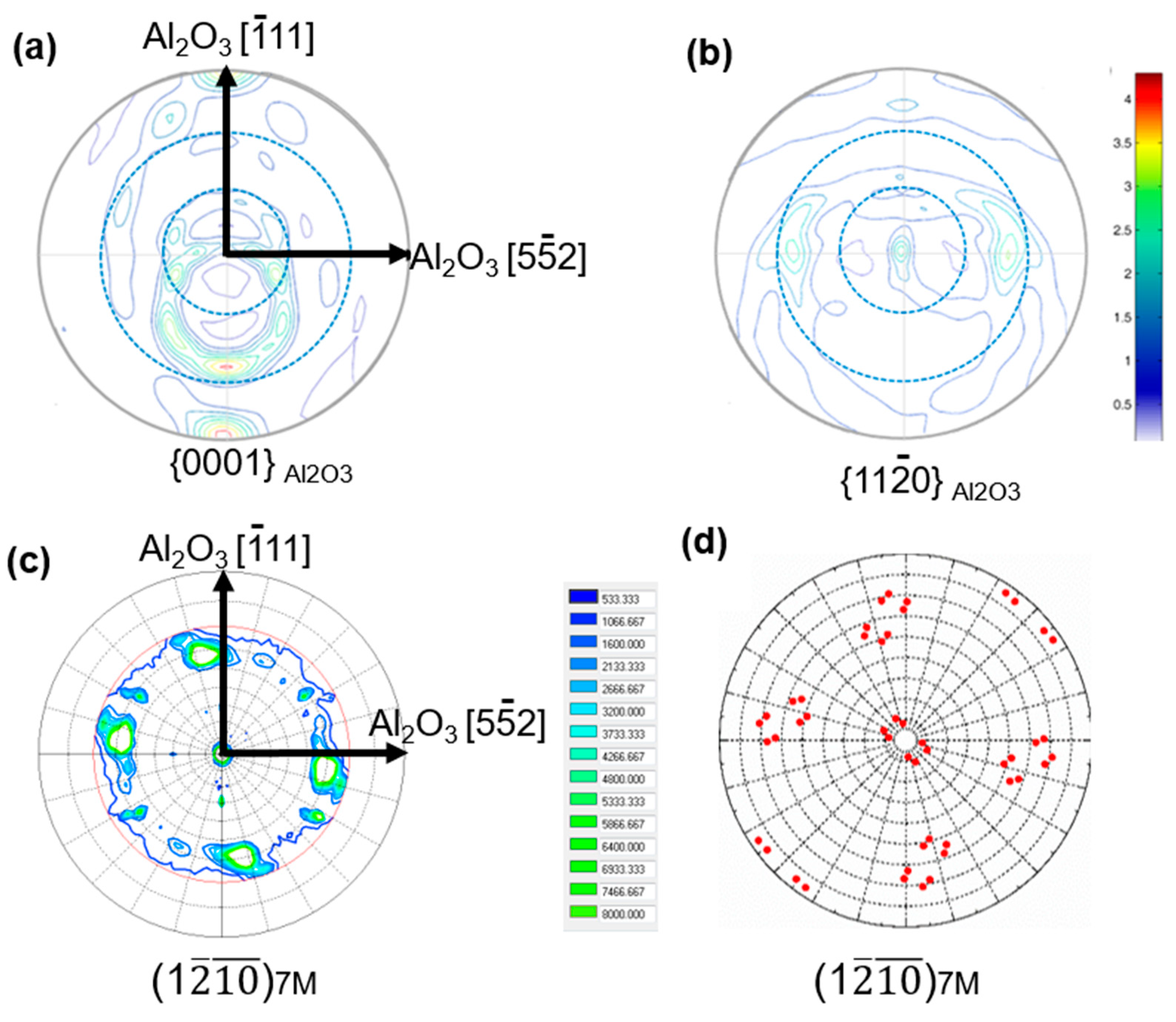
3.2. Macroscopic Texture

In order to determine the orientation relationship between the variants and the substrate, X-ray diffraction was used to measure the pole figure of the Ni51.0Mn27.5Ga21.5 thin film with a thickness of 600 nm and the Al2O3 (1 1 2 ¯ 0) substrate. As shown in Figure 5a,b, two symmetrically distributed spots appear at the {0 0 0 1} pole figures of the Al2O3 substrate and three spots appear at the {1 1 2 ¯ 0} pole figure of the Al2O3 substrate. Thus, it can be determined that the edges of the substrate are parallel to Al2O3 [5 5 ¯ 2] and Al2O3 [ 1 ¯ 1 1]. Thus, we choose Al2O3 [5 5 ¯ 2], Al2O3 [ 1 ¯ 1 1] and Al2O3 [1 1 0] as the coordinate direction of the macroscopic sample coordinate system.
As shown in Figure 5c, four spots appear at the {1 2 ¯   10 ¯ }7M pole figures of the Ni51.0Mn27.5Ga21.5 thin film. The measured pole figure from the image of Figure 5c is in agreement with the theoretically predicted pole figure from the image of Figure 5d. Since the (1 2 ¯   10 ¯ )7M plane of 7M martensite is parallel to the (2 2 0)A plane of Austenite, we can conclude that the films were epitaxially grown on the Al2O3 (1 1 2 ¯ 0) substrate, which justified that there is one crystallographic orientation for the Austenite in the present thin films. The crystallographic orientation of the Austenite can be described using Euler angle (60°, 90°, 45°). The mismatch relationship and the orientation of Austenite can also be determined, as shown in Figure 6. The Al2O3 [5 5 ¯ 2] and Al2O3 [ 1 ¯ 1 1] direction parallel to the [1 1 ¯ 1]A and [1 1 ¯   2 ¯ ]A direction of the Austenite of Ni-Mn-Ga thin films.

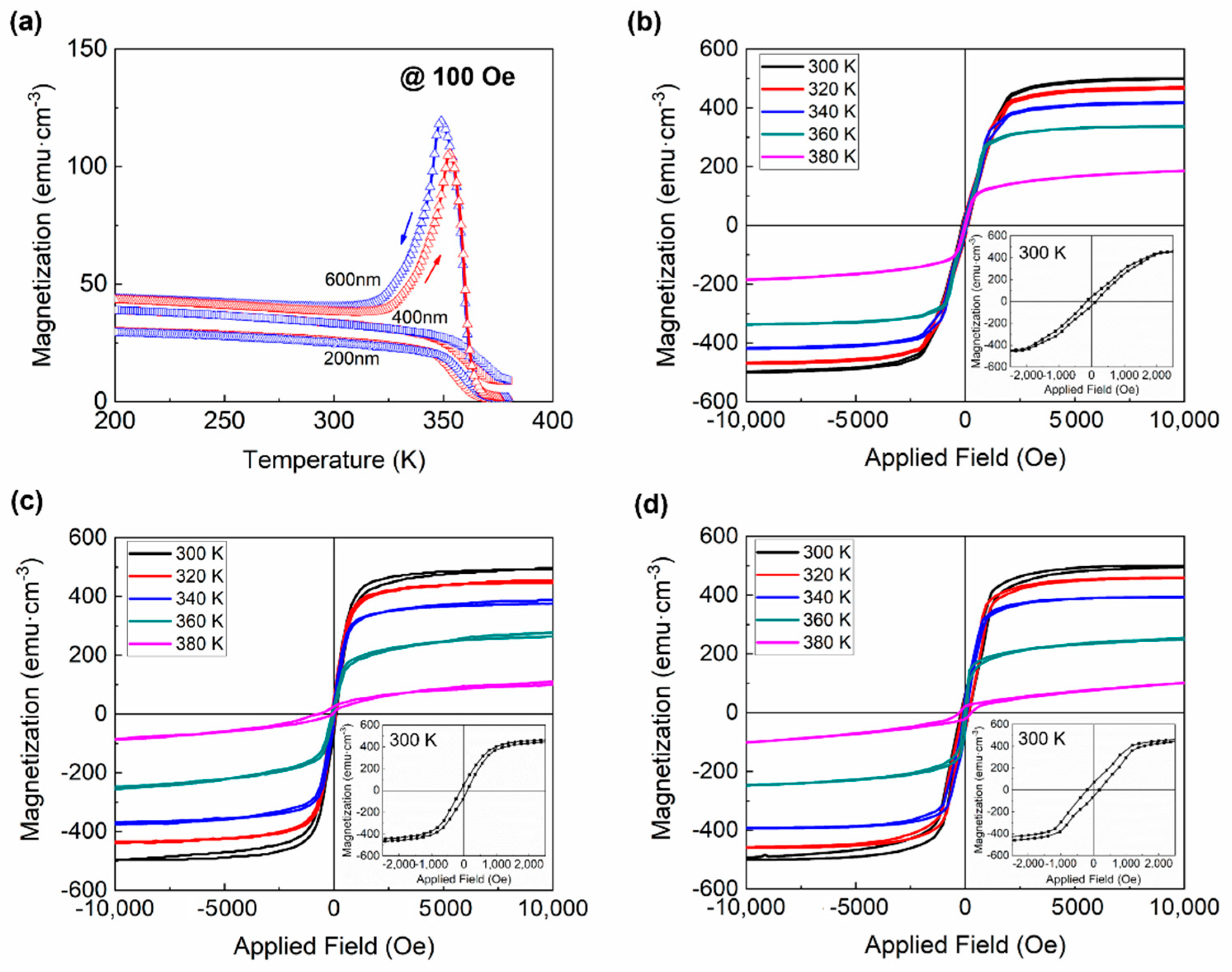
3.3. Magnetic Properties

Magnetization measurements are employed to identify the magnetic field-induced variant reorientation of the twin related martensitic variants in Ni-Mn-Ga thin films. Before the measurement of M-H loops, temperature-dependent magnetization cures were carried out to determine the martensitic transformation temperature as shown in Figure 7a. The M-T curves confirmed that the martensitic transformation temperature of the Ni51.0Mn27.5Ga21.5 thin films are above the ambient temperature, which suggested that these thin films are of martensite state at ambient temperature.
The magnetization hysteresis (M-H) loop of Ni-Mn-Ga thin films was measured at different temperatures, as shown in Figure 7b–d. “Magnetization jumps” in magnetization hysteresis loops can be a sign of the magnetic field-induced reorientation of martensitic variants. However, no obvious “magnetization jumps” can be found in magnetization hysteresis loops of the present Ni51.0Mn27.5Ga21.5 thin films with different thicknesses, from Figure 7b–d, which may be attributed to the high stress from the substrate.

4. Conclusions

In summary, we deposited <1 1 0>A-oriented Ni51.0Mn27.5Ga21.5 thin films from 200 nm to 600 nm which are of monoclinic 7M at room temperature on the Al2O3 (1 1 2 ¯ 0). X-ray diffraction and transmission electron microscopy characterization revealed that the martensite-related twin interfaces are parallel to the substrate, and each group possesses a maximum of four-oriented variants. Only one crystallographic orientation for the Austenite is in the thin film. The characterization of magnetic properties shows that the magnetic field-induced variant reorientation was not obvious in the present thin films.

Author Contributions

Conceptualization, B.Y.; validation. Z.L. and H.Y.; investigation, B.Y. and Y.Z.; writing—review and editing, C.E., X.Z. and L.Z.; supervision, C.E.; project administration, X.Z.; funding acquisition, L.Z. All authors have read and agreed to the published version of the manuscript.

Funding

This work was supported by the National Natural Science Foundation of China (Grant No. 52071071, 52171005), the Program of Introducing Talents of Discipline Innovation to Universities 2.0 (the 111 Project of China 2.0, No. BP0719037), the Fundamental Research Funds for the Central Universities of China (grant no. N2102006).

Institutional Review Board Statement

Not applicable.

Informed Consent Statement

Not applicable.

Data Availability Statement

The data presented in this study are available on request from the corresponding author.

Conflicts of Interest

The authors declare no conflict of interest.

References

  1. Murray, S.J.; Marioni, M.; Allen, S.M.; O’Handley, R.C.; Lograsso, T.A. 6% magnetic-field-induced strain by twin-boundary motion in ferromagnetic Ni-Mn-Ga. Appl. Phys. Lett. 2000, 77, 886–888. [Google Scholar] [CrossRef] [Green Version]
  2. Sozinov, A.; Likhachev, A.A.; Lanska, N.; Ullakko, K. Giant magnetic-field-induced strain in NiMnGa seven-layered martensitic phase. Appl. Phys. Lett. 2002, 80, 1746–1748. [Google Scholar] [CrossRef]
  3. Sozinov, A.; Lanska, N.; Soroka, A.; Zou, W. 12% magnetic field-induced strain in Ni-Mn-Ga-based non-modulated martensite. Appl. Phys. Lett. 2013, 102, 021902. [Google Scholar] [CrossRef]
  4. Gaitzsch, U.; Pötschke, M.; Roth, S.; Rellinghaus, B.; Schultz, L. A 1% magnetostrain in polycrystalline 5M Ni-Mn-Ga. Acta Mater. 2009, 57, 365–370. [Google Scholar] [CrossRef]
  5. Li, Z.Z.; Li, Z.B.; Yang, B.; He, X.J.; Gan, W.M.; Zhang, Y.L.; Li, Z.; Zhang, Y.D.; Esling, C.; Zhao, X.; et al. Over 2% magnetic-field-induced strain in a polycrystalline Ni50Mn28.5Ga21.5 alloy prepared by directional solidification. Mater. Sci. Eng. A 2020, 780, 139170. [Google Scholar] [CrossRef]
  6. Guan, Z.; Jiang, X.; Gu, J.; Bai, J.; Liang, X.; Yan, H.; Zhang, Y.; Esling, C.; Zhao, X.; Zuo, L. Large magnetocaloric effect and excellent mechanical properties near room temperature in Ni-Co-Mn-Ti non-textured polycrystalline alloys. Appl. Phys. Lett. 2021, 119, 051904. [Google Scholar] [CrossRef]
  7. Kuang, Y.F.; Ai, Z.R.; Yang, B.; Hao, X.W.; Li, Z.B.; Yan, H.L.; Zhang, Y.D.; Esling, C.; Zhao, X.; Zuo, L. Simultaneously achieved good mechanical properties and large magnetocaloric effect in spark plasma sintered Ni-Mn-In alloys. Intermetallics 2020, 124, 106868. [Google Scholar] [CrossRef]
  8. Cong, D.Y.; Huang, L.; Hardy, V.; Bourgault, D.; Sun, X.M.; Nie, Z.H.; Wang, M.G.; Ren, Y.; Entel, P.; Wang, Y.D. Low-field-actuated giant magnetocaloric effect and excellent mechanical properties in a NiMn-based multiferroic alloy. Acta Mater. 2018, 146, 142–151. [Google Scholar] [CrossRef]
  9. Zuo, L.; Li, Z.; Yan, H.; Yang, B.; Zhao, X. Texturation and Functional Behaviors of Polycrystalline Ni-Mn-X Phase Transformation Alloys. Acta Metall. Sin. 2021, 57, 1396–1415. [Google Scholar]
  10. Liu, J.; Gottschall, T.; Skokov, K.P.; Moore, J.D.; Gutfleisch, O. Giant magnetocaloric effect driven by structural transitions. Nat. Mater. 2012, 11, 620–626. [Google Scholar] [CrossRef]
  11. Kainuma, R.; Imano, Y.; Ito, W.; Sutou, Y.; Morito, H.; Okamoto, S.; Kitakami, O.; Oikawa, K.; Fujita, A.; Kanomata, T.; et al. Magnetic-field-induced shape recovery by reverse phase transformation. Nature 2006, 439, 957–960. [Google Scholar] [CrossRef] [PubMed]
  12. Krenke, T.; Duman, E.; Acet, M.; Wassermann, E.F.; Moya, X.; Manosa, L.; Planes, A. Inverse magnetocaloric effect in ferromagnetic Ni-Mn-Sn alloys. Nat. Mater. 2005, 4, 450–454. [Google Scholar] [CrossRef] [PubMed] [Green Version]
  13. Buschbeck, J.; Niemann, R.; Heczko, O.; Thomas, M.; Schultz, L.; Fähler, S. In situ studies of the martensitic transformation in epitaxial Ni–Mn–Ga films. Acta Mater. 2009, 57, 2516–2526. [Google Scholar] [CrossRef]
  14. Eichhorn, T.; Hausmanns, R.; Jakob, G. Microstructure of freestanding single-crystalline Ni2MnGa thin films. Acta Mater. 2011, 59, 5067–5703. [Google Scholar] [CrossRef] [Green Version]
  15. Ranzieri, P.; Fabbrici, S.; Nasi, L.; Righi, L.; Casoli, F.; Chernenko, V.A.; Villa, E.; Albertini, F. Epitaxial Ni–Mn–Ga/MgO(100) thin films ranging in thickness from 10 to 100nm. Acta Mater. 2013, 61, 263–272. [Google Scholar] [CrossRef]
  16. Backen, A.; Yeduru, S.R.; Diestel, A.; Schultz, L.; Kohl, M.; Fähler, S. Epitaxial Ni-Mn-Ga Films for Magnetic Shape Memory Alloy Microactuators. Adv. Eng. Mater. 2012, 14, 696–709. [Google Scholar] [CrossRef]
  17. Ranzieri, P.; Campanini, M.; Fabbrici, S.; Nasi, L.; Casoli, F.; Cabassi, R.; Buffagni, E.; Grillo, V.; Magen, C.; Celegato, F.; et al. Achieving Giant Magnetically Induced Reorientation of Martensitic Variants in Magnetic Shape-Memory Ni-Mn-Ga Films by Microstructure Engineering. Adv. Mater. 2015, 27, 4760–4766. [Google Scholar] [CrossRef]
  18. Heczko, O.; Thomas, M.; Buschbeck, J.; Schultz, L.; Fähler, S. Epitaxial Ni–Mn–Ga films deposited on SrTiO3 and evidence of magnetically induced reorientation of martensitic variants at room temperature. Appl. Phys. Lett. 2008, 92, 072502. [Google Scholar] [CrossRef]
  19. Thomas, M.; Heczko, O.; Buschbeck, J.; Rößler, U.K.; McCord, J.; Scheerbaum, N.; Schultz, L.; Fähler, S. Magnetically induced reorientation of martensite variants in constrained epitaxial Ni–Mn–Ga films grown on MgO(001). New J. Phys. 2008, 10, 023040. [Google Scholar] [CrossRef]
  20. Zhang, Y.P.; Hughes, R.A.; Britten, J.F.; Preston, J.S.; Botton, G.A.; Niewczas, M. Self-activated reversibility in the magnetically induced reorientation of martensitic variants in ferromagnetic Ni-Mn-Ga films. Phys. Rev. B 2010, 81, 054406. [Google Scholar] [CrossRef]
  21. Yang, B.; Soldatov, I.; Chen, F.; Zhang, Y.; Li, Z.; Yan, H.; Schäfer, R.; Wang, D.; Esling, C.; Zhao, X.; et al. Observation of magnetic domain evolution in constrained epitaxial Ni–Mn–Ga thin films on MgO(001) substrate. J. Mater. Sci. Technol. 2022, 102, 56–65. [Google Scholar] [CrossRef]
  22. Li, Z.B.; Yang, B.; Zou, N.F.; Zhang, Y.D.; Esling, C.; Gan, W.M.; Zhao, X.; Zuo, L. Crystallographic Characterization on Polycrystalline Ni-Mn-Ga Alloys with Strong Preferred Orientation. Materials 2017, 10, 463. [Google Scholar] [CrossRef]
  23. Ghahfarokhi, M.T.; Casoli, F.; Fabbrici, S.; Nasi, L.; Celegato, F.; Cabassi, R.; Trevisi, G.; Bertoni, G.; Calestani, D.; Tiberto, P.; et al. Martensite-enabled magnetic flexibility: The effects of post-growth treatments in magnetic-shape-memory Heusler thin films. Acta Mater. 2020, 187, 135–145. [Google Scholar] [CrossRef]
  24. Yang, B.; Li, Z.B.; Zhang, Y.D.; Qin, G.W.; Esling, C.; Perroud, O.; Zhao, X.; Zuo, L. Microstructural features and orientation correlations of non-modulated martensite in Ni-Mn-Ga epitaxial thin films. Acta Mater. 2013, 61, 6809–6820. [Google Scholar] [CrossRef]
  25. Yang, B.; Zhang, Y.D.; Li, Z.B.; Qin, G.W.; Zhao, X.; Esling, C.; Zuo, L. Insight into variant selection of seven-layer modulated martensite in Ni-Mn-Ga thin films grown on MgO(001) substrate. Acta Mater. 2015, 93, 205–217. [Google Scholar] [CrossRef]
  26. Jakob, G.; Eichhorn, T.; Kallmayer, M.; Elmers, H.J. Correlation of electronic structure and martensitic transition in epitaxial Ni2MnGa films. Phys. Rev. B 2007, 76, 174407. [Google Scholar] [CrossRef] [Green Version]
  27. Jakob, G.; Elmers, H.J. Epitaxial films of the magnetic shape memory material Ni2MnGa. J. Magn. Magn. Mater. 2007, 310, 2779–2781. [Google Scholar] [CrossRef]
  28. Sharma, A.; Mohan, S.; Suwas, S. Structural transformations in highly oriented seven modulated martensite Ni-Mn-Ga thin films on an Al2O3 (11–20) substrate. J. Mater. Res. 2016, 31, 3016–3026. [Google Scholar] [CrossRef]
  29. Sharma, A.; Mohan, S.; Suwas, S. Evidence of adaptive modulation and magnetic field induced reorientation of variants in epitaxially grown Ni-Mn-Ga thin film on Al2O3 (11–20) substrate. Mater. Sci. Eng. B 2019, 242, 6–16. [Google Scholar] [CrossRef]
Figure 1. (a) X-ray diffraction pattern of Ni51.0Mn27.5Ga21.5 thin films with different thicknesses and (bd) the corresponding temperature-dependent XRD patterns. (b) 200 nm, (c) 400 nm, and (d) 600 nm. In (bd), A and M represent the Austenite and Martensite, respectively.
Materials 15 01916 g001
Figure 2. X-ray diffraction patterns of Ni51.0Mn27.5Ga21.5 thin films at various azimuth angles (Phi) and tilt angles (Psi). (a) 200 nm, (b) 400 nm, and (c) 600 nm.
Materials 15 01916 g002
Figure 3. SEM images of Ni51.0Mn27.5Ga21.5 thin films. (a) 200 nm, (b) 400 nm, and (c) 600 nm.
Materials 15 01916 g003
Figure 4. Cross-sectional TEM bright-field images of Ni51.0Mn27.5Ga21.5 thin films. (a) 200 nm, (b) 400 nm, and (c) 600 nm. (df) Detailed analysis of the Ni51.0Mn27.5Ga21.5 thin films with a thickness of 600 nm.
Materials 15 01916 g004
Figure 5. (a) {0001}Al2O3 and (b) {11 2 ¯ 0}Al2O3 pole figures of the Al2O3 substrate, (c) measured and (d) theoretically calculated {1 2 ¯   10 ¯ }7M pole figures of the Ni51.0Mn27.5Ga21.5 thin films with thicknesses of 600 nm.
Materials 15 01916 g005
Figure 6. The mismatched relationship between the Al2O3 substrate and the Austenite of Ni-Mn-Ga.
Materials 15 01916 g006
Figure 7. (a) Temperature-dependent magnetization cures and (bd). Magnetization hysteresis loops at different temperatures of the Ni51.0Mn27.5Ga21.5 thin films. (b) 200 nm, (c) 400 nm, and (d) 600 nm.
Materials 15 01916 g007
Table 1. The lattice parameters of Ni51.0Mn27.5Ga21.5 thin films on Al2O3 substrate.
Thickness MartensiteCSa (Å)b (Å)c (Å)β (°)V (Å3)
200 nm7MMonoclinic4.1885.44642.4693.12966.9
400 nm7MMonoclinic4.1955.48342.3493.07972.5
600 nm7MMonoclinic4.1645.56642.6393.03986.7
Publisher’s Note: MDPI stays neutral with regard to jurisdictional claims in published maps and institutional affiliations.

