Article

One-Stage Hydrothermal Growth and Characterization of Epitaxial LaMnO3 Films on SrTiO3 Substrate

College of Materials Science and Engineering, Nanjing Tech University, Nanjing 211800, China
*
Authors to whom correspondence should be addressed.
Materials 2022, 15(17), 5928; https://doi.org/10.3390/ma15175928
Submission received: 4 August 2022 / Revised: 21 August 2022 / Accepted: 22 August 2022 / Published: 27 August 2022

Abstract

:
Epitaxial LaMnO3 thin films were grown on SrTiO3 substrate using a one-stage hydrothermal route from La(NO3)3, MnCl2 and KMnO4 in an aqueous solution of 10 M KOH at 340 °C. Scanning electron microscopy (SEM) and energy dispersive X-ray spectroscopy (EDS) indicate full coverage of LaMnO3 on the substrate. X-ray diffraction in the symmetric ω/2θ mode suggests the film has an out-of-plane preferred orientation along the [001] direction of the substrate. The LaMnO3 epitaxial thin film growth mechanism is proposed based on the analysis of the atomic sharp interface formed between LaMnO3 and the SrTiO3 substrate, as seen by aberration−corrected scanning transmission electron microscopy (AC−STEM) imaging in combination with electronic energy loss spectroscopy (EELS). Compared with the conventional vapor deposition methods, the one-stage hydrothermal route opens up a new way to fabricate complex oxide epitaxial heterostructures.

1. Introduction

Lanthanum manganite perovskite (LaMnO3) and derivatives are appealing complex oxides for their functional applications in high-temperature solid oxide fuel cells (SOFCs) [1,2,3], supercapacitors [4,5,6], volatile organic compound (VOC) elimination [7,8], photocatalysis [9] and spintronic devices [10]. In particular, LaMnO3 films have been subjected to intensive research as a highly competitive material for information storage since the discovery of the colossal magnetoresistance effect [11,12,13,14]. Bulk LaMnO3 displays an orthorhombic ( P n m a ; a= 5.6792 Å, b = 7.7030 Å, c = 5.5410 Å) or a trigonal ( R 3 ¯ c ; a= 5.6792 Å, b = 7.7030 Å, c = 5.5410 Å) symmetry at room temperature depending on the substitution or synthesis conditions [15,16]. Both Pnma and R 3 ¯ c are subgroups of the space group of idealized LaMnO3 ( P m 3 ¯ m ; a ≈ 3.90 Å) [17]. The idealized bulk LaMnO3 itself possesses only high-spin Mn3+ ( t 2 g 3 e g 1 ), resulting in an antiferromagnetic insulator. When Mn4+ cations are introduced to LaMnO3 via either divalent cation substitution for La3+ cations or annealing in an oxygen atmosphere, lanthanum manganite exhibits ferromagnetism, which can be explained by the double exchange mechanism [18]. In addition to doping or heat treatment, growing LaMnO3 thin films on substrates has been an efficient way to manipulate the physical properties of LaMnO3. Symmetry breaking at the interface is mainly the source of novel properties. Interfacial mismatches in lattice parameters, oxygen octahedral torsions and tilts, chemical composition or valence states induce emerging physical and chemical properties that are distinct to the bulk LaMnO3.
When antiferromagnetic LaMnO3 forms a heterostructure with paramagnetic LaNiO3, electrons are transferred from LaMnO3 to LaNiO3, leading to magnetism in LaNiO3 [19]. For atomic layer superlattices of LaMnO3(n)/SrTiO3(n), with n being the number of corresponding layers, the magnetization in LaMnO3 was enhanced when n = 8, while n = 1 or 2 superlattices showed decreased magnetization. LaMnO3 thin films tend to be ferromagnetic insulators when grown on SrTiO3 substrate [20]. The La3+ and O2- stoichiometries can be improved by using a reducing atmosphere during the growth process, and therefore, the bulk-like electronic and magnetic state of LaMnO3 can be stabilized [21].
While most heterostructures involving LaMnO3 are fabricated using vapor deposition methods, such as pulsed laser deposition (PLD) [22,23,24], physical vapor deposition (PVD) [25], chemical vapor deposition (CVD) [26] and molecular beam deposition (MBE) [27,28], we have been working on developing hydrothermal routes to synthesize LaMnO3 thin films on appropriate substrates. Hydrothermal routes have intrinsic advantages of low temperature, low cost and the ability to control the grain size and polymorph of the target product compared with the well-developed vapor methods [29]. Our one-stage hydrothermal approach to perovskite oxides differs from the conventional chemical solution deposition (CSD) method, which involves the deposition of precursors on the substrate, followed by heat treatment to induce pyrolysis and subsequent nucleation and growth of desired perovskite [30]. Nano islands are usually formed on substrates in CSD. On the contrary, in the one-stage hydrothermal route, high-quality thin films can be obtained directly by choosing appropriate hydrothermal conditions. T. Morita’s group successfully grew PbTiO3 epitaxial films with piezoelectric properties on the (001) surface of single-crystal SrTiO3 substrates [31]. High-quality BiFeO3 multiferroic single-crystal films can also be deposited onto SrTiO3 single-crystal substrates using the hydrothermal method [32,33]. Despite the success of applying hydrothermal routes to produce a variety of perovskite thin films on substrates, growing LaMnO3 thin films using a one-stage hydrothermal route is rarely reported.
Single-crystal SrTiO3 ( P m 3 ¯ m ; a = 3.905 Å) provides a suitable lattice match to LaMnO3 and was chosen as the substrate for LaMnO3 film growth. In this work, we prepared LaMnO3 thin films on a [001] SrTiO3 substrate under hydrothermal conditions. Epitaxial LaMnO3 film was obtained, and the film growth mechanism was investigated. We evaluated the quality of the LaMnO3 thin film and the interfacial structure using a combination of X-ray diffraction, surface morphology and scanning transmission electron microscopy.

2. Materials and Methods

2.1. Synthesis of Bulk LaMnO3 Powders

Prior to LaMnO3 film growth, hydrothermal conditions for preparing bulk LaMnO3 powders were explored in terms of precursor type, temperature, mineralizer concentration and length of time for hydrothermal treatment. In this work, La(NO3)3·6H2O (99.0%, Aladdin Biochemical Technology, Shanghai, China) was used as a lanthanum precursor, and MnCl2·4H2O (99.0%,Shanghai Macklin Biochemical, Shanghai, China) and KMnO4 (99.5%, Shanghai Lingfeng, Shanghai, China) were used as manganese precursors. KOH (85.0%, Sinopharm, Beijing, China) served as a mineralizer. Solutions of 0.05 M KMnO4, 0.2 M MnCl2·4H2O and 0.25 M La(NO3)3·6H2O were prepared with deionized water before the synthesis. First, 20 mL of KMnO4 solution and 20 mL of La(NO3)3 solution were mixed, and then 44.88 g of KOH was added in several portions to the above mixture. After cooling to room temperature, the resultant mixture was mixed dropwise with 20 mL of MnCl2 solution. The resulting brown suspension was diluted by adding deionized water to a total volume of 70 mL and transferred to the Hastelloy autoclave (100 mL in volume) for the subsequent hydrothermal treatment. Powder samples were filtered after a certain time period during the hydrothermal reaction at 340 °C. The final product was washed with deionized water and dried with ethanol.

2.2. Preparation of LaMnO3 thin Films on SrTiO3 Substrate

LaMnO3 thin films were prepared using a similar procedure, except a SrTiO3 substrate (5.0 × 5.0 × 0.5 mm3) was placed at the bottom of the autoclave before the hydrothermal treatment. The film obtained after a period of 2 h was subject to ultrasonic cleaning for 10 min, and washed with deionized water and ethanol.

2.3. Material Characterization

The morphology of the thin film was characterized by a field emission scanning electron microscope (FESEM; Ultra-55, Zeiss, Oberkochen, Germany). X-ray diffraction patterns were recorded using a Rigaku Smartlab diffractometer (Cu target, 40 kV and 100 mA). Powder diffraction data for bulk LaMnO3 powders were collected in the conventional Bragg Brentano mode using a linear position-sensitive detector covering 6° in 2θ for phase quantitative analysis. For LaMnO3 thin films, X-ray diffraction data were obtained in the symmetric ω/2θ mode using a 2-bounce Ge(220) monochromator and a scintillation detector. The STEM sample was prepared by a focused ion beam (FIB; Crossbeam 350, Zeiss, Germany) using gallium ions, and mounted onto the Ominiprobe half grid for analysis. STEM images were recorded by a double Cs-corrected JEOL ARM-300CF electron microscope equipped with a cold field emission gun. ADF images were recorded with the inner and outer collector semi-angle settings at 38 and 153 mrad, respectively. The locations of the A-site and B-site atomic columns were obtained by Gaussian fitting in the CalAtom software package. The displacement vector was calculated by the deviation of the B-site atom from the ideal mass center of surrounding A-site atoms. Electron energy loss spectroscopy was used to analyze the elemental distribution at the interface, and the strain at the interface was calculated in Digital Micrograph software using the GPA plug-in.

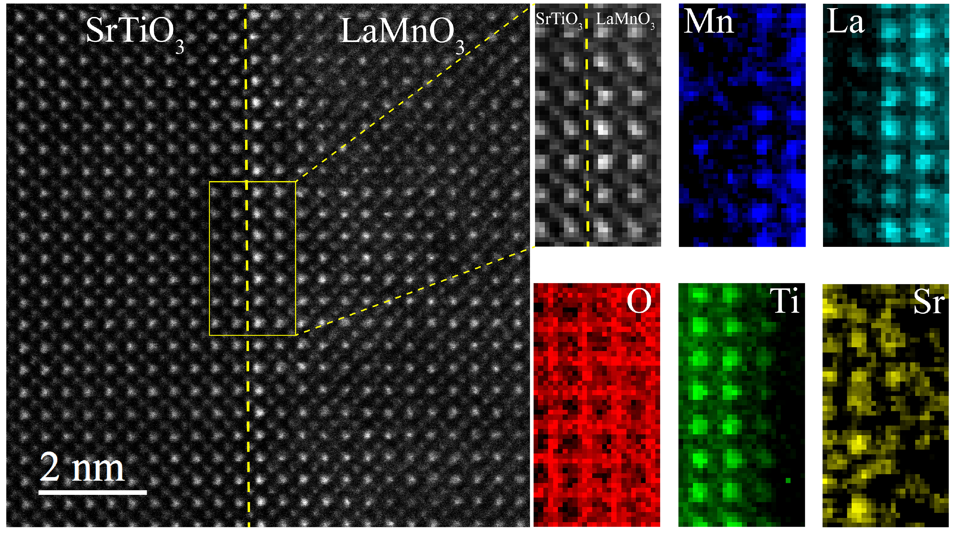
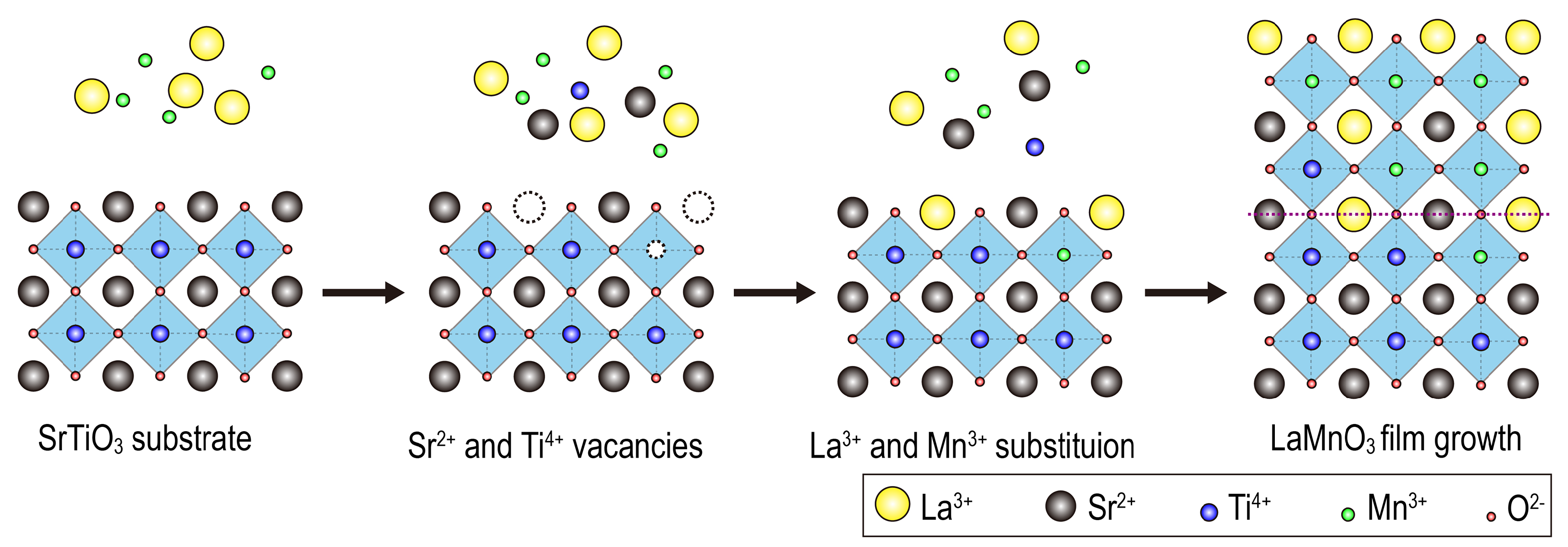
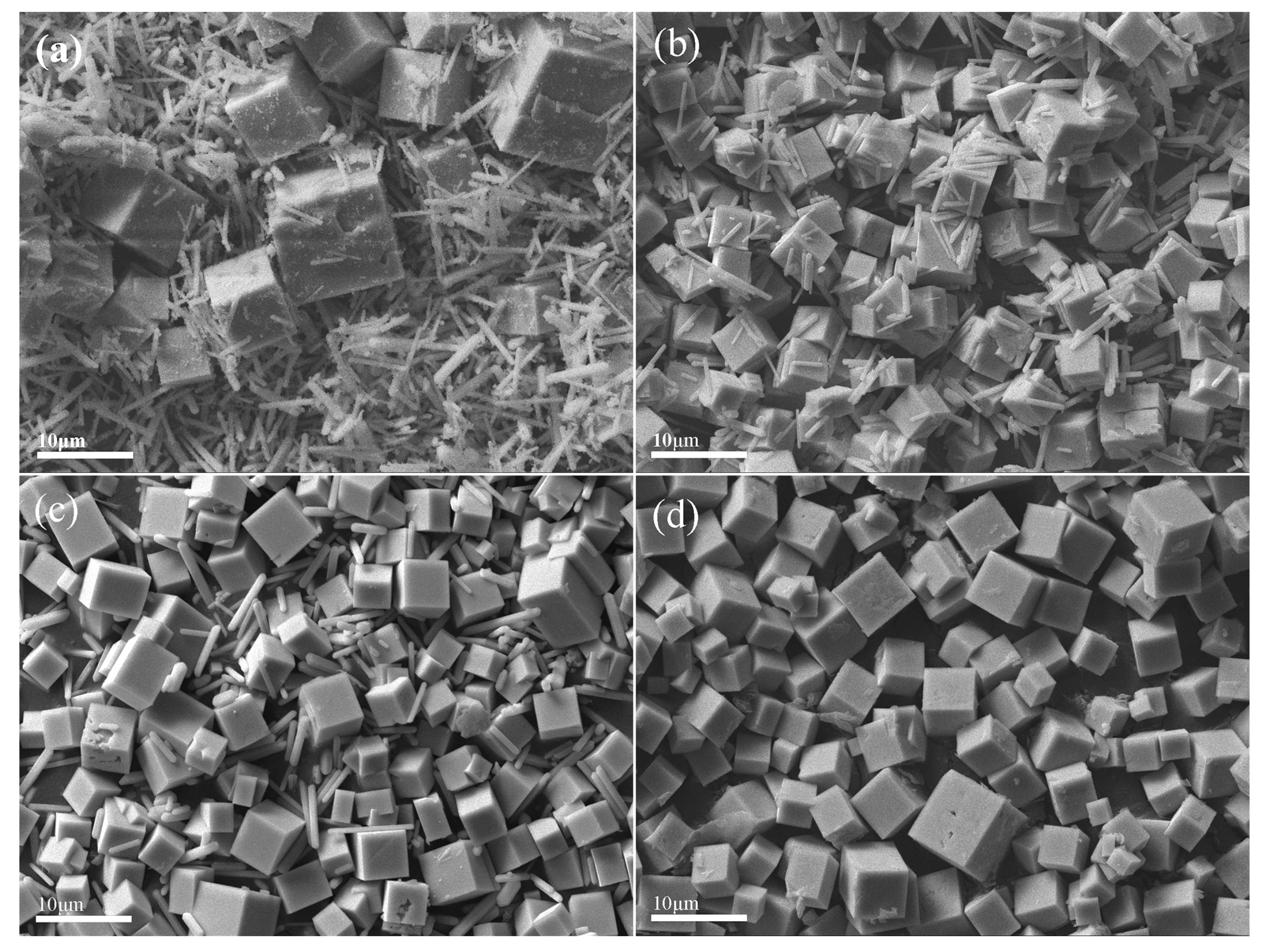
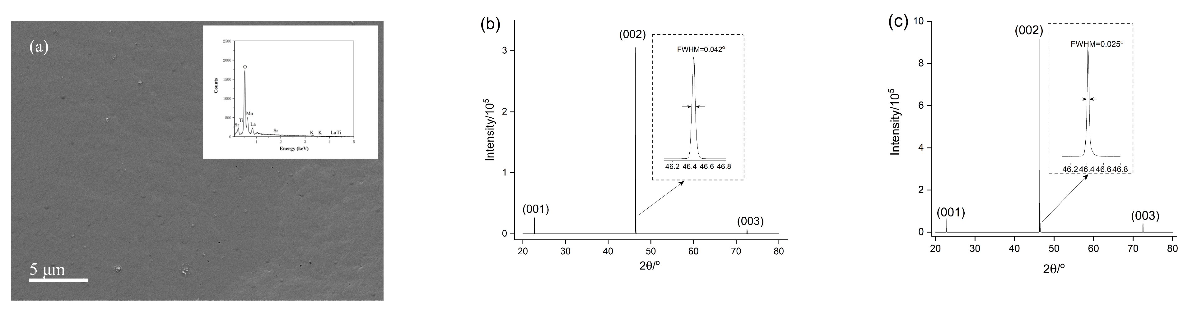
3. Results and Discussion

Figure 1 shows the powder X-ray diffraction patterns of samples obtained after a period of 2, 3, 6, 12 and 18 h under hydrothermal treatment. The low background in the diffraction patterns indicates adequate crystallinity of the bulk powders. As shown in Figure 1, the samples at 2 h and 3 h contain impurities, namely, K-rich birnessite and La(OH)3.
For the product at 6 h, a small amount of La(OH)3 can be identified, and no reflections due to K-rich birnessite are observed. No reflections from La(OH)3 or K-rich birnessite are detected in the sample after a period of 12 h. The powder X-ray diffraction patterns for the samples at 12 and 18 h indicate the presence of the trigonal phase of LaMnO3. Since most reflections due to the orthorhombic phase coincide with the reflections from the trigonal phase, the identification of the orthorhombic phase cannot be made unambiguously. Rietveld quantitative analyses including both trigonal and orthorhombic phases of LaMnO3 were then carried out using the GSASII software package [34] in order to evaluate the phase composition at each stage of the hydrothermal process. In Rietveld refinement, the instrumental parameters were obtained using the standard reference material LaB6 (SRM-660b) from the National Institute of Standards and Technology (NIST) and remained the same during Rietveld refinement. Lattice parameters and scale factors were involved in the refinement while atomic positions or thermal displacement parameters remained fixed. Anisotropic peak broadening was employed to interpret the sample micro strain effects. The powder sample at 2 h was not included in Rietveld refinement because no satisfying refinement can be obtained due to the lack of a proper model to describe the strong preferred orientation of the sample. The final Rietveld phase quantitative refinement of the sample at 18 h is shown in Figure 2 as a representative, and other Rietveld plots are in Figure S1. The phase compositions for samples at 3, 6, 12 and 18 h are listed in Table 1.
As indicated by the trigonal to orthorhombic phase ratio listed in Table 1, the trigonal phase dominates in the early stage of the hydrothermal synthesis. Previous research suggested that the trigonal phase can be stabilized by improving the Mn4+ content in LaMnO3 [35]. In our case, KMnO4 is used as a manganate precursor and Mn4+ is ready to be incorporated into LaMnO3 at the early stage of synthesis. As the synthesis proceeds, the amount of available Mn4+ decreases, resulting in a relatively more orthorhombic phase being formed.
Powder samples were further examined by SEM, and images for samples at 2, 3, 6 and 12 h are shown in Figure 3. Cube-shaped LaMnO3 was formed together with rod-shaped La(OH)3 at 2, 3 and 6 h, and the amount of La(OH)3 decreased gradually in the process of hydrothermal synthesis. The K-rich birnessite phase was not directly observed, but EDS shows that potassium was uniformly distributed across the sample surface (Figure S2). Pure LaMnO3 was yielded at 12 h.
After successfully obtaining LaMnO3 powders using the one-stage hydrothermal method, similar hydrothermal conditions were employed to prepare LaMnO3 films on SrTiO3 substrate. The lanthanum and manganese precursor concentrations were half of those used for the synthesis of bulk LaMnO3 powders and the film growth was carried out for a period of 2 h. The obtained LaMnO3 films on the SrTiO3 substrate were characterized by SEM/EDS, shown in Figure 4a. SEM images suggest that the film covered the substrate completely and EDS shows that La, Mn and O elements are uniformly distributed across the surface. High-resolution X-ray diffraction was employed to characterize the LaMnO3 film using a two-bounce Ge(220) monochromator. A symmetric ω/2θ scan of the LaMnO3 film shows only (00l) peaks. For comparison, a symmetric ω/2θ scan of the substrate was also obtained under the same instrumental conditions. Despite the similar (00l) peak positions from the LaMnO3 film and the substrate, the difference in the full width at the half maximum (FWHM), as shown in Figure 4b,c, indicates that pseudo-cubic LaMnO3 films with an out-of-pane preferred orientation were obtained on the SrTiO3 substrate.
To further demonstrate the film orientation with respect to the substrate, the interfacial structure was examined using a double-spherical differential correction electron microscope. Figure 5a shows the atomic arrangement at the interface between the LaMnO3 film and the SrTiO3 substrate, confirming that the epitaxial LaMnO3 film was grown on the substrate. Figure 5b shows the cross-section of the LaMnO3 film along the [100]pc (pc denotes pseudo-cubic) direction. In the ADF-STEM images, the contrast is approximately proportional to Z1.67, with Z being the atomic number. Therefore, the brightest atom columns correspond to the position of the heaviest element, i.e., lanthanum (Z = 57). Strontium (Z = 38) columns are formed by the second brightest features, which are in the horizontal line formed by La columns shown in Figure 5b. Mn and Ti columns are recognized as those in between La columns in LaMnO3 and Sr columns in SrTiO3. The O columns are largely invisible in the ADF mode. EELS mapping confirmed the assignment as discussed in the following section. Figure 5c displays a pixel intensity line scan of one row of atoms in the horizontal direction (white rectangle in Figure 5b) to differentiate the atoms at the interface. ADF-STEM imaging reveals that an atomic flat and sharp interface formed between LaMnO3 films and the SrTiO3 substrate.
Once the epitaxial heterostructure was identified, geometrical phase analysis (GPA) was performed to understand the lattice distortion of the LaMnO3 film. A wider field of view of the ADF−STEM images was chosen for GPA. The warm coloration on GPA represents a lattice expansion relative to the reference that was chosen from the core region of the substrate, while the cool coloration indicates that the lattice is contracted. If the film conforms to the substrate interplanar distance via epitaxy, there should be no obvious warm or cool coloration region on GPA. As shown in Figure 6a, the contrast between the substrate and the film in the in-plane direction of the film is relatively similar, suggesting no obvious lattice distortion along the [100]pc direction. On the contrary, Figure 6b exhibits a significant large out-of-plane lattice spacing of the LaMnO3 film, as indicated by the warm coloration on the LaMnO3 side. The average lattice spacing d001 for SrTiO3 and LaMnO3 is 3.96 and 4.01Å, respectively. The average lattice spacing d001 is slightly larger than the ideal bulk LaMnO3 (≈3.90 Å). GPA suggests that the LaMnO3 film is in a tensile strain state.
Lattice strain affects the orbital ordering and magnetism at the interface between LaMnO3 and SrTiO3. In the meantime, B-site atomic displacement also has a profound influence on the physical properties of the LaMnO3 film. Therefore, the B-site atomic displacements for both LaMnO3 films and the SrTiO3 substrate were investigated. The arrows shown in Figure 7a represent the magnitude and direction of the displacement of the B-site atoms from the ideal positions, and the color indicates the coherent direction of the B-site displacement.
The polar plot of the B-site atomic displacement (Figure 7b) shows that the direction is not restricted to any particular direction but is distributed over a wide range. The majority of the B-site displacement falls in the range of 2.5–10 pm and −30–120°. The B-site displacement analysis suggests the polarity of the LaMnO3 film.
To further investigate the LaMnO3 film growth mechanism, the elemental distribution at the interface was analyzed using electronic energy loss spectroscopy. Figure 8 shows an atomic resolution of the ADF−STEM images, along with EELS mapping. As shown in Figure 8, Mn, Ti, La and Sr atoms have diffusion to varying degrees in between the LaMnO3 film and the SrTiO3 substrate, indicating a certain degree of cation intermixing at the interface.
The cation diffusion may relate to the strong alkaline environment that causes some corrosion of the substrate to form Sr2+ vacancies. The resultant vacancies may be substituted by La3+ and therefore become active sites for nucleation and growth of LaMnO3 thin films. The free Sr2+ due to corrosion in the vicinity of the substrate may be incorporated into the newly formed LaMnO3 films. The diffusion of La and Sr is easily identified in the La and Sr maps shown in Figure 8. Furthermore, Figure 5b shows a bright feature of one atom column (middle) on the SrTiO3 side, which also indicates that La3+ is incorporated into the SrTiO3 substrate. Figure 9 shows a schematic view of the growth mechanism of LaMnO3 films. Mn3+ and Ti4+ have a weaker diffusion compared with La3+ and Sr2+, as seen in the Mn3+ and Ti4+ maps in Figure 8. Meanwhile, the O atomic columns on both sides of the interface are not as well-defined as the other elements, indicating the presence of octahedral distortions at the interface.

4. Conclusions

In summary, bulk LaMnO3 powders were successfully prepared by choosing appropriate lanthanum and manganese precursors under hydrothermal conditions. Trigonal LaMnO3 formation dominates the early stage of synthesis when a large amount of high valent manganese is available, while orthorhombic LaMnO3 has a preference over trigonal LaMnO3 at later stages. Epitaxial LaMnO3 film on SrTiO3 can be obtained using the hydrothermal route with growth parameters obtained from the bulk LaMnO3 powder synthesis. A highly out-of-plane preferred orientation of the LaMnO3 film was revealed by the high-resolution ω/2θ X-ray diffraction. A flat and sharp interface between LaMnO3 and SrTiO3 was verified by ADF−STEM imaging in combination with electronic energy loss spectroscopy. Anisotropic strain states were observed for the LaMnO3 film with no apparent in-plane strain and significant out-of-plane strain states. The B-site atomic displacement field displays a preferred position indicating the polarity of the LaMnO3 film. These findings open up a new way to fabricate complex oxide heterostructures with a well-defined interface using the one-stage hydrothermal route. Further work will include the investigation of the film formation process and morphology changes. Characterization of the magnetic properties of the epitaxial LaMnO3 film on the SrTiO3 substrate is in progress.

Supplementary Materials

The following supporting information can be downloaded at: https://www.mdpi.com/article/10.3390/ma15175928/s1, Figure S1: Rietveld phase quantitative analysis of the LaMnO3 powder at 3, 6 and 12 h; Figure S2: SEM image of hydrothermal product at 2 h together with EDS elemental mapping.

Author Contributions

Conceptualization, Y.L. (Yinong Lyu); Formal analysis, Y.L. (Yunfei Liu); Funding acquisition, Y.T.; Investigation, K.G.; Methodology, Y.L. (Yunfei Liu); Resources, Y.L. (Yinong Lyu); Software, Z.P.; Supervision, Y.L. (Yinong Lyu) and Z.P.; Writing – original draft, K.G.; Writing – review & editing, Y.T. and Z.P. All authors have read and agreed to the published version of the manuscript.

Funding

This research was funded by the Natural Science Funds for Young Scholar of China (grant no. 21107049) and the priority academic program development of Jiangsu Higher Education Institution (PAPD).

Institutional Review Board Statement

Not applicable.

Informed Consent Statement

Not applicable.

Conflicts of Interest

The authors declare no conflict of interest.

References

  1. Carda, M.; Budáč, D.; Paidar, M.; Bouzek, K. Current trends in the description of lanthanum strontium manganite oxygen electrode reaction mechanism in a high-temperature solid oxide cell. Curr. Opin. Electrochem. 2022, 31, 100852. [Google Scholar] [CrossRef]
  2. Park, B.-K.; Song, R.-H.; Lee, S.-B.; Lim, T.-H.; Park, S.-J.; Jung, W.; Lee, J.-W. Conformal bi-layered perovskite/spinel coating on a metallic wire network for solid oxide fuel cells via an electrodeposition-based route. J. Power Sources 2017, 348, 40–47. [Google Scholar] [CrossRef]
  3. Shimada, H.; Yamaguchi, T.; Sumi, H.; Nomura, K.; Yamaguchi, Y.; Fujishiro, Y. Extremely fine structured cathode for solid oxide fuel cells using Sr-doped LaMnO3 and Y2O3-stabilized ZrO2 nano-composite powder synthesized by spray pyrolysis. J. Power Sources 2017, 341, 280–284. [Google Scholar] [CrossRef]
  4. Mo, H.; Nan, H.; Lang, X.; Liu, S.; Qiao, L.; Hu, X.; Tian, H. Influence of calcium doping on performance of LaMnO3 supercapacitors. Ceram. Int. 2018, 44, 9733–9741. [Google Scholar] [CrossRef]
  5. Hu, Q.; Yue, B.; Yang, F.; Shao, H.; Bao, M.; Wang, Y.; Liu, J. Electrochemical and magnetic properties of perovskite type RMnO3 (R = La, Nd, Sm, Eu) nanofibers. J. Alloys Compd. 2021, 872, 159727. [Google Scholar] [CrossRef]
  6. Zhang, C.; Zhang, W.; Li, X.; Zhu, Z.; Wang, Q.; Luo, S.; Xie, A. Honeycomb LaMnO3 Perovskite Synthesized by a Carbon Sphere as a Self-Sacrificing Template for Supercapacitors. Energy Fuels 2021, 35, 13457–13465. [Google Scholar] [CrossRef]
  7. Spinicci, R.; Faticanti, M.; Marini, P.; De Rossi, S.; Porta, P. Catalytic activity of LaMnO3 and LaCoO3 perovskites towards VOCs combustion. J. Mol. Catal. A Chem. 2003, 197, 147–155. [Google Scholar] [CrossRef]
  8. Musialik-Piotrowska, A.; Landmesser, H. Noble metal-doped perovskites for the oxidation of organic air pollutants. Catal. Today 2008, 137, 357–361. [Google Scholar] [CrossRef]
  9. Shaterian, M.; Enhessari, M.; Rabbani, D.; Asghari, M.; Salavati-Niasari, M. Synthesis, characterization and photocatalytic activity of LaMnO3 nanoparticles. Appl. Surf. Sci. 2014, 318, 213–217. [Google Scholar] [CrossRef]
  10. Cossu, F.; Jilili, J.; Schwingenschlögl, U. 2D Electron Gas with 100% Spin-Polarization in the (LaMnO3)2/(SrTiO3)2 Superlattice under Uniaxial Strain. Adv. Mater. Interfaces 2014, 1, 1400057. [Google Scholar] [CrossRef]
  11. Tokura, Y.; Tomioka, Y. Colossal magnetoresistive manganites. J. Magn. Magn. Mater. 1999, 200, 1–23. [Google Scholar] [CrossRef]
  12. Zhang, X.Y.; Tao, Y.J.; Jiao, P.J.; Wang, J.; Zhang, J.; Luo, Z.L.; Gu, Z.B.; Zhou, J.; Chen, Y.B.; Zhang, S.T. Robust ferromagnetic insulating and large exchange bias in LaMnO3:CoO composite thin films. J. Phys. D Appl. Phys. 2022, 55, 195003. [Google Scholar] [CrossRef]
  13. Chen, Y.F.; Wang, G.M.; Sun, Z.F.; Wang, S.G.; Mao, Y.W.; Deng, Q.R.; Yang, J.J. Magnetic characteristics of LaMnO3+δ thin films deposited by RF magnetron sputtering in an O2/Ar mixture gas. Mater. Res. Express 2021, 8, 016101. [Google Scholar]
  14. Niu, W.; Liu, W.Q.; Gu, M.; Chen, Y.D.; Zhang, X.Q.; Zhang, M.H.; Chen, Y.Q.; Wang, J.; Du, J.; Song, F.Q.; et al. Direct Demonstration of the Emergent Magnetism Resulting from the Multivalence Mn in a LaMnO3 Epitaxial Thin Film System. Adv. Electron. Mater. 2018, 4, 1800055. [Google Scholar] [CrossRef]
  15. Hibble, S.J.; Cooper, S.P.; Hannon, A.C.; Fawcett, I.D.; Greenblatt, M. Local distortions in the colossal magnetoresistive manganates La0.70Ca0.30MnO3, La0.80Ca0.20MnO3 and La0.70Sr0.30MnO3 revealed by total neutron diffraction. J. Phys. Condens. Mat. 1999, 11, 9221–9238. [Google Scholar] [CrossRef]
  16. Guo, Y.Q.; Zhang, X.H.; Wappling, R. Crystal structure of La1-xSrxMnO3-2x+deltaF2x. J. Alloys Compd. 2000, 306, 133–140. [Google Scholar] [CrossRef]
  17. Wollan, E.O.; Koehler, W.C. Neutron Diffraction Study of the Magnetic Properties of the Series of Perovskite-Type Compounds [(1-x)La,xCa]MnO3. Phys. Rev. 1955, 100, 545–563. [Google Scholar] [CrossRef]
  18. Zener, C. Interaction between the d-Shells in the Transition Metals. II. Ferromagnetic Compounds of Manganese with Perovskite Structure. Phys. Rev. 1951, 82, 403–405. [Google Scholar] [CrossRef]
  19. Hoffman, J.; Tung, I.C.; Nelson-Cheeseman, B.B.; Liu, M.; Freeland, J.W.; Bhattacharya, A. Charge transfer and interfacial magnetism in (LaNiO3) n/(LaMnO3)2 superlattices. Phys. Rev. B 2013, 88, 144411. [Google Scholar] [CrossRef]
  20. Zhai, X.F.; Mohapatra, C.S.; Shah, A.B.; Zuo, J.M.; Eckstein, J.N. Magnetic properties of the (LaMnO3)N/(SrTiO3)Natomic layer superlattices. J. Appl. Phys. 2013, 113, 173913. [Google Scholar] [CrossRef]
  21. Marton, Z.; Seo, S.S.A.; Egami, T.; Lee, H.N. Growth control of stoichiometry in LaMnO3 epitaxial thin films by pulsed laser deposition. J. Cryst. Growth 2010, 312, 2923–2927. [Google Scholar] [CrossRef]
  22. Ogugua, S.N.; Ntwaeaborwa, O.M.; Swart, H.C. Latest Development on Pulsed Laser Deposited Thin Films for Advanced Luminescence Applications. Coatings 2020, 10, 1078. [Google Scholar] [CrossRef]
  23. Li, T.; Ma, D.; Li, K.; Hu, Z. Self-biased magnetoelectric coupling effect in the layered La0.7Sr0.3MnO3/BaTiO3/La0.7Sr0.3MnO3 multiferroic heterostructure. J. Alloy. Compd. 2018, 747, 558–562. [Google Scholar] [CrossRef]
  24. Yu, T.; Chen, P.; Zhou, L.; Ning, X.; Deng, B.; Shi, X.; He, H. Cluster glass induced unconventional exchange bias in epitaxial LaMnO3/SrMnO3 bilayers. Mater. Lett. 2019, 242, 95–98. [Google Scholar] [CrossRef]
  25. Panjan, P.; Drnovsek, A.; Gselman, P.; Cekada, M.; Panjan, M. Review of Growth Defects in Thin Films Prepared by PVD Techniques. Coatings 2020, 10, 447. [Google Scholar] [CrossRef]
  26. Dunnill, C.W.; Kafizas, A.; Parkin, I.P. CVD Production of Doped Titanium Dioxide Thin Films. Chem. Vapor Depos. 2012, 18, 89–101. [Google Scholar] [CrossRef]
  27. Saha, I.; Tomohiro, Y.; Kanazawa, K.; Nitani, H.; Kuroda, S. Structural and Magnetic Properties of Nitrogen Acceptor Co-doped (Zn,Fe)Te Thin Films Grown in Zn-Rich Condition by Molecular Beam Epitaxy (MBE). J. Electron. Mater. 2020, 49, 5739–5749. [Google Scholar] [CrossRef]
  28. Wang, T.; Thoutam, L.R.; Prakash, A.; Nunn, W.; Haugstad, G.; Jalan, B. Defect-driven localization crossovers in MBE-grown La-doped SrSnO3 films. Phys. Rev. Mater. 2017, 1, 06160. [Google Scholar] [CrossRef]
  29. Morita, T. Piezoelectric Materials Synthesized by the Hydrothermal Method and Their Applications. Materials 2010, 3, 5236. [Google Scholar] [CrossRef]
  30. Ngida, R.E.A.; Zawrah, M.F.; Khattab, R.M.; Heikal, E. Hydrothermal synthesis, sintering and characterization of nano La-manganite perovskite doped with Ca or Sr. Ceram. Int. 2019, 45, 4894–4901. [Google Scholar] [CrossRef]
  31. Morita, T.; Cho, Y. Epitaxial PbTiO3 thin films on SrTiO3(100) and SrRuO3/SrTiO3(100) substrates deposited by a hydrothermal method. Jpn. J. Appl. Phys. 2004, 43, 6535–6538. [Google Scholar] [CrossRef]
  32. Rout, D.; Han, S.H.; Moon, K.S.; Kim, H.G.; Cheon, C.I.; Kang, S.J.L. Low temperature hydrothermal epitaxy and Raman study of heteroepitaxial BiFeO3 film. Appl. Phys. Lett. 2009, 95, 122509. [Google Scholar] [CrossRef]
  33. Huang, A.; Handoko, A.D.; Goh, G.K.L.; Shannigrahi, S.R.; Tan, C.K. Hydrothermal epitaxy of BiFeO3 films on SrTiO3 substrates. Prog. Cryst. Growth Charact. Mater. 2011, 57, 109–116. [Google Scholar] [CrossRef]
  34. Toby, B.H.; Von Dreele, R.B. GSAS-II: The genesis of a modern open-source all purpose crystallography software package. J. Appl. Crystallogr. 2013, 46, 544–549. [Google Scholar] [CrossRef]
  35. Mahendiran, R.; Mahesh, R.; Raychaudhuri, A.K.; Rao, C.N.R. Giant magnetoresistance in bulk samples of LaMnO3 with varying Mn4+ content. PRAMANA-J. Phys. 1995, 44, 393–396. [Google Scholar] [CrossRef]
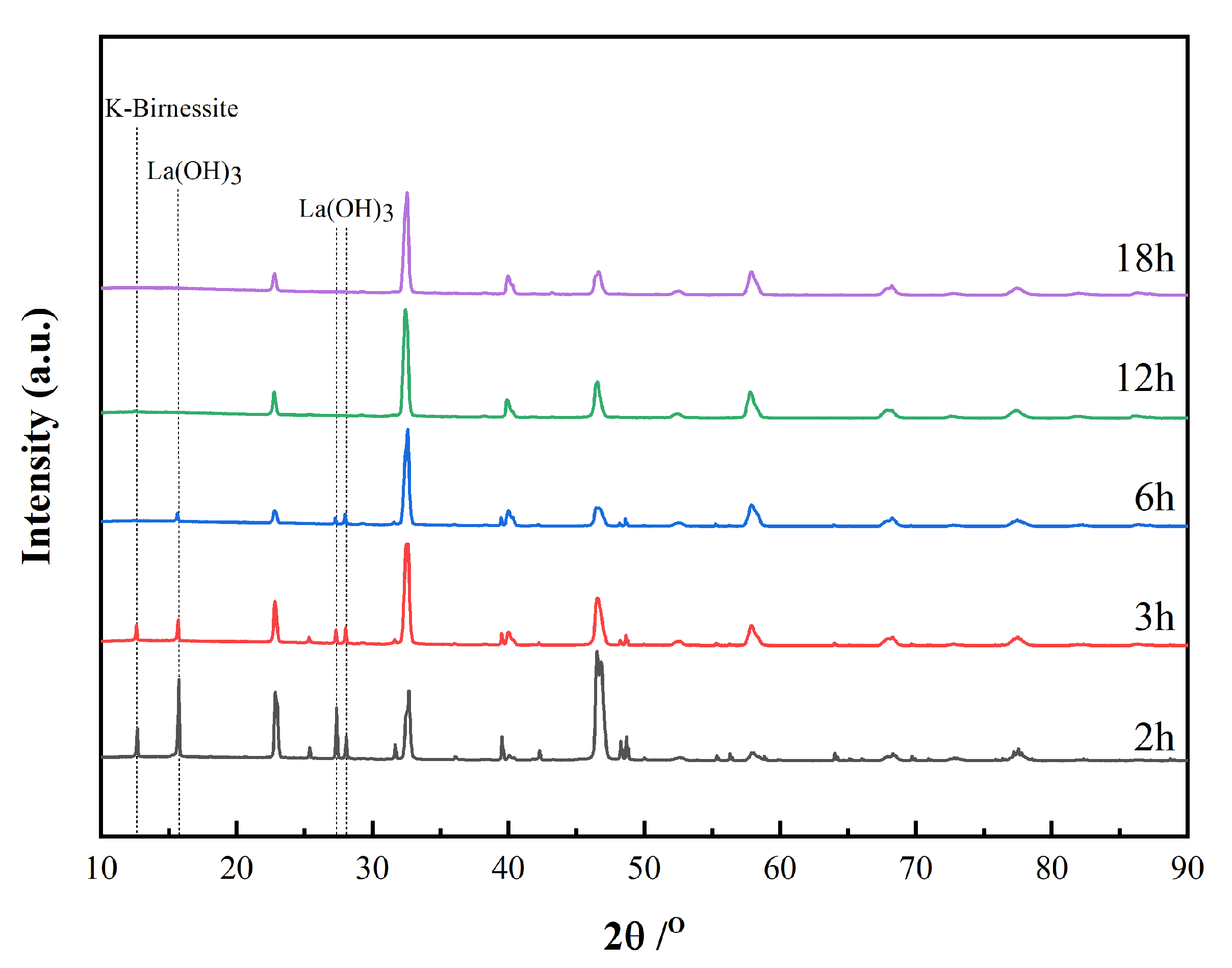
Figure 1. XRD patterns of LaMnO3 powders for periods of 2, 3, 6, 12 and 18 h. Reflections due to K-rich birnessite and La(OH)3 are indicated by dotted lines.
Materials 15 05928 g001
Figure 2. Rietveld phase quantitative analysis of the LaMnO3 powder at 18 h. The experimental powder X-ray diffraction data (black + marks), calculated powder X-ray diffraction data (black solid line) and the difference between experimental and calculated data (black lower line) are shown. Tick marks the reflection positions for trigonal (red, upper) and orthorhombic (blue, lower) LaMnO3.
Materials 15 05928 g002
Figure 3. SEM images of products at different hydrothermal synthesis stages: (a) 2 h, (b) 3 h, (c) 6 h, (d) 12 h.
Materials 15 05928 g003
Figure 4. Characterization of the LaMnO3 film on the SrTiO3 substrate: (a) SEM image showing uniform and flat film surface. Inset: EDS spectrum; (b) symmetric ω/2θ scan of the LaMnO3 film. Inset: FWHM of the LaMnO3 film (002) peak; (c) symmetric ω/2θ scan of the SrTiO3 substrate. Inset: FWHM of the substrate (002) peak.
Materials 15 05928 g004
Figure 5. (a) ADF-STEM images of the interfaces between LaMnO3 films and the SrTiO3 substrate. Yellow dotted lines indicate the positions of the atomically sharp interface; (b) ADF−STEM images of the cross-section of LaMnO3 films with five unit cells of thickness in the [100] direction; (c) pixel in-tensity profile of an ADF line scan.
Materials 15 05928 g005
Figure 6. Geometrical phase analysis of LaMnO3 film showing in-plane (a) and out-of-plane (b) strain states.
Materials 15 05928 g006
Figure 7. (a) ADF−STEM image with superimposed arrows representing the displacement of the Mn atoms with respect to the center of four neighboring A-site atoms. (b) The polar plot of the displacement of B-site atoms.
Materials 15 05928 g007
Figure 8. ADF−STEM images of the LaMnO3/SrTiO3 heterostructure with selected area for core-loss EELS mapping (outlined by a yellow rectangle) and the resulting map showing the spatial distribution of Mn (blue), La (cyan), O (red), Ti (green) and Sr (yellow) atoms.
Materials 15 05928 g008
Figure 9. Schematic view of LaMnO3 epitaxial thin film growth mechanism.
Materials 15 05928 g009
Table 1. Phase compositions of the hydrothermal products at different stages.
TimeTrigonal
LaMnO3/%
Orthorhombic LaMnO3/%K-Rich Birnessite/%La(OH)3/%Trigonal/
Orthorhombic
3 h62.523.97.16.42.61
6 h61.433.1-5.51.85
12 h63.736.3--1.75
18 h61.738.3--1.61
Publisher’s Note: MDPI stays neutral with regard to jurisdictional claims in published maps and institutional affiliations.

