Review

Magnesium for Implants: A Review on the Effect of Alloying Elements on Biocompatibility and Properties

1
Department of Mechanical Engineering, King Fahd University of Petroleum and Minerals, Dhahran 31261, Saudi Arabia
2
Interdisciplinary Research Center for Advanced Materials, King Fahd University of Petroleum and Minerals, Dhahran 31261, Saudi Arabia
*
Author to whom correspondence should be addressed.
Materials 2022, 15(16), 5669; https://doi.org/10.3390/ma15165669
Submission received: 21 April 2022 / Revised: 31 May 2022 / Accepted: 11 June 2022 / Published: 18 August 2022

Abstract

:
An attempt is made to cover the whole of the topic of biodegradable magnesium (Mg) alloys with a focus on the biocompatibility of the individual alloying elements, as well as shed light on the degradation characteristics, microstructure, and mechanical properties of most binary alloys. Some of the various work processes carried out by researchers to achieve the alloys and their surface modifications have been highlighted. Additionally, a brief look into the literature on magnesium composites as also been included towards the end, to provide a more complete picture of the topic. In most cases, the chronological order of events has not been particularly followed, and instead, this work is concentrated on compiling and presenting an update of the work carried out on the topic of biodegradable magnesium alloys from the recent literature available to us.

1. Introduction

Biodegradable materials for implants have been in clinical use for some time now [1]. These materials have come into prominence in lieu of non-biodegradable, permanent implants, which have had temporary applications to afford the body a healing period after which the implants are no longer required. The use of permanent implants for temporary applications meant a secondary operation is carried out to remove the implant. Apart from the obvious trauma and the inconvenience suffered by the patients and their families, it also meant added medical costs and medical resources are spent in this ordeal, not to mention the economic value of time spent by all parties involved. These permanent implants were mostly either titanium or steel-based alloys. Common issues of the resulting ‘effects’ by the permanent orthopedic implants include inflammation, infection, stress shielding, and consequent bone loss. Stress shielding is due to the higher stiffness of the implants, leading to the dis-use of adjacent bones and further leading to a gradual loss of bone structure and weakening of the bones. The solution has been the use of biodegradable materials for implants, which would corrode naturally within the body, after or during affording the required time for the healing. Biodegradable materials such as biodegradable polymers (with polyglycolic acid (PGA) and polylactic acid (PLA) being most common), bioceramics (Tricalcium phosphate (TCP), Hydroxyapatite (HA)), and biodegradable Mg alloys [2] have been in increasing use for this reason. The applications included bone fixtures such as nails [3], screws [4], clips [5], wires [6], and stents [7]. Of interest has been applications involving bones, which is where magnesium and magnesium-based alloys have been researched dominantly.
Magnesium [8] has the electron configuration of 1s22s22p63s2 [8], with a hexagonal close-packed (HCP) crystal structure. At 1.7 g/cm3 it has the lowest density amongst all structural metals. It has a Young’s Modulus of approximately 45 GPa. In comparison, cortical bone has a density of 1.8–2.0 g/cm3 and a Young’s modulus of 7–30 GPa [9]. This closeness in properties is an advantage for which magnesium and its alloys have been of significant research as a biodegradable implant material.
Magnesium has been found to have special osteoconductive properties, which is much appreciated when used as bone implants. It has been reported that Mg, as a cofactor of the alkaline phosphatase isozymes, helps in the healing and remodeling of the bone tissue [10]. Magnesium-based stents are also useful where biodegradable nasal stents could help avoid treatment failure that occurs due to the secondary operations that are required of traditional implants [11]. In the dental field, the use of scaffolds made of Mg/PLGA porous composites to improve bone healing following tooth extraction has been used [12]. More recently, in the dental and orthopedic fields, Mg in bone cement composites have been used to obtain high-strength cement with both biodegradability and bioactivity in the form of magnesium calcium phosphate/sodium alginate composite cement [13].
Others still have used Mg in magnesium/calcium phosphate cements to induce an improved cellular response of bone marrow stromal cells (BMSCs) [14]. Mg ions have been known to promote osteogenic activity of bone marrow stromal cells [15]. Furthermore, various studies have been conducted to observe the effect of Mg alloys on mesenchymal stem cells [16,17]. B. Kanter et al. [18] has demonstrated the suitability of magnesium phosphate cements in partially load-bearing defects of a sheep model. However, Magnesium phosphate is also one of the mineral components of kidney stones [19], although any potential relationship between that and the biodegradable Mg-based cement is not clear.
The potential application of biodegradable magnesium alloys is not limited to its use as temporary implants. Research has been carried out to investigate the utilization of the fast degradation characteristics of magnesium with aqueous media, or in this case, blood, to provide the thrust in magnesium-based micromotors [20]. Moreover, research into a magnesium-based biodegradable battery has been conducted [21] to investigate the potential applications to power bioresorbable transient implants.
There has also been a number of research studies carried out to improve the surface properties of the traditional implants with the use of magnesium to achieve better performance. These include the co-implantation of Zn/Mg ions on titanium dental implant surfaces to improve osteogenesis [22] properties of the implant, and the Mg ion implantation on micro and nano-structured titanium surfaces to improve its osteogenic differentiation and proliferation of bone marrow cells [23] so as to achieve greater contact of the corroding implant and the healing bone tissue.
With all this being said, the effect of the individual alloying elements on the biocompatibility, biodegradability, microstructure, and mechanical properties of pure magnesium need to be understood clearly and is therefore discussed in detail in this study, and the effects of work history, heat treatment, and coating have been briefly discussed.

2. Requirements for Bio Application of Mg Alloys and Their Corrosion Strengths

The biocompatibility of magnesium biodegradable alloy is determined via cytotoxicity tests conducted either in vitro or in vivo. The cytotoxicity tests are mainly designed to test for either the cell proliferation in a given medium of magnesium-degraded products or cell adhesion to the magnesium alloy being tested. The standard of the in vitro tests is usually performed as in standard ISO 10993 part 5 [24,25,26,27].
However, there has been poor correlation between in vitro and in vivo studies when using the ISO 10993 standard [24,27] due to which, in recent years, there have been studies to propose a modified approach to the standards of in vitro testing of cytotoxicity. Some researchers suggested dilution of the extracts to achieve more accurate in vitro tests. J. Wang et al. [24] proposed a 6- to 10-times dilution of extracts while J. Fischer [25] proposed a 10-times dilution for as-cast material. Others, such as L. Scheideler [27], proposed the use of a bovine serum instead of the standard recommended cell culture medium. However, X. Liu et al. [28] studied the extraction parameters for its influence in the predictability of in vivo tests via in vitro tests and they specifically discounted the addition of bovine serum albumin (BSA) and fetal bovine serum (FBS) for greater predictability because of their acceleration of corrosion of magnesium samples during extraction and their effects on cell health during the test. Their proposed solution was the establishment of a database recording the tolerance of cells towards the main hazards of metal ions, pH, and H2 gas along with a set of in vitro corrosion tests with high similarity to in vivo tests.
O. Jung et al. [29] reported an optimized procedure for testing the in vitro cytocompatibility, within the standard DIN EN ISO 10993-5:2009. Their study recommended the use a combination of an indirect assay and a complementary direct live-dead staining of cells grown on magnesium materials, for testing cell proliferation, viability, and cytotoxicity. It was also suggested that the exposed surface area of Mg in the in vitro testing media may have played a role in the discrepancies in results. This is also a possible reason for the differences between in vitro and in vivo cytotoxicity results discussed above. A higher exposure of the magnesium surface has been recommended to give better results [29].
The release of gases to the surrounding vicinity, i.e., tissue [30] and bone, is investigated to determine its influence and effects. Slower degradation rates are necessary for low gas evolution rates. Higher rates of evolution of H2 gas could lead to inflammation and swelling of the surrounding tissue. Similarly, gas pockets and the pressure associated with the release of H2 could lead to deformations during the osteogenesis applications of magnesium alloys.
Due to the above-mentioned limitations, only those elements with positive or negligibly negative effects to the human body should be considered for use as in applications involving biodegradable magnesium alloys. The requirements for bio application of the magnesium alloys are (i) uniform corrosion degradation, (ii) a slow and controlled degradation rate, and (iii) good cytotoxicity

3. Biotoxicity of Common Alloying Elements

3.1. Toxicology and Pathophysiology

The nutritional requirements of the human body and therefore, by extension, the tolerance of the body to different elements, vary by ethnicity—which can be said to be differentiated by geography and culture. For instance, it has been reported by the World Health Organization (WHO) [31] that there exists wide differences in dietary intake of calcium levels between the different regions of the world, which has been attributed to the different diets based on food culture and availability. This is an indication of the differences in allowances when considering each specific group of people. Therefore, this variation in tolerance needs to be respected when choosing the magnesium alloy for use as implants. The degradation rate should be within these tolerances to avoid unnecessary complications.
The nature of the chemical reaction and thereby the toxicity of the alloying elements is dependent on identifying the speciation of the elements in the extracellular fluid, which is the medium of their transport to various other locations. This is, however, a difficult task as the metals in the biological environment could form complexes with any of the available potential ligands and as many of the metal complexes are kinetically labile, i.e., susceptible to being altered [32]. For this reason, the toxicology of the alloying elements will be discussed on the basis of their general toxicology and nutritional value rather than their specific effects on individual species. Moreover, for the purpose of relevancy to the topic, the toxicology of the inhalation route of exposure to the metals is not discussed.

3.1.1. Magnesium (Mg)

The human skeleton contains approximately 50–60% of the body’s magnesium, with 1% in the extracellular fluid and the remaining in the muscles and tissues [31]. It is also an essential element involved in the regulation of potassium fluxes and the metabolism of calcium [31]. The magnesium forms a surface constituent of the hydroxyapatite mineral component in the bone and acts in the regulation of magnesium content in the serum at times of deficiency. At times of plenty in erythrocyte magnesium, the bone mineral density increases, while in times of deficiency, it helps make up the magnesium amount to a certain extent, and this form of accessible magnesium availability has been reported to decrease significantly with age [31].

3.1.2. Iron (Fe)

Iron is an essential element for the human body [33], and it is one of the most abundant metals in the body. Iron is essential for oxygen transport (Hemoglobin) and cellular functions such as the synthesis of RNA and DNA, as well as the synthesis of proteins, and is also involved in the regulation of gene expression among many other functions [33]. Additionally, low levels of Iron in the body have also been known to cause anemia. However, an excess of iron, or an iron overload, has also been reported to lead to abnormal interference in the body and cause serious health issues including iron complications arising from altered iron content in cells and tissues, leading to death if left untreated [33].

3.1.3. Calcium (Ca)

Calcium helps provide rigidity to the human skeleton, and its ions are involved in many aspects of the body’s metabolism [31]. It is the fifth most abundant element in the human body and constitutes approximately 2% of an adult’s lean body mass [31], of which almost all are found in the human skeletal system including the teeth and the soft tissues. Approximately 0.1% of the total Calcium in the body is available in the extracellular fluids and are present in the form of ions (1.20 mmol/L or 4.8 mg/100 mL) and complexes (1.6 mg/100 mL or 0.4 mmol/L [31]. Low levels of Ca are regulated via bone resorption, and a higher intake of Ca is absorbed back while the excess unabsorbed Ca is excreted out in the feces [31]. The bone mineral serves as a reservoir in this process. However, very high doses of Ca in carbonate form have been reported to lead to the precipitation of Ca salts in the renal tissue, while Ca deficiency leads to osteoporosis [31].

3.1.4. Zinc (Zn)

Zinc is an essential element, with its presence in all body tissues and fluids, though the plasma zinc accounts for only approximately 0.1% of the total zinc content in the body. It is essential for the enzymes involved in the metabolism of nutrients such as carbohydrates, lipids, proteins, as well as nucleic acids [31]. Furthermore, it is even essential for genetic expression and helps to stabilize the molecular structures of membranes and other components of the cells. Zn has a concentration presence of 0.46 μmol/g (30 μg/g) in the lean body mass [31]. The plasma zinc is regulated via homeostatic control.

3.1.5. Copper (Cu)

Cu is essential and is the third most abundant trace element in the human body [34,35]. Cu2+ affects gene expression in mammals [36]. It is an important catalyst for the synthesis of heme and absorption of iron [34]. The body commonly obtains it via ingestion, i.e., as water contaminants or as nutritional components of food. This pathway of absorption of Cu into the body is well-regulated, and most of the follow-up reactions are well-documented [35]. However, when used in biodegradable material, the cellular tissues could be exposed to free Cu ions, which, at concentrated levels, could lead to cellular damage due to its inherent highly reactive nature. Although transmembrane transporters and metallochaperones exist to control the levels of intra cellular copper [35], care should be taken in designing implants so as not to dissociate too much Cu into the surrounding contact fluid. Freely available Cu can be potentially toxic if it oxidizes lipids and proteins leading to the formation of intracellular and extracellular toxic free radicals [36]. Copper toxicity is rare, and when it does occur, it primarily affects the liver [34]. Above 3 mg/L of the whole blood concentration of Cu it is reported to lead to gastrointestinal symptoms of toxicity [34].

3.1.6. Silicon (Si)

Forrest H. Nielson [37] has reported circumstantial evidence that Silicon is an essential nutrient for the human body. Its deficiency has been reported to cause abnormal metabolism of connective tissue and bone in animal tests. Furthermore, tests conducted on rats have shown that Si helps to avoid the accumulation of Al in the brain—which has been connected to Alzheimer’s [37]. Moreover, Si as a polymer, in the form of polydimethysiloxane, has been a popular breast implant material for some time now. However, though these Silicones have been thought to be biologically inert, they have caused inflammation and other complications over time [38,39,40,41]. On the other hand, the intravenous administration of Si nanoparticles has reportedly resulted in relative biocompatibility as far as acute toxicity was concerned but showed granuloma formation (See Figure 1), indicating inflammation in reticulo-endothelial organs such as the liver and spleen [42]. Therefore, the use of elemental Si in implants needs more research, and if it is included as an alloying element to Mg, it must be included cautiously.

3.1.7. Tin (Sn)

Sn is not regarded as an essential element, although multivitamin and mineral supplements reportedly contain up to 0.01 mg in a daily recommended dose [43]. Sn is mostly ingested into the body via canned food consumption, where the cans are lined with or is made of Sn. At a high dosage, Sn accumulates in the bones, liver, and kidney, with bones being the primary site of deposition when Sn is injected intramuscularly and during extended exposure to Sn even through other means such as ingestion [43].

3.1.8. Manganese (Mn)

Mn is an essential element for the human body, which is regulated via homeostasis where excess Mn is mainly excreted via bile and feces. The portion that forms conjugates with the bile is also ultimately excreted out mostly via feces and only a small amount is excreted through urine. It is mainly concentrated in the liver, pancreas, and kidney, but notably, it has its lowest concentrations in fat and bones, wit the latter being of significance in the aspect of its effect in the current discussion. A Mn overload could affect the motor and cognitive abilities of the central nervous system, while prolonged Mn deficiencies have been reported to lead to greater occurrence of symptoms related to Parkinsonism [44].

3.1.9. Aluminum (Al)

Aluminum is rated as Generally Regarded As Safe (GRAS) by the US FDA [32], due to which it is widely available in many foods and medications. Previous studies have reported the lack of proof for Al accumulation in the brain [32]. However, it has been associated with neurotoxicity in recent years and has been reported to lead to Alzheimer’s disease, although there is still skepticism with this conclusion [45].

3.1.10. Lithium (Li)

Lithium is a non-essential trace element. Due to it is similarity with Sodium and Potassium, it can cross all biological barriers, and since it is not protein-bound, it is excreted by the kidneys and does not accumulate in the tissues too much. However, long-term use of Li in therapeutic medicine has been recorded to show toxic effects on the kidney, thyroid, and the Central Nervous System [46]. Taking into account the biodegradable nature of the implants, and the excretion of the Li from the body, the long-term exposure effects can be disregarded, and as such, Li is potentially useable as an alloying element of Mg pending further site-specific tests.

3.1.11. Nickel (Ni)

There is reportedly circumstantial evidence of Ni as an essential element [37], where it is necessary for certain activities of metabolism, and in animal studies conducted, its deficiency has been shown to show negative effects on growth, reproductive performance, and plasma glucose [37]. Ni deficiency has been also reported to affect the distribution of other essential elements in the body such as Ca, Zn, and Fe. However, exposure of Ni in some of the forms other than oral administration have been reported to be a potential source of cancer in animals and humans [37]. It has been suspected of interfering in the functions of Vitamin B12 and Folic acid [37].

3.1.12. Indium (In)

Indium is a non-essential element that produces wide-ranging toxic effects depending on its form. Ionic Indium led to renal failure upon its concentration in the kidney. It is mainly excreted in the urine. The colloidal form of Indium causes damage to the liver and spleen, and this form is mostly excreted via feces [47]. If poorly absorbed when ingested, indium is mostly stored in muscles, skin, and bones [47].
The toxicology of some of the common Mg alloying elements along with their daily allowances as well as the whole blood levels (based on French, Swedish, and Benin populations) and the blood serum levels (based on French and Swedish populations) are given in Table 1 below. The data are as that reported for adults, and it may vary for different age groups. Notably, the reported Mg daily allowance limits from two sources are very different from each other. Again, this could be due to differences in the population, which was considered in the literature.

3.2. In Vitro Biotoxicity

In vitro testing has been carried out in laboratory facilities with the Mg alloy specimens immersed in a cell culture of preferred contents and tested for the cell viability and cytotoxicity after the test duration. Different groups have used a number of different cell mediums in this regard. Biotoxicity is measured in terms of cell viability (%), hemolysis, % and cell adhesion. These parameters are dependent on culture time. It is perhaps noteworthy to mention that the characteristic rates of these parameters need careful investigation when designing the implant itself. This is due to the non-linearity of these parameters. As per the ISO 10993-5:2009, cell viability below 30% is deemed as cytotoxic in nature [60].
Cytotoxicity: Measured by Relative Growth Rates (RGR) of the cells as determined by a method such as gradation optical density measurements [61]. The graded scale is used as a means of determining the cytotoxicity of the alloy to the tested cell line.
Cell viability: Viability of the cell can be measured by determining the count of live and dead cells. This has been performed by exposing the experimental cell line to the aqueous extract of the alloy [61].
Cell Adhesion: This is useful to measure the BIC or the Bone-to-Implant Contact of the Mg alloy, useful in osteogenesis applications. A greater BIC score is useful to avoid the formation of voids between the degrading implant surface and the forming bone tissue.
When the Mg alloys are tested in vitro for cell viability, a range of cell cultures are available for use. The type of cells used depend mostly on the type of design application of the alloy. For instance, those relating to osteosynthesis would be tested with hBMMSCs (human bone marrow mesenchymal stem cells) [4] or MC3T3-E1 (osteoblasts) [62], or U-2OS (human osteosarcoma cells) [63] have been used when testing for osteosynthesis potentiality. Similarly, those that may be exposed to blood or lymph vessels, as is the case of the dissolute ions in the blood, could be tested using VSMC (rodent vascular smooth muscle cells) [62] for animal models. Table 2 has been composed using such in vitro cytotoxicity results of the alloy elements themselves or as binary alloys of Mg in extract media as found in various literature. The elements’ effect on various cell lines are listed along with a summary of the test result. It has to be noted that the cytotoxicity may not always be detected during the period of either in vitro or in vivo tests and could be due to the absence of degradation products’ effects on the specific organs tested in vivo/ex vivo, and this by no means can be taken as having no adverse effect of any alloying elements on the body. Rather, in the case of alloys containing specifically toxic elements in terms of both the toxicity and pathophysiology, it is the view of the Authors that a better way is to refer to the biological aspects of element absorption, accumulation, and excretion pathways during in vivo studies (see Table 3).

3.3. In Vivo Biotoxicity

3.3.1. Clinical Trials: Animal Tests

Recently, J. Zhang et al. [70] studied the follow-up of an Mg alloy stent [71] in a Rabbit model and reported the ion diffusion and replacement that takes place after the Mg alloy stent was completely degraded, at 20 months after implantation. In vivo tests conducted on a rabbit model by P. Han et al. [72] using high-purity magnesium screws determined some qualitative aspects of the mechanical properties of the screws. As shown in Figure 2, the screw used as a fixature for the femoral fracture with a gap of 3 mm was seen to bend at 4 weeks post-operation. This localized mechanical stress has been observed to lead to a localized corrosion which was detected at 16 weeks. However, these effects were concluded not to have an observable difference in the osseointegration and the overall corrosion behavior of the implant, and during this period, bone tissue formation and fracture healing were also observed, leading to increased prospects of HP Mg-based implants for load-bearing bone fractures.
Recently, H. Bai et al. [73] tested Mg3Zn0.2Ca clips both in vitro and in vivo and reported its suitability as a surgical hemostatic clip achieving complete closure of the blood vessel tested. The clip produced by hot-extrusion followed by blanking and subsequent annealing was found to be biocompatible with no adverse effects 2 weeks after degradation, with a uniform degradation.

3.3.2. Clinical Trials: Human Tests

D. Zhao et al. [74] has conducted a clinical trial on the application of Mg alloy screws in fixing the bone grafts during hip-preserving surgery as shown in Figure 3. The method was used to overcome the slight slip and displacement, which may occur otherwise and has claimed significant improvement in the Harris Hip Score (HHS) used as a measure of the rate of recovery. Moreover, they did not find any adverse reactions on the bone tissue around the screws via CT imaging and the levels of degradation products in the serum postoperative was found to be same as the control and within the allowable limits.
In 2013, an Mg alloy consisting of MgYREZr and industrially trademarked as MAGNEZIX® was clinically tested for use in hallux valgus surgery as screw implants and found to be similar to currently popular titanium screws both radiographically and clinically [75]. The hollow screws (Ø2 mm and Ø1.3 mm cannulation) of MAGNEZIX® were made using the Powder Metallurgy process, and this short pilot study showed no observable inflammation or symptoms of foreign body reaction. D. Dziuba et al. [76] conducted long-term in vivo studies of ZEK100 and has reported conflicting in vivo results where there were pathological effects observed on the host tissue, despite achieving good in vitro biocompatibility results.
Table 3. In vivo test results for some of the biodegradable alloys as found in literature.
AlloyCompositionProcessing HistoryComments on the ResultsAnimalLocationDurationType of ImplantImplant DimensionsRef.
ZEK100Mg-0.96 wt.% Zn-0.21 wt.% Zr-0.3 wt.% REGravity die casting followed by direct extrusion.ZEK100 did not show good biocompatibility.
Pathological effects on the host tissue during complete degradation. This comes in spite of the favorable initial degradation and biocompatibility results.
RabbitIntramedullary tibia9 months, 12 monthsCylindrical implants2.5 mm dia, 25 mm length[76]
ZX50Mg-5 wt.% Zn-0.25 wt.% Ca-0.15 wt.% MnDirect Chill Casted (DCC) followed by hot extrusionGood tolerance is observed.Rat (Sprague-Dawley)Femoral bone24 weeks and 36 weeks
25 weeks and 36 weeks
Cylindrical pins1.6 mm dia, 8 mm length[30]
WZ21Mg-1 wt.% Zn-2 wt.% Y-0.25 wt.% Ca-0.15 wt.% MnDirect Chill Casted (DCC) followed by hot extrusionWZ21 encourages bone formation and gives evidence of osteoinductivity and osteoconductivity around magnesium.
LAE442Mg-3.7 wt.% Li-3.62 wt.% Al-0.73 wt.% Ce-0.38 wt.% La-0.16 wt.% Nd-0.03 wt.% PrDie casting followed by hot extrusionModerate gas formation and inflammatory reaction observed. Clinical tolerance deemed slightly lower than the austenitic stainless steel used as reference. Good regulation of the Mg levels by the body is observed, but Al and RE detected in the kidney, liver, and spleen.
Conclusion: Prospective suitability of the alloy, although study was inconclusive on the final biocompatibility of the alloy.
SheepRight tibia24 weeksIntramedullary Interlocked Nailing system (nails/screws)9 mm/3.5 mm dia, 130 mm/15–40 mm length[3]
HP Mg99.99 wt.% MgCast, hot extruded, Rolled and Heat TreatedGood osseointegration compared to commercial PLLA screw, resulting in fracture healing 8 weeks after operation with increased bone density and mineralization.RabbitLeft femoral condyle24 weeksScrewsMajor dia: 2.7 mm, Core dia: 2.1 mm, length: 27 mm, Pitch: 1 mm[4]
Mg0.8CaMg0.8 wt.% CaMachined from Extruded bar stockWell-tolerated generally, although showed signs of slight reddening near the wound, which disappeared by 14 days after implantation. Mild to moderate amounts of gas accumulation was observed throughout the 8-week period.RabbitLateral cortex of tibia (both legs)2,4,6,8 weeksScrewsMajor dia: 4 mm, length: 6 mm. Thread length: 5 mm, Core dia: 3 mm, Pitch: 1 mm.[77]
LAE442Mg-4.26 wt.% Li-3.30 Al-1.03 Ce-0.46 La-0.27 Nd-0.09 PrCast and Extruded.Clinically acceptable. No signs of deformity leading to lameness, swelling, pain, or gas formation was observed. Mg was well-degraded by 99.76%, but even after 3.5 years, the RE was not regulated or excreted out of the body, and Al, while present, had a decreased presence although Li was not detected.Rabbit (New Zealand White Rabbits)Intramedullary cavity of tibia9 months, 3.5 yearsCylindrical pinsDia: 2.5 mm, Length: 25 mm.[78,79]
AZ31Mg-2.5–3.5 wt.% Al-0.6–1.4 wt.% Zn-0.2–1.0 wt.% MnCommercial bought and hot extruded.ZJ41 > WKX41 > AZ31 in terms of both degradation rates and volume of H2 evolution. Histological study showed no significant toxic effects on kidney, spleen, liver, lung, intestine, skin, skull, heart, and brain within the period.Athymic Nude MouseSubcutaneous pocket on the back.1 monthDisc5 mm dia, 1.4 mm thickness[80]
ZJ41Mg-4 wt.% Zn-1 wt.% Sr-0.5 wt.% ZrCast and hot extruded.
WKX41Mg-4 wt.% Y-1 wt.% Zr-0.6 wt.% Ca
JDBMMg-2.1 Nd-0.21 Zn-0.5 Zr (0.009Mn-0.006Si-0.005Cu-0.002Fe as impurities)Alloy billet is machined, extruded, rolled, annealed, drawn and annealedStudy confirmed the safe metabolization of Mg and Zn. No sign of continuous accumulation of Nd and Zr in the organs (brain, lung, heart, liver, spleen, and kidney), although after the 1-month period, a sharp increase was detected in the liver and spleen. Lower aggregation of inflammatory cells compared to 316 L SS stent after 14 days. Endothelial cell recovery completed by 28 days. Ca concentration and degradation products decreased overtime without calcification of the vessel.Rabbit (New Zealand White Rabbits)Common carotid artery1,4,12 months. 20 monthsStent3 mm dia, 16 mm length, Stent strut thickness: 150 μm[70,71]
Mg-Zn-SrMg-6 wt.% Zn-0.5 wt.%SrMold cast, Solution Treated and hot extrudedIncreased peri-tunnel bone mass 16 weeks after ACL reconstruction surgery. Release of metal ions during degradation helps to heal. While the release of gases was expected to cause voids, no such large accumulation of gases was observed, which has been attributed to the excretion of the gas to local tissue via diffusion owing to the buffering role played by the knee-joint space.Rabbit (New Zealand White Rabbits)—MaleACL (Femur and tibia)16 weeksHollow interference screw3 mm outer dia, 8 mm length[81]
AZ91Mg-9 wt.% Al-0.9 wt.% Zn-0.1 wt.% Si-0.2 wt.% Mn-0.002 wt.% Fe-0.0005 wt.% NiExtruded, T6 heat treatedThe good in vitro antimicrobial property is not found in vivo, tested against A. baumaniiLong Evans Rats (male)Humeral head7 daysRods1.6 mm dia, 16 mm length[82]
WE43Mg-4Y-3RE-ZrCast ingot hot extruded and machined.No allergic or systemic reactions or complications during healing were observed. However, near the implant, foreign body reactions were observed.Rabbit (New Zealand White Rabbits)—MaleRight tibia16 weeks (4-week intervals)Screws (w/pads)Screw head dia: 3 mm, thread dia: 1.5 mm, core dia: 1.1 mm, length: 3 mm, pad thickness: 1 mm.[83]
Mg-Ag-YMg-0.95 wt.% Ag-0.92 wt.% YCast, Homogenized, Hot extruded, Wire Drawn w/annealing after every two passes.No abnormal effects detected after 6 weeks in liver, heart, and lungs. More than double the bone volume compared with PMg was detected.Rat (Sprague-Dawley)Distal femoral metaphysis (perpendicular to axis)6 weeksRods (Intramedullary Nails)1 mm dia,[61]

4. Biodegradation

Degradation characteristics of the alloys can be classified as early fast degradation or slow degradation [30]. Slow degradation leads to the conservation of the structural integrity of the implant [30]. Fast degradation, on the other hand, is usually detrimental to the tissue and bone cells as demonstrated by the evolution of gases and non-uniform degradation.

4.1. Different Aspects of Biocorrosion Tests

4.1.1. Electrochemical Measurements

Electrochemical tests were performed by a three-electrode system where the counter or control rod is composed of graphite [84] or, in other cases, it is platinum [64], the reference electrode is a saturated calomel electrode (SCE) [62,84,85,86], and the alloy specimen is the working electrode. Simulated body fluid (SBF) has been used as the solution medium [69].

4.1.2. Potentiodynamic Polarization (PDP)

Potentiodynamic Polarization tests are used as an indication of how fast the corrosion reaction is [87]. This is useful in determining the rate of the degradation of a specific Mg alloy under the conditions tested. Mg alloys used as implants undergo biodegradation, forming Mg(OH)2 as a corrosion product [88]. Polarization tests using the Rotating Disk Electrode (RDE) have been employed by some researchers [85]. PDP scan rates of 0.5 mV/s [84,86] and 1 mV/s [64,88] have been most commonly used, and the corrosion potential is obtained from the PDP curves of Potential (V) vs. Current density (A·cm2).

4.1.3. Immersion Tests

Immersion tests are usually governed by the ASTM-G31-72 standard [62,84,85,86,88]. Immersion durations have commonly varied between 30 days [84] and 7 days or as low as a 24 h. Immersion tests have been conducted to observe the pH change during degradation [86] and are also performed to weigh the mass loss of the alloy during degradation from which the corrosion rate (CR) can be calculated as per the standard mentioned above.
The common media used for immersion tests have been a variety of Simulated Body Fluids (SBF), including Hank’s solution, Dulbecco’s Modified Eagle Medium (DMEM), and Kokubo solution with, at times, slight modifications in composition. Hank’s solution has been found to be widely used in research as a medium for testing the degradation rates of biodegradable Mg alloys. Electrochemical and immersion tests were performed in this medium [84]. In some cases, Dulbecco’s Modified Eagle Medium (DMEM), as well as with 10% Fetal Bovine Serum (FBS), has been used for conducting immersion tests [85].
The temperature of the corrosion media also affects the corrosion results [89], which is why those corrosion studies concerned with biodegradation tests are usually carried out at the normal average human body temperature of 37 °C [62,84,85,86].

4.1.4. Hydrogen Evolution Rates Measurement

Some studies have used the method of measuring the hydrogen gas evolved during the degradation of Mg alloys as an indicator of its rate of degradation. The hydrogen evolution is usually measured in mL/cm2/day. It has been used to deduce the corrosion rate in an immersion test by assuming the equivalency of 1 mL of H2 evolution to 1 mg of Mg dissolution [69]. In this method, the temperature difference between the standard atmospheric temperature and the body temperature is ignored for this assumption. However, it has also been reported that it cannot be correlated to the production of Mg ions as the stoichiometry of the redox reaction that it undergoes and is not fully understood [90]. For this reason, the hydrogen evolution measurements have not been discussed much in this review as a quantitative measure of biocorrosion.

4.1.5. Modes of Corrosion

The main modes of corrosion, as found in the literature, appear to be either (a) the galvanic corrosion of the precipitates along the grain boundaries leading to the erosion of the grains, (b) micro-galvanic corrosion due to galvanic coupling of the α-Mg matrix and any solutes leading to corrosion within the grain itself, and (c) a combination of both. It is in this first case that grain refinement gives the advantage of encouraging uniform degradation.
D. Liu et al. [91] have discussed the correlation between the grain size and corrosion resistance. For instance, the microstructure development resulting from multiple hot extrusions were observed to achieve better corrosion resistance. This has been further discussed in the heat treatment section.

4.1.6. Effect of pH

Biocorrosion is more complex than simply considering the effects of a stagnant corrosive media alone. In reality, the biocorrosion environment to which the implants would be exposed depends a great deal on the metabolic activities of the surrounding tissues and their absorption rates of the degradation products. This, in turn, means that the surrounding cell types need consideration when testing for the application/site specific degradation rates. A. Witecka et al. [92] studied the corrosion behavior of ZM21 in the presence of SaOS2 cells and has observed increased degradation, which has been attributed to the decrease in pH of the medium due to the metabolic activities of the cells.

5. Effect of Individual Alloying Element on Mg Alloy Biodegradation

5.1. Magnesium

A wide range of data are available on the corrosion characteristics of Mg due to degradation tests performed on Pure Mg as a reference in virtually all the corrosion tests conducted in this field. The test results usually vary from one to the next due to the difference in testing parameters, work history, and, more importantly, due to differences in the purity of the Mg. The duration of the test also affects the results due to the change in corrosion rate over time, otherwise known as corrosion characteristics. J. Hoffstetter et al. [93] conducted an immersion test in NaHCO3/CO2-buffered SBF and determined the degradation characteristics on the basis of the hydrogen evolution rates for high purity (HP) and ultra-high purity (XHP) Mg. The study determined that the as-cast XHP Mg had an average degradation rate of ~10 μm/year and the HP Mg degraded at ~28 μm/year while the HP Mg, after annealing, degraded at ~39 μm/year. Though these rates are deductions based on the hydrogen evolution rate, they are mentioned as a qualitative assessment. It shows that both the purity and the work history affect the degradation rates of the Mg. In direct studies involving the use of Hank’s solution, corrosion rates between 0.22 mm/year (as-rolled) amd 0.36 mm/year (as-cast) have been obtained for the same duration of study (500 h) [62]. Meanwhile, the weight loss method using DMEM + 10% FBS has yielded as low as 0.66 ± 0.36 mm/year for 168 h [65].

5.2. Iron

G. Xie et al. [94] reported that, in a binary Mg-Fe alloy, specifically the Mg30Fe70 alloy, alloying with Fe decreased the degradation rate significantly. However, Fe, which is usually present as impurities in many of the Mg alloys, has been said to cause an increase in degradation.

5.3. Calcium

An increase in Ca content in as-cast Mg-Ca alloys have been observed to result in increased corrosion rates [95]. It has been reported by R.-C. Zeng et al. [96] that the effect of Ca on the biodegradation of binary Mg-Ca alloys is dual in nature. As a grain refiner, it decreases the corrosion rate of the alloy, but at the same time, it accelerates the corrosion rate due to galvanic coupling between the Mg2Ca phase and the α-Mg matrix. However, modifying this secondary phase has been reported to result in decreased corrosion rates compared with pure Magnesium [97].

5.4. Zinc

The Zn effect on Mg degradation is dual in nature. Zn added to form Mg-6Zn reduced the corrosion rate due to grain refinement leading to a more uniform corrosion surface [98]. This could also be due to the solid solution treatment and hot working performed on the alloy leading to a uniform single phase. Similarly, Mg2+ dissolution into Hank’s solution has been reportedly reduced in a Mg6.5 wt.%Zn alloy produced by mechanical milling [99]. However, when tested in SBF, the addition of Zn has been reported to lead to decreased corrosion resistance [100]. The increase in corrosion rate with the increase in Zn content, as immersion tested in 3.5 wt.%NaCl solution, has also been reported in the literature [101]. Y. Yan et al. [102] reported that an increase in Zn content achieved a reduction of corrosion potential, albeit an increased corrosion current owing to microgalvanic corrosion resulting from the increased sizes of Mg-Zn intermetallics. So, the overall effect of Zn on Mg corrosion would depend on the work history and the design of the implant in addition to the specific environment it is exposed to.

5.5. Copper

The addition of Cu to Mg results in increased degradation rates, which increases along with the Cu content [65]. This has been attributed to the presence of Mg2Cu precipitates acting as a cathode in the galvanic couple formed between the Mg matrix and the secondary phase. Binary Mg-Cu alloys have shown to increase pH in in vitro studies, which is expected to inhibit bacterial growth. Though in vivo, it is thought that the homeostatic regulation of the pH would take place at the site of implant.

5.6. Silicon

Si has been reported to decrease the corrosion potential of binary Mg1Si alloy compared to unalloyed Mg [62]. Although the corrosion rate of as-cast alloys increased significantly, after rolling, it achieved lower corrosion rates than unalloyed Mg. This change in corrosion rate after working could be the result of the modification of the secondary-phase Mg2Si, which is formed when alloyed with Mg [103], the presence of which provides cathodic corrosion initiation sites [104].

5.7. Tin

J. Kubásek et al. [63] has reported that in binary Mg-Sn alloys, Sn improves the degradation rate when existing as less than 1 wt.% concentrations, while worsening the degradation rate when present in higher concentrations due to the galvanic effect of secondary-phase Mg2Sn. Increasing the presence of the Mg2Sn intermetallic while promoting passivity also acted as initiation sites for pitting corrosion, as has been reported by H.-Y. Ha et al. [105]. They also noted an increase in the H2 gas evolution rate accompanied with the increased secondary phase, while at the same time, Sn dissolved in the matrix was noted to reduce the H2 gas evolution rate.

5.8. Strontium

An Sr content of up to 2 wt.% has been observed to produce decreased corrosion rates in hot-rolled binary Mg-Sr alloys [88]. A further increase in Sr content reportedly increased corrosion rates of the alloys. M. Bornapour et al. [106] reported that, in the binary alloy of as-cast Mg-0.5Sr, heat treatment had resulted in significantly increased rates of degradation in SBF. L. Chen et al. [107] reported that there is an increase in susceptibility to IGSCC with an increase in Sr from 0.4 wt.% to 1.6 wt.% in ZK40 alloys. This was attributed to the increase in micro-galvanic corrosion between the grain boundary precipitates and the α-Mg as shown in Figure 4. Similar results of increasing Sr content leading to increased corrosion rates have also been reported by C. Zhao et al. [108]. However, X.N. Gu et al. [88] reported a decrease in the corrosion rate of binary Mg-xSr (x: 1–4 wt.%) alloys produced via casting and subsequent multi-pass rolling when compared with pure Mg. The study of J. Han et al. [109] also mentioned the contribution of the intergranular precipitates to the micro-galvanic corrosion and further noted that, by extruding the alloy, it resulted in slower degradation rates, which have been attributed to the breaking and redistribution of the Mg17Sr2 secondary phase. Homogenization treatment of Mg-Sr alloys at 450 °C for 12 h followed by water quenching have also been reported to reduce the quantity of the secondary-phase Mg17Sr2 and achieve reduced corrosion rates [110].

5.9. Manganese

Decreased corrosion resistance with the addition of Mn to Mg has been reported in a binary alloy of Mg-Mn, tested both in Hank’s solution and SBF [62]. However, D. H. Cho et al. [111] has observed that, in tests, conducted on Mg4Zn0.4Ca in Hank’s solution, the addition of Mn resulted in a decrease in the depth of the corrosion product attributed to the formation of a Mn oxide film providing protection against the attack of chloride ions.

5.10. Aluminum

Aluminum contributes to corrosion inhibition by providing a passivating film. Most of the literature on the effect of Al concerns the effect of secondary phases formed in the presence of other alloying elements, and as such, the sole contribution of Al to Mg degradation is somewhat unclear. X. Gu et al. [62] concluded that, overall, the addition of Al decreases the corrosion rate of an as-cast binary Mg-1Al alloy [62] both in SBF and Hank’s solution compared to unalloyed pure Mg. The hydrogen evolution rate of Mg-1Al was lower in both SBF and Hank’s solution of as-cast alloys. When the alloys were tested after rolling, it was found to have accelerated H2 evolution rates [62]. However, the reported electrochemical data suggests otherwise, albeit the decrease in corrosion performance of as-cast alloy is slight compared to unalloyed Mg. The tests conducted on TZ81 alloys (Mg-8 wt.%Sn-1 wt.%Zn) have demonstrated that Al forms an intermetallic phase of Al5Fe2 with the impurity Fe content of the alloy, and between 1 and 3 wt.% of Al has been reported to lead to increased corrosion rates, though at 0.3 wt.% Al, it has been reported to have slightly reduced corrosion rates [112].

5.11. Bismuth

It has been reported by S. Remennik et al. [113] that Mg-5Bi-1Si and Mg-5Bi-1Ca (all in wt.%) alloys produced via Rapid Solidification followed by extrusion showed rapid degradation, although the Mg-Bi-Ca alloy have been noted for their lack of observable gas formation in rabbit femur implants. More recently, H. Y. Tok et al. [114] has also reported its grain refinement with increasing Bi content in Mg-1.2 wt.%Ca. The secondary-phase Mg3Bi2 forms below 3 wt.% of Bi, and Mg2Bi2Ca forms as precipitates above 5 wt.% of Bi [114]. Their conclusion was that the secondary-phase Mg2Bi2Ca formed as precipitates contributed to the increased corrosion rate by galvanic coupling with the α-Mg matrix. However, with 0.5 wt.% Bi, the corrosion rate was reduced, attributed to its presence inside the Mg matrix as a solute due to the low Bi concentration. Despite these conclusions, the results of their study did not indicate any significant decrease degradation rates compared with Mg-1.2 wt.%Ca, i.e., with no Bi alloyed.

5.12. Scandium

T. Li et al. [115] reported that the addition of 0.2 wt.% Sc to ZK21 (ZK21-0.2Sc) resulted in reduced hydrogen evolution and decreased degradation rates compared with pure Mg and ZK21.

5.13. Gallium

Gallium has been reported to lower the corrosion rate at low concentrations (below 1 wt.%) while it increased the degradation at higher content when studied in binary Mg-Ga alloys [63]. The secondary phase, Mg5Ga2, acts as the cathode compared to Mg, and galvanic corrosion takes place at the vicinity of this secondary phase.
Table 4 below shows the summary of the elemental effects on the degradation rates of Mg alloys as found in the literature. The summarized results are based on the reported effect of the element on the Mg alloys as discussed above and considers the various work histories through which the alloys were produced and the different test durations of the corrosion tests. These differing factors would explain the differing corrosion rates that have been reported for each element. Figure 5 presents the effect of work history on the degradation rates of binary alloys of biodegradable Mg alloys in SBF and Hanks solution for an immersion time of 500 h. It can be observed that the corrosion rates of all the alloys decreases upon hot-rolling except for the Mg-1Al alloy. The accelerated corrosion rate is attributed to the precipitation of a eutectic α phase during hot-rolling [62]. Among all the Mg-1X alloys, the as-rolled Mg-1Mn alloy exhibited not only the lowest corrosion rate but also a lower corrosion rate compared to as-rolled pure Mg in both SBF and Hank’s solution.

6. Microstructure and Mechanical Properties Mg Alloys

6.1. Effect of the Alloying Elements

The microstructures of Mg-alloys are mainly composed of an α-Mg matrix with some amount of alloying element in them, followed by secondary phases. These secondary phases are primarily located in the grain boundaries. The mode of formation when casting is that the secondary phases accumulate in advance of the forming grains. The secondary phases appear as precipitates along the grain boundary [88]. However, in some cases, the secondary phases are also present in the dendritic structures, if present [110].
The microstructure of the Mg alloy is dependent on the alloying elements as well as the work history of the alloy. This can be seen from the results of Table 5.

6.1.1. Mg

Mg in its pure form exists in an α-Mg phase, which has hexagonal close pack (HCP) structures with dimensions of a = 0.32 nm and c = 10.3 nm [88]. The addition of further alloying elements render the different characteristics associated with the formation of their respective secondary and tertiary phases. These initially formed phases undergo additional changes during further processing of the Mg alloy. Therefore, it is necessary to look at some of the alloying elements and the commonly formed phases with Mg found in the literature. For this purpose, only the elements with sufficient biocompatibility have been chosen for further discussion. Pure Mg has an ultimate compressive strength (UCS) of approximately 185.67 MPa and an ultimate tensile strength (UTS) of approximately 63 MPa [65].

6.1.2. Zn

Zinc forms the intermetallic phase MgZn, which is mainly present in the grain boundary [116]. It has a solubility limit of approximately 2.6 wt.% in Mg [117]. It has been reported by C.J. Boehlert and K. Knittel [118] that Zn of 4 wt.% produced the highest refinement of the grain size in the Mg binary alloy. Similarly, S. Cai et al. [116] indicated that the addition of Zn up to 5 wt.%. increased the mechanical properties of Mg alloys, which has been attributed to the grain refinement, solid solution strengthening, and second-phase strengthening. Based on this, optimal Zn content can be said to be between 4 and 5 wt.% for grain refinement. On the other hand, the elongation % was the highest when 1 wt.%. Zn was used [116].
Mg-Zn alloy produced from powder metallurgy produced fine, equiaxed grains and row elongated grains. Strike-like coarse intermetallic phases were also reportedly produced with increased Zn concentrations [119]. Zn addition to Mg-6 wt.%Sn was investigated by N. El Mahallawy et al. [120] with the addition of Zn wt.% of 2 and 4. In as-cast alloys, Zn acted as a grain refiner to the Sn, further complimenting the grain-refining characteristic of Sn itself. Zn addition increased the grain size of as-rolled and as-extruded alloys as well.

6.1.3. Ca

Y. C. Lee et al. [121] reported that the addition of Ca up to 0.4 wt.% resulted in significant grain refinement of approximately 270 μm, and any additional grain refinement was reported to have only minor changes in grain size. Research conducted by Z. Li et al. [64] on Mg-Ca binary alloys with 1–3 wt.% of Ca found that the yield strength (YS), UTS, and elongation of the binary Mg alloy decreased with increasing Ca content for the as-cast alloys. The UTS and elongation were successively increased after hot rolling and hot extrusion [64]. The loss in mechanical properties has been attributed to the embrittlement of the alloy owing to the secondary phase Mg2Ca. An increase in this Mg2Ca was found to enhance the corrosion rate of the alloy. The formation of the Mg2Ca phase, in proximity to Fe and Si, have been reported to lead to pitting corrosion [96].
Meanwhile, H.R.B. Rad et al. reported that increasing the Ca content from 0.5 wt.% to 10 wt.% significantly increased the hardness of binary Mg-Ca alloys [86]. Mg-0.79Ca with its high hardness, UTS, YS, and corrosion resistance was found to be the most promising Mg-Ca composition for use as a biodegradable material by R.-C. Zeng et al. [96]. In the presence of Si, the CaMgSi phase is present in higher-order alloys [122].

6.1.4. Cu

Recent works by C. Liu et al. [65] produced Mg-Cu alloys of approximate grain size 100 μm. Mg2Cu secondary-phase precipitates form with an increasing Cu content (0.05, 0.1, 0.5 wt.%.) and have been reported to be present as a discontinuous distribution along the grain boundaries as well as in the grains as particles [65]. A similar study by Y. Li et al. [123] in their supplementary data reported obtaining a Mg-Cu alloy grain size of approximately 300 μm, and with an increasing Cu content (0.05, 0.1, 0.25 wt.%.), the presence of secondary-phase Mg2Cu becomes more visible in the form of a globular presence, mainly at the grain boundaries and a small amount inside the grains.
The presence of the secondary-phase Mg2Cu has also been credited with better mechanical properties of the Mg-Cu alloys compared with pure Mg [65]. Mg-0.03Cu has been reported to have a UCS of 199.67 MPa and a UTS of approximately 83 MPa, with these values decreasing with the increase in Cu content [65].
Cu has also been added to Mg-Zn alloys to enhance the mechanical properties, which gives rise to the intermetallic phases of Mg(Zn,Cu) and Mg(Zn,Cu)2 [124]. M. Lotfpour et al. [124] reported that when added to Mg-2 wt.%Zn, there is an increase in mechanical properties of UTS and ductility until approximately 0.5 wt.% Cu, after which it decreases drastically, even well below that of the original alloy. This behavior has been attributed to the embrittlement of the alloy due to an increase in cleavage planes resulting in brittle fracture [124].

6.1.5. Si

Si is also a grain refiner of Mg [121]. Si has been reported to have produced approximately 240 μm. Si forms the secondary phase of Mg2Si, which, after annealing, becomes finer and more homogenized [122]. If the Si concentration becomes more than the eutectic concentration limits, that secondary phase crystallizes in the form of needles, resulting in increased brittleness [125]. Si, when added to Mg alloys containing Ca, forms CaMgSi [122].

6.1.6. Mn

The maximum solubility of manganese in magnesium is only approximately 2.2 wt.% [126]. It has been reported by Gu et al. [62] that the addition of Mn below this volume results in complete solubility of Mn in Mg and only a purely α-Mg matrix is formed. Moreover, they also noted that the addition of Mn does not contribute to an enhancement of strength, but rather lowers the elongation.

6.1.7. Sn

Tin has a solid solubility limit of 14.5 wt.% in Mg [127] and forms the secondary-phase of Mg2Sn in binary alloys [63,67,128], which may not be detected by XRD in low volumes (<3 wt.%) [67]. This eutectic phase is found as particles between the α-Mg dendrites [63]. C. Zhao et al. [67] reported that with 1 wt.% Sn, a near equiaxed grain structure was formed while Sn content of 3 wt.% and higher gave rise to dendrites of α-Mg where the secondary dendrite arm spacing of the alloys decreased with increasing Sn content. Binary alloys of Mg-Sn are composed of an α-Mg matrix, a eutectic composition of α-Mg + Mg2Sn, and Mg2Sn in a devoiced manner, as well as a distribution of tiny white particles [128]. For 5 wt.% Sn, UTS of over 130 MPa (40.7% increase), and an elongation of approximately 120 MPa (39.3% increase) compared with pure Mg, has been reported by H. Liu et al. [128]. Further increase in Sn content reduces both the UTS and ductility.

6.1.8. Al

Al has a maximum solid solubility of 13 wt.% in Mg at eutectic temperatures [129]. Though the eutectic phase should theoretically appear at approximately 13 wt.% Al, it is present in as low as 2 wt.% Al during non-equilibrium cooling processes such as casting [129]. A β-Mg17Al12 phase forms in the grain boundaries and inter-dendritic regions of the alloy, with the dendrites being α-Mg. Additionally, the eutectic α-Mg with high concentrations of Al is also expected to be found in grain boundaries [129]. It has been reported that the addition of Al increases the porosity of the Mg alloy, until approximately 11% wt of Al is reached, after which the porosity decreases [conference paper]. The increase in Al content also correlated with an increase in the pore size of the alloy. Additionally of note is the undesirable interaction between zirconium and aluminum, which seems to limit the use of Zr along with Al in Mg alloys [129].

6.1.9. Sr

Sr is known as a grain refiner of Magnesium alloys whereby it has been reported that an increase in its wt.% of 0.5–2% decreased the average grain size of binary Mg-Sr alloys [110]. Sr has a maximum solid solubility of 0.11 wt.% at a eutectic temperature [110] and its secondary phase with Mg is present as Mg17Sr2 [122] but is mostly concentrated in the grain boundaries [110]. These Mg17Sr2 have been reportedly been present in binary alloys of Mg-Sr (with wt.% 1–4) in a hexagonal structure where the base sides measure 10.469 nm with a c value of 10.3 nm [88]. For binary alloys of Mg-Sr, an increase in Sr wt.% of up to 2% has been found to increase the TYS and UTS [88]. H. Liu et al. [130] reported that the addition of Sr to the as-cast alloy Mg-5 wt.%Sn refined the microstructure and produced rod-shaped and bone-shaped secondary-phase MgSnSr, and the optimum mechanical properties were achieved with 2.14 wt.% Sr content. Additional Sr content resulted in decreased UTS and elongation, although TYS increased.

6.1.10. Zr

This is normally added as a grain refiner, though its use in Mg alloys containing Al is not advised due to undesirable interactions between Zr and Al [129].

6.1.11. Bi

Bismuth is known to be a grain refiner of Mg alloys [113,114,131]. Increasing Bi increased the grain refinement. For Bi up to 3 wt.%, a Mg3Bi2 phase was formed, and when used alongside Ca, Mg2Ca was formed, while between 5 and 12 wt.%, a Mg2Bi2Ca phase was present along with the Mg3Bi2 [114]. Greater than 0.5 wt.% of Bi has been attributed to a greater role of galvanic corrosion between the primary α-Mg and the secondary phases [114].
Table 5 and Figure 6 presents the effect of alloying elements on tensile properties of Mg-1X alloys in both as-cast and as-rolled conditions. While the addition of the alloying elements has improved the tensile properties, the addition of Mn and Y has reduced UTS and the percentage elongation of the alloy in the as-cast condition. On one hand, the addition of alloying elements has improved YS and UTS of all the as-rolled Mg-1X alloys, but on the other, has reduced percentage elongation except for Zr. Hot-rolling significantly improved the strength properties of as-cast Mg-1X alloys with an obvious reduction in percentage elongation.

6.1.12. Sc

0.2 wt.% Scandium added to ZK21 [115] has been reported to result in the grain refinement of ZK21.The alloy was as-cast and the resultant XRD analysis showed a single primary phase similar to the pure Mg pattern, indicating an α-Mg matrix with dissolved alloying elements.

6.2. Effect of Processing

6.2.1. Liquid Metallurgy

Liquid metallurgy has the major advantage of producing bulk alloys and producing the starting material for various alloys. It is a principal process through which Mg alloy billets are produced for further processing. It has the chief advantage of being economically scalable for the large-scale industrial output of Mg alloys. All the alloys mentioned in this paper, except that which has been produced by Powder Metallurgy (PM) or the newly emerging field of Additive Manufacturing (AM), have been produced by any one of the various casting processes.
O. Hakimi et al. [132] have reported an improvement in the resistance to SCC (Stress Corrosion Cracking) of the Mg alloy EW62 (Mg-6Nd-2Y-0.5Zr, all in wt.%) produced by the RS (Rapid Solidification) process followed by extrusion.

6.2.2. Powder Metallurgy

In recent years, a number of researchers [133,134,135,136,137,138,139] that produced Mg alloys used the Powder Metallurgy process route. This process is usually followed by extrusion to form the final alloy. PM has the advantage of uniform dispersal of the elements, and thereby uniformity of the secondary phases and a greater dissolution of the alloy elements in the αMg matrix.
J. Kubásek et al. [134] reported the superior properties of TYS and hardness of the WE43 (Mg-4Y-3RE-Zr) alloy produced via extrusion of the alloy prepared by powder metallurgy when compared with that of the extruded alloy prepared using casting. In particular, the effects of solid-solution strengthening and precipitate strengthening achieved as a result of the more uniform distribution of elements via the PM method has been noted. The ability to produce a uniform distribution of the secondary phases has also been reported by M. Rashad et al. [135] using the conventional PM route followed by hot extrusion. Grain size refinement of as much as 500 nm has been reported for Mg-6Zn-5Ca produced via PM [137].

6.2.3. Extrusion and Rolling

Secondary mechanical processing are commonly used in Mg alloys to improve mechanical properties by modification of microstructure. In the study conducted by N. El Mahallawy et al. [120] on Mg-6Sn-xZn alloys (x: 0, 2, 4 wt.%), it was determined that grain sizes were vastly refined after extrusion and after rolling when compared with as-cast alloys [120], with the as-rolled alloys having the most refinement. Furthermore, the results of mechanical testing determined that the highest YS and UTS were produced for extruded alloys, followed by rolled alloys, with each of the processes producing superior strength for all values of Zn [120]. J. Su et al. [140] reported that a higher rolling speed (1000 m/min) achieved better rollability and weaker texture of the AZ31 alloys compared to the slower speed of 15 m/min. These results have been attributed to the dynamic recrystallization taking place owing to the increased temperature of the alloy and the activation of a larger number of twinning and slip systems—in particular, the <c + a> pyramidal slip system.
However, in the case of the extrusion process, researchers have used both the direct extrusion process [141,142] and the indirect extrusion process [105,143,144] to process the Mg alloys. Extrusion has the advantage of grain refinement of the Mg alloy via plastic deformation, and in the case of the alloy being produced via the Powder Metallurgy route, it also offers the advantage of increasing the bonding between the sinter-bonded particles, i.e., decreasing the porosity of the alloy.

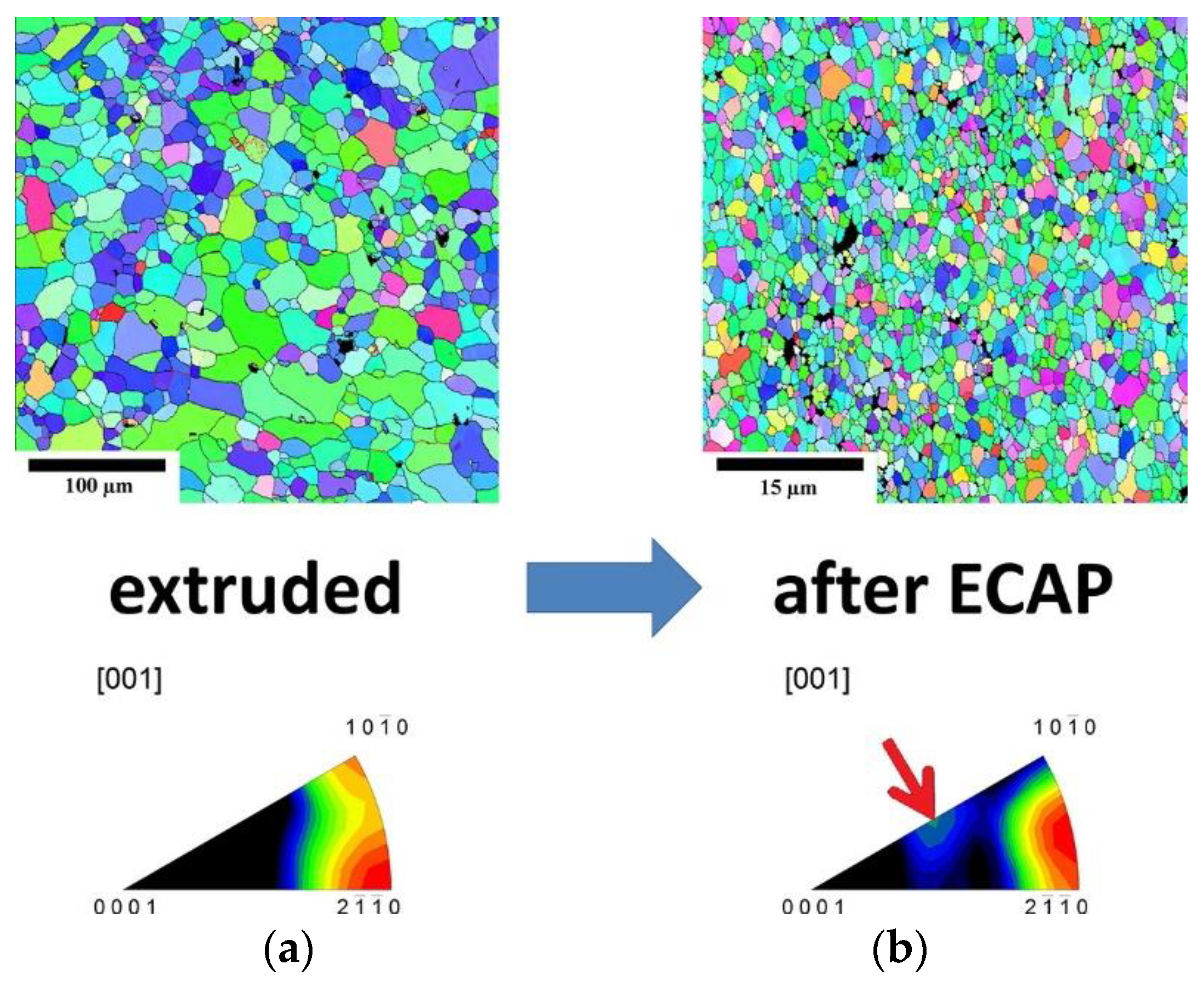
6.2.4. Equal Channel Angular Pressing/Extrusion (ECAP/ECAE)

ECAP or ECAE [145], on the other hand, has the added advantage of producing grain refinement as well as equiaxed grains in comparison with extrusion via the change in direction of plastic deformation of the alloy. Through work carried out on LAE442 by P. Minárik et al. [146], it was found that the grain size of ~1 mm obtained by casting was refined down to ~1.7 μm after hot extrusion (350 °C) at a ratio of 22:1 followed by 12 passes of ECAP at 90° (See Figure 7). Similarly, P. Minárik et al. [147] reported achieving higher degradation resistance using the ECAP process for LAE442 as compared with extruded LAE442 with no adverse effects in cytocompatibility noticed due to the additional ECAP process. Y. Tan et al. [148] conducted multiple passes of ECAP on a Mg–2Y–0.6Nd–0.6Zr alloy at different temperatures and reported improved tensile strength of the material.

6.3. Effect of Post-Processing Treatment

The heat treatment and surface modification of the Mg alloys are two of the main post-alloy processing methods found in the literature.

6.3.1. Effect of Heat Treatment

D. Liu et al. [91] attributed the grain refinement and solid solution strengthening to improved mechanical properties of the extruded alloy compared to the as-cast alloy.
It has been reported that the age-hardening response increases with increasing Sr content [110]. Homogenization of as-cast Mg-xSr (x: 0.5, 1, 2 wt.%) at 450 °C for 12 h followed by quenching in water dissolved the dendritic structures of the as-cast Mg-Sr alloy but it had no effect on the grain size [110]. Aging at 160 °C for 30 to 300 h showed that there was a reduction in hardness though it increased the UTS, TYS, and CYS [110]. H. Ibrahim et al. [149] performed solution treatment (510 °C, 3 h) age hardening in an oil bath (200 °C; 1 to 10 h) of a cast Mg alloy (Mg-1.2 wt.% Zn-0.5 wt.% Ca) and found that the heat treatment improved both the mechanical and degradation properties of the alloy. As an example of thermomechanical processing, H. B. Henderson et al. [150] recently reported an improvement in both the mechanical and corrosion properties of an as-cast Mg-1 wt.%Ca-0.5 wt.%Sr alloy billet by hot extrusion (275, 340, and 400 °C) at a ratio of 25:1. Extrusion served the purpose of providing the large driving force required to break the structure of the interconnected as-cast eutectic phase, as well as bringing about further grain refinement resulting from the severe plastic deformation it undergoes. Grain sizes as low as 1.6–6.3 μm were obtained at an extrusion temperature of 275 °C. Moreover, the process lowered the degradation rate while at the same time showed low toxicity for mouse osteoblasts. On the other hand, it also improved the maximum tensile yield strength up to 304 MPa, which is even higher than annealed 316L SS.
D.-J. Lin et al. [151] used both the solid solution treatment (345 °C, 10 h) and a strain-induced melting activation (SIMA) heat treatment (355 °C and 370 °C) on the extruded ZAX1330 alloy and also noted the role of grain refinement in improving the mechanical strength and corrosion resistance. Heating the alloys decreased the elastic modulus. Improved UTS and elongation, despite decreased YS, following the T4 treatment was observed when compared with the as-extruded alloy. Heat treatment up to 355 °C resulted in a uniform distribution of the secondary phases, whereas higher heating produced segregated regions of secondary phases due to the matrix melting at 370 °C and the solid-solution Ca diffusion into the liquid phase [151]. T4 produced the lowest corrosion density due to the near elimination of Ca2Mg6Zn3 precipitates by the solid solution effect of Ca dissolving into the matrix, leading to the dispersion of Mg-Zn precipitates.
An example of the corrosion mechanism being altered due to heat treatment is found in the case of a MgAlGd alloy, whose main corrosion mechanism of as-cast and T4-treated alloys changed from filiform and intergranular corrosion to that of needle-like precipitates within the grains, acting as a barrier to corrosion of the matrix [152].

6.3.2. Surface Modifications

The development of Mg alloys is many times complemented by the development of various surface modification characteristics of the alloys/implants. The research in these areas concern the alteration of the implant surface characteristics in order to affect the corrosion characteristics as well as improve the initial cell adhesion and proliferation characteristics. These surface alterations are mainly of either the application of a coating on the alloy/composite surface or modification of the implant surface properties by various means. Both these methods impart different physical characteristics of the surface. A solution to develop biodegradable Mg alloy implants according to the different physical environments required by the various applications and locations could lie in tailoring application-specific surface modifications on high-performance Mg alloys.

Surface Coating

One of the most common coating materials have been Hydroxyapatite (HA) [153]. The use of Micro Arc Oxidation techniques (MAO) for surface coating are also commonly found in the literature [153,154,155,156]. Tang et al. used this method to coat AZ31 with HA (Hydroxyapatite) and reported that it has induced additional resistance of the alloy to corrosion in Simulated Body Fluid by the barrier effect and the degradation of the coating itself. HA coating has also been said to result in enhanced osteoblast development compared to uncoated Mg alloy samples along with a significant reduction in the rate of degradation [157]. Furthermore, N. Yu et al. [158] reported that doping Strontium into the HA coatings to produce a dual layer of SrHA by microwave irradiationled to increased corrosion resistance of the initial stages of the Mg alloy. They also worked on commercially available AZ31 alloy.
Others such as H.R. Baksheshi-Rad et al. [159] have coated Mg-1.2Ca-2Zn samples with a coating of HA and ostacalcium phosphate (OCP) followed by a layer of polycaprolactone (PCL) and reported a significant reduction in corrosion current densities as compared with uncoated samples. They used a combination of chemical solution deposition and the dip coating method to apply the coating on the samples. Su et al. [160] fabricated a composite coating of calcium phosphate (CaP) and collagen (Col) on the surface of AZ60 Mg alloy by chemical conversion and dip-coating methods. The composite coating not only reduced the in vitro degradation rate of Mg alloy effectively because the collagen coating sealed the pores in the CaP coating but also improved the biocompatibility via effective promotion of cell adhesion and proliferation. Li et al. [161] applied a multilayered coating on a Mg substrate. The coating contained a chemical conversion layer of fluoride or phosphate, an adhesion layer, and a layer of biodegradable polylactic acid (PLA). The corrosion rate of the coated Mg has been reduced to one-tenth that of the uncoated bare Mg.
One-step electrodeposition of a Ce-based hydrophobic surface on Mg alloy was carried out by Yan liu et al. [162]. S. Shen et al. [157] used a technique known as the Rapid microwave aqueous chemical route to produce an HA bilayer on Mg alloy by microwave irradiation. The use of the electrospinning method to coat AZ31 with composite of Polycaprolactone (PCL) and ZnO nano particles of 1 wt.% and 3 wt.%, used by J. Kim et al. [163], produced increased biocompatibility and corrosion resistance. P. Tian et al. [164] combined the use of plasma electrolytic oxidization (PEO) and Hydrothermal treatment to apply a coating of HA using the latter on the coating applied using the former. The additional secondary treatment has been attributed to the improved surface biocompatibility via improved cell adhesion and proliferation.
J. Tang et al. [165] attempted a comparison between micro arc oxidation (MAO) and electrophoresis deposition (EPD) methods. Though both coating methods provided corrosion resistance for the Mg-Zr pins used, the study was not performed on the same coating components and the difference in reported results may have been due to a component factor rather than a definitive statement of the processes used. Deposition of a CaP (Calcium Phosphate) ceramic coating on Pure Mg, Mg-0.6 wt.%Ca and Mg-0.55 wt.%Ca-1.74 wt.%Zn alloys by MAO was reported to yield a better corrosion protection in SBF, though in Tris-HCl immersion, it had the opposite effect of increased corrosion compared with uncoated alloys [166]. Furthermore, Razavi et al. [167] deposited a nanocomposite coating made of diopside, bredigite, and fluoridated hydroxyapatite bioceramics on a micro-arc oxidation-treated surface of a biodegradable AZ91 Mg alloy by the electrophoretic deposition method. Improved corrosion resistance and implant osteointegration were reported to be achieved. X. Yu et al. [168] reported on improved corrosion resistance achieved by oxidizing Y alloys of Mg, i.e., Mg-1.5 wt.%Y and Mg-1.5 wt.%-0.25 wt.%Sn. This improvement has been attributed to the formation of a Y-enriched oxide film on the alloy surface. Lin et al. [169] synthesized a bifunctional TiO2/Mg2TiO4 nano layer on the functionalized surface of a WE43 magnesium implant by using the dual titanium and oxygen plasma immersion ion implantation technique. A significant improvement in corrosion resistance of Mg substrates was reported together with the enhancement in the in vitro osteoblastic differentiation capability due to the controlled release of magnesium ions. Guo et al. [170] electrochemically synthesized a multifunctional polypyrrole/zinc oxide (Ppy/ZnO) composite coating on AZ31 Mg alloy by the cyclic voltammetry method. The multifunctional coating was reported to achieve a balance of good corrosion resistance, cytocompatibility, and excellent antibacterial property.

Ion Implantation and Stress Impartation Methods

V. K. Caralapatti and S. Narayanswamy [171] used High-Repetition Laser Shock Peening (HRLSP) to impart compressive residual stress on the surface of the Mg alloys to improve its degradation and biocompatibility. W. Jin et al. [172] carried out Nd ion implantation on WE43 using metal ion implanter with a Nd cathodic arc source. The retardation of the degradation rate and improvement in biocompatibility was attributed to a hydrophobic surface layer of mostly Nd2O3 and MgO. Y. Zhao et al. [173] used a dual Zirconium and Oxygen ion implantation method to create a hydrophobic surface film containing ZrO2 to achieve better cytocompatibility and corrosion resistance of the Mg-Ca and Mg-Sr alloys. It had the added advantage of having better antibacterial capability as well.
Most, if not all, the studies above focused on improving the surface characteristics of commercially available or common Mg alloys, and as such, there remains to be seen a concentrated effort in combining the development of high-performance biodegradable Mg alloys along with its surface modification tailored for specific applications/locations.

7. Effect of Implant Geometry on the Biodegradable Characteristics

Development of biodegradable Mg alloys in itself does not represent the practical application of it as an implant without discussing the physical dimensions of the alloy as an implant. This is because, despite the simplified models of a symmetric shape being used to test many of the degradation characteristics and mechanical performance in many of the studies, in practice, implants could come in a variety of complex shapes. Moreover, with regard to bone-grafting implants, the physical geometry and porosity of the implant are crucial factors in encouraging osteogenesis. Porosity is a result of enclosed/entrapped gaps within a sample. The more porous the alloy, the weaker the alloy is due to the stress concentration at these points. Additionally, the availability of gaps for the body’s contact fluid to seep into the implant offers an increased surface area for the degradation to take place, further weakening the implant. However, despite these inherent disadvantages, the complex implant geometry is a crucial aspect that is required for many bone-grafting and repair applications, and therefore the design of the implants needs to take into consideration the effect of its geometry on the final degradation characteristics and its subsequent deterioration of the mechanical properties. Mei Li et al. [174] reported the positive effects of hollow 3D implants for bone grafting [175]. Recently, additive manufacturing has gained focus in the fabrication of scaffolds exhibiting optimal porosity, fully interconnected structures, suitable compressive properties, and moderate corrosion behavior, thus meeting the basic requirements for tissue engineering scaffolds [176,177,178,179].

8. Prospects of Some of the Commercially Popular Mg Alloys

The commercially available Mg alloys have the chief advantage of being economically favorable due to the common availability of the alloys. These alloys were primarily made for industrial uses to take advantage of the high strength-to-weight ratio of the Mg alloys. Much research has been carried out to investigate the potential of these alloys as biodegradable implant materials. This includes their use as master alloys in casting and further processing of these alloys such as heat treatment, coating, etc. However, the chief disadvantage of some of these alloys is that since they have been developed for industrial use, they may contain one or more biotoxic elements, making them unpractical for use as a biodegradable material. Of these commercial alloys, those with aluminum are the most popular, and a number of studies have been carried out to determine its potential for use as a biodegradable Mg alloy.
An experiment conducted by Chen Ying Liang et al. [180] found that the corrosion resistance in increasing order is AZ91 < AZ31 < AM60 < ZK60 when tested on commercially available rolled plates of these Mg alloys. When tested in a 3.5% NaCl solution, the corrosion rate increased in terms of weight loss: Mg < AZ91 < AZ31, though after 3 h, the corrosion rate of AZ91 increased above that of AZ31, attributed to the start of galvanic corrosion [181]. The lower corrosion rate is attributed to the formation of Mg(OH)2 on its surface. In vitro tests conducted in SBF by J. Fu et al. [182] on as-cast Mg-Zn-Ca alloys concluded that micro-galvanic corrosion between the eutectic products of (Mg + Ca2Mg6Zn3) and the Mg matrix were the main form of corrosion. Hence, the biocorrosion rate increases with the increase in the volume fraction of the secondary phase. Additionally, they have also observed that large-grained samples corroded faster.

9. Conclusions

A wide range of Mg alloys are available with Al as the primary alloying element, which has a neurotoxic effect and causes Alzheimer’s disease. Reports indicate that the presence of silicon in these alloys helps to lower the accumulation of Al in mice and is perhaps conceivable to address the neurotoxic effect with an Mg-Al-Si alloy system, which needs more research both in vitro and in vivo.
Manufacturing processes such as ECAE/ECAP can be utilized in the future to achieve equiaxed grains in all directions, i.e., axially as well as radially. This could achieve better symmetry of tension vs. compression strength.
Various studies prove the degradation resistance enhancement of ‘as-made’ alloys with post processing operations. It is conceivable to address the issues related to the initial corrosion characteristics and its resultant weakening of the implant by adopting suitable heat treatment and surface modification regimes to achieve a very realistic clinical biodegradable Mg alloy.
The Mg alloys being developed and tested for application-specific implants need to be tested by clinical studies. Biodegradability is inherent in nature and, as such, the local environment of exposure would dictate the biological mechanisms in interaction with the implant alloy. Clinical trials of the biodegradable alloys are critically important as there is concern of the effect of the degradation product, which does not arise in in the case of non-biodegradable or inert implants.
With the various available biodegradable Mg alloys, each with its own set of characteristics, and the various available heat treatments, surface modifications, and coatings, the design of implants with functional gradations could provide a breakthrough in the clinical use of implants. For example, a high-strength, high-degradation core complemented by low degradation, yet relatively low strength can be used for osteogenesis applications requiring gap filling. This would ensure sufficient core strength of the implant while bone formation takes place, while, by the time the fast degradation of the core occurs, sufficient healing would have occurred to provide the final disintegration of the Mg implant.

Author Contributions

Conceptualization, S.F.H.; methodology, S.F.H. and M.T.I.; formal analysis, S.F.H. and M.T.I.; investigation, S.F.H. and M.T.I.; resources, S.F.H., M.M.A.B. and N.S.; data curation, S.F.H., M.T.I. and N.S.; writing—original draft preparation, M.T.I. and S.F.H.; writing—review & editing, S.F.H., M.M.A.B. and N.S.; supervision, S.F.H.; project administration, S.F.H.; funding acquisition, S.F.H. All authors have read and agreed to the published version of the manuscript.

Funding

The authors would like to acknowledge the support provided by the Deanship of Scientific Research (DSR) at King Fahd University of Petroleum and Minerals (KFUPM) for funding this work through project No. IN151019.

Institutional Review Board Statement

Not applicable.

Informed Consent Statement

Not applicable.

Data Availability Statement

Not applicable.

Conflicts of Interest

The authors declare no conflict of interest.

References

  1. Witte, F. Reprint of: The history of biodegradable magnesium implants: A review. Acta Biomater. 2015, 23, S28–S40. [Google Scholar] [CrossRef] [PubMed]
  2. Sheikh, Z.; Najeeb, S.; Khurshid, Z.; Verma, V.; Rashid, H.; Glogauer, M. Biodegradable Materials for Bone Repair and Tissue Engineering Applications. Materials 2015, 8, 5744–5794. [Google Scholar] [CrossRef] [PubMed]
  3. Song, L.; Gao, M.; Tan, L.; Ma, Z.; Ni, P.; Zhou, M.; Na, D. Application Potential of Mg–Zn–Nd Alloy as a Gas-trointestinal Anastomosis Nail Material. In Acta Metallurgica Sinica (English Letters); Springer: Berlin/Heidelberg, Germany, 2022; Volume 35, pp. 609–620. [Google Scholar]
  4. Chen, Y.T.; Hung, F.Y.; Lin, Y.L.; Lin, C.Y. Biodegradation ZK50 magnesium alloy compression screws: Mechanical properties, biodegradable characteristics and implant test. J. Ortho. Sci. 2020, 25, 1107–1115. [Google Scholar] [CrossRef] [PubMed]
  5. Ghiotti, A.; Bruschi, S.; Bertolini, R.; Perzynski, K.; Madej, L. Forming of bioabsorbable clips using magnesium alloy strips with enhanced characteristics. CIRP Ann. 2020, 69, 257–260. [Google Scholar] [CrossRef]
  6. Rizvi, S.H.A.; Che, J.; Mehboob, A.; Zaheer, U.; Chang, S.-H. Experimental study on magnesium wire–polylactic acid biodegradable composite implants under in vitro material degradation and fatigue loading conditions. Compos. Struct. 2021, 272, 114267. [Google Scholar] [CrossRef]
  7. Li, Y.; Wang, Y.; Shen, Z.; Miao, F.; Wang, J.; Sun, Y.; Zhu, S.; Zheng, Y.; Guan, S. A biodegradable magnesium alloy vascular stent structure: Design, optimisation and evaluation. Acta Biomater. 2022, 142, 402–412. [Google Scholar] [CrossRef]
  8. Callister, W.D.; Rethwisch, D.G. Materials Science and Engineering: An. Introduction, 7th ed.; Wiley: New York, NY, USA, 2007. [Google Scholar]
  9. Zhao, D.; Witte, F.; Lu, F.; Wang, J.; Li, J.; Qin, L. Current status on clinical applications of magnesium-based orthopaedic implants: A review from clinical translational perspective. Biomaterials 2017, 112, 287–302. [Google Scholar] [CrossRef]
  10. Nakahata, I.; Tsutsumi, Y.; Kobayashi, E. Mechanical Properties and Corrosion Resistance of Magnesium–Hydroxyapatite Composites Fabricated by Spark Plasma Sintering. Metals 2020, 10, 1314. [Google Scholar] [CrossRef]
  11. Durisin, M.; Reifenrath, J.; Weber, C.M.; Eifler, R.; Maier, H.J.; Lenarz, T.; Seitz, J.M. Biodegradable nasal stents (MgF2-coated Mg–2 wt %Nd alloy)—A long-term in vivo study. J. Biomed. Mater. Res. Part. B Appl. Biomater. 2017, 105, 350–365. [Google Scholar] [CrossRef]
  12. Brown, A.; Zaky, S.; Ray, H.; Sfeir, C. Porous magnesium/PLGA composite scaffolds for enhanced bone regeneration following tooth extraction. Acta Biomater. 2015, 11, 543–553. [Google Scholar] [CrossRef]
  13. He, H.; Ren, H.; Ding, Z.; Ji, M.; Chen, H.; Yan, Y. Developing a novel magnesium calcium phosphate/sodium alginate composite cement with high strength and proper self-setting time for bone repair. J. Biomater. Appl. 2021, 36, 346–357. [Google Scholar] [CrossRef] [PubMed]
  14. Wu, J.; Liu, F.; Wang, Z.; Liu, Y.; Zhao, X.; Fang, C.; Leung, F.; Yeung, K.W.K.; Wong, T.M. The Development of.a Magnesium-Releasing and Long-Term Mechanically Stable Calcium Phosphate Bone Cement Possessing Osteogenic and Immunomodulation Effects for Promoting Bone Fracture Regeneration. Front. Bioeng. Biotechnol. 2022, 9, 803723. [Google Scholar] [CrossRef] [PubMed]
  15. Yoshizawa, S.; Brown, A.; Barchowsky, A.; Sfeir, C. Magnesium ion stimulation of bone marrow stromal cells enhances osteogenic activity, simulating the effect of magnesium alloy degradation. Acta Biomater. 2014, 10, 2834–2842. [Google Scholar] [CrossRef] [PubMed]
  16. Wang, S.-H.; Lee, S.-P.; Yang, C.-W.; Lo, C.-M. Surface Modification of Biodegradable Mg-Based Scaffolds for Human Mesenchymal Stem Cell Proliferation and Osteogenic Differentiation. Materials 2021, 14, 441. [Google Scholar] [CrossRef]
  17. Ni, S.; Xiong, X.; Ni, X. MgCl2 promotes mouse mesenchymal stem cell osteogenic differentiation by activating the p38/Osx/Runx2 signaling pathway. Mol. Med. Rep. 2020, 22, 3904–3910. [Google Scholar] [CrossRef]
  18. Kanter, B.; Vikman, A.; Brückner, T.; Schamel, M.; Gbureck, U.; Ignatius, A. Bone regeneration capacity of magnesium phosphate cements in a large animal model. Acta Biomater. 2018, 69, 352–361. [Google Scholar] [CrossRef]
  19. Nabiyouni, M.; Ren, Y.; Bhaduri, S.B. Magnesium substitution in the structure of orthopedic nanoparticles: A comparison between amorphous magnesium phosphates, calcium magnesium phosphates, and hydroxyapatites. Mater. Sci. Eng. C 2015, 52, 11–17. [Google Scholar] [CrossRef]
  20. Xu, C.; Wang, S.; Wang, H.; Liu, K.; Zhang, S.; Chen, B.; Liu, H.; Tong, F.; Peng, F.; Tu, Y.; et al. Magnesium-Based Micromotors as Hydrogen Generators for Precise Rheumatoid Arthritis Therapy. Nano Lett. 2021, 21, 1982–1991. [Google Scholar] [CrossRef]
  21. Fanelli, A.; Ghezzi, D. Transient electronics: New opportunities for implantable neurotechnology. Curr. Opin. Biotechnol. 2021, 72, 22–28. [Google Scholar] [CrossRef]
  22. Yu, Y.; Jin, G.; Xue, Y.; Wang, D.; Liu, X.; Sun, J. Multifunctions of dual Zn/Mg ion co-implanted titanium on oste-ogenesis, angiogenesis and bacteria inhibition for dental implants. Acta Biomater. 2017, 49, 590–603. [Google Scholar] [CrossRef]
  23. Wang, G.; Li, J.; Zhang, W.; Xu, L.; Pan, H.; Wen, J.; Wu, Q.; She, W.; Jiao, T.; Liu, X.; et al. Magnesium ion implantation on a micro/nanostructured titanium surface promotes its bioactivity and osteogenic differentiation function. Int. J. Nanomed. 2014, 2387, 2387–2398. [Google Scholar]
  24. Wang, J.; Witte, F.; Xi, T.; Zheng, Y.; Yang, K.; Yang, Y.; Zhao, D.; Meng, J.; Li, Y.; Li, W.; et al. Recommendation for modifying current cytotoxicity testing standards for biodegradable magnesium-based materials. Acta Biomater. 2015, 21, 237–249. [Google Scholar] [CrossRef] [PubMed]
  25. Fischer, J.; Pröfrock, D.; Hort, N.; Willumeit, R.; Feyerabend, F. Reprint of: Improved cytotoxicity testing of magnesium materials. Mater. Sci. Eng. B 2011, 176, 1773–1777. [Google Scholar] [CrossRef]
  26. Li, Z.; Shang, Z.; Wei, X.; Zhao, Q. Corrosion resistance and cytotoxicity of AZ31 magnesium alloy with N+ ion im-plantation. Mater. Technol. 2019, 34, 730–736. [Google Scholar] [CrossRef]
  27. Scheideler, L.; Füger, C.; Schille, C.; Rupp, F.; Wendel, H.-P.; Hort, N.; Reichel, H.; Geis-Gerstorfer, J. Comparison of different in vitro tests for biocompatibility screening of Mg alloys. Acta Biomater. 2013, 9, 8740–8745. [Google Scholar] [CrossRef]
  28. Liu, X.; Xi, T.; Zheng, Y. Influence of the extraction parameters on the cytotoxicity test results of Mg materials. Prog. Nat. Sci. 2014, 24, 507–515. [Google Scholar] [CrossRef]
  29. Jung, O.; Smeets, R.; Porchetta, D.; Kopp, A.; Ptock, C.; Müller, U.; Heiland, M.; Schwade, M.; Behr, B.; Kröger, N.; et al. Optimized in vitro procedure for assessing the cytocompatibility of magnesium-based biomaterials. Acta Biomater. 2015, 23, 354–363. [Google Scholar] [CrossRef]
  30. Kraus, T.; Fischerauer, S.F.; Hänzi, A.C.; Uggowitzer, P.J.; Löffler, J.F.; Weinberg, A.M. Magnesium alloys for temporary implants in osteosynthesis: In vivo studies of their degradation and interaction with bone. Acta Biomater. 2012, 8, 1230–1238. [Google Scholar] [CrossRef]
  31. World Health Organization. Vitamin and Mineral Requirements in Human Nutrition, 2nd ed.; World Health Organization: Geneva, Switzerland, 2004; pp. 340–342. [Google Scholar]
  32. Hofmann, F.B.; Board, E.; Beavo, J.a.; Busch, a.; Ganten, D.; Michel, M.C.; Page, C.P.; Rosenthal, W. Handbook of Experimental Pharmacology: Methylxanthines; Springer: Berlin/Heidelberg, Germany, 2011. [Google Scholar]
  33. Hashimoto, Y.; Kim, D.J.; Adams, J.C. The roles of fascins in health and disease. J. Pathol. 2011, 224, 289–300. [Google Scholar] [CrossRef]
  34. Barceloux, D.G.; Barceloux, D. Copper. J. Toxicol. Clin. Toxicol. 1999, 37, 217–230. [Google Scholar] [CrossRef]
  35. Ellingsen, D.G.; Møller, L.B.; Aaseth, J. Handbook on the Toxicology of Metals, 4th ed.; Elsevier: Amsterdam, The Netherlands, 2014; Volume 1, pp. 765–786. [Google Scholar]
  36. Cuatrecasas, P.; Arbor, A.; Ganten, D.; Herken, H.; Melman, K.L. Handbook of Experimental Pharmacology; Springer: Berlin/Heidelberg, Germany, 2006. [Google Scholar]
  37. Nielsen, F.H. Importance of making dietary recommendations for elements designated as nutritionally beneficial, pharmacologically beneficial, or conditionally essential. J. Trace Elem. Exp. Med. 2000, 13, 113–129. [Google Scholar] [CrossRef]
  38. Pavan, C.; Delle Piane, M.; Gullo, M.; Filippi, F.; Fubini, B.; Hoet, P.; Turci, F. The puzzling issue of silica toxicity: Are silanols bridging the gaps between surface states and pathogenicity? Part. Fibre Tox. 2019, 16, 32. [Google Scholar] [CrossRef] [PubMed]
  39. Borenstein, D. Siliconosis: A spectrum of illness. Semin. Art. Rheum. 1994, 24, 1–7. [Google Scholar] [CrossRef]
  40. Kossovsky, N.; Stassi, J. A pathophysiological examination of the biophysics and bioreactivity of silicone breast implants. Semin. Art. Rheum. 1994, 24, 18–21. [Google Scholar] [CrossRef]
  41. Onnekink, C.; Kappel, R.M.; Boelens, W.C.; Pruijn, G.J.M. Low molecular weight silicones induce cell death in cultured cells. Sci. Rep. 2020, 10, 9558. [Google Scholar] [CrossRef]
  42. Ivanov, S.; Zhuravsky, S.; Yukina, G.; Tomson, V.; Korolev, D.; Galagudza, M. In Vivo Toxicity of Intravenously Administered Silica and Silicon Nanoparticles. Materials 2012, 5, 1873–1889. [Google Scholar] [CrossRef]
  43. Ostrakhovitch, E.A. Tin. In Handbook on the Toxicology of Metals, 4th ed.; Elsevier: Amsterdam, The Netherlands, 2014; Volume 2, pp. 1241–1285. [Google Scholar]
  44. Lucchini, R.G.; Aschner, M.; Kim, Y.; Šarić, M. Manganese. In Handbook on the Toxicology of Metals, 4th ed.; Elsevier: Amsterdam, The Netherlands, 2014; Volume 1, pp. 975–1011. [Google Scholar]
  45. Sjögren, B.; Iregren, A.; Montelius, J.; Yokel, R.A. Aluminum, 4th ed.; Elsevier: Amsterdam, The Netherlands, 2015. [Google Scholar]
  46. Bernard, A. Lithium. In Handbook on the Toxicology of Metals, 4th ed.; Elsevier: Amsterdam, The Netherlands, 2014; Volume 2, pp. 969–974. [Google Scholar]
  47. Fowler, B.A.; Maples-Reynolds, N. Indium. In Handbook on the Toxicology of Metals, 4th ed.; Elsevier: Amsterdam, The Netherlands, 2014; Volume 2, pp. 845–853. [Google Scholar]
  48. Cesbron, A.; Saussereau, E.; Mahieu, L.; Couland, I.; Guerbet, M.; Goullé, J.-P. Metallic Profile of Whole Blood and Plasma in a Series of 106 Healthy Volunteers. J. Anal. Toxicol. 2013, 37, 401–405. [Google Scholar] [CrossRef]
  49. Schultze, B.; Lind, P.M.; Larsson, A.; Lind, L. Whole blood and serum concentrations of metals in a Swedish population-based sample. Scand. J. Clin. Lab. Investig. 2013, 74, 143–148. [Google Scholar] [CrossRef]
  50. Goullé, J.-P.; Le Roux, P.; Castanet, M.; Mahieu, L.; Guyet-Job, S.; Guerbet, M. Metallic Profile of Whole Blood and Plasma in a Series of 99 Healthy Children. J. Anal. Toxicol. 2015, 39, 707–713. [Google Scholar] [CrossRef]
  51. Ernst, W. Overview of naturally occurring Earth materials and human health concerns. J. Southeast Asian Earth Sci. 2012, 59, 108–126. [Google Scholar] [CrossRef]
  52. European Food Safety Authority, Tolerable Upper Intake Levels Scientific Committee on Food Scientific Panel on Dietetic Products, Nutrition and Allergies. February 2006. Available online: https://www.efsa.europa.eu/sites/default/files/efsa_rep/blobserver_assets/ndatolerableuil.pdf (accessed on 20 August 2021).
  53. Querido, W.; Rossi, A.L.; Farina, M. The effects of strontium on bone mineral: A review on current knowledge and microanalytical approaches. Micron 2016, 80, 122–134. [Google Scholar] [CrossRef] [PubMed]
  54. Rim, K.T.; Koo, K.H.; Park, J.S. Toxicological Evaluations of Rare Earths and Their Health Impacts to Workers: A Literature Review. Saf. Health Work 2013, 4, 12–26. [Google Scholar] [CrossRef] [PubMed]
  55. Slikkerveer, A.; de Wolff, F.A. Pharmacokinetics and Toxicity of Bismuth Compounds. Med. Toxicol. Adverse Drug Exp. 1989, 4, 303–323. [Google Scholar] [CrossRef] [PubMed]
  56. Holler, J.S.; Fowler, B.A.; Nordberg, F.G. Nordberg, Silver. In Handbook on the Toxicology of Metals, 4th ed.; Elsevier: Amsterdam, The Netherlands, 2014; Volume 2, pp. 1209–1216. [Google Scholar]
  57. Ostrakhovitch, E.A.; Cherian, M.G. Tin. In Handbook on the Toxicology of Metals, 3rd ed.; Elsevier: Amsterdam, The Netherlands, 2007; Volume 2, pp. 839–860. [Google Scholar]
  58. Blaschke, C.; Volz, U. Soft and hard tissue response to zirconium dioxide dental implants: A clinical study in man. Neuro Endocrinol Lett. 2006, 27 (Suppl. S1), 69–72. [Google Scholar]
  59. Greiter, M.B.; Giussani, A.; Höllriegl, V.; Li, W.B.; Oeh, U. Human biokinetic data and a new compartmental model of zirconium—A tracer study with enriched stable isotopes. Sci. Total Environ. 2011, 409, 3701–3710. [Google Scholar] [CrossRef]
  60. Li, N.; Zheng, Y. Novel Magnesium Alloys Developed for Biomedical Application: A Review. J. Mater. Sci. Technol. 2013, 29, 489–502. [Google Scholar] [CrossRef]
  61. Yu, K.; Dai, Y.; Luo, Z.; Long, H.; Zeng, M.; Li, Z.; Zhu, J.; Cheng, L.; Zhang, Y.; Liu, H.; et al. In vitro and in vivo evaluation of novel biodegradable Mg-Ag-Y alloys for use as resorbable bone fixation implant. J. Biomed. Mater. Res. Part A 2018, 106, 2059–2069. [Google Scholar] [CrossRef]
  62. Gu, X.; Zheng, Y.; Cheng, Y.; Zhong, S.; Xi, T. Biomaterials In vitro corrosion and biocompatibility of binary magnesium alloys. Biomaterials 2009, 30, 484–498. [Google Scholar] [CrossRef]
  63. Kubásek, J.; Vojtěch, D.; Lipov, J.; Ruml, T. Structure, mechanical properties, corrosion behavior and cytotoxicity of biodegradable Mg-X (X = Sn, Ga, In) alloys. Mater. Sci. Eng. C 2013, 33, 2421–2432. [Google Scholar] [CrossRef]
  64. Li, Z.; Gu, X.; Lou, S.; Zheng, Y. The development of binary Mg–Ca alloys for use as biodegradable materials within bone. Biomaterials 2008, 29, 1329–1344. [Google Scholar] [CrossRef]
  65. Liu, C.; Fu, X.; Pan, H.; Wan, P.; Wang, L.; Tan, L.; Wang, K. Biodegradable Mg-Cu alloys with enhanced osteogenesis, angiogenesis, and long-lasting antibacterial effects. Nat. Publ. Gr. 2016, 6, 27374. [Google Scholar] [CrossRef] [PubMed]
  66. Zhu, S.; Huang, N.; Xu, L.; Zhang, Y.; Liu, H.; Sun, H.; Leng, Y. Biocompatibility of pure iron: In vitro assessment of degradation kinetics and cytotoxicity on endothelial cells. Mater. Sci. Eng. C 2009, 29, 1589–1592. [Google Scholar] [CrossRef]
  67. Zhao, C.; Pan, F.; Zhao, S.; Pan, H.; Song, K.; Tang, A. Microstructure, corrosion behavior and cytotoxicity of bio-degradable Mg-Sn implant alloys prepared by sub-rapid solidification. Mater. Sci. Eng. C 2015, 54, 245–251. [Google Scholar] [CrossRef]
  68. Sanchez, A.H.M.; Feyerabend, F.; Laipple, D.; Willumeit, R.; Weinberg, A.; Luthringer, B. Chondrogenic differentiation of ATDC5-cells under the influence of Mg and Mg alloy degradation. Mater. Sci. Eng. C 2017, 72, 378–388. [Google Scholar] [CrossRef] [PubMed]
  69. Willbold, E.; Gu, X.; Albert, D.; Kalla, K.; Bobe, K.; Brauneis, M.; Janning, C.; Nellesen, J.; Czayka, W.; Tillmann, W.; et al. Effect of the addition of low rare earth elements (lanthanum, neodymium, cerium) on the biodegradation and biocompatibility of magnesium. Acta Biomater. 2014, 11, 554–562. [Google Scholar] [CrossRef] [PubMed]
  70. Zhang, J.; Li, H.; Wang, W.; Huang, H.; Pei, J.; Qu, H.; Yuan, G.; Li, Y. Acta Biomaterialia The degradation and transport mechanism of a Mg-Nd-Zn-Zr stent in rabbit common carotid artery: A 20-month study. Acta Biomater. 2018, 69, 372–384. [Google Scholar] [CrossRef]
  71. Liu, F.; Chen, C.; Niu, J.; Pei, J.; Zhang, H.; Huang, H.; Yuan, G. The processing of Mg alloy micro-tubes for biodegradable vascular stents. Mater. Sci. Eng. C 2014, 48, 400–407. [Google Scholar] [CrossRef]
  72. Han, P.; Cheng, P.; Zhao, C.; Zhang, S.; Zhou, R.; Zhang, X.; Chai, Y. Comparative Study about Degradation of High-purity Magnesium Screw in Intact Femoral Intracondyle and in Fixation of Femoral Intracondylar Fracture. J. Mater. Sci. Technol. 2017, 33, 305–310. [Google Scholar] [CrossRef]
  73. Bai, H.; He, X.; Ding, P.; Liu, D.; Chen, M. Fabrication, microstructure, and properties of a biodegradable Mg-Zn-Ca clip. J. Biomed. Mater. Res. Part B Appl. Biomater. 2019, 107, 1741–1749. [Google Scholar] [CrossRef]
  74. Zhao, D.; Huang, S.; Lu, F.; Wang, B.; Yang, L.; Qin, L.; Yang, K.; Li, Y.; Li, W.; Wang, W.; et al. Vascularized bone grafting fixed by biodegradable magnesium screw for treating oste-onecrosis of the femoral head. Biomaterials 2016, 81, 84–92. [Google Scholar] [CrossRef]
  75. Windhagen, H.; Radtke, K.; Weizbauer, A.; Diekmann, J.; Noll, Y.; Kreimeyer, U.; Schavan, R.; Stukenborg-Colsman, C.; Waizy, H. Biodegradable magnesium-based screw clinically equivalent to titanium screw in hallux valgus surgery: Short term results of the first prospective, randomized, controlled clinical pilot study. Biomed. Eng. Online 2013, 12, 62. [Google Scholar] [CrossRef] [PubMed]
  76. Dziuba, D.; Meyer-Lindenberg, A.; Seitz, J.M.; Waizy, H.; Angrisani, N.; Reifenrath, J. Long-term in vivo degradation behaviour and biocompatibility of the magnesium alloy ZEK100 for use as a biodegradable bone implant. Acta Biomater. 2013, 9, 8548–8560. [Google Scholar] [CrossRef] [PubMed]
  77. Erdmann, N.; Angrisani, N.; Reifenrath, J.; Lucas, A.; Thorey, F.; Bormann, D.; Meyer-Lindenberg, A. Biomechanical testing and degradation analysis of MgCa0.8 alloy screws: A comparative in vivo study in rabbits. Acta Biomater. 2011, 7, 1421–1428. [Google Scholar] [CrossRef] [PubMed]
  78. Krause, A.; Von Der Höh, N.; Bormann, D.; Krause, C.; Bach, F.-W.; Windhagen, H.; Meyer-Lindenberg, A. Degradation behaviour and mechanical properties of magnesium implants in rabbit tibiae. J. Mater. Sci. 2010, 45, 624–632. [Google Scholar] [CrossRef]
  79. Angrisani, N.; Reifenrath, J.; Zimmermann, F.; Eifler, R.; Meyer-Lindenberg, A.; Vano-Herrera, K.; Vogt, C. Bio-compatibility and degradation of LAE442-based magnesium alloys after implantation of up to 3.5 years in a rabbit model. Acta Biomater. 2016, 44, 355–365. [Google Scholar] [CrossRef]
  80. Zhao, D.; Wang, T.; Nahan, K.; Guo, X.; Zhang, Z.; Dong, Z.; Chen, S.; Chou, D.T.; Hong, D.; Kumta, P.N.; et al. In vivo characterization of magnesium alloy biodegradation using electrochemical H2monitoring, ICP-MS, and XPS. Acta Biomater. 2017, 50, 556–565. [Google Scholar] [CrossRef]
  81. Wang, J.; Wu, Y.; Li, H.; Liu, Y.; Bai, X.; Chau, W.; Zheng, Y.; Qin, L. Magnesium alloy based interference screw developed for ACL reconstruction attenuates peri-tunnel bone loss in rabbits. Biomaterials 2018, 157, 86–97. [Google Scholar] [CrossRef]
  82. Brooks, E.K.; Ahn, R.; Tobias, M.E.; Hansen, L.A.; Luke-Marshall, N.R.; Wild, L.; Campagnari, A.A.; Ehrensberger, M.T. Magnesium alloy AZ91 exhibits antimicrobial properties in vitro but not in vivo. J. Biomed. Mater. Res. Part B Appl. Biomater. 2017, 106, 221–227. [Google Scholar] [CrossRef]
  83. Levorova, J.; Duskova, J.; Drahos, M.; Vrbová, R.; Vojtech, D.; Kubásek, J.; Bartoš, M.; Dugova, L.; Ulmann, D.; Foltan, R. In vivo study on biodegradable magnesium alloys: Bone healing around WE43 screws. J. Biomater. Appl. 2017, 32, 886–895. [Google Scholar] [CrossRef]
  84. Zhang, B.; Hou, Y.; Wang, X.; Wang, Y.; Geng, L. Mechanical properties, degradation performance and cytotoxicity of Mg-Zn-Ca biomedical alloys with different compositions. Mater. Sci. Eng. C 2011, 31, 1667–1673. [Google Scholar] [CrossRef]
  85. Gil-Santos, A.; Marco, I.; Moelans, N.; Hort, N.; Van der Biest, O. Microstructure and degradation performance of biodegradable Mg-Si-Sr implant alloys. Mater. Sci. Eng. C 2017, 71, 25–34. [Google Scholar] [CrossRef] [PubMed]
  86. Rad, H.R.B.; Idris, M.H.; Kadir, M.R.A.; Farahany, S. Microstructure analysis and corrosion behavior of biode-gradable Mg-Ca implant alloys. Mater. Des. 2012, 33, 88–97. [Google Scholar] [CrossRef]
  87. Pourbaix, M.; Staehle, R.W. Lectures on Electrochemical Corrosion; PWN: Warszawa, Poland, 1978. [Google Scholar]
  88. Gu, X.; Xie, X.; Li, N.; Zheng, Y.; Qin, L. In vitro and in vivo studies on a Mg–Sr binary alloy system developed as a new kind of biodegradable metal. Acta Biomater. 2012, 8, 2360–2374. [Google Scholar] [CrossRef] [PubMed]
  89. Choi, Y.I.; Kuroda, K.; Okido, M. Temperature-dependent corrosion behaviour of flame-resistant, Ca-containing AZX911 and AMX602 Mg alloys. Corros. Sci. 2016, 103, 181–188. [Google Scholar] [CrossRef]
  90. Witte, F.; Hort, N.; Vogt, C.; Cohen, S.; Kainer, K.U.; Willumeit, R.; Feyerabend, F. Degradable biomaterials based on magnesium corrosion. Curr. Opin. Solid State Mater. Sci. 2008, 12, 63–72. [Google Scholar] [CrossRef]
  91. Liu, D.; Guo, C.; Chai, L.; Sherman, V.R.; Qin, X.; Ding, Y.; Meyers, M.A. Mechanical properties and corrosion resistance of hot extruded Mg-2.5Zn-1Ca alloy. Mater. Sci. Eng. B 2015, 195, 50–58. [Google Scholar] [CrossRef]
  92. Witecka, A.; Yamamoto, A.; Święszkowski, W. Influence of SaOS-2 cells on corrosion behavior of cast Mg-2.0Zn0.98Mn magnesium alloy. Colloids Surf. B Biointerfaces 2017, 150, 288–296. [Google Scholar] [CrossRef]
  93. Hofstetter, J.; Martinelli, E.; Weinberg, A.M.; Becker, M.; Mingler, B.; Uggowitzer, P.J.; Löffler, J.F. Assessing the degradation performance of ultrahigh-purity magnesium in vitro and in vivo. Corros. Sci. 2015, 91, 29–36. [Google Scholar] [CrossRef]
  94. Xie, G.; Takada, H.; Kanetaka, H. Development of high performance MgFe alloy as potential biodegradable ma-terials. Mater. Sci. Eng. A 2016, 671, 48–53. [Google Scholar] [CrossRef]
  95. Harandi, S.E.; Mirshahi, M.; Koleini, S.; Idris, M.H.; Jafari, H.; Kadir, M.R.A. Effect of calcium content on the microstructure, hardness and in-vitro corrosion behavior of biodegradable Mg-Ca binary alloy. Mater. Res. 2012, 16, 11–18. [Google Scholar] [CrossRef]
  96. Zeng, R.; Qi, W.; Cui, H.; Zhang, F.; Li, S.; Han, E. In vitro corrosion of as-extruded Mg-Ca alloys-The influence of Ca concentration. Corros. Sci. 2015, 96, 23–31. [Google Scholar] [CrossRef]
  97. Seong, J.; Kim, W. Development of biodegradable Mg–Ca alloy sheets with enhanced strength and corrosion properties through the refinement and uniform dispersion of the Mg2Ca phase by high-ratio differential speed rolling. Acta Biomater. 2015, 11, 531–542. [Google Scholar] [CrossRef] [PubMed]
  98. Zhang, S.; Zhang, X.; Zhao, C.; Li, J.; Song, Y.; Xie, C. Acta Biomaterialia Research on an Mg–Zn alloy as a degradable biomaterial. Acta Biomater. 2010, 6, 626–640. [Google Scholar] [CrossRef]
  99. Salleh, E.M.; Zuhailawati, H.; Noor, S.N.F.M.; Othman, N.K. In Vitro Biodegradation and Mechanical Properties of Mg-Zn Alloy and Mg-Zn-Hydroxyapatite Composite Produced by Mechanical Alloying for Potential Application in Bone Repair. Met. Mater. Trans. A 2018, 49, 5888–5903. [Google Scholar] [CrossRef]
  100. Hu, Y.; Guo, X.; Qiao, Y.; Wang, X.; Lin, Q. Preparation of medical Mg–Zn alloys and the effect of different zinc contents on the alloy. J. Mater. Sci. Mater. Med. 2022, 33, 9. [Google Scholar] [CrossRef] [PubMed]
  101. Song, Y.; Han, E.H.; Shan, D.; Yim, C.D.; You, B.S. The effect of Zn concentration on the corrosion behavior of Mg-xZn alloys. Corros. Sci. 2012, 65, 322–330. [Google Scholar] [CrossRef]
  102. Yan, Y.; Cao, H.; Kang, Y.; Yu, K.; Xiao, T.; Luo, J.; Deng, Y.; Fang, H.; Xiong, H.; Dai, Y. Effects of Zn concentration and heat treatment on the microstructure, mechanical properties and corrosion behavior of as-extruded Mg-Zn alloys produced by powder metallurgy. J. Alloys Compd. 2016, 693, 1277–1289. [Google Scholar] [CrossRef]
  103. Zhou, X.; Guo, T.; Wu, S.; Lü, S.; Yang, X.; Guo, W. Effects of Si Content and Ca Addition on Thermal Conductivity of As-Cast Mg–Si Alloys. Materials 2018, 11, 2376. [Google Scholar] [CrossRef]
  104. Zhang, E.; Yang, L.; Xu, J.; Chen, H. Microstructure, mechanical properties and bio-corrosion properties of Mg-Si(-Ca, Zn) alloy for biomedical application. Acta Biomater. 2010, 6, 1756–1762. [Google Scholar] [CrossRef]
  105. Ha, H.-Y.; Kang, J.-Y.; Kim, S.G.; Kim, B.; Park, S.S.; Yim, C.D.; You, B.S. Influences of metallurgical factors on the corrosion behaviour of extruded binary Mg–Sn alloys. Corros. Sci. 2014, 82, 369–379. [Google Scholar] [CrossRef]
  106. Bornapour, M.; Celikin, M.; Pekguleryuz, M. Thermal exposure effects on the in vitro degradation and mechanical properties of Mg–Sr and Mg–Ca–Sr biodegradable implant alloys and the role of the microstructure. Mater. Sci. Eng. C 2015, 46, 16–24. [Google Scholar] [CrossRef] [PubMed]
  107. Chen, L.; Bin, Y.; Zou, W.; Wang, X.; Li, W. The influence of Sr on the microstructure, degradation and stress corrosion cracking of the Mg alloys—ZK40xSr. J. Mech. Behav. Biomed. Mater. 2017, 66, 187–200. [Google Scholar] [CrossRef] [PubMed]
  108. Zhao, C.; Pan, F.; Zhang, L.; Pan, H.; Song, K.; Tang, A. Microstructure, mechanical properties, bio-corrosion properties and cytotoxicity of as-extruded Mg-Sr alloys. Mater. Sci. Eng. C 2017, 70, 1081–1088. [Google Scholar] [CrossRef] [PubMed]
  109. Han, J.; Wan, P.; Ge, Y.; Fan, X.; Tan, L.; Li, J.; Yang, K. Tailoring the degradation and biological response of a magnesium–strontium alloy for potential bone substitute application. Mater. Sci. Eng. C 2015, 58, 799–811. [Google Scholar] [CrossRef] [PubMed]
  110. Wang, Y.; Tie, D.; Guan, R.; Wang, N.; Shang, Y.; Cui, T.; Li, J. Microstructures, mechanical properties, and degradation behaviors of heat-treated Mg-Sr alloys as potential biodegradable implant materials. J. Mech. Behav. Biomed. Mater. 2017, 77, 47–57. [Google Scholar] [CrossRef]
  111. Cho, D.H.; Lee, B.W.; Park, J.Y.; Cho, K.M.; Park, I.M. Effect of Mn addition on corrosion properties of biodegradable Mg-4Zn-0.5Ca-xMn alloys. J. Alloys Compd. 2017, 695, 1166–1174. [Google Scholar] [CrossRef]
  112. Kim, H.J.; Kim, B.; Baek, S.-M.; Sohn, S.-D.; Shin, H.-J.; Jeong, H.Y.; Yim, C.D.; You, B.S.; Ha, H.-Y.; Park, S.S. Influence of alloyed Al on the microstructure and corrosion properties of extruded Mg–8Sn–1Zn alloys. Corros. Sci. 2015, 95, 133–142. [Google Scholar] [CrossRef]
  113. Remennik, S.; Bartsch, I.; Willbold, E.; Witte, F.; Shechtman, D. New, fast corroding high ductility Mg–Bi–Ca and Mg–Bi–Si alloys, with no clinically observable gas formation in bone implants. Mater. Sci. Eng. B 2011, 176, 1653–1659. [Google Scholar] [CrossRef]
  114. Tok, H.Y.; Hamzah, E.; Bakhsheshi-Rad, H.R. The role of bismuth on the microstructure and corrosion behavior of ternary Mg-1.2Ca-xBi alloys for biomedical applications. J. Alloys Compd. 2015, 640, 335–346. [Google Scholar] [CrossRef]
  115. Li, T.; Zhang, H.; He, Y.; Wen, N.; Wang, X. Microstructure, Mechanical Properties and InVitro Degradation Behavior of a Novel Biodegradable Mg-1.5Zn-0.6Zr-0.2Sc Alloy. J. Mater. Sci. Technol. 2015, 31, 744–750. [Google Scholar] [CrossRef]
  116. Cai, S.; Lei, T.; Li, N.; Feng, F. Effects of Zn on microstructure, mechanical properties and corrosion behavior of Mg–Zn alloys. Mater. Sci. Eng. C 2012, 32, 2570–2577. [Google Scholar] [CrossRef]
  117. Jiang, P.; Blawert, C.; Zheludkevich, M.L. The Corrosion Performance and Mechanical Properties of Mg-Zn Based Alloys—A Review. Corros. Mater. Degrad. 2020, 1, 92–158. [Google Scholar] [CrossRef]
  118. Romzi, M.; Alias, J.; Ramli, M. Effect of zinc (Zn) on the microstructure and corrosion behaviour of magnesium (Mg). Mater. Today Proc. 2021, 48, 1873–1879. [Google Scholar] [CrossRef]
  119. Jingyuan, Y.; Jianzhong, W.; Qiang, L.; Jian, S.; Jianming, C.; Xudong, S. Effect of Zn on Microstructures and Properties of Mg-Zn Alloys Prepared by Powder Metallurgy Method. Rare Met. Mater. Eng. 2016, 45, 2757–2762. [Google Scholar] [CrossRef]
  120. El Mahallawy, N.; Diaa, A.A.; Akdesir, M.; Palkowski, H. Effect of Zn addition on the microstructure and mechanical properties of cast, rolled and extruded Mg-6Sn-xZn alloys. Mater. Sci. Eng. A 2017, 680, 47–53. [Google Scholar] [CrossRef]
  121. Lee, Y.C.; Dahle, A.K.; StJohn, D.H. The role of solute in grain refinement of magnesium. Met. Mater. Trans. A 2000, 31, 2895–2906. [Google Scholar] [CrossRef]
  122. Wang, W.; Han, J.; Yang, X.; Li, M.; Wan, P.; Tan, L.; Zhang, Y.; Yang, K. Novel biocompatible magnesium alloys design with nutrient alloying elements Si, Ca and Sr: Structure and properties characterization. Mater. Sci. Eng. B 2016, 214, 26–36. [Google Scholar] [CrossRef]
  123. Li, Y.; Liu, L.; Wan, P.; Zhai, Z.; Mao, Z.; Ouyang, Z.; Yu, D.; Sun, Q.; Tan, L.; Ren, L.; et al. Biodegradable Mg-Cu alloy implants with antibacterial activity for the treatment of osteomyelitis: In vitro and in vivo evaluations. Biomaterials 2016, 106, 250–263. [Google Scholar] [CrossRef]
  124. Lotfpour, M.; Emamy, M.; Dehghanian, C.; Tavighi, K. Influence of Cu Addition on the Structure, Mechanical and Corrosion Properties of Cast Mg-2 % Zn Alloy. J. Mater. Eng. Perform. 2017, 26, 2136–2150. [Google Scholar] [CrossRef]
  125. Hanus, A.; Pawlica, L. Structure and mechanical properties of Mg-Si alloys at elevated temperatures. J. Achiev. Mater. Manuf. 2009, 35, 37–46. [Google Scholar]
  126. Okamoto, H. Mg-Mn (Magnesium-Manganese). J. Phase Equilibria Diffus. 2012, 33, 496. [Google Scholar] [CrossRef]
  127. Martienssen, W.; Warlimont, H. Springer Handbook of Condensed Matter and Materials Data, 1st ed.; Springer: Berlin/Heidelberg, Germany, 2005. [Google Scholar]
  128. Liu, H.; Chen, Y.; Tang, Y.; Wei, S.; Niu, G. The microstructure, tensile properties, and creep behavior of as-cast Mg-(1-10)%Sn alloys. J. Alloys Compd. 2007, 440, 122–126. [Google Scholar] [CrossRef]
  129. Dahle, A.K.; Lee, Y.C.; Nave, M.D.; Schaffer, P.L.; StJohn, D. Development of the as-cast microstructure in magnesium–aluminium alloys. J. Light Met. 2001, 1, 61–72. [Google Scholar] [CrossRef]
  130. Liu, H.; Chen, Y.; Zhao, H.; Wei, S.; Gao, W. Effects of strontium on microstructure and mechanical properties of as-cast Mg-5 wt.%Sn alloy. J. Alloys Compd. 2010, 504, 345–350. [Google Scholar] [CrossRef]
  131. Tingting, G.; Xuerong, L.; Kumar, V.R.; Cheng, Z.; Jun, W.; Jianwei, Y.; Jian, C. Influence of Coarse Mg3Bi2 Particles on Deformation Behaviors of Mg-Bi Alloys. Front. Mater. 2021, 8, 633789. [Google Scholar]
  132. Hakimi, O.; Aghion, E.; Goldman, J. Improved stress corrosion cracking resistance of a novel biodegradable EW62 magnesium alloy by rapid solidification, in simulated electrolytes. Mater. Sci. Eng. C 2015, 51, 226–232. [Google Scholar] [CrossRef]
  133. Kumar, A.; Pandey, P.M. Development of Mg based biomaterial with improved mechanical and degradation properties using powder metallurgy. J. Magnes. Alloys 2020, 8, 883–898. [Google Scholar] [CrossRef]
  134. Kubásek, J.; Dvorský, D.; Čavojský, M.; Vojtěch, D.; Beronská, N.; Fousová, M. Superior Properties of Mg–4Y–3RE–Zr Alloy Prepared by Powder Metallurgy. J. Mater. Sci. Technol. 2017, 33, 652–660. [Google Scholar] [CrossRef]
  135. Rashad, M.; Pan, F.; Asif, M. Room temperature mechanical properties of Mg–Cu–Al alloys synthesized using powder metallurgy method. Mater. Sci. Eng. A 2015, 644, 129–136. [Google Scholar] [CrossRef]
  136. Galindez, Y.; Correa, E.; Zuleta, A.A.; Escobar, A.V.; Calderon, D.; Toro, L.; Chacón, P.; Echeverría, F.E. Improved Mg–Al–Zn Magnesium Alloys Produced by High Energy Milling and Hot Sintering. Met. Mater. Int. 2021, 27, 1113–1130. [Google Scholar] [CrossRef]
  137. Zhou, T.; Yang, M.; Zhou, Z.; Hu, J.; Chen, Z. Microstructure and mechanical properties of rapidly solidified/powder metallurgy Mg–6Zn and Mg–6Zn–5Ca at room and elevated temperatures. J. Alloys Compd. 2013, 560, 161–166. [Google Scholar] [CrossRef]
  138. Hu, Y.; Dong, D.; Wang, X.; Chen, H.; Qiao, Y. Synthesis and Properties of Mg-Mn-Zn Alloys for Medical Applications. Materials 2021, 14, 1855. [Google Scholar] [CrossRef] [PubMed]
  139. Panigrahi, S.K.; Mishra, R.S.; Brennan, R.C.; Cho, K. Achieving extraordinary structural efficiency in a wrought magnesium rare earth alloy. Mater. Res. Lett. 2020, 8, 151–157. [Google Scholar] [CrossRef]
  140. Su, J.; Sanjari, M.; Kabir, A.; Jung, I.-H.; Jonas, J.; Yue, S.; Utsunomiya, H. Characteristics of magnesium AZ31 alloys subjected to high speed rolling. Mater. Sci. Eng. A 2015, 636, 582–592. [Google Scholar] [CrossRef]
  141. She, J.; Pan, F.; Zhang, J.; Tang, A.; Luo, S.; Yu, Z.; Song, K.; Rashad, M. Microstructure and mechanical properties of Mg-Al-Sn extruded alloys. J. Alloys Compd. 2016, 657, 893–905. [Google Scholar] [CrossRef]
  142. Hu, G.S.; Zhang, D.F.; Zhao, D.Z.; Shen, X.; Jiang, L.Y.; Pan, F.S. Microstructures and mechanical properties of extruded and aged Mg-Zn-Mn-Sn-Y alloys. Trans. Nonferrous Met. Soc. China (Engl. Ed.) 2014, 24, 3070–3075. [Google Scholar] [CrossRef]
  143. Chen, D.; Ren, Y.-P.; Guo, Y.; Pei, W.-L.; Zhao, H.-D.; Qin, G.-W. Microstructures and tensile properties of as-extruded Mg-Sn binary alloys. Trans. Nonferrous Met. Soc. China 2010, 20, 1321–1325. [Google Scholar] [CrossRef]
  144. Cheng, W.; Park, S.; You, B.; Koo, B. Microstructure and mechanical properties of binary Mg–Sn alloys subjected to indirect extrusion. Mater. Sci. Eng. A 2010, 527, 4650–4653. [Google Scholar] [CrossRef]
  145. Mg-ga, T.; Mgsga, M.; Mgga, G.M. Ga-Mg (Gallium-Magnesium) H-Hf (Hydrogen-Hafnium). J. Phase Equilibria 1991, 12, 119–120. [Google Scholar]
  146. Minárik, P.; Král, R.; Pešička, J.; Daniš, S.; Janeček, M. Microstructure characterization of LAE442 magnesium alloy processed by extrusion and ECAP. Mater. Charact. 2016, 112, 1–10. [Google Scholar] [CrossRef]
  147. Minárik, P.; Jablonska, E.; Král, R.; Lipov, J.; Ruml, T.; Blawert, C.; Hadzima, B.; Chmelík, F. Effect of equal channel angular pressing on in vitro degradation of LAE442 magnesium alloy. Mater. Sci. Eng. C 2017, 73, 736–742. [Google Scholar] [CrossRef] [PubMed]
  148. Tan, Y.; Li, W.; Li, A.; Shi, X. Microstructure and properties evolution of Mg–2Y–0.6Nd–0.6Zr alloy rolled at room and liquid nitrogen temperature. Sci. Rep. 2021, 11, 20893. [Google Scholar] [CrossRef] [PubMed]
  149. Ibrahim, H.; Klarner, A.D.; Poorganji, B.; Dean, D.; Luo, A.A.; Elahinia, M. Microstructural, mechanical and corrosion characteristics of heat-treated Mg-1.2Zn-0.5Ca (wt.%) alloy for use as resorbable bone fixation material. J. Mech. Behav. Biomed. Mater. 2017, 69, 203–212. [Google Scholar] [CrossRef] [PubMed]
  150. Henderson, H.; Ramaswamy, V.; Wilson-Heid, A.E.; Kesler, M.; Allen, J.B.; Manuel, M.V. Mechanical and degradation property improvement in a biocompatible Mg-Ca-Sr alloy by thermomechanical processing. J. Mech. Behav. Biomed. Mater. 2018, 80, 285–292. [Google Scholar] [CrossRef]
  151. Lin, D.-J.; Hung, F.-Y.; Lui, T.-S.; Yeh, M.-L. Heat treatment mechanism and biodegradable characteristics of ZAX1330 Mg alloy. Mater. Sci. Eng. C 2015, 51, 300–308. [Google Scholar] [CrossRef]
  152. Lu, F.; Ma, A.; Jiang, J.; Guo, Y.; Yang, D.; Song, D.; Chen, J. Significantly improved corrosion resistance of heat-treated Mg–Al–Gd alloy containing profuse needle-like precipitates within grains. Corros. Sci. 2015, 94, 171–178. [Google Scholar] [CrossRef]
  153. Tang, H.; Wu, T.; Wang, H.; Jian, X.; Wu, Y. Corrosion behavior of HA containing ceramic coated magnesium alloy in Hank’s solution. J. Alloys Compd. 2017, 698, 643–653. [Google Scholar] [CrossRef]
  154. Zhu, Y.; Liu, W.; Ngai, T. Polymer coatings on magnesium—based implants for orthopedic applications. J. Appl. Polym. Sci. 2021, 60, 32–51. [Google Scholar] [CrossRef]
  155. Echeverry-Rendon, M.; Allain, J.P.; Robledo, S.M.; Echeverria, F.; Harmsen, M.C. Coatings for biodegradable magnesium-based supports for therapy of vascular disease: A general view. Mater. Sci. Eng. C 2019, 102, 150–163. [Google Scholar] [CrossRef]
  156. Wang, J.; Xu, J.; Hopkins, C.; Chow, D.H.; Qin, L. Biodegradable Magnesium Based Implants in Orthopedics—A General Review and Perspectives. Adv. Sci. 2020, 7, 1902443. [Google Scholar] [CrossRef]
  157. Shen, S.; Cai, S.; Li, Y.; Ling, R.; Zhang, F.; Xu, G.; Wang, F. Microwave aqueous synthesis of hydroxyapatite bilayer coating on magnesium alloy for orthopedic application. Chem. Eng. J. 2017, 309, 278–287. [Google Scholar] [CrossRef]
  158. Yu, N.; Cai, S.; Wang, F.; Zhang, F.; Ling, R.; Li, Y.; Jiang, Y.; Xu, G. Microwave assisted deposition of strontium doped hydroxyapatite coating on AZ31 magnesium alloy with enhanced mineralization ability and corrosion resistance. Ceram. Int. 2017, 43, 2495–2503. [Google Scholar] [CrossRef]
  159. Bakhsheshi-Rad, H.; Hamzah, E.; Ismail, A.; Sharer, Z.; Abdul-Kadir, M.; Daroonparvar, M.; Saud, S.; Medraj, M. Synthesis and corrosion behavior of a hybrid bioceramic-biopolymer coating on biodegradable Mg alloy for orthopaedic implants. J. Alloys Compd. 2015, 648, 1067–1071. [Google Scholar] [CrossRef]
  160. Guo, Y.; Su, Y.; Gu, R.; Zhang, Z.; Li, G.; Lian, J.; Ren, L. Enhanced corrosion resistance and biocompatibility of biodegradable magnesium alloy modified by calcium phosphate/collagen coating. Surf. Coat. Technol. 2020, 401, 126318. [Google Scholar] [CrossRef]
  161. Li, Y.; Gao, J.; Yang, L.; Shen, J.; Jiang, Q.; Wu, C.; Zhu, D.; Zhang, Y. Biodegradable and Bioactive Orthopedic Magnesium Implants with Multilayered Protective Coating. ACS Appl. Bio Mater. 2019, 2, 3290–3299. [Google Scholar] [CrossRef] [PubMed]
  162. Liu, Y.; Xue, J.; Luo, D.; Wang, H.; Gong, X.; Han, Z.; Ren, L. One-step fabrication of biomimetic superhydrophobic surface by electrodeposition on magnesium alloy and its corrosion inhibition. J. Colloid Interface Sci. 2017, 491, 313–320. [Google Scholar] [CrossRef] [PubMed]
  163. Kim, J.; Mousa, H.M.; Park, C.H.; Kim, C.S. Enhanced corrosion resistance and biocompatibility of AZ31 Mg alloy using PCL/ZnO NPs via electrospinning. Appl. Surf. Sci. 2017, 396, 249–258. [Google Scholar] [CrossRef]
  164. Tian, P.; Liu, X.; Ding, C. In vitro degradation behavior and cytocompatibility of biodegradable AZ31 alloy with PEO/HT composite coating. Colloids Surfaces B Biointerfaces 2015, 128, 44–54. [Google Scholar] [CrossRef]
  165. Tang, J.; Wang, J.; Xie, X.; Zhang, P.; Lai, Y.; Li, Y.; Qin, L. Surface coating reduces degradation rate of magnesium alloy developed for orthopaedic applications. J. Orthop. Transl. 2013, 1, 41–48. [Google Scholar] [CrossRef]
  166. Pan, Y.; He, S.; Wang, D.; Huang, D.; Zheng, T.; Wang, S.; Dong, P.; Chen, C. In vitro degradation and electrochemical corrosion evaluations of microarc oxidized pure Mg, Mg-Ca and Mg-Ca-Zn alloys for biomedical applications. Mater. Sci. Eng. C 2015, 47, 85–96. [Google Scholar] [CrossRef]
  167. Razavi, M.; Fathi, M.; Savabi, O.; Tayebi, L.; Vashaee, D. Biodegradable Magnesium Bone Implants Coated with a Novel Bioceramic Nanocomposite. Materials 2020, 13, 1315. [Google Scholar] [CrossRef] [PubMed]
  168. Yu, X.; Jiang, B.; Yang, H.; Yang, Q.; Xia, X.; Pan, F. High temperature oxidation behavior of Mg-Y-Sn, Mg-Y, Mg-Sn alloys and its effect on corrosion property. Appl. Surf. Sci. 2015, 353, 1013–1022. [Google Scholar] [CrossRef]
  169. Lin, Z.; Zhao, Y.; Chu, P.K.; Wang, L.; Pan, H.; Zheng, Y.; Wu, S.; Liu, X.; Cheung, K.M.C.; Wong, T.; et al. A functionalized TiO2/Mg2TiO4 nano-layer on biodegradable magnesium implant enables superior bone-implant integration and bacterial dis-infection. Biomaterials 2019, 219, 119372. [Google Scholar] [CrossRef] [PubMed]
  170. Guo, Y.; Jia, S.; Qiao, L.; Su, Y.; Gu, R.; Li, G.; Lian, J. A multifunctional polypyrrole/zinc oxide composite coating on biodegradable magnesium alloys for orthopedic implants. Colloids Surfaces B Biointerfaces 2020, 194, 111186. [Google Scholar] [CrossRef] [PubMed]
  171. Caralapatti, V.K.; Narayanswamy, S. Effect of high repetition laser shock peening on biocompatibility and corrosion resistance of magnesium. Opt. Laser Technol. 2017, 88, 75–84. [Google Scholar] [CrossRef]
  172. Jin, W.; Wu, G.; Feng, H.; Wang, W.; Zhang, X.; Chu, P.K. Improvement of corrosion resistance and biocompatibility of rare-earth WE43 magnesium alloy by neodymium self-ion implantation. Corros. Sci. 2015, 94, 142–155. [Google Scholar] [CrossRef]
  173. Zhao, Y.; Jamesh, M.I.; Li, W.K.; Wu, G.; Wang, C.; Zheng, Y.; Yeung, K.W.; Chu, P.K. Enhanced antimicrobial properties, cytocompatibility, and corrosion resistance of plasma-modified biodegradable magnesium alloys. Acta Biomater. 2013, 10, 544–556. [Google Scholar] [CrossRef]
  174. Yazdimamaghani, M.; Razavi, M.; Vashaee, D.; Moharamzadeh, K.; Boccaccini, A.R.; Tayebi, L. Porous magne-sium-based scaffolds for tissue engineering. Mater. Sci. Eng. C 2017, 71, 1253–1266. [Google Scholar] [CrossRef]
  175. Li, M.; Yang, X.; Wang, W.; Zhang, Y.; Wan, P.; Yang, K.; Han, Y. Evaluation of the osteo-inductive potential of hollow three-dimensional magnesium-strontium substitutes for the bone grafting application. Mater. Sci. Eng. C 2017, 73, 347–356. [Google Scholar] [CrossRef]
  176. Kamrani, S.; Fleck, C. Biodegradable magnesium alloys as temporary orthopaedic implants: A review. BioMetals 2019, 32, 185–193. [Google Scholar] [CrossRef]
  177. Wang, Y.; Fu, P.; Wang, N.; Peng, L.; Kang, B.; Zeng, H.; Yuan, G.; Ding, W. Challenges and Solutions for the Additive Manufacturing of Biodegradable Magnesium Implants. Engineering 2020, 6, 1267–1275. [Google Scholar] [CrossRef]
  178. Sezer, N.; Evis, Z.; Koç, M. Additive manufacturing of biodegradable magnesium implants and scaffolds: Review of the recent advances and research trends. J. Magnes. Alloy. 2020, 9, 392–415. [Google Scholar] [CrossRef]
  179. Xie, K.; Wang, N.; Guo, Y.; Zhao, S.; Tan, J.; Wang, L.; Li, G.; Wu, J.; Yang, Y.; Xu, W.; et al. Additively manufactured biodegradable porous magnesium implants for elimination of implant-related infections: An in vitro and in vivo study. Bioact. Mater. 2021, 8, 140–152. [Google Scholar] [CrossRef]
  180. Ying-liang, C.; Ting-wei, Q.I.N.; Hui-min, W.; Zhao, Z. Comparison of corrosion behaviors of AZ31, AZ91, AM60 and ZK60 magnesium alloys. Trans. Nonferrous Met. Soc. China 2008, 19, 517–524. [Google Scholar]
  181. Singh, I.B.; Singh, M.; Das, S. A comparative corrosion behavior of Mg, AZ31 and AZ91 alloys in 3. 5 % NaCl solution. J. Magnes. Alloy. 2015, 3, 142–148. [Google Scholar] [CrossRef]
  182. Fu, J.; Du, W.; Liu, K.; Du, X.; Zhao, C.; Liang, H.; Mansoor, A.; Li, S.; Wang, Z. Effect of the Ca2Mg6Zn3 Phase on the Corrosion Behavior of Biodegradable Mg-4.0Zn-0.2Mn-xCa Alloys in Hank’s Solution. Materials 2022, 15, 2079. [Google Scholar] [CrossRef]
Figure 1. Histological evaluation of organs from rats treated with SiNPs or SiO2NPs. Liver, spleen, and kidney samples were collected at 5 and 60 days after intravenous administration of SiNPs or SiO2NPs at a dose of 7 mg/kg and fixed with paraformaldehyde, followed by staining with hematoxylin and eosin. (ac) Vehicle-treated animals (controls); (df) 5 days after SiNP treatment (SiNP5); (gi) 5 days after SiO2NP treatment (SiO2NP5); (jl) 60 days after SiNP treatment (SiNP60); and (mo) 60 days after SiO2NP treatment (SiO2NP60). The arrows indicate granulomas in the liver and spleen. The asterisks indicate microgranulation in the liver. The tissue sections were observed under a microscope at 400×. The scale bar is 25 μm for all images. The pictures are representative of at least four independent sections [42].
Materials 15 05669 g001
Figure 2. 3D images of the gross morphology (upper) and coronal sections (lower) of femoral intracondyle fixed by HP Mg screws at 4, 8, and 16 weeks. Red arrowheads mark the specific screw portion exposed to fracture gap [72].
Materials 15 05669 g002
Figure 3. The temporal changes in biodegradation rate of Mg screws interpreted by a decrease in screw diameter. (AD) X-ray imaging of femoral head in patients implemented with Mg screws at 1 (A), 3 (B), 6 (C), and 12 (D) months postoperatively. (ad) Magnified surgical regions in (AD) for measurement of screw diameter at different time points. Scale bar represents 10 mm [74].
Materials 15 05669 g003
Figure 4. Schematic diagrams for intergranular stress corrosion cracking (IGSCC) propagation in m-SBF: (a) Galvanic corrosion of Mg matrix with grain boundaries; (b) galvanic corrosion developed through the partially Mg(OH)2 film; (c) accelerated galvanic corrosion along the grain boundaries under stress [107].
Materials 15 05669 g004
Figure 5. Effect of work history on the degradation rates of binary alloys of biodegradable Mg alloys in SBF and Hank’s solution for the immersion time of 500 h [62].
Materials 15 05669 g005
Figure 6. Tensile properties of (a) as-cast and (b) as-rolled pure Mg and Mg–1X alloy (X = Al, Ag, In, Mn, Si, Sn, Y, Zn, and Zr) samples at room temperature [62].
Materials 15 05669 g006
Figure 7. Electron back-scattered diffraction orientation maps of LAE442 (a) after extrusion and (b) after ECAP, with orientation triangles [146].
Materials 15 05669 g007
Table 1. Summary of toxicology of the common Mg alloying elements.
ASTM CodeChemical SymbolWhole Blood Level (Mean)Blood Serum Levels (Mean)General Daily Allowance for Adults (mg)Toxicology and Pathophysiology
French, μg/L [48] *Elderly Swedish, (Mean ± SD) μM/L [49] ***Benin, μg/L (Male) [50] **French, μg/L [48] *Elderly Swedish, (Mean ± SD) μM/L [49] ***MaleFemale
Mg--27858--260 [31]/400–420 [51]220 [31]/310–320 [51]Non-toxic except at high levels [31]. However, the upper limit is dependent on various factors such as gender/age/diet.
XCa-----1000 [31,51]1000–1300 [31,51]An essential element of the body [31]. Makes up the human skeletal system.
CCu152312.9 ± 1.91874.925164215.1 ± 2.920.9 [51]0.9 [51]It is an essential trace element of the body [34]. Above 3 mg/L of whole blood concentration of Cu leads to gastrointestinal symptoms of toxicity [34].
FFe--472457--8 [51]18 [51]Essential for normal metabolism of cells [33]. Have been reported to be toxic to cells under certain conditions [36].
MMn33.80.144 ± 0.04319.93614.20.0284 ± 0.0212.3 [51]1.8 [51]It is a trace element, Mn2+ is the predominant form in human body [52]. Leads to neurotoxic effects (manganism) only when exposed to inhalation [52]. Almost entirely excreted in the feces [52].
ZZn666395.9 ± 12.74937.58152911.2 ± 1.711 [51]8 [51]Zn is an essential [31] trace element [36]. The plasma zinc is regulated via homeostatic control [31].
JSr9.6-31.79223.8- Sr is not an essential element. Strontium ranelate is used for treatment of osteoporosis [53].
LLi0.268-0.4745749---Non-Essential trace element without which the human body can lead a healthy life [46]. Long-term dosage and high dosage can be toxic. Excreted almost completely via kidneys with low tissue accumulation [46].
WY ------Water-insoluble Y compounds are non-toxic but water-soluble compounds are mildly toxic [54]. Y and its compounds have been reported to have caused liver and lung damage in animals [54].
VGd-------Highly toxic as a free ion [54]. It is used after chelation as MRI contrast agents. The strength of the chelation determines the toxicity [54].
AAl-0.709 ± 0.5393.726 0.424 ± 0.752--Aluminum is rated as Generally Regarded As Safe (GRAS) by US FDA [32]. However, concerns of its role in Alzheimer’s due to its accumulation in the brain exist, although the consensus for this is disputed [45].
NNi18.80.144 ± 0.175-5.940.0446 ± 0.0527--Circumstantial evidence as an essential element [37]. Potentially leads to Cancer in forms administered other than orally [37].
BBi4.72-<0.0100.01---Found to be toxic in high doses [55].
QAg0.127--0.234---Ag is reported to be extremely toxic and is potentially fatal in the case of ingestion in the form of silver salts [56].
TSn5.59-0.2570.443---Not an essential element [57]. Found in cans and some vitamin supplements. Relatively non-toxic (barring respiratory forms), but in chronic doses, tends to accumulate in bones, kidney, and liver and may cause liver and kidney problems [57].
KZr-------Zr dental implants have been found to be biocompatible with good osseointegration with good soft tissue response [58]. Majority is excreted via urine while absorption is dependent on the species of Zr [59].
SS-------Circumstantial evidence as an essential element has been reported [37]. Previously considered as biologically inert (breast implants), but exposure to severe and long-term doses could lead to inflammation of liver and spleen [42].
ENd-------Low to moderate toxicity has been observed [54].
ELa-------Animal tests involving injection of La in solution form has been reported to cause low blood pressure, hyperglycemia, hepatic alterations, and degeneration of the spleen [54].
ECe--<0.010----Experiments involving high dosage of Cerium injection in animals have led to fatal cardiovascular collapse [54].
* Reported from 106 Adults in a French hospital population (Male and Female). ** Arithmetic mean values reported for 70 healthy Males from Benin (Cotonou) with no occupational history of exposure to the tested elements. *** Reported from 1016 70-year-old Swedish population.
Table 2. In vitro cytotoxicity test results for various cell lines as found in literature.
Astm CodeElementIn Vitro Test ResultsReported ConcentrationCell Line(S)/Cell DescriptionDuration (Day)MediumRef.
-MgCells were fully viable.160 × 103 ng/mLU-2OS/(human osteosarcoma) [63]
Increased cell viability of cells. Viability was 171.1 ± 11.6% after day 5.7 × 10⁴ cells/μLhBMMSCs/(human bone marrow mesenchymal stem cells)1, 3, 5α-MEM[4]
AAlHemolysis and adhered platelets decreased for Mg-Al alloy as compared with Mg element.20 ± 7 μM/LPlatelets7DMEM[62]
Al showed no decrease in cell viabilityL929/NIH3T3/(fibroblasts)
MC3T3-E1/(osteoblasts)
Al alloyed Mg showed no observed negative effects on cell viabilities.ECV304/VSMC/blood vessel related cell
XCaNot toxic when Mg-1Ca was tested on cells. It also showed good viability.-L9297DMEM[64]
CCuLow Cu concentration, i.e., Cu wt.% of 0.03 and 0.19 stimulates growth of tested cells and promotes initial cell adhesion and spreading.0.03–0.19 wt.%HUVEC/MC3T3-E11α-MEM and Endothelial cell medium, respectively[65]
High Cu concentration such as Cu wt.% above 0.57% is slightly toxic for cell proliferation.>0.57%HUVEC/(Human Umbilical Vein Endothelial cells) and MC3T3-E11–5
FFeLow iron concentrations are favorable for metabolism of cells.<10 μg/mLHUVEC/(Human Umbilical Vein Endothelial cells)3(Cell proliferation agent) WST-8[66]
No difference in metabolism of cells compared to zero Fe concentration.50 μg/mL1
High concentrations are cytotoxic to cells.>50 μg/mL1, 3
MMnSerious toxic effect to the tested cell lines.1.8 μM/LL929/(fibroblasts)7DMEM[62]
NIH3T3/(fibroblasts)
MC3T3-E1/(osteoblasts)
ECV304/blood vessel related cell
VSMC/blood vessel related cell
SSiIncreased cell viability of cells.71 ± 27 μM/LMC3T3-E1/(osteoblasts)
Toxic for the cells tested.ECV304/VSMC/blood vessel related cell
ZZnHemolysis and adhered platelets decreased for Mg-Zn as compared with Mg element.2.6 ± 1 μM/LPlatelets
Mg-Zn showed no decrease in cell viabilityL929/NIH3T3/(fibroblasts)
MC3T3-E1/(osteoblasts)
ECV304/blood vessel related cell
Zn alloyed Mg showed no observed negative effects on cell viabilitiesVSMC/blood vessel related cell
KZrSerious toxic effect to the tested cell line6.9 ± 1 μM/LNIH3T3/L929/(fibroblasts)
ECV304/VSMC/blood vessel related cell
Platelets
TSnHemolysis and adhered platelets decreased for Mg-Sn as compared with Mg element.15.8 ± 7.8 μM/LL929/NIH3T3/(fibroblasts)
Sn showed no decrease in cell viability.
MC3T3-E1/(osteoblasts)
VSMC/ECV304/blood vessel related cell
Showed negative effects on blood-vessel-related cell viabilities. Especially toxic from Mg-1Sn alloy extract.
Showed negative effects on blood-vessel-related cell viabilities.ECV304 cells
Toxic at the tested concentrationMG63
Mg-1Sn, Mg-3Sn are harmless to tested cells.1–3 wt.%ATDC56[67]
VGdGood cell viability results when tested with ATDC5 cells [9]. Mg10Gd promoted cell maturation and hypertrophy in vitro indicating enhanced healing possibilities in vivo [9].-L929/(fibroblasts)7α-MEM + DMEM/F12-HAM[68]
QAgSerious toxic effect to the tested cell lines.0.9 ± 0.5 μM/LNIH3T3/(fibroblasts)7DMEM
[62]
MC3T3-E1/(osteoblasts)
ECV304/blood vessel related cell
ATDC5
Good cell viability when tested with ATDC5 cells.-L929/(fibroblasts)α-MEM + DMEM/F12-HAM[68]
InNo significant viability changes.5 ± 1.8 μM/LNIH3T3/(fibroblasts)7DMEM[62]
MC3T3-E1/(osteoblasts)
Toxic for tested cells.ECV304/VSMC/blood vessel related cell
MC3T3-E1/(Murine calvarial preosteoblasts)
ENd and
La
Cytotoxic for tested cells at high concentrations. Cytotoxicity decreased with lower concentrations.-MC3T3-E1/(Murine calvarial preosteoblasts)5[69]
CeSeverely cytotoxic for MC3T3-E1 cells even in low concentrations.-
DMEM: Dulbecco’s Modified Eagle Medium. α-MEM: Alpha- Minimum Essential Medium. F12-HAM: Nutrient Mixture (Sigma-Aldrich).
Table 4. A compilation of some of the favorable degradation rates of binary alloys of biodegradable Mg alloys along with their work history as found in literature.
Electrochemical Test
Composition (wt.%)Work HistoryDurationCorrosion PotentialCorrosion Current DensityCorrosion RateImmersion Test, CR (Mass Loss)Ref
SBFHank’sSBFHank’sSBFHank’sHank’s(DMEM + 10%FBS)
HRSEcorr (V)Icorr (μA/cm2)mm/yr
Pure MgAs-Cast168----9.55 ± 1.19 P--0.66 ± 0.36[85]
720----- 2.08 ± 0.2-[84]
72----- 1.318681-[65]
168----- 1.507064-
5001.8861.53386.0615.981.940.36--[62]
As-Rolled5001.7961.54437.249.580.840.22--
Mg–1AlAs-Cast5001.7771.52291.8117.582.070.4--
1.7641.5360.251.398.121.34--
As-Rolled1.6851.391136.8172.93.093.9--
1.7081.51453.95261.220.59--
Mg–1InAs-Cast1.9051.56110319.482.320.44--
As-Rolled1.8631.47242.6160.960.36--
Mg–1MnAs-Cast1.8111.511109.124.272.460.55--
As-Rolled1.8251.48620.155.710.450.13--
Mg–1SiAs-Cast1.5681.51329647.956.681.08--
As-Rolled1.6341.45228.3621.170.640.48--
Mg–1SnAs-Cast1.8931.621108.816.32.450.37--
As-Rolled1.7871.47154.8413.761.240.31--
Mg–1YAs-Cast1.7031.4914027.673.160.62--
As-Rolled1.8481.50273.0616.631.650.38--
Mg–1ZnAs-Cast1.8221.60967.310.471.520.24--
As-Rolled1.8051.54940.787.550.920.17--
Mg–1ZrAs-Cast1.8861.5597.6921.732.20.49--
As-Rolled1.6331.52240.212.150.910.27 -
Mg0.03CuAs-Cast72----- 10.54945-[65]
As-Cast168----- 9.230769-
Mg0.5SrAs-cast372 *------1.157076 H-[110]
Cast (Homogenized at 450 °C + Quenched)372 *------0.777605 H-
Cast (Aged 150 °C, 360 h + Quenched)372 *------0.827372 H-
Mg–0.5CaAs-Cast84−1.986 K 186 K 1.52 K -[86]
Mg-Fe (Mg30Fe70)Ball-milled, SPS (500 °C, 600 MPa, 10 min)240------0.00292 d-
Mg-0.69LaAs-Cast250- ---14.7 ± 0.92 **---[69]
Mg-1.27Ce----9.6 ± 0.78 **---
Mg-2.13Nd----4.1 ± 0.29 **---
* including the 12 h pre-corrosion exposure to corrosive media. ** as derived from H2 evolution rates from immersion test (included due to scarcity of other corrosion tests of binary alloys of the respective elements). H—HBSS immersed. K—Kokubo solution. P—PBS: Dulbecco’s Phosphate Buffered Saline without Calcium and Magnesium salts. d—Calculated as per ASTM-G1-72 for 1.6% weight loss during immersion period.
Table 5. A summary of the mechanical properties of some of the Mg binary alloys as reported in various literature.
MaterialsYS, MPaUTS, MPaUCS, MPaElongation, %
As-CastAs-RolledAs-ExtrudedAs-CastAs-RolledAs-ExtrudedAs-CastAs-RolledAs-ExtrudedAs-CastAs-RolledAs-ExtrudedRef
Mg20.83113.2-86.69169.6----13.0612.26-[62]
Mg-1Al42.34168.8-159.94230.1----16.586.09-
Mg-1Ag23.86126.9-116.26196.6----13.346.687-
Mg-1In35.62133.5-145.82191.6----14.969.473-
Mg-1Mn28.9116.5-82.99172.1----7.5363.741-
Mg-1Si80.3120.7-194.21196.1----14.853.582-
Mg-1Sn35.28146-149.18203.2----20.046.647-
Mg-1Y25.54146.8-74.59199.9----9.9929.154-
Mg-1Zn25.54160.5-133.39239.7----18.257.124-
Mg-1Zr67.2131-172.03182.9----27.0217.27-
Mg-1Ca40.26123.7136.271.54166.8240.13---1.9113.19610.81
Mg-0.57Cu---104.14--167.48-----[65]
Publisher’s Note: MDPI stays neutral with regard to jurisdictional claims in published maps and institutional affiliations.

