Article

Spectroscopic Characterization and Antioxidant Properties of Mandelic Acid and Its Derivatives in a Theoretical and Experimental Approach

by
Monika Parcheta
,
Renata Świsłocka
*,
Grzegorz Świderski
,
Marzena Matejczyk
and
Włodzimierz Lewandowski
Department of Chemistry, Biology and Biotechnology, Bialystok University of Technology, Wiejska 45E, 15-351 Bialystok, Poland
*
Author to whom correspondence should be addressed.
Materials 2022, 15(15), 5413; https://doi.org/10.3390/ma15155413
Submission received: 6 July 2022 / Revised: 28 July 2022 / Accepted: 2 August 2022 / Published: 5 August 2022

Abstract

:
The following article discusses the antioxidant properties of mandelic acid and its hydroxy and methoxy derivatives. The antioxidant capacity of these compounds is determined by DPPH, FRAP, CUPRAC and ABTS. The mechanisms underlying the antioxidant properties are described by BDE, IP, PDE, ETE and PA calculation method values and referenced to experimental data. Thermochemistry, HOMO/LUMO energies, dipole moments, charge distribution, IR, RAMAN, NMR frequencies, binding lengths and angles were calculated using the B3LYP method and the 6-311++G(d,p) basis set. The structure of mandelic acid and its derivatives was determined experimentally using IR and RAMAN spectroscopy.

1. Introduction

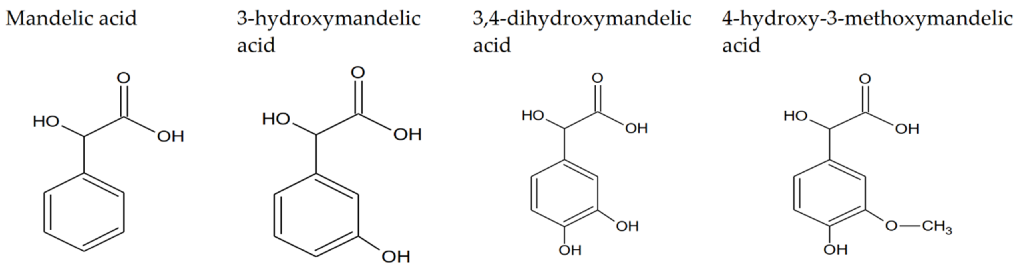
1.1. Mandelic Acid and Its Derivatives—Properties and Applications of Studied Compounds

Mandelic acid (2-hydroxy-2-phenylacetic acid, MA) is a white, crystalline solid that belongs to a group of aromatic α-hydroxy carboxylic acids. The molar mass and water solubility are equal at 152.147 g/mol and 0.158 g/mL, respectively [1]. DL-mandelic acid can be derived from the hydrolysis of an extract of bitter almond [2]; it can also be isolated from Aesculus indica fruit [3]. Due to the presence of the chirality center in moiety, mandelic acid exists in enantiomeric forms and racemic forms [4]. Chirality underlie pharmaceutical industry applications of mandelic acid. Mandelic acid is used as a reactant in semi-synthetic penicillins, cephalosporins and antiobesity and antitumor agents production [5]. Despite its antibacterial activity, it is also used as a skincare modality agent, precursor for the pharmaceutical industry and sensing substrate for molecule recognition research [6]. Mandelic acid exhibits antibacterial properties, and due to its lack of toxic effect on organisms, it finds usage as a medicament for urinary infections and acne. The condensation reaction produces a mandelic acid condensation polymer (SAMMA). SAMMA is useful as an inhibitor of HIV, herpes viruses 1 and 2 and is active against Neisseria gonorrhoeae and Chlamydia trachomatis. It also shows activity against Gram-positive bacteria (Listeria monocytogenes and Staphylococcus aureus) and Gram-negative bacteria (Klebsiella pneumoniae and Pseudomonas aeruginosa) [7]. MA is a raw material developed in the production of polymers and rubber [8]. It also serves as a biomarker of exposure to styrene. As a biomarker, it is created in metabolic pathways and excreted through urine [9]. Triorganotin (IV) derivatives of mandelic acid have shown potent in vitro anticancer activity against mammary, liver and prostate cancers. Diorganotin (IV) derivatives of mandelic acid are more cytotoxic than triorganotin analogues [10]. The simplest derivative of mandelic acid, 3-hydroxymandelic acid, also known as m-hydroxymandelic acid (MHMA), is a product of the phenylephrine metabolism in the human body [11]. Midgley et al., showed that MHMA is a normal constituent of human urine [12]. Another mandelic acid derivative is 3,4-dihydroxymandelix acid, also known as DHMA. This compound can be obtained in the thermophilic reaction cascade using thermostable enzymes obtained from Thermococus barophilus and Thermomonospora curvata, of low-cost phenylpyruvic acid (PPA) and 2-phenylglyoxylic acid (PGA), as subtracts [13]. It also occurs in mammalian tissues, especially in the heart, as a decarboxylated noradrenaline metabolite [14]. Another ligand discussed in this paper is 3-methoxy-4-hydroxymandelic acid, also known as vanillymandelic acid-VMA. It can be obtained as an undesirable byproduct of the condensation reaction of glyoxalic acid and guaiacol [15]. The presence of vanillymandelic acid, produced almost exclusively in the human liver, present in human urea indicates the presence of tumor cells such as PCC, paraganglioma or neuroblastoma [16]. Since the 1970s, VMA has also been used as a biomarker of metabolic disorders such as dopamine excretion disorders, as well as neurological disorders such as autism, post-traumatic stress disorder, Parkinson’s disease or depression [17]. Mandelic acid derivatives are still poorly described in the literature. The aim of this study was to compare the structure of these compounds and their antioxidant properties in relation to mandelic acid. Discussion of the antioxidant properties of mandelic acid and its derivatives also includes the description of reaction mechanisms with free radicals and radical cations. The structures of the studied compounds are presented in Figure 1.

1.2. The Antioxidant Reaction Mechanisms Description

The DPPH antioxidant assay is described with different mechanisms of reaction, so it is not possible to assign one mechanism of reaction unequivocally to this assay. According to the literature, the mechanism of the reaction between the antioxidant molecule and DPPH radical depends on the used solvent. In ionizing solvents such as methanol and ethanol, DPPH reacts with phenolic compounds following the SPLET mechanism of the reaction. In ionizing solvents, this mechanism follows the HAT mechanism, which is slower than SPLET [18]. Due to the strong hydrogen atom bonding capacity of methanol solutions, electron SET (single electron transfer mechanism) is favorized over HAT (hydrogen atom transfer) [19]. The presence of acids in solution also has an influence on the mechanism of the reaction between radicals and antioxidants, e.g., hydrogen atom transfer in the HAT mechanism is inhibited in the presence of acetic acid, which suppresses the ionization of the hydrogen group of the antioxidant compound, thus proton transfer to DPPH radical is inhibited [20]. DPPH is also considered an assay with mixed mechanisms regarding HAT, SPLET, PCET (proton-coupled electron transfer) and ET-PT (electron transfer followed by proton transfer), and the mechanism of the DPPH reaction depends not only on solvent polarity but also on the structure of antioxidant and pH [21]. ABTS assay reactions are described with mixed reaction mechanisms, consisting of HAT and ET combinations [22]. FRAP and CUPRAC are both described with ET reaction mechanisms [23]. In vitro antioxidant test results are compared with thermodynamical parameters describing the mechanisms of antioxidant assays. Scavenging of radicals undergo different reaction mechanisms, among which HAT (hydrogen atom transfer) is the key reaction in biology and chemistry [24]. In the HAT mechanism, protons and electrons are simultaneously transferred from the donor to the acceptor in a one-step reaction, without any intermediate stage. This reaction does not involve significant charge distribution [25]. The HAT reaction mechanism can be described with the following equation [26]:
Ar(OH) + R → ArO + RH
The HAT mechanism is described by the BDE value (bond dissociation energy) of the hydroxyl group of the antioxidant compound. The lower the BDE value is, the higher the ability of the hydrogen abstraction grom hydroxyl group, and the higher the antioxidant capacity of the compound. HAT does not involve charge separation; hence this mechanism is favorable in a non-polar environment [27]. Below, the equation for BDE value calculation is presented [28].
BDE = H(ArO) + H(H) − H(ArOH)
where H(ArO) is the enthalpy of aromatic radical formation in the reaction of hydrogen abstraction, H(H) is the enthalpy of the hydrogen atom and H(ArOH) is the enthalpy of the neutral molecule.
Another mechanism involved in providing antioxidant capacity is the sequential proton loss electron transfer (SPLET) mechanism. This reaction can be described by the following reactions [29]:
Ar(OH) → ArO + H+
ArO +R → ArO + R
R + H+ → RH.
The SPLET mechanism is described with PA (the proton affinity) and the electron transfer enthalpy (ETE). Values of these thermochemistry parameters can be obtained according to Equations (6) and (7), respectively.
PA = H(ArO) + H(H+) − H(ArOH)
ETE = H(ArO) + H(e) − H(ArO)
where H(ArO) is the enthalpy of the aromatic anion, H(H+) is enthalpy of the proton and H(e) is the enthalpy of the electron.
The last mechanism underlying antioxidant properties is single electron transfer followed by proton transfer (SET-PT). This mechanism is described with reactions (8) and (9):
ArOH + R → ArO+• + R
ArO+• + R →RH + ArO•.
The SET-PT mechanism is described by the proton dissociation enthalpy (PDE), according to Equation (10) and the ionization potential (IP) provided as Equation (11) [29].
PDE = H(ArO•) + H(H+) − H(ArO+•)
where H(ArO+•) is the enthalpy of the antioxidant radical cation.
IP = H(ArO+•) + H(e-) − H(ArOH).
All the transfer processes cited above are known as proton-coupled electron transfer (PCET) [30].

2. Materials and Methods

2.1. Materials

Mandelic acid 98%, 3-hydroxymandelic acid ≥ 97% and 3,4-dihydroxymandelic acid 95% were purchased from Sigma Aldrich (Saint Louis, MO, USA), 4-hydroxy-3methoxymandelic acid 98% was purchased from Alfa Aesor (Kandel, Germany), CuCl2 × 2H2O, FeCl3 × 6H2O, ABTS (2,2′-azino-bis(3-ethylbenzothiazoline-6-sulfonic acid), Trolox and H2O2, were purchased from Sigma Aldrich and used without purification. KBr, DPPH (2,2-diphenyl-1-picrylhydrazyl), TPTZ (2,4,6—trypyridyl-s-tirazine), FeCl3, FeSO4 × 7H2O, neocuproine and ammonium acetate were purchased from Sigma Aldrich. Hydrochloric acid (35%), methanol and ethanol (analytical grade) were purchased from Chempur (Poland).

2.2. FTIR and Raman Spectra

The Ft-IR spectra were registered in KBr matrix pellets on an Alfa Bruker spectrometer (Bremen, Germany) within the range of 400–4000 cm−1 with a resolution of 4 cm−1. FT-Raman spectra of solid samples were recorded with a MultiRam (Bruker, Bremen, Germany) spectrometer in the range of 400–4000 cm−1.

2.3. NMR Spectra

1H NMR and 13C NMR spectra of the DMSO samples solution of the studied compound were recorded with a Bruker Avance II 400 MHz unit at room temperature with TMS as an internal reference

2.4. Evaluation of Antioxidant Activity

In this paper, the antioxidant properties of the tested compounds were evaluated using DPPH, ABTS, FRAP and CUPRAC assays. The antioxidant capacity of compounds can be measured using stable and intensely colored radical 2,2-diphenyl-1-picrylhydrazyl (DPPH). The DPPH assay consists of measuring the ability of antioxidant compounds to quench DPPH radicals, expressed as a percentage of DPPH turned into hydrazine DPPH-H form. The reaction of reducing the unpaired electrons of nitrogen atoms in DPPH is visualized by a change in color of the examined solution from violet to yellow [31]; hence the antioxidant capacity of antioxidant can be measured using the spectrophotometric method as a decrease in absorbance value at about 515–520 nm [32]. H–atom donation by an antioxidant molecule to DPPH can be described using a single electron transfer or hydrogen atom transfer mechanism, depending on the reaction environment [33]. The initial water and ethanolic solutions were prepared at concentrations of 0.25 µM–5 µM for the investigated mandelic acids. The methanolic solution of DPPH was prepared at a concentration of 15 µM. Tested compounds were prepared in testing tubes, where the appropriate dilutions were made to obtain the abovementioned scope of concentrations, with the final volume after dilution equal to 1 mL for water and ethanol series separately. Then, 2 mL of DPPH solution were added to each testing tube. All samples were incubated in darkness for an hour. The absorbance of the samples was measured at 516 nm against water and ethanol as a blank for water and ethanol series, respectively, using an Agilent Carry 5000 spectrophotometer (Santa Clara, CA, USA). Since DPPH is not highly soluble in hydrophobic solvents, to determine antioxidant capacity in organic media, the 2,2’-azinobis(3-ethylbenzothiazoline-6-sulfonate) radical cation (ABTS•+) can be applied. Apart from good solubility in organic solvents, the ABTS assay can be implemented over a wide range of pH values [34]. Green radical cation ABTS•+ chromophore formed in the reaction of ABTS with potassium persulfate is reduced by an antioxidant to an extent depending on the concentration of the antioxidant and the duration of the reaction. The total antioxidant activity of the compound in the ABTS assay is designated using the spectrophotometric method [35]. ABTS water solution was prepared at a concentration of 5.4 mM and mixed with potassium persulfate at a concentration of 1.74 mM at a 1:1 ratio. After 12 h of incubation, the solution was diluted with methanol to achieve an absorbance value between 0.7 and 0.8 at 734 nm. The scope of the concentrations of the tested compounds was the same as at the DPPH assay, but dilutions were made to achieve a final volume of 1.5 mL for both water and ethanolic series. To prepare the concentration scope of water and ethanolic solutions of mandelic acids compounds in this way, 1.5 mL of ABTS solution were added. The absorbance value was measured at 734 nm against 1.5 mL of appropriate solvents mixed with 1.5 mL of ABTS solution as a blank. Results of both the DPPH and ABTS assays are expressed as IC50 values, corresponding to the concentration of antioxidant that is required to decrease the initial concentration of DPPH radical or ABTS radical cations by 50% [36]. The % of inhibition in DPPH and ABTS assays was calculated using the following equation:
%   I = A c o n t r o l A s a m p l e A c o n t r o l
where % I is the % of inhibition of DPPH or ABTS radical, A c o n t r o l is the absorbance of the control and A s a m p l e is the absorbance of the sample.
IC50 was designated as the dependence between the concentrations of tested compounds and their % I values. Another antioxidant capacity assay is CUPRAC (cupric reducing antioxidant capacity). In this method, the working solution contains CuCl2 × 2 H2O (0.01 M), neocuproine (0.0075 M) and ammonium acetate at pH 7 (1.07 M), mixed at a 1:1:1 ratio. Absorbance values are measured at 450 nm [37] using an Agilent Carry 5000 spectrometer. To the 3 mL of CUPRAC solution, 0.5 mL of tested compound (at 1 mM for mandelic acid and 3-hydroxymanelic acid, 0.1 mM for 4-hydroxy-3-methoxymandelic acid and 3,4-dihydroxymandelic acid) and 0.6 mL of distillated water were added. The CUPRAC assay consists of the creation of a copper (II)–neocuproine complex, which is reduced by an antioxidant compound to a colored copper(I)–neocuproine chelate complex. This method is used in both oil and water solutions [38]. Antioxidant activity was expressed as the Trolox equivalents [µM], using the calibration curve prepared over the range of 0.05–0.35 mM. FRAP (Ferric reducing activity power assay) evaluates total antioxidant activity in the reaction of reducing the ferric tripyridyl triazine (Fe(III) TPTZ) complex to blue ferrous tripyridyl triazine (Fe(II)-TPTZ) form at low pH, in the presence of an antioxidant, which can be monitored by measuring the change in absorption at 593 nm [39]. To prepare the FRAP reagent acetate buffer (300 mM), TPTZ (10 mM) and FeCl3 (20 mM) were mixed together at a 10:1:1 ratio. Next, 0.4 mL of each of the tested compounds at the same concentrations as in the CUPRAC assay were mixed with 3 mL of the FRAP reagent. Then, samples were incubated in darkness for 8 min. Antioxidant activity was expressed as Fe2+ equivalents [µM], using the calibration curve prepared over the range of 0.05–0.3 mM. All measurements in the performed antioxidant assays were taken in two series of five repetitions for every compound and every concentration in two different solutions.

2.5. Computational Details

All calculations were performed using the Gaussian09 program package. Optimization and geometries of the studied mandelic acids and corresponding radicals, anions and radical cations were calculated using the B3LYP method and the 6-311++G(d,p) basis set. All calculations were performed for vacuum and for water and ethanol solvents using the same method and basis set. Enthalpies for BDE, IP, PDE, PA and ETE were calculated for 298.15 K and 1.0 atmospheric pressure. The calculated gas-phase enthalpy for proton, electron and hydrogen atoms were taken from the literature and were equal to 6.197 kJ/mol, 3.146 kJ/mol [40] and −1306 kJ/mol, respectively [41]. The solvent phase calculations in the water of the proton, electron and hydrogen atoms were, respectively: −1058 kJ/mol [40], −101 kJ/mol and −4 kJ/mol [41]. Additionally, for the solvent phase in ethanol, these values were equal to −1068.4 kJ/mol, −73.6 kJ/mol [40] and −3.7 kJ/mol for hydrogen atoms [41]. The HOMO and LUMO energies were calculated. HOMA aromaticity indices for vacuum, water and ethanol were calculated. Parameters such as softness, hardness, electronegativity and electrophilicity were also calculated.
The aromaticity of studied acids was designated as the HOMA index on the basis of the following Formula [42]:
HOMA = 1     [ α ( R opt     R ar   ) 2 + α n   ( R ar R i ) 2 ] = 1     EN GEO
where:
  • Ropt is the optimal value of a bond length. For C-C type of bonds in a benzene ring, the Ropt value is equal to 1.334;
  • Ri is the length of the ith bond;
  • n is the number of bond lengths in the ring;
  • Rar is the average bond length;
  • α is the normalization factor necessary to obtain a HOMA value equal to 1 for ideally aromatic benzene or 0 for an ideally alternating cyclohexatriene Kekulé ring.
In this study, I6 and BAC aromaticity indexes are also calculated. The I6 aromaticity index, also known as Bird’s aromaticity index, is defined as:
I 6   = 100   ( 1 V 33.3 )  
where
V = 100 N a v N N N a v 2
N is the bonds order provided by (a/R2 − b), where a and b are empirical constants, and R is the bond length [43]. Another approach to determining the aromaticity is the so-called bond alteration coefficient BAC, defined as BAC = r ( R r   R r + 1 ), where Rr and Rr+1 are consecutive bond lengths in the ring [44].

3. Results

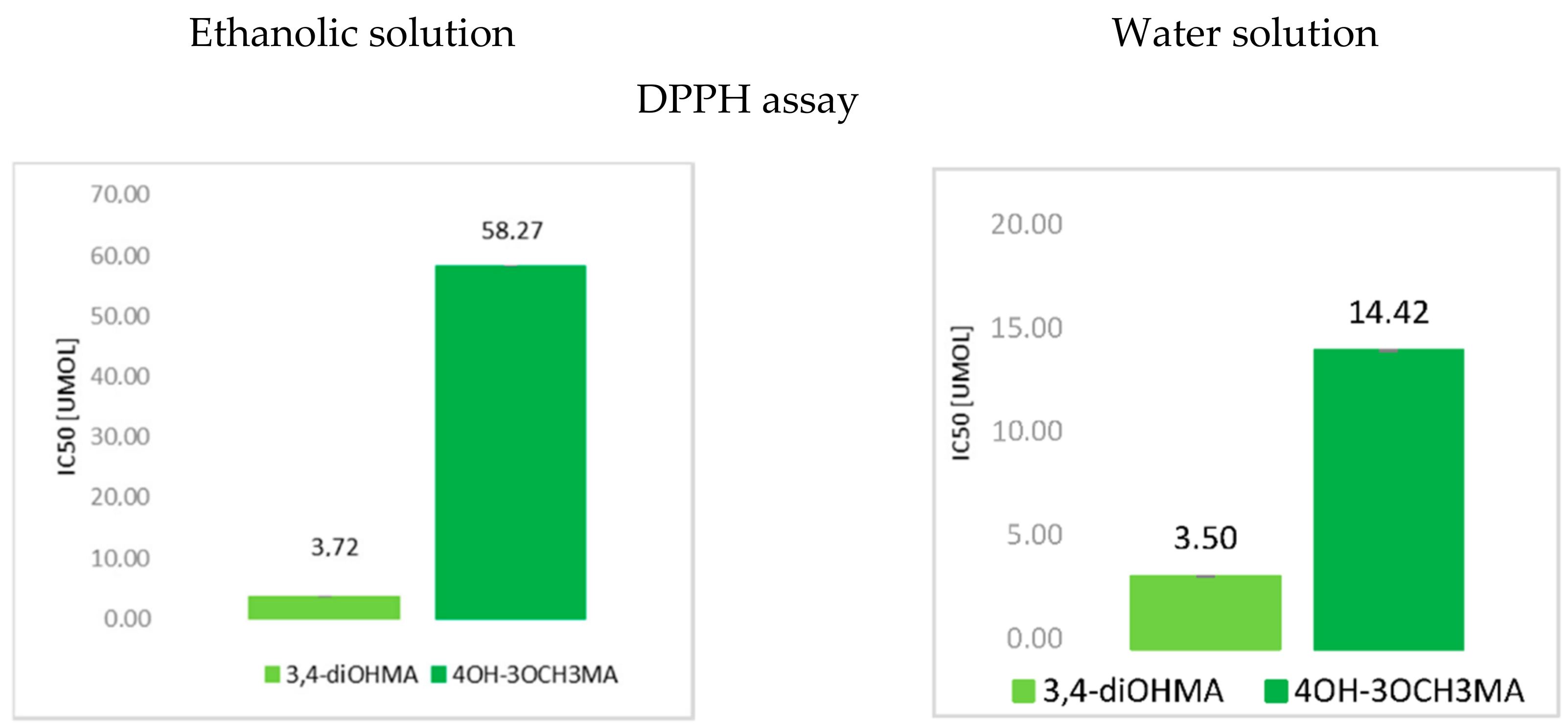
3.1. The Antioxidant Activity of Mandelic Acid and Its Derivatives

The lower the value of IC50 in DPPH and ABTS assays, the better antioxidant properties of the tested compounds. The higher FRAP and CUPRAC values, the higher ferric and cupric reducing activities of these compounds (Figure 2). Thus, according to the DPPH and ABTS assays, 4-hydroxy-3-metoxymandelic acid exhibits weaker antioxidant properties than 3,4-di-hydroxymandelic acid. 3-hydroxymandelic acid and mandelic acid did not exhibit antiradical activity in these assays. According to the FRAP and CUPRAC assays, the antioxidant properties of the tested compounds grew as follows: 3-hydroxymandelic acid < mandelic acid < 4-hydroxy-3-metoksymandelic acid < 3,4-dihydroxymandelic acid.

3.2. Computational Results

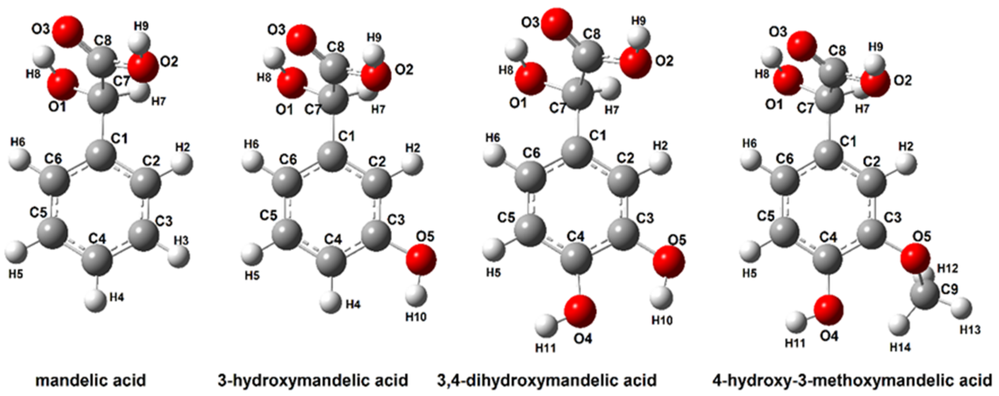
3.2.1. Structure of Mandelic Acid and Their Derivatives

The structures of mandelic acid and its derivatives were optimized by the B3LYP/6-311++G (d, p) method. Calculations of the NBO electron charge distribution, thermodynamic parameters, theoretical NMR and IR spectra and energy of HOMO and LUMO molecular orbitals were performed for optimized conformer structures of modeled molecules. Table 1 show the energy values and dipole moments of the optimized structures. Figure 3 show the optimized molecules with atom numbering used for the description of NMR, NBO and other parameters.

3.2.2. Bond Dissociation Enthalpy, Ionization Potentials, Proton Dissociation Enthalpies, Proton Affinities and Electron Transfer Enthalpies for Mandelic Acid and Its Derivatives

BDE, IP, PDA, PA and ETE were calculated using the abovementioned equations. In Table 2, the results are presented.
The BDE energy value parameter describes the ability to donate H atoms. The minimum BDE value indicates the greatest possibility of hydrogen abstraction in the substituent, thus which substituent is the most susceptible to radical attack [30]. The calculations of thermodynamical parameters related to the reactivity of the studied compounds (in relation to free radical) showed that substituted aromatic ring position derivatives of mandelic acid require less energy expenditure in reactions related to the antioxidant activity of these compounds. The values of the energy of the dissociation process (BDE) are the lowest for 4-hydroxy-3-methoxymandelic acid. In reactions with free radicals, mandelic acid requires the highest energy expenditure among the other tested compounds.

3.2.3. Aromaticity

For the optimized mandelic acid structures (Figure 3) (calculated in gas, water and ethanolic phase with B3LYP-6-311++G(d,p)) method), the aromaticity indices were calculated. Three calculation models (HOMA, I6 and BAC indices) based on the bond lengths in aromatic rings, were used. The calculation results are presented in Table 3.
The Aromaticity indices of hydroxy and methoxy derivatives of mandelic acid are lower than those of pure mandelic acid. The aromaticity of the π-electron aromatic ring is reduced by the attachment of one hydroxyl group, two hydroxyl groups or both hydroxyl groups simultaneously. It is consistent with the calculated aromaticity indices in each of the tested solvents (water and ethanol) and in the aqueous phase. The attachment of two hydroxyl groups to the aromatic ring causes a greater decrease in aromaticity values than the attachment of one hydroxyl group. An even greater decrease in aromaticity is observed when a methoxy group in the aromatic ring of the hydroxymandelic acid is substituted for the second hydroxyl group. 4-hydroxy-3-methoximandelic acid is characterized by the lowest aromaticity. Substitution of the methoxy group causes a greater increase in the disruption of the π-electron system in the aromatic ring than in the hydroxyl group. The aromaticity of the studied compounds changes in series: Mandelic acid > 3-hydroxymandelic acid > 3,4-dihydroxymandelic acid > 4-hydroxy-3-methoxy mandelic acid.

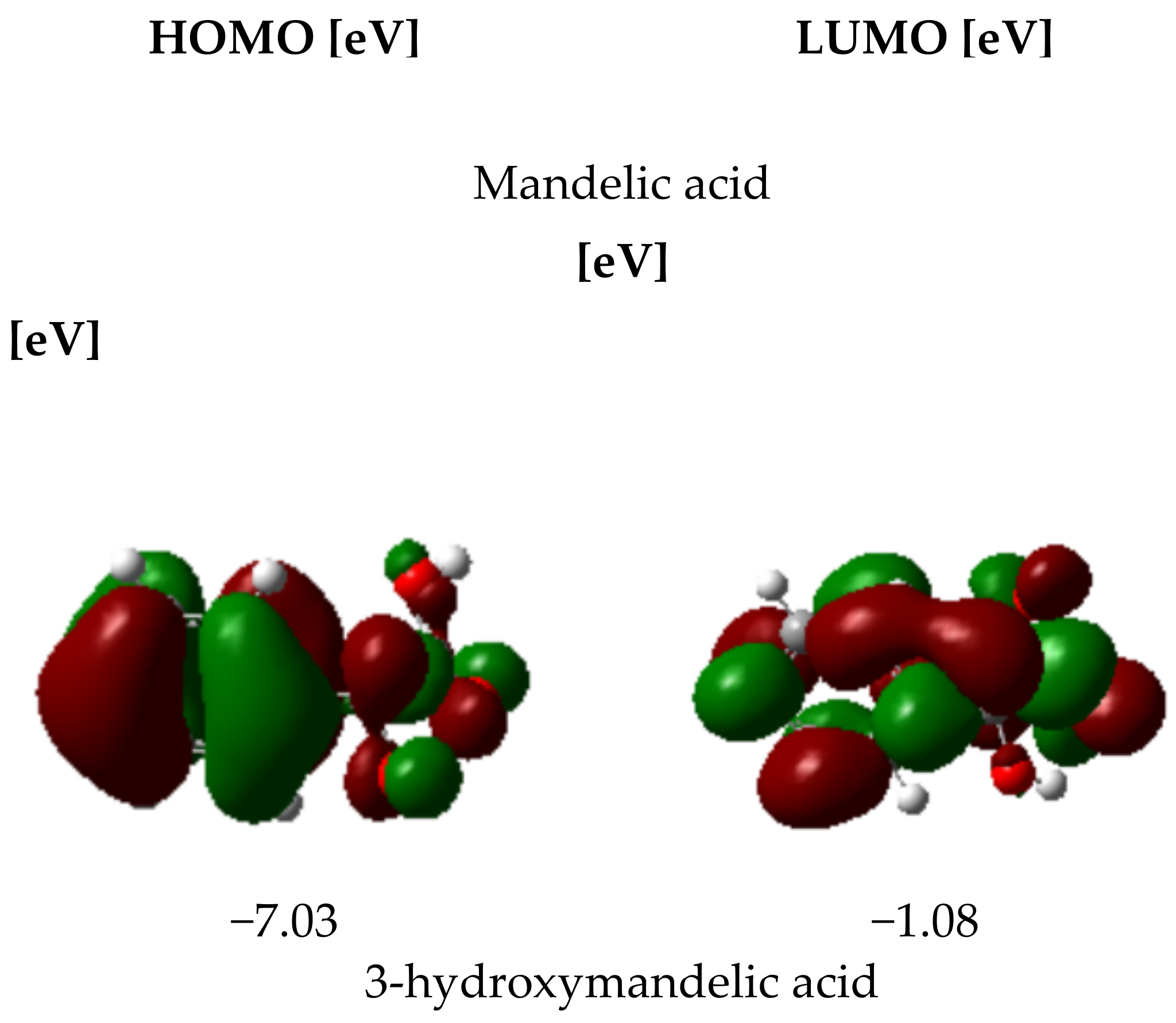
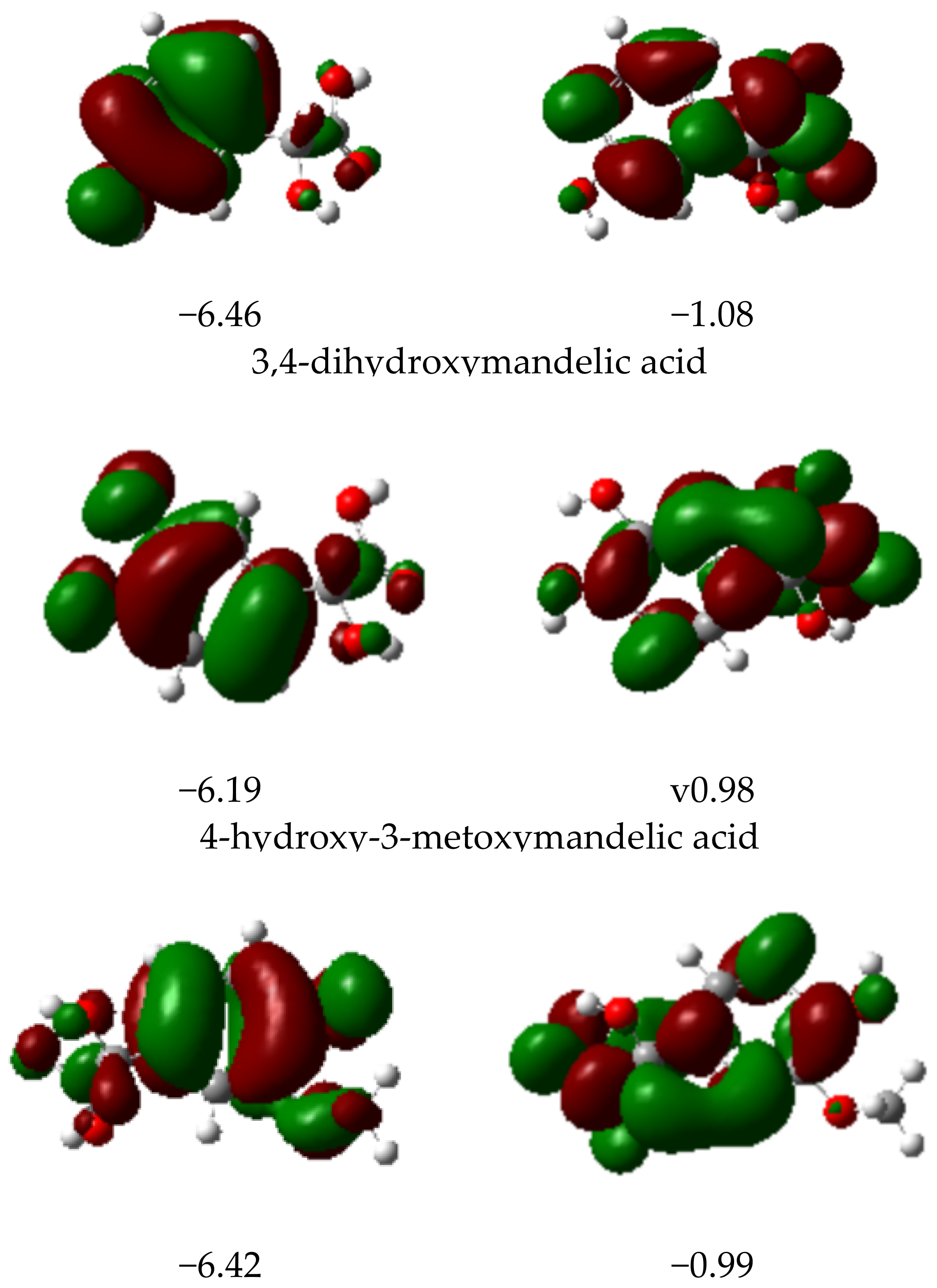
3.2.4. HOMO and LUMO Parameters

For every tested compound, HOMO and LUMO energies in vacuum, ethanol and water were calculated, and then other electronic parameters such as energy gap, electroaffinity, electronegativity, chemical hardness and softness were designated. Below, in Figure 4, the HOMO and LUMO energies are presented.
The designation of HOMO and LUMO parameters is a very helpful tool in the provision of antioxidant properties of tested compounds. The HOMO orbital energy value describes the electron-donating properties of the molecule. The higher the HOMO value, the better the antiradical properties of the compound. The ionization potential provides information regarding the electron removing facility. The lower the ionization potential, the lower the energy required to remove an electron. The reactiveness and stability of compounds can be predicted by assessing the difference value between HOMO orbital energy and LUMO orbital energy (ΔE). The higher the value of that difference, the lower the reactivity and stability of the compounds. In Table 4, the values of the calculated electronic parameters are provided. According to the HOMO and LUMO energy values, the mandelic acid derivatives analyzed in the frame of that work are characterized by lower antioxidant activity than mandelic acid. The ΔE parameter shows that the difference in HOMO and LUMO energy levels is reduced due to the substitution of the aromatic ring with –OH and –OCH3 functional groups, which leads to the distribution of electronic charge in the aromatic ring. Further analyses such as the NBO electron charge distribution and EPS electrostatic potential distribution maps provide information on the reactivity of individual fragments of the studied molecules.

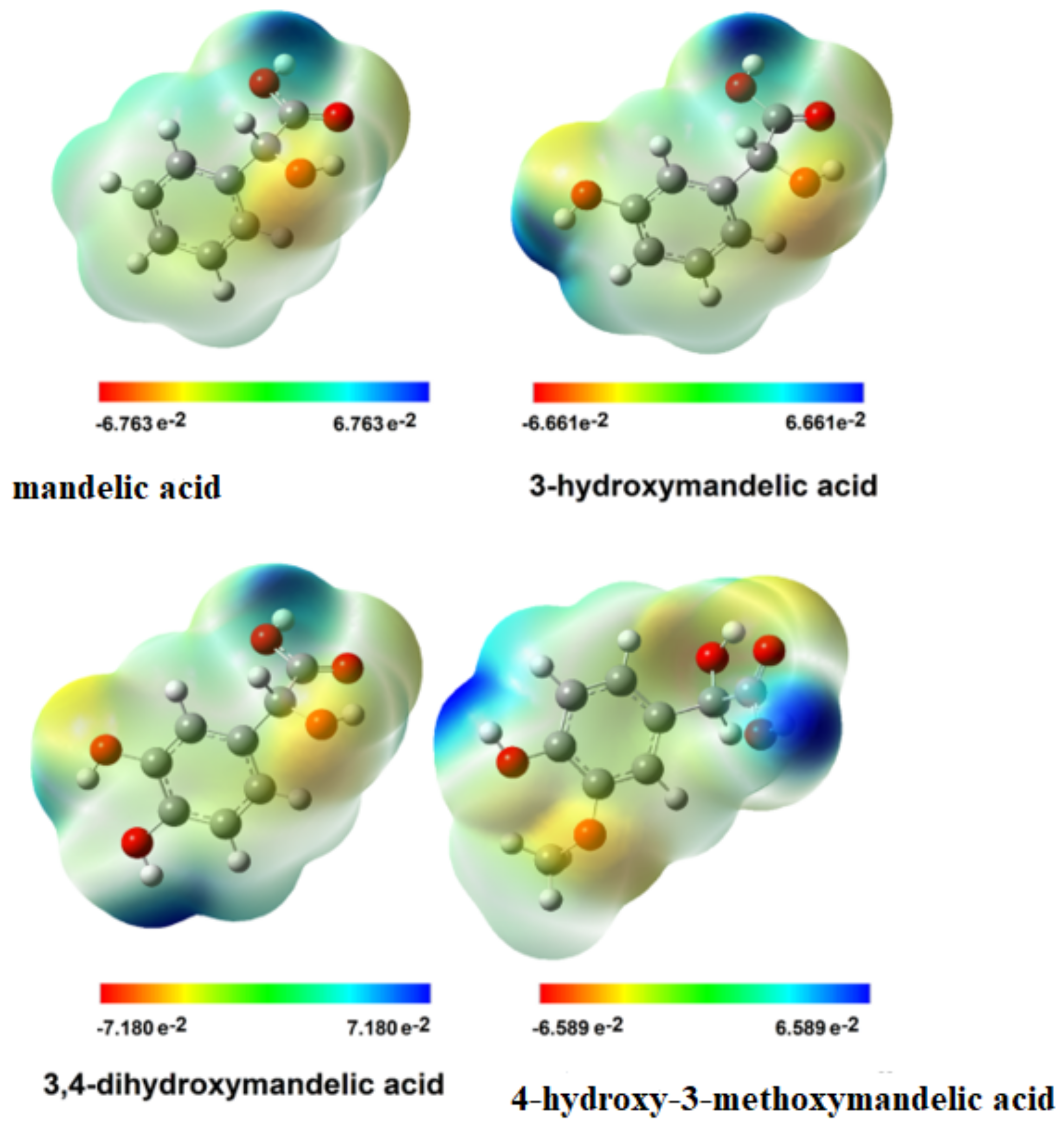
3.2.5. Electron Charge Distribution and EPS Distribution

The electron charge distribution calculated by the Natural Bond Orbital method for the structures optimized with the B3LYP/6-311++G(d,p) method of the investigated mandelic acid derivatives is presented in Table 5. The calculations were performed for two solvents and structures optimized in the gas phase. The analysis of changes in the distribution of electronic charge, with particular emphasis on the aromatic ring, showed that the electronic system of the aromatic ring in the mandelic acid molecule is disturbed (aromaticity decrease) due to the substitution of hydroxyl/methoxy groups in this aromatic ring. The electronegative oxygen atoms substituted in the aromatic ring shift the electron cloud of the aromatic ring towards the substituents, which changes the reactivity of the aromatic ring. The electron charge values of NBO on the aliphatic carbon atoms designated as C7 and C8 in mandelic acid derivative structures remain the same in relation to the atomic charge value in mandelic acid. The greatest changes in the charge distribution of NBO are observed at the carbon atoms substituted in the C3 and C4 positions. In the case of monosubstituted hydroxymandelic acid, a slight increase in the electronic charge distribution around the C3 and C4 atoms is observed, while in the case of disubstituted mandelic acid derivatives, e.g., 3,4-dihydroxymandelic acid and 3-methoxy-4-hydroxy mandelic acid, values of the electron charge decrease significantly. The distribution of electronic charge in the aromatic ring of the mandelic acid molecule substituted with one or two hydroxyl groups (or hydroxyl and methoxy group) demonstrates the reduced aromaticity of these ligands compared to the ligand molecule unsubstituted in the aromatic ring. The results are consistent with the observations made with the calculated aromaticity indices. The NBO electron charge distribution does not change significantly due to the substitution of the aromatic ring of the mandelic acid molecule.
The electrostatic potential map shows the areas of a molecule related to its electrophilic (red) and nucleophilic (blue) reactivity (Figure 5).
The Electrostatic potential map shows the regions of the molecules related to their electrophilic (red) and nucleophilic (blue) reactivity (Figure 5). In mandelic acid, 3-hydroxy mandelic acid and 3,4-dihydroxymandelic acid, the hydroxyl group of the carboxylic moiety is susceptible to nucleophilic attack which cannot be observed in 4-hydroxy-3-methoxy mandelic acid. In the case of the latter molecule, the methoxy group is susceptible to nucleophilic attack. In the studied molecules, the hydrogen atoms in the aromatic ring are susceptible to nucleophilic substitution. The protons of these groups are electron-poor due to the shift of the electron cloud towards electronegative oxygen atoms, making them susceptible to the nucleophilic attack, which affects the reactivity of these groups in reaction with free radicals having an unpaired electron. Increasing the amount of hydroxyl (methoxy) groups contributes to the greater reactivity of these molecules in the reaction with free radicals. The maps of the distribution of electrostatic charges show that mandelic acid is the least susceptible to attack by free radicals because it has only one nucleophilic center, the carboxylic acid proton.

3.3. FT-IR and Raman Spectroscopy

The characteristic bands of the carbonyl stretching vibrations appear in both the IR and Raman spectra of mandelic acid and its derivatives (Table 6). In the IR spectra of mandelic acid, this band is placed at 1716 cm−1. In a similar placement, the stretching band νC=O in 3-hydroxymandelic acid occurred (1715 cm−1). The greatest shift in the localization of these bands is observed in 34-dihydroxymandelic acid and 4-hydroxy-3-methoxymandelic acid. In the case of 3,4-dihydroxymandelic acid, this band is shifted toward a lower wavenumber (1708 cm−1) compared to mandelic acid, and in the IR spectra of 4-hydroxy-3-methoxymandelic acid, this band is shifted towards a higher wavenumber (1743 cm−1). The bands assigned to the stretching vibrations between aromatic carbon atoms are shifted toward growing wavenumbers in the series: 3-hydroxymandelic acid (1466 cm−1) → mandelic acid (1497 cm−1) → 4-hydroxy-3-methoxymandelic acid (1517 cm−1) → 3,4-dihydroxymandelic acid (1537 cm−1). In the Raman spectra, the band originating from the stretching vibrations of the carbonyl group in mandelic acid occurred at 1719 cm−1, in 4-hydroxy-3-methoxymandelic acid at 1716 cm−1. In 3,4-dihydroxymandelic acid, this band is shifted to the lower wavenumber and occurred at 1648 cm−1, whereas in the spectra of 3-hydroxymandelic acid, this band did not occur. In the Raman spectra, the bands derived from the stretching bonds νCC in aromatic rings appeared only in mandelic acid at 1588 cm−1 and in 3,4-dihydroxymandelic acid at 1605 cm−1. In the IR and Raman spectra of mandelic acid derivatives, the bands derived from hydroxyl and methoxy groups also appeared. In 3,4-dihydroxymandelic acid and 4-hydroxy-3-methoxymandelic acid, the bands of stretching vibrations of aromatic hydroxyl groups appeared at 3420 cm−1 and 3402 cm−1, respectively. This band is absent in the Raman spectra of 4-hydroxy-3-methoxymandelic acid, but the stretching bands of the methoxy group appeared at 954 cm−1 in the IR spectra and at 953 cm−1 in the Raman spectra. The location of aromatic bands, related to the vibrations of the π-electron system, determines the influence of the substituents on electron charge distribution in the molecule. The decrease in intensity, the fading of the bands or the shift toward lower wavenumbers indicates the disturbance in the charge distribution in the ligand’s aromatic ring. In the case of bonds in the aromatic ring system (19a, 19b, 8b, 9a, 18b,18a), the spectra of the mandelic acid derivatives showed higher wavenumbers than the corresponding bands in mandelic acid. It was also observed that a number of bands in the mandelic acid spectrum (e.g., 16a, 16b, 17a,9b) shifted towards lower wavenumbers, demonstrating that the substitution of mandelic acid with the hydroxyl/methoxy group in the aromatic ring increases the aromaticity disturbance of the ligand.

NMR Study

13CNMR and 1HNMR chemical shifts for mandelic acid and their derivatives are presented in Table 7.
  • 13C NMR
Substitution of the aromatic ring with a hydroxyl group, two hydroxyl groups or with a methoxy group in the 3- and 4-position in the mandelic acid molecule does not change the electron density of carbon atoms marked as C7 and C8 (Figure 1). A slight chemical shift of these atoms can only be noticed when comparing the 13C NMR spectrum with the spectra of hydroxy and methoxy derivatives of mandelic acid. Substitution with a hydroxyl group in the 3-position of the aromatic ring of mandelic acid causes a slight decrease in electron density around the C1 and C6 carbon atoms, which can be observed in the 13C NMR spectrum of 3-hydroxymandelic acid as a shift towards higher chemical shift values, while substitution with another hydroxyl or methoxy group increases electron density (change in the value of chemical shifts in the reverse direction). The C2 carbon chemical shift in substituted mandelic acids is significantly lower than in the ligand deprived substituents in the aromatic ring. It proves the increase of electron density around the C2 atom of mandelic acid after introducing the substituents. The introduction of substituents to the aromatic ring of mandelic acid causes significant changes in the electron density around the atoms to which the substituents are attached. Around the 3C atom in 3-hydroxymandelic acid, there is a significant decrease in electron density (a significant increase in the value of chemical shifts in the 13C NMR spectra) in relation to the mandelic acid. Substituting another substituent, the change in density around 3C in disubstituted acids versus mandelic acid is slightly less due to the attraction of the electron cloud by the second substituent. Changes in the electron charge distribution (charge distribution disturbance) in substituted mandelic acid derivatives in relation to the unsubstituted ligand affect its reactivity. The aromaticity of these systems is lower than that of mandelic acid.
  • 1H NMR
The calculated aromaticity indexes indicate that the most stable electron system of the aromatic ring is found in mandelic acid. Substitution with a hydroxyl group, two hydroxyl groups and a methoxy group reduces the aromaticity of mandelic acid. Proton chemical shifts in the 1H NMR spectra show the same effect. A decrease in the value of aromatic proton shifts (2a, 5a and 6a) were observed in hydroxymandelic acid, 3,4,-dihydroxymandelic acid and 4-hydroxy-3-methoxymandelic acid. A decrease in the value of the chemical shift of the aliphatic proton 7a was also observed, while the value of the chemical shifts of 8a proton increased in the mandelic acid derivatives substituted in the aromatic ring. The experimentally determined values of the chemical shift of aromatic and aliphatic protons are similar to the theoretical calculations determined by the GIAO method for optimized structures by the B3LYP/6-311++G(d,p) method using the DMSO solvent model. Significant differences between the experimental and theoretical values of chemical shifts of hydroxyl and carboxyl protons probably result from the influence of hydrogen interactions in the structure of tested compounds, which does not occur in the calculated monomers.

4. Conclusions

The study of the electronic structure of 3-hydroxymandelic acid, 3,4-dihydroxymandelic acid and 4-hydroxy-3-methoxymandelic acid showed that the substitution of the aromatic ring with a hydroxyl group/groups and methoxy group changed the π-electron system in the mandelic acid structure. The analyses were carried out by several experimental (FTIR, FT-Raman and NMR) and quantum theoretical methods (calculations of the structure, aromaticity and NBO electronic charge distribution). The results of the experimental and theoretical methods are consistent. Substitution in the aromatic ring of mandelic acid with substituents containing an electronegative oxygen atom increases the disturbance of the electronic system of the aromatic ring (decrease in aromaticity) of this acid. The decrease in ring aromaticity increases the reactivity of the molecules, which is consistent with the theoretical calculations of the HOMO and LUMO energies as well as other descriptors such as electro-affinity and calculated reactivity parameters such as bond dissociation enthalpies, ionization potential, proton dissociation enthalpies, proton affinity and electron transfer enthalpies. The presence of hydroxyl and methoxy groups in the aromatic ring of carboxylic acids may influence their reactivity towards free radicals, which was also investigated. The antioxidant activity of the studied compounds was tested using DPPH and ABTS radicals and the reduction abilities in the FRAP and CUPRAC assays. The conducted research shows that 3-hydroxymandelic acid, like unsubstituted mandelic acid, shows very weak antioxidant properties in ABTS and DPPH assays. The best antioxidant properties are demonstrated by 3,4-dihydroxymandelic acid, and slightly weaker antioxidant properties are shown by 4-hydroxy-3-methoxymandelic acid. Additionally, 3,4-dihydroxymandelic acid presented the highest reduction potential in FRAP and DPPH assays. The presence of hydroxyl and methoxy substituents changes the antioxidant potential of mandelic acid, while the presence of two hydroxyl groups has a greater effect than the introduction of a methoxy group. One hydroxyl group substituted in the aromatic ring does not significantly increase the reduction potential of mandelic acid or its antioxidant activity. The change in electron charge distribution in the aromatic ring of mandelic acid caused by substitution increases the reactivity of this acid, including its antioxidant potential. For the structures optimized by the B3LYP/6-311++G(d,p) method, electron potential maps were carried out with the EPS method. EPS maps show that hydroxyl groups are reactive centers, susceptible to attack by nucleophile molecules (which are free radicals having one unpaired electron). The methoxy group is less susceptible to attack by nucleophilic molecules. In the model system calculated theoretically by the DFT method (B3LYP/6-311++G(d,p)), the influence of the solvent on the analyzed molecules was compared with the experimental results and calculations performed in the gas phase. The solvent does not significantly affect the results of the calculations; in particular, it affects the calculations of the structure and electron charge distribution of the studied molecules. The calculations of the thermodynamic parameters related to the reactivity of the tested compounds (in relation to free radicals) show that the substituted mandelic acid derivatives require less energy expenditure in the reactions related to the antioxidant activity of these compounds.

Author Contributions

Conceptualization, R.Ś. and W.L.; Formal analysis, R.Ś. and G.Ś.; Funding acquisition, W.L.; Investigation, M.P.; Project administration, R.Ś.; Supervision, M.M.; Writing—original draft, M.P.; Writing—review & editing, G.Ś., M.M. All authors have read and agreed to the published version of the manuscript.

Funding

This work was financially supported by National Science Centre, Poland, under the research project number 2018/31/B/NZ7/03083.

Institutional Review Board Statement

Not applicable.

Informed Consent Statement

Not applicable.

Data Availability Statement

Not applicable.

Conflicts of Interest

The authors declare no conflict of interest.

Abbreviations

ABTS2,2-azino-bis(3-ethylbenzothiazoline-6-sulfonic acid
DPPH 2,2-diphenyl-1-picrylhydrazyl radical
BDEbond dissociation energy
CUPRACCupric ion reducing antioxidant capacity
DFTdensity functional theory
ETEelectron transfer enthalpy
FRAPferric reducing ability of plasma
HAThydrogen atom transfer
IPionization potential
MAMandelic acid
PAproton affinity
PCETproton-coupled electron transfer
SAMMAmandelic acid condensation polymer
SPLETsequential proton loss electron transfer
3OH-MA3-hydroxymandelic acid
3,4-diOH-MA3,4-dihydroxymandelic acid
4OH-3OCH3-MA4-hydroxy-3-methoxymandelic acid
PDEproton dissociation enthalpy
HOMAharmonic oscillator model of aromaticity
HOMO highest Occupied Molecular Orbital
LUMOlowest Occupied Molecular Orbital

References

  1. Kiris, B.; Asç, Y.S. Parametric Analysis of Mandelic Acid Separation from Aqueous Solutions by Using Secondary Amine Mixture (Amberlite LA-2) in Various Diluents. J. Chem. Eng. Data 2019, 64, 3331–3336. [Google Scholar] [CrossRef]
  2. Tang, L.; Cheng, H.; Cui, S.; Wang, X.; Song, L.; Zhou, W.; Li, S. DL-mandelic acid intercalated Zn-Al layered double hydroxide: A novel antimicrobial layered material. Colloids Surf. B 2018, 165, 111–117. [Google Scholar] [CrossRef] [PubMed]
  3. Zahoor, M.; Shafiq, S.; Ullah, H.; Sadiq, A.; Ullah, F. Isolation of quercetin and mandelic acid from Aesculus indica fruit and their biological activities. BMC Biochem. 2018, 19, 5. [Google Scholar] [CrossRef] [PubMed] [Green Version]
  4. Brittain, H.G. Mandelic Acid. Anal. Profiles Drug Subst. Excip. 2002, 29, 197–211. [Google Scholar]
  5. Ni, K.; Wang, H.; Zhao, L.; Zhang, M.; Zhang, S.; Ren, Y.; Wei, D. Efficient production of (R)-(−)-mandelic acid in biphasic system by immobilized recombinant E. coli. J. Biotechnol. 2013, 167, 433–440. [Google Scholar] [CrossRef]
  6. Yang, X.; Liu, X.; Shen, K.; Fu, Y.; Zhang, M.; Zhu, C.; Cheng, Y. Enantioselective fluorescent recognition of mandelic acid by unsymmetrical salalen and salan sensors. Org. Biomol. Chem. 2011, 9, 6011–6021. [Google Scholar] [CrossRef]
  7. Xiaa, M.; Yanga, M.; Wanga, Y.; Tiana, F.; Hua, J.; Yanga, W.; Taoa, S.; Lub, L.; Dinga, X.; Jianga, S.; et al. DL-Mandelic acid exhibits high sperm-immobilizing activity and low vaginal irritation: A potential non-surfactant spermicide for contraception. Biomed. Pharmacother. 2020, 126, 110114–110121. [Google Scholar] [CrossRef]
  8. Emel’yanenkoa, V.N.; Turovtsevc, V.V.; Fedina, Y.A. Experimental and theoretical thermodynamic properties of RS-(±)- and S-(+)-mandelic acids. Thermochim. Acta 2018, 665, 37–42. [Google Scholar] [CrossRef]
  9. Rahimpoor, R.; Bahrami, A.; Nematollahi, D.; Shahna, F.G.; Farhadian, M. Facile and sensitive determination of urinary mandelic acid by combination of metal organic frameworks with microextraction by packed sorbents. J. Chromatogr. B Biomed. Appl. 2019, 1114, 45–54. [Google Scholar] [CrossRef]
  10. Nath, M.; Roy, P.; Mishra, R.; Thakur, M. Structure-cytotoxicity relationship for apoptotic inducers organotin(IV) derivatives of mandelic acid and L-proline and their mixed ligand complexes having enhanced cytotoxicity. Appl. Organomet. Chem. 2018, 33, e4663. [Google Scholar] [CrossRef]
  11. Gumbhirt, K.; Mason, W.D. Determination of m-hydroxymandelic acid, mhydroxyphenylglycol and their conjugates in human plasma using liquid chromatography with electrochemical detection. J. Pharm. Biomed. Anal. 1994, 12, 943–949. [Google Scholar] [CrossRef]
  12. Davis, B.A.; Boulton, A.A. Excretion of m-hydroxymandehc acid in human urine. J. Chromatogr. 1981, 222, 271–275. [Google Scholar] [CrossRef]
  13. Li, C.; Zhang, Z.; Wang, J. A Thermophilic Biofunctional Multienzyme Cascade Reaction for Cell-Free Synthesis of Salvianic Acid A and 3,4-Dihydroxymandelic Acid. ACS Sustain. Chem. Eng. 2019, 22, 18247–18253. [Google Scholar] [CrossRef]
  14. Ley, J.P.; Engelhart, K.; Bernhardt, J.; Bertram, H.J. 3,4-Dihydroxymandelic Acid, a Noradrenalin Metabolite with Powerful Antioxidative Potential. J. Agric. Food Chem. 2002, 50, 5897–5902. [Google Scholar] [CrossRef]
  15. Niu, D.; Li, H.; Zhang, X. Improved synthesis of 3-methoxy-4-hydroxymandelic acid by glyoxalic acid method. Tetrahedron 2013, 69, 8174–8177. [Google Scholar] [CrossRef]
  16. Soler Arias, E.A.; Trigo, R.H.; Miceli, D.D.; Vidal, P.N.; Hernandez Blanco, M.F.; Castillo, V.A. Urinary vanillylmandelic acid: Creatinine ratio in dogs with pheochromocytoma. Domest. Anim. Endocrinol. 2021, 74, 106559–106566. [Google Scholar] [CrossRef]
  17. Hrdlicka, V.; Barek, J.; Navratil, T. Differential pulse voltammetric determination of homovanillic acid as a tumor biomarker in human urine after hollow fiber-based liquid-phase microextraction. Talanta 2021, 221, 121594–121601. [Google Scholar] [CrossRef]
  18. Xie, J.; Schaich, K.M. Re-evaluation of the 2,2-Diphenyl-1-picrylhydrazyl Free Radical (DPPH) Assay for Antioxidant Activity. J. Agric. Food Chem. 2014, 62, 4251–4260. [Google Scholar] [CrossRef]
  19. Schaich, K.M.; Tian, X.; Xie, J. Reprint of Hurdles and pitfalls in measuring antioxidant efficacy: A critical evaluation of ABTS, DPPH, and ORAC assays. J. Funct. Foods 2015, 18, 782–796. [Google Scholar] [CrossRef]
  20. Tang, Y.; Liu, Z. Free-Radical-Scavenging Effect of Carbazole Derivatives on DPPH and ABTS Radicals. J. Am. Oil Chem. Soc. 2007, 84, 1095–1100. [Google Scholar] [CrossRef]
  21. Lewandowski, W.; Lewandowska, H.; Golonko, A.; Świderski, G.; Świsłocka, R.; Kalinowska, M. Correlations between molecular structure and biological activity in “logical series” of dietary chromone derivatives. PLoS ONE 2020, 15, e0229477. [Google Scholar] [CrossRef] [PubMed]
  22. Esin Celik, S.; Özyürek, M.; Güclü, K.; Apak, R. Solvent effects on the antioxidant capacity of lipophilic and hydrophilic antioxidants measured by CUPRAC, ABTS/persulphate and FRAP methods. Talanta 2010, 81, 1300–1309. [Google Scholar] [CrossRef] [PubMed]
  23. Apak, R.; Özyürek, M.; Guclu, K.; Capanoglu, E. Antioxidant activity/capacity measurement: I. Classification, physicochemical principles, mechanisms and electron transfer (ET)-based assays. J. Agric. Food Chem. 2016, 64, 997–1027. [Google Scholar] [CrossRef] [PubMed]
  24. Weinberg, D.R.; Gagliardi, C.J.; Hull, J.F.; Fecenko Murphy, C.; Kent, C.A.; Westlake, B.C.; Paul, A.; Ess, D.H.; Granville McCafferty, D.; Meyer, T.J. Proton-Coupled Electron Transfer. Chem. Rev. 2012, 112, 4016–4093. [Google Scholar] [CrossRef]
  25. Hammes-Schiffer, S.; Stuchebrukhov, A.A. Theory of Coupled Electron and Proton Transfer Reactions. Chem. Rev. 2010, 110, 6939–6960. [Google Scholar] [CrossRef] [PubMed] [Green Version]
  26. Chen, Y.; Xiao, H.; Zheng, J.; Liang, G. Structure-Thermodynamics-Antioxidant Activity Relationships of Selected Natural Phenolic Acids and Derivatives: An Experimental and Theoretical Evaluation. PLoS ONE 2015, 10, e0121276. [Google Scholar] [CrossRef] [PubMed]
  27. Wright, J.S.; Johnson, E.R.; DiLabio, G.A. Predicting the Activity of Phenolic Antioxidants: Theoretical Method, Analysis of Substituent Effects, and Application to Major Families of Antioxidants. J. Am. Chem. Soc. 2001, 123, 1173–1183. [Google Scholar] [CrossRef]
  28. Saqiba, M.; Iqbala, S.; Mahmooda, A.; Akrama, R. Theoretical investigation for exploring the antioxidant potential of chlorogenic acid: A DFT study. Int. J. Food Prop. 2015, 19, 745–751. [Google Scholar] [CrossRef] [Green Version]
  29. Wang, G.; Liu, Y.; Zhang, L.; An, L.; Chen, R.; Liu, Y.; Luo, Q.; Li, Y.; Wang, H.; Xue, Y. Computational study on the antioxidant property of coumarin-fused coumarins. Food Chem. 2020, 304, 125446. [Google Scholar] [CrossRef]
  30. Amić, A.; Marković, Z.; Marković, J.M.D.; Stepanić, V.; Lučić, B.; Amic, D. Towards an improved prediction of the free radical scavenging potency of flavonoids: The significance of double PCET mechanisms. Food Chem. 2014, 152, 578–585. [Google Scholar] [CrossRef]
  31. Chen, X.; Liang, L.; Han, C. Borate suppresses the scavenging activity of gallic acid and plant polyphenol extracts on DPPH radical: A potential interference to DPPH assay. Lwt-Food Sci. Technol. 2020, 131, 109769–109802. [Google Scholar] [CrossRef]
  32. Dawidowicz, A.L.; Wianowska, D.; Olszowy, M. On practical problems in estimation of antioxidant activity of compounds by DPPH method (Problems in estimation of antioxidant activity). Food Chem. 2012, 131, 1037–1043. [Google Scholar] [CrossRef]
  33. Leopoldini, M.; Marino, T.; Russo, N.; Toscano, M. Antioxidant Properties of Phenolic Compounds: H-Atom versus Electron Transfer Mechanism. J. Phys. Chem. A 2004, 108, 4916–4922. [Google Scholar] [CrossRef]
  34. Magalhaes, L.M.; Barreiros Salette, L.; Marcela, R.; Segundo, A. Kinetic matching approach applied to ABTS assay for high-throughput determination of total antioxidant capacity of food products. J. Food Compos. Anal. 2014, 33, 187–194. [Google Scholar] [CrossRef]
  35. Re, R.; Pellegrini, N.; Proteggente, A.; Pannala, A.; Yang, M.; Rice-Evans, C. Antioxidant Activity Applying an Improved Abts Radical Cation Decolorization Assay. Free Radic. Biol. Med. 1999, 26, 1231–1237. [Google Scholar] [CrossRef]
  36. Sridhar, K.; Linton Charles, A. In vitro antioxidant activity of Kyoho grape extracts in DPPH and ABTS assays: Estimation methods for EC50 using advanced statistical programs. Food Chem. 2018, 275, 41–49. [Google Scholar] [CrossRef]
  37. Apak, R.; Guclu, K.; Ozyurek, M.; Esin Celik, S. Mechanism of antioxidant capacity assays and the CUPRAC (cupric ion reducing antioxidant capacity) assay. Microchim. Acta. 2008, 160, 413–419. [Google Scholar] [CrossRef]
  38. Ozyurek, M.; Guclu, K.; Apak, R. The main and modified CUPRAC methods of antioxidant measurement. Trends Anal. Chem. 2011, 30, 652–664. [Google Scholar] [CrossRef]
  39. Elsa Madhu, S.; Sreeja, H.; Sudendara Priya, J. A preliminary study on phytochemical, antioxidant and cytotoxic activity of leaves of Naregamia alata Wight & Arn. Mater. Today 2020, 25, 343–348. [Google Scholar]
  40. Marković, Z.; Tošović, J.; Milenković, D.; Marković, S. Revisiting the solvation enthalpies and free energies of the proton and electron in various solvents. Comput. Theor. Chem. 2015, 1077, 11–17. [Google Scholar] [CrossRef]
  41. Szeląg, M.; Urbaniak, A.; Bluyssen, H.A.R. A theoretical antioxidant pharmacophore for natural hydroxycinnamic acids. De Gruyter 2015, 13, 17–31. [Google Scholar] [CrossRef]
  42. Świderski, G.; Łyszczek, R.; Wojtulewski, S.; Kalinowska, M.; Świsłocka, R.; Lewandowski, W. Comparison of structural, spectroscopic, theoretical and thermal properties of metal complexes (Zn(II), Mn(II), Cu(II), Ni(II) and Co(II)) of pyridazine-3- carboxylic acid and pyridazine-4-carboxylic acids. Inorg. Chim. Acta 2020, 512, 119865. [Google Scholar] [CrossRef]
  43. Bird, C.W. A New Aromaticity Index and its Application to Fivemembered Ring Heterocycles. Tetrahedron 1985, 41, 1409. [Google Scholar] [CrossRef]
  44. Katritzky, A.R.; Barczynski, P.; Musumarra, G.; Pisano, D.; Szafran, M. Aromaticity as a Quantitative Concept. 1. A Statistical Demonstration of the Orthogonality of “Classical” and “Magnetic” Aromaticity in Five- and Six-Membered Heterocycles. J. Am. Chem. Soc. 1989, 111, 7–15. [Google Scholar] [CrossRef]
  45. Varsanyi, G. Vibrational Spectra of 700 Benzene Derivatives, Vol. I–II.; Academic Kiado: Budapest, Hungary, 1973. [Google Scholar]
Figure 1. The structure of mandelic acid and its hydroxy and methoxy derivatives.
Materials 15 05413 g001
Figure 2. Comparison of antioxidant activities of mandelic acid and its derivatives in water in ethanolic solutions using DPPH, ABTS, FRAP and CUPRAC assays.
Materials 15 05413 g002aMaterials 15 05413 g002b
Figure 3. Optimized structures of mandelic acid and their derivatives calculated in B3LYP/6-311++G(d,p).
Materials 15 05413 g003
Figure 4. HOMO and LUMO energies [eV] distribution in mandelic acid and its derivatives in vacuum calculated at B3LYP/6-311++G (d,p) level of theory.
Materials 15 05413 g004aMaterials 15 05413 g004b
Figure 5. Maps of the electrostatic potential distribution of EPS in mandelic acid and its derivatives.
Materials 15 05413 g005
Table 1. Structural parameters of studied mandelic acid and its derivatives.
Mandelic Acid3-Hydroxymanndelic Acid3,4-Dihydroxymandelic Acid4-Hydroxy-3-Metoxymandelic Acid
Energy [hartree]−535.51−610.76−685.99−725.31
Energy [kJ/mol]−1,406,909.87−1,604,604.26−1,802,266.09−1,905,551.83
Dipole moment [De]2.411.920.271.47
Table 2. Thermodynamical parameters of mandelic acid and its derivatives in vacuum, water and ethanolic solutions obtained at the B3LYP/6-311++G(d,p) level of theory.
BDE [kcal/mol]
CompoundVacuumWaterEthanol
Solvent
MA97.65407.23409.11
3OH-MA77.06396.1389.21
3,4-diOH-MA
3-OH radical70.74382.56384.45
4-OH radical62.21382.45384.33
4OH-3OCH3-MA69.9382.16383.9
IP [kcal/mol]
MA199.33131.92139.63
3OH-MA188.75136.01142.10
3,4-diOH-MA178.05113.81121.47
4OH-3OCH3-MA176.92112.78120.36
PDE [kcal/mol]
MA212.480.06−4.18
3OH-MA202.49−15.16−13.31
3,4-diOH-MA
3-OH radical206.85−6.49−10.67
4-OH radical198.32−6.60−10.78
4OH-3OCH3-MA207.14−5.86−10.10
PA [kcal/mol]
MA318.2142.1240.92
3OH-MA331.6830.3828.85
3,4-diOH-MA
3-OH radical329.3429.8428.28
4-OH radical315.3923.2521.30
4OH-3OCH3-MA326.1128.6426.99
ETE [kcal/mol]
MA93.6089.8694.55
3OH-MA59.5590.4786.71
3,4-diOH-MA
3-OH radical55.5677.4782.53
4-OH radical61.0183.9689.39
Table 3. Aromaticity indices (HOMA, Bird’s indices (I6) and BAC) for mandelic acid and its derivatives for gas phase, water and ethanolic solutions.
Aromaticity IndiceSolution/Gas PhaseMandelic Acid 3-Hydroxy-
Mandelic Acid
3,4-Dihydroxy-
Mandelic Acid
4-Hydroxy-3-Methoxy
Mandelic Acid
HOMAWater0.9890.9880.9800.986
Gas phase0.9890.9880.9880.984
Ethanol0.9840.9890.9800.980
I6Water98.9097.7496.5795.84
Gas phase98.9097.7497.7595.81
Ethanol98.5398.6596.5395.84
BACWater0.9840.9590.9400.930
Gas phase0.9840.9590.9590.918
Ethanol0.9750.9800.9390.930
Table 4. Values of electronic parameters of studied ligands at the B3LYP/6-311 ++ G (d,p) level.
Mandelic Acid
SolventΔE [eV]Hardness [eV]Softness [eV]Electrophilicity [eV]Electronegativity [eV]
Ethanol0.2230.1128.9650.1020.151
Water0.2230.1128.9540.1020.151
Vacuum0.2180.1099.1540.0500.149
3-hydroxymandelic acid
Ethanol0.2030.1029.8300.0970.140
Water0.2040.1029.8140.0970.140
Vacuum0.1980.09910.1200.0970.139
3,4-dihydroxymandelic acid
Ethanol0.1900.10010.4060.0900.133
Water0.1930.10010.3880.0900.133
Vacuum0.1910.10010.4490.0901.132
4-hydroxy-3-methoxymandelic acid
Ethanol0.2000.10010.0040.0900.137
Water0.2010.1009.9700.1370.137
Vacuum0.2000.10010.0160.0900.136
Table 5. NBO Atom Charge Distribution for mandelic acid and its hydroxy end methoxy derivatives.
NBO Atom Charge Distribution
Mandelic Acid
Atom *EthanolWaterVacuum
C1−0.063−0.064−0.060
C2−0.196−0.198−0.192
C3−0.202−0.201−0.197
C4−0.205−0.205−0.200
C50.201−0.202−0.194
C6−0.108−0.1970.190
C70.0300.0290.036
C80.8100.8100.799
H20.2150.2190.207
H30.2150.2150.205
H40.2140.2150.205
H50.2140.2150.206
H60.2180.2150.220
H70.2120.2120.201
H80.4870.4870.481
H90.5850.5060.488
O1−0.750−0.751−0.729
O2−0.670−0.670−0.671
O3−0.636−0.637−0.613
3-hydroxymandelic acid
C1−0.042−0.043−0.04
C2−0.273−0.273−0.273
C30.3190.318−0.323
C4−0.257−0.258−0.250
C5−0.181−0.182−0.176
C6−0.229−0.230−0.226
C70.0310.0300.037
C80.8100.8100.800
H20.2230.2230.218
H40.2230.2230.218
H50.2160.2160.206
H60.2160.2170.209
H70.2130.2130.203
H80.4870.4880.481
H90.5060.5060.488
H100.4870.4880.468
O1−0.758−0.751−0.732
O2−0.669−0.669−0.669
O3−0.636−0.637−0.613
O5−0.692−0.692−0.672
3,4-dihydroxymandelic acid
C1−0.073−0.074−0.081
C2−0.253−0.253−0.213
C30.2740.2730.254
C40.2710.2710.283
C5−0.257−0.257−0.265
C6−0.208−0.208−0.178
C70.0320.0310.039
C80.8090.8180.799
H20.2180.2190.217
H50.2190.2200.284
H60.2200.2200.221
H70.2110.2120.200
H80.4860.4970.481
H90.5050.5050.488
H100.4880.4890.470
H110.4880.4890.466
O1−0.751−0.752−0.730
O2−0.671−0.671−0.671
O3−0.638−0.639−0.613
O4−0.686−0.687−0.659
O5−0.686−0.687−0.711
4-hydroxy-3-methoxymandelic acid
C1−0.082−0.083−0.077
C2−0.212−0.213−0.207
C30.2640.2630.273
C40.2820.2820.277
C5−0.262−0.262−0.265
C6−0.190−0.190−0.187
C70.0320.0320.039
C80.8090.8100.799
H20.2230.2240.219
H50.2190.2200.201
H60.2210.2210.222
H70.2110.2120.200
H80.4860.4870.480
H90.5050.5060.487
H100.1880.1880.183
H120.4900.4910.470
H130.1710.1710.162
H140.1800.1790.182
O1−0.751−0.752−0.731
O2−0.670−0.671−0.670
O3−0.638−0.639−0.515
O4−0.687−0.687−0.676
O5−0.589−0.591−0.569
* Atoms numbers as Figure 3.
Table 6. Wavenumbers and intensities of selected bands in mandelic acid and its derivatives spectra.
Mandelic Acid3-Hydroxymandelic Acid3,4-Dihydroxymandelic Acid4-Hydroxy-3-Methoxymandelic acidAssignment
IRKBrIRATRRamanTheor. (1)IRKBrIRATRRamanTheor.IRKBrIRATRRamanTheor.IRKBrIRATRRamanTheor.
cm−1 (int.)cm−1 (int.)cm−1 (int.)cm−1Int.cm−1 (int.)cm−1 (int.)cm−1 (int.)cm−1Int.cm−1 (int.)cm−1 (int.)cm−1 (int.)cm−1Int.cm−1 (int.)cm−1 (int.)cm−1 (int.)cm−1Int. [45]
3420 s3408 m3427 w385093.83402 s 385093.8νOHar
3338 s3327 m 383462.73335 s3330 m 3792113.0 νOHar
3400 s3401 m 375586.1 375588.0 375686.13353 vs3329 m 375689.4νOH
3734109.5 3730112.2 3732112.3 372878.9νOH
3070 m3074 w3064 vs31975.43062 vw3066 vw3070 s31985.8 3198 w3175 w32091.13087 vw 3069 vs32031.7ν(CH)2
3031 m3038 w3049 m318816.963035 w3032 vw 31859.4 3032 w3031 s31861.63034 vw 3034 s31822.9ν(CH)20a
2967 m 2972 m317719.9 31736.3 2974 w2973 w2974 m ν(CH)20b
2927 m2936 w 31672.1 31705.22945 w2911 w2916 m315414.42935 w2932 w2935 s314618.6ν(CH)7b
2716 m2722 m 301517.42628 m2622 w 301616.5 301718.1 301438.5νCH
1716 vs1711 vs1719 m1797335.61715 vs1713 vs 1796333.41708 vs
1695 vs
1692 vs1648 m1795336.21743 vs
1718 s
1743 s
1715 s
1716 m1796317.3νC=O
1603 m16424.21605 s1603 s1609 m164431.11622 m1620 w1618 s16598.01611 m1610 m1609 s164722.7ν(CC)8a
1588 w 1588 w16270.4 164186.11606 s1603 m1605 s164646.7 163422.9ν(CC)8b
1497 w1497 w 152410.51466 vs1465 s 152912.01537 s1534 m1530 vw1544183.71517 vs1515 s 1548225.6ν(CC)19a
1460 sh1460 sh1461 m15108.9δas(CH3)
1451 m 14886.7δas(CH3)
1452 m1453 m 14839.2 148596.81452 m1450 m1449 vw14912.21439 s1437 s1447 m14816.7ν(CC)19b
1378 m1377 m 142318.31420 m1420 m 142319.51431 s1428 s1414 vw142618.9 βOH; δCHOH
13660.1 136818.8 1377 sh138914.81380 m1377 m1376 w ν(CC)3
13484.01359 w 134833.1 135185.4 ν(CC); βCH14
1299 s1296 s1295 w134092.01268 vs1265 s1265 w134277.81350 s1347 s1355 m134188.81365 sh βOH; νC–OH
1253 w1253 vw1256 vw13072.51249 vs1245 vs 13247.11283 s1280 s1293 m131925.01270 vs1267 s1265 m τCHOH(CH2); β(CH)
1303237.0 1305203.3νC–OH; α(CCC); νC–CH3
1229 m1228 m1222 vw125327.5 1232 sh 125437.41259 s1256 s1261 w125154.21237 vs1234 s 125155.7ωCHOH(CH2)
1192 m1192 m1192 m12076.4 1214 vs1208 vs 1220 s1219 vs1193 m β(CH); βOHar9a
1156 vw1154 vw1155 w11958.71168 s1167 m1167 w119647.21151 s1148 s1155 m120843.31150 vs1148 vs1147 w120727.5β(CH); ρ(CH3)9b
1170166.8 117939.5 118075.2 118548.4βOH
1132 m1131 s 11715.0ρ(CH3)
1062 s1062 s1058 vw108979.71083 s1079 vs1082 m108566.01119 s1116 s1111 m110286.31061 s1057 s1061 m1101116.1β(CH)18a
1028 w1030 w1030 m10487.1 1089 vs1084 vs1085 w 1032 s1031 s1032 m β(CH)18b
10180.6 10134.8 96018.2 α(CCC)13
954 w954 w953 w104089.4νO–(CH3)
1004 w1004 w1004 s1118104.81001 m1001 w1000 vs111883.1982 m980 m 1124208.4 α(CCC); νC–OH12
940 m940 m 9850.1869 s 9820.1920 sh923 sh 9371.9 924 w913 m9470.3γ(CH)17a
889 m887 m 89222.1932 m967 w 91916.2881 s880 s908 m91123.4880 m 93612.6νC–COOH; α(CCC); γOH
855 w854 w858 w8692.0826 m826 m821 w8606.6868 m867 m 8698.3863 m861 s 8787.8βC=O
8161.7 83618.2 83520.1γC=O; α(CCC)
768 w768 w768 vw76316.7732 s731 vs725 m78828.5835 m833 m 79236.6825 m822 s824 w81128.1γ(CH)11
733 s731 s732 w72750.9697 s695 s 72054.8807 s801 s784 vs7251.8775 m773 s777 m7461.4φ(CC)4
697 s 697 s 70933.2674 m671 s 69526.7732 m731 m718 m70636.1732 m731 m 70731.8α(CCC)1
609 m608 m617 w66035.1 637 w66039.8646 m662 m 66143.8707 m693 s701 m66227.0γC=O; βOH
528 m 57052.2505 m 54520.4603 w 585 w54928.8634 w 54619.8γOH
494 m 501 w4984.1462 m 47514.4519 m 47520.3533 w 47121.8φ(CC); γOH16b
467 w 4134.4 41353.2468 m 40348.3465 w 40235.2φ(CC); βOH16a
s—strong; m—medium; w—weak; v—very; sh—shoulder; ν: stretching; β: in-plane deformations; γ: out of plane deformations; δ: scissoring; α: the aromatic ring in-plane bending modes; φ: the aromatic ring out-of-plane ones; τ—bending off the plane-twisting; ω—bending off the plane-fan; ρ—bending in the plane-swinging. Fundamental modes of the phenyl ring are numbered according to Varsányi [45].
Table 7. Experimental and theoretical chemical shifts ƍ [ppm] for mandelic acid and its hydroxy and methoxy derivatives.
Compound
Mandelic Acid3-OH-Mandelic Acid3,4-Dihydoxymandelic Acid4-Hydroxy-3-Methoxymandelic Acid
13C NMR
assignmentCalc.Exp.Calc.Exp.Calc.Exp.Calc.Exp.
C1 *146.02140.39147.49141.62138.31131.10137.91131.13
C2129.71126.85113.89113.47118.61114.14128.73110.80
C3133.36128.34163.51157.13150.89144.90153.63146.12
C4133.39127.87118.26114.51149.05144.90156.53147.26
C5133.15128.34134.76129.00117.19115.06119.51115.01
C6133.58126.85124.10117.31120.68117.83125.42119.33
C775.5672.6375.0872.3374.7472.1374.8172.21
C8182.50174.36182.60174.04182.56174.48182.73174.39
C9------60.0755.56
1H NMR
H27.897.457.176.827.256.807.356.96
H37.697.35-------
H47.627.347.237.11----
H57.617.356.987.116.956.646.906.72
H67.757.457.477.137.256.657.456.95
H73.345.083.174.893.144.803.154.88
H85.475.085.466.815.335.555.335.69
H96.6012.696.0512.416.5512.386.5112.44
H10--4.579.364.548.90--
H11----5.338.824.538.93
H12------3.923.74
H13------3.443.74
H14 3.153.74
* Atoms numbers as Figure 3.
Publisher’s Note: MDPI stays neutral with regard to jurisdictional claims in published maps and institutional affiliations.

