
X-ray Dose Rate and Spectral Measurements during Ultrafast Laser Machining Using a Calibrated (High-Sensitivity) Novel X-ray Detector

 ,
,
Abstract
:1. Introduction
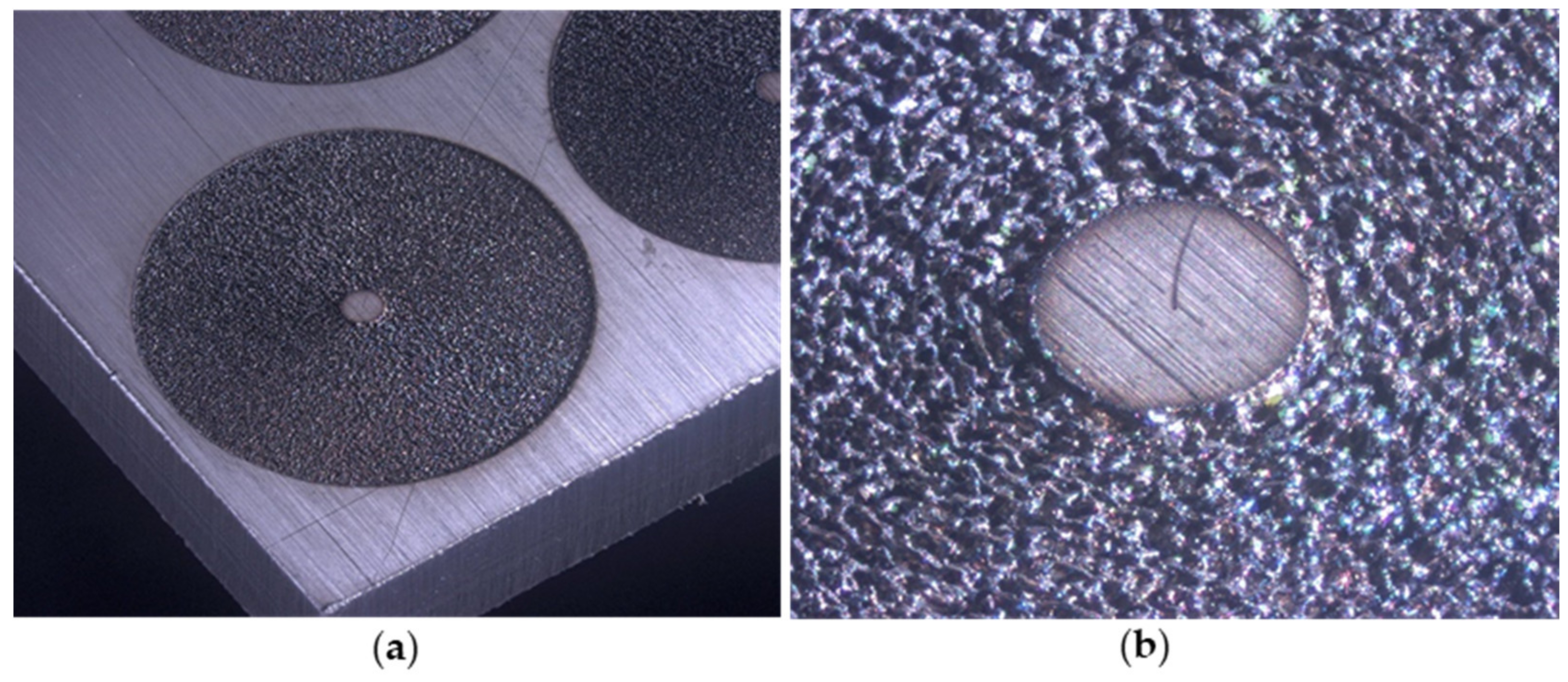
2. Materials and Methods
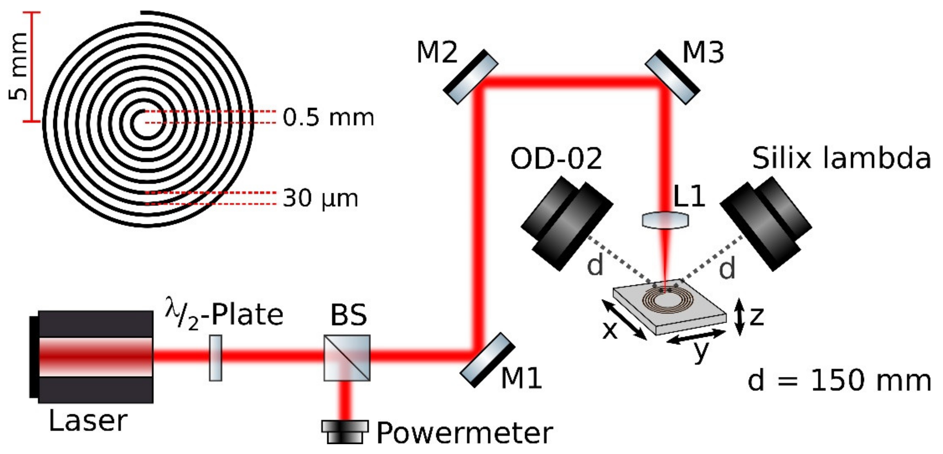
2.1. Optical Setup
2.2. Silix Lambda X-ray Spectrodosimeter
- Directional dose equivalent rate for the skin Ḣ′(0.07).
- Directional dose equivalent rate for the eyes Ḣ′(3).
- Ambient dose equivalent rate Ḣ*(10).
3. Results and Discussion
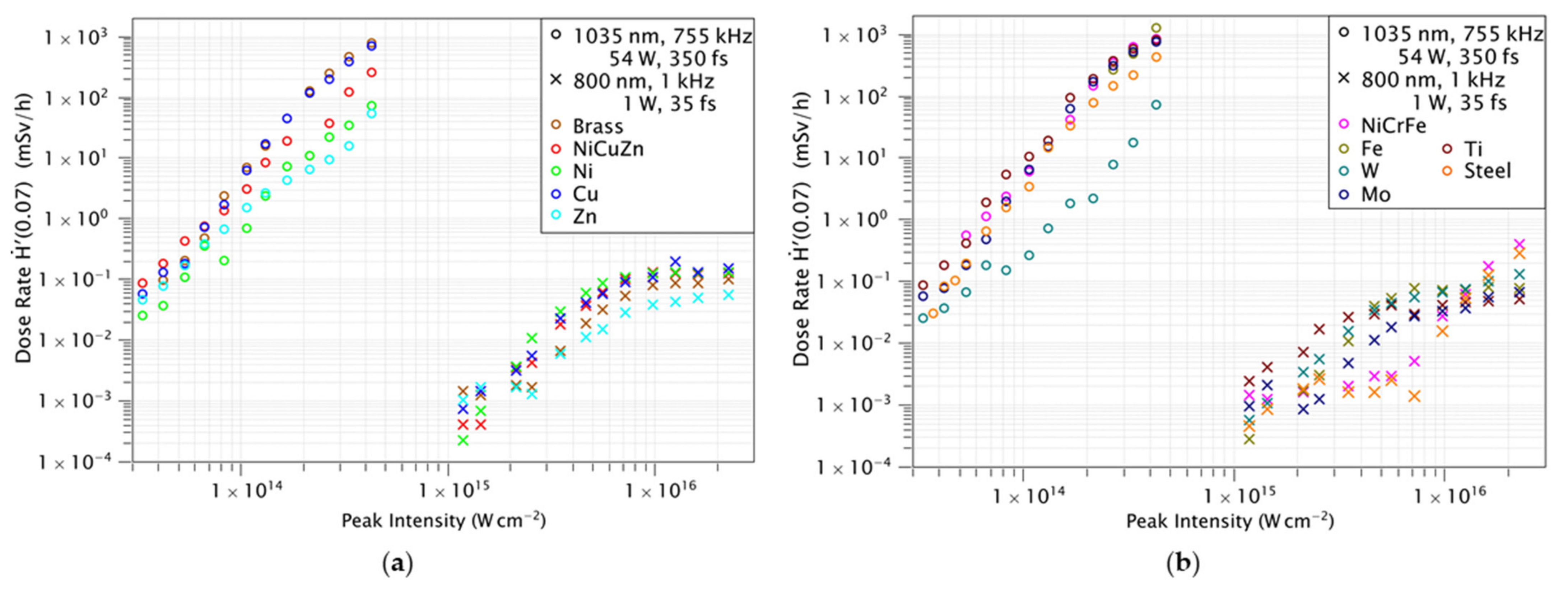
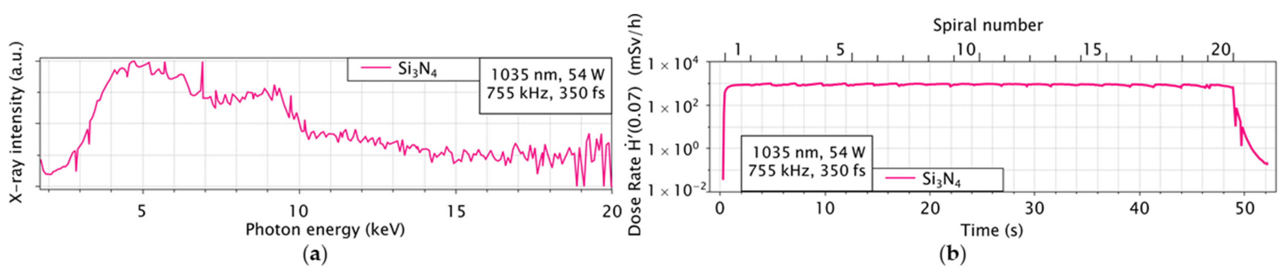
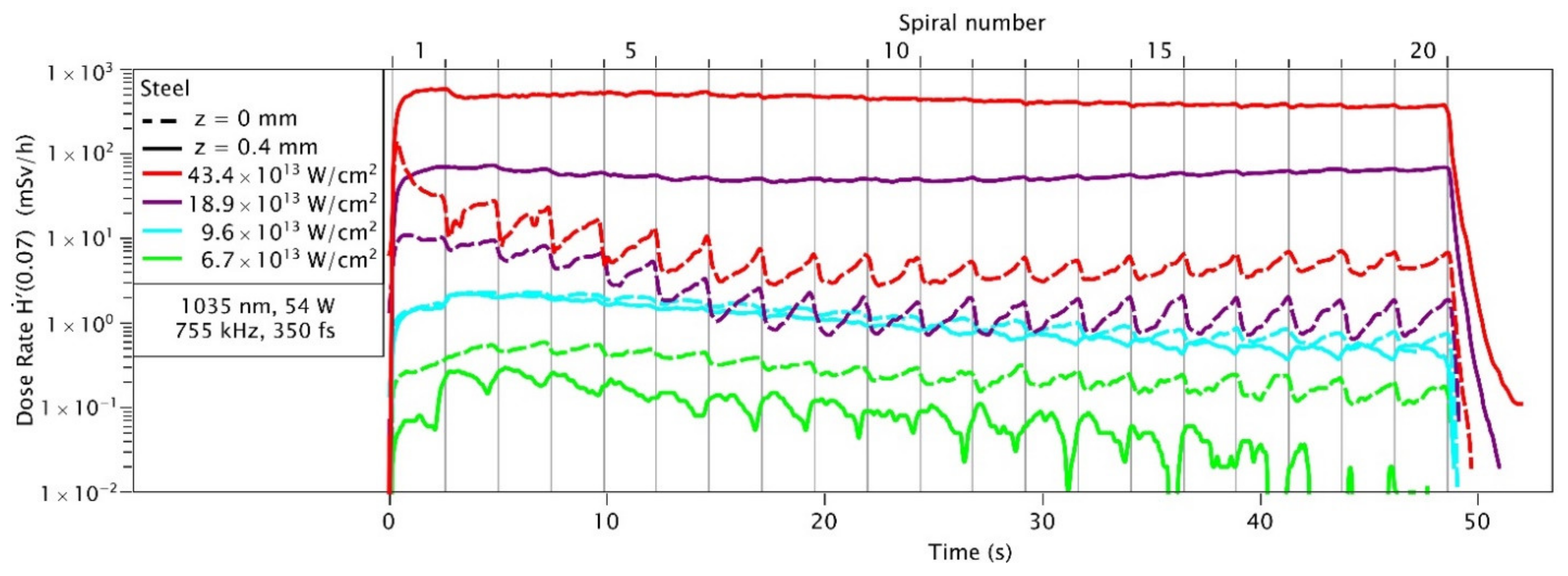
3.1. Dose Rates
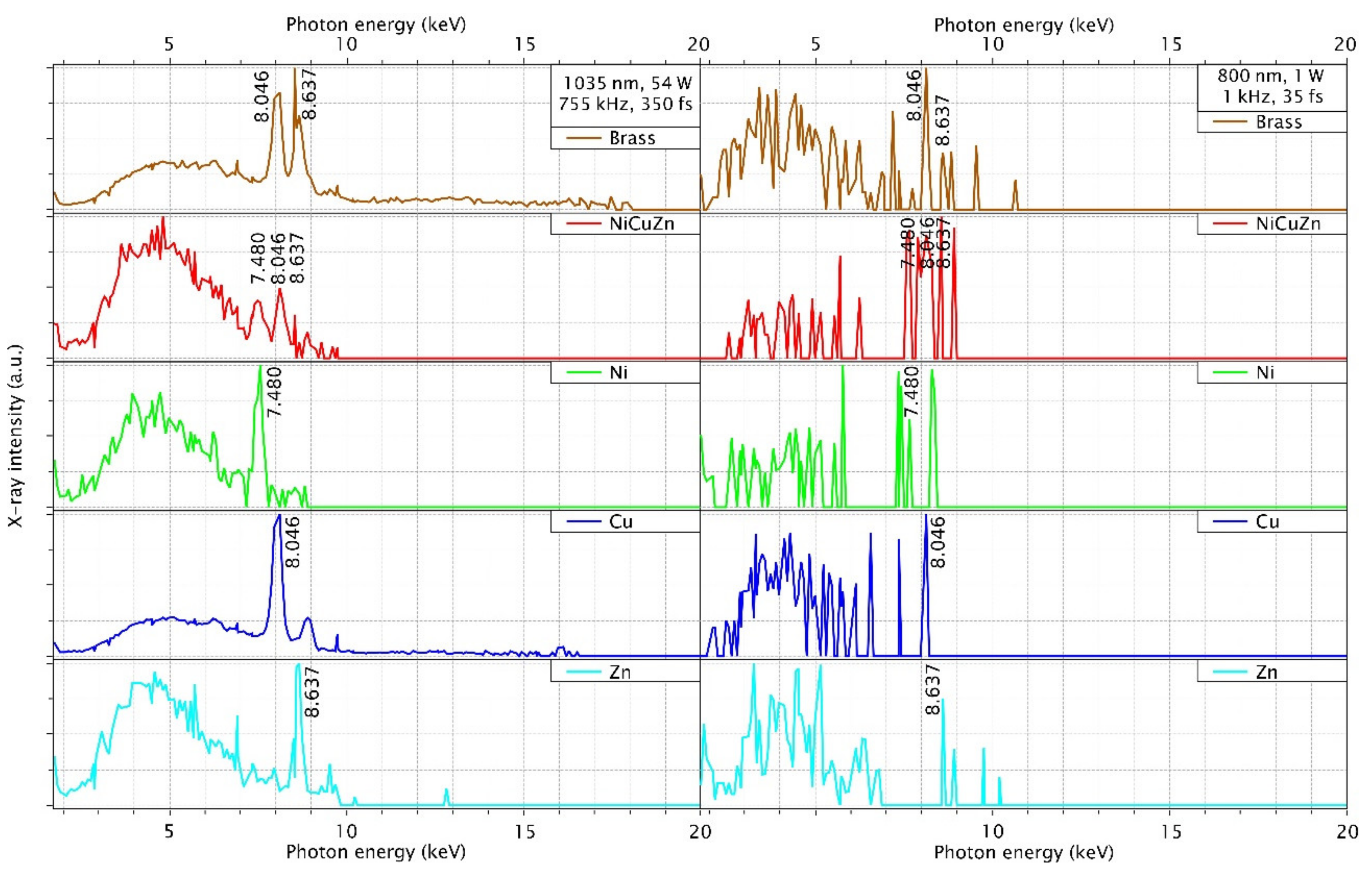
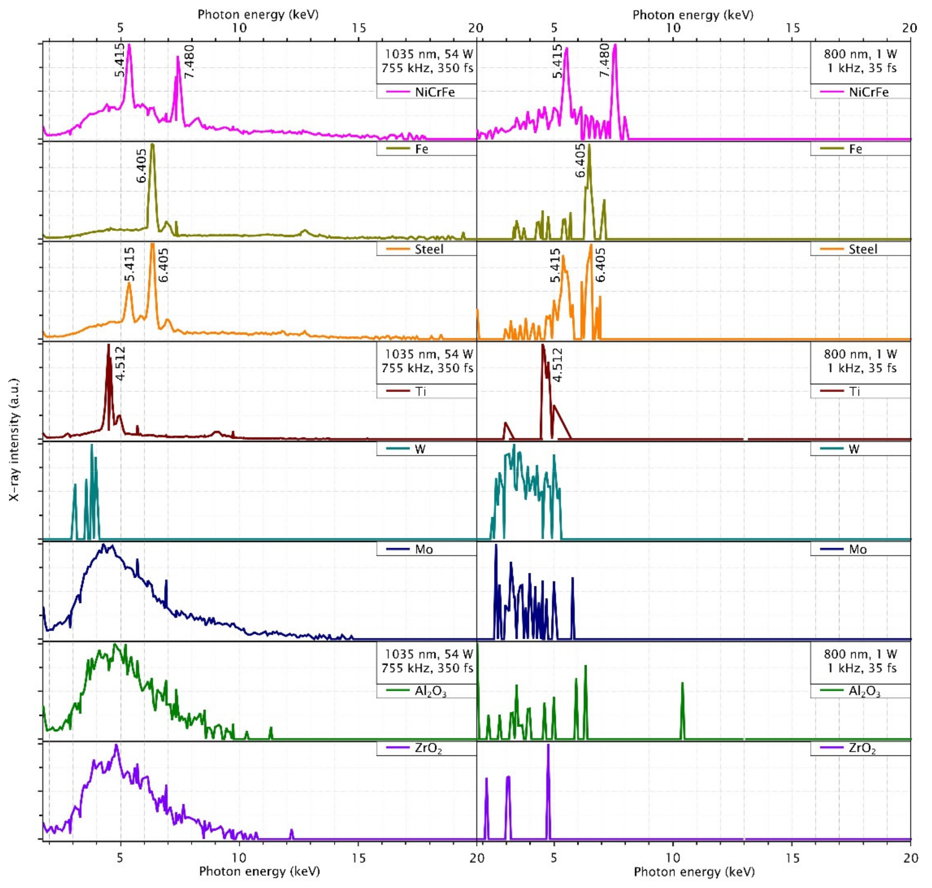
3.2. Spectra
3.3. Discrepency in Dose Rates
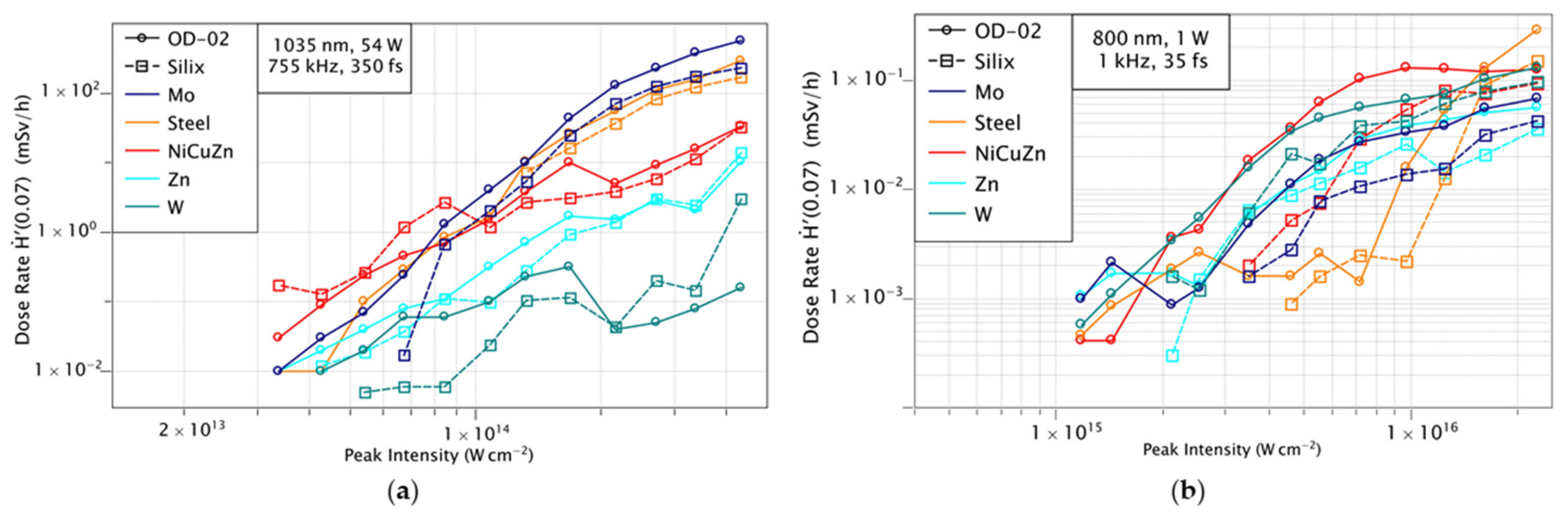
3.4. Comparison Silix and OD-02
4. Conclusions
Author Contributions
Funding
Institutional Review Board Statement
Informed Consent Statement
Data Availability Statement
Acknowledgments
Conflicts of Interest
References
- Gower, M.C. Industrial applications of laser micromachining. Opt. Express 2000, 7, 56–67. [Google Scholar] [CrossRef] [PubMed]
- Cheng, J.; Liu, C.-S.; Shang, S.; Liu, D.; Perrie, W.; Dearden, G.; Watkins, K. A review of ultrafast laser materials micromachining. Opt. Laser Technol. 2013, 46, 88–102. [Google Scholar] [CrossRef]
- Thogersen, J.; Borowiec, A.; Haugen, H.K.; McNeill, F.E.; Stronach, I.M. X-ray emission from femtosecond laser micromachining. Appl. Phys. A 2001, 73, 361–363. [Google Scholar] [CrossRef]
- Weber, R.; Giedl-Wagner, R.; Förster, D.J.; Pauli, A.; Graf, T.; Balmer, J.E. Expected x-ray dose rates resulting from industrial ultrafast laser applications. Appl. Phys. A 2019, 125, 1–12. [Google Scholar] [CrossRef] [Green Version]
- Legall, H.; Bonse, J.; Krüger, J. Review of x-ray exposure and safety issues arising from ultra-short pulse laser material processing. J. Radiol. Prot. 2021, 41, R28. [Google Scholar] [CrossRef] [PubMed]
- Bunte, J.; Barcikowski, S.; Püster, T.; Burmester, T.; Brose, M.; Ludwig, T. Secondary Hazards: Particle and X-Ray Emission. In Femtosecond Technology for Technical and Medical Applications; Dausinger, F., Lubatschowski, H., Lichtner, F., Eds.; Springer: Berlin, Germany, 2004; Volume 96, pp. 309–321. [Google Scholar]
- Kephart, J.F.; Godwin, R.P.; McCall, G.H. Bremsstrahlung emission from laser—Produced plasmas. Appl. Phys. Lett. 1974, 25, 108–109. [Google Scholar] [CrossRef]
- Legall, H.; Schwanke, C.; Pentzien, S.; Dittmar, G.; Bonse, J.; Krüger, J. X-ray emission as a potential hazard during ultrashort pulse laser material processing. Appl. Phys. A 2018, 124, 1–8. [Google Scholar] [CrossRef] [Green Version]
- Legall, H.; Schwanke, C.; Bonse, J.; Krüger, J. X-ray radiation protection aspects during ultrashort laser processing. J. Laser Appl. 2020, 32, 022004. [Google Scholar] [CrossRef]
- Behrens, R.; Pullner, B.; Reginatto, M. X-ray emission from materials processing lasers. Radiat. Prot. Dosim. 2019, 183, 361–374. [Google Scholar] [CrossRef]
- Kruer, W.L. The Physics of Laser Plasma Interactions, 1st ed.; CRC Press: Boca Raton, FL, USA, 2019. [Google Scholar]
- Weisshaupt, J.; Juvé, V.; Holtz, M.; Ku, S.; Woerner, M.; Elsaesser, T.; Ališauskas, S.; Pugžlys, A.; Baltuška, A. High-brightness table-top hard X-ray source driven by sub-100-femtosecond mid-infrared pulses. Nat. Photonics 2014, 8, 927–930. [Google Scholar] [CrossRef]
- Schroeder, W.A.; Omenetto, F.G.; Borisov, A.B.; Longworth, J.W.; McPherson, A.; Jordan, C.; Boyer, K.; Kondo, K.; Rhodes, C.K. Pump laser wavelength-dependent control of the efficiency of kilovolt x-ray emission from atomic clusters. J. Phys. B At. Mol. Opt. Phys. 1998, 31, 5031–5051. [Google Scholar] [CrossRef]
- Di Niso, F.; Gaudiuso, C.; Sibillano, T.; Mezzapesa, F.P.; Ancona, A.; Lugarà, P.M. Role of heat accumulation on the incubation effect in multi-shot laser ablation of stainless steel at high repetition rates. Opt. Express 2014, 22, 12200–12210. [Google Scholar] [CrossRef]
- Vorobyev, A.Y.; Guo, C. Direct observation of enhanced residual thermal energy coupling to solids in femtosecond laser ablation. Appl. Phys. Lett. 2005, 86, 011916. [Google Scholar] [CrossRef]
- König, J.; Nolte, S.; Tünnermann, A. Plasma evolution during metal ablation with ultrashort laser pulses. Opt. Express 2005, 13, 10597–10607. [Google Scholar] [CrossRef] [PubMed]
- Chen, F.F. Introduction to Plasma Physics; Plenum Press: New York, NY, USA, 1974. [Google Scholar]
- Attwood, D. Soft X-rays and Extreme Ultraviolet Radiation: Principles and Applications; Cambridge University Press: Cambridge, UK, 2000. [Google Scholar]
- Eliezer, S. The Interaction of High-Power Lasers with Plasmas; CRC Press: Boca Raton, FL, USA, 2002. [Google Scholar]
- Forslund, D.W.; Kindel, J.M.; Lee, K. Theory of hot-electron spectra at high laser intensity. Phys. Rev. Lett. 1977, 39, 284–288. [Google Scholar] [CrossRef]
- German Federal Office for Radiation Protection. Limit Values in Radiation Protection. Available online: https://www.bfs.de/EN/topics/ion/radiation-protection/limit-values/limit-values_node.html (accessed on 7 July 2021).
- Sankar, P.; Thomas, J.; Shashikala, H.D.; Philip, R. Enhanced bremsstrahlung X-ray emission from Ag nanoparticles irradiated by ultrashort laser pulses. Opt. Mater. 2019, 92, 30–35. [Google Scholar] [CrossRef]


| Laser Parameter | KMLabs Dragon | Coherent Monaco |
|---|---|---|
| Wavelength | 800 nm | 1035 nm |
| Rep. rate | 1 kHz | 755 kHz |
| Pulse energy | 1 mJ | 72 µJ |
| Avg. power | 1 W | 54 W |
| Pulse duration | 35 fs | 350 fs |
| Focus diameter (1/e2) | 12 ± 2 µm | 22 ± 2 µm |
| Rayleigh length | 0.2 mm | 0.4 mm |
| Scanning speed | 1.3 mm/s | 1000 mm/s |
| Pulse overlap | 10.7 µm | 20.7 µm |
| Polarization | Linear | Linear |
| Beam profile | Gaussian | Gaussian |
| Angle of incidence | Perpendicular | Perpendicular |
| Scanning pattern | 20 Spirals | 20 Spirals |
| Scanning time | 49 s | 49 s |
| CuZn | Cu 58% | Zn 39% | Pb 3% |
| NiCuZn | Cu 47–64% | Ni 10–25% | Zn 15–42% |
| NiCrFe | Ni 72–76% | Cr 18–21% | Fe 5% |
| Steel 1.4310 | Fe 70–77% | Cr 16–19% | Ni 6–9.5% |
| Dose Rate H′(0.07) mSv/h | KMLabs Dragon 2.26 × 10¹⁶ W/cm2 | Coherent Monaco 4.22 × 10¹⁴ W/cm2 | ||||
|---|---|---|---|---|---|---|
| OD-02 | OD-02 (Mean) | Silix Lambda | OD-02 | OD-02 (Mean) | Silix Lambda | |
| Brass | 0.10 | 0.05 | 0.09 | 748 | 305 | 171 |
| NiCuZn | 0.13 | 0.05 | 0.09 | 248 | 33 | 33 |
| Cu | 0.15 | 0.11 | 0.12 | 678 | 229 | 212 |
| Ni | 0.13 | 0.06 | 0.08 | 70 | 0.77 | 5.46 |
| Zn | 0.06 | 0.04 | 0.04 | 53 | 10 | 14 |
| NiCrFe | 0.40 | 0.13 | 0.30 | 822 | 351 | 215 |
| Steel | 0.29 | 0.09 | 0.15 | 432 | 295 | 169 |
| Fe | 0.08 | 0.05 | 0.05 | 1220 | 453 | 237 |
| Mo | 0.07 | 0.06 | 0.04 | 745 | 610 | 231 |
| Ti | 0.05 | 0.04 | 0.05 | 793 | 166 | 159 |
| W | 0.13 | 0.12 | 0.10 | 72 | 0.16 | 2.98 |
| Al2O3 | 0.0024 | 0.0011 | - | 21 | 20 | 10 |
| ZrO2 | 0.0031 | 0.0007 | - | 25 | 18 | 12 |
| Si3N4 | - | - | 988 | 889 | 268 | |
Publisher’s Note: MDPI stays neutral with regard to jurisdictional claims in published maps and institutional affiliations. |
© 2021 by the authors. Licensee MDPI, Basel, Switzerland. This article is an open access article distributed under the terms and conditions of the Creative Commons Attribution (CC BY) license (https://creativecommons.org/licenses/by/4.0/).
Share and Cite
Mosel, P.; Sankar, P.; Düsing, J.F.; Dittmar, G.; Püster, T.; Jäschke, P.; Vahlbruch, J.-W.; Morgner, U.; Kovacev, M. X-ray Dose Rate and Spectral Measurements during Ultrafast Laser Machining Using a Calibrated (High-Sensitivity) Novel X-ray Detector. Materials 2021, 14, 4397. https://doi.org/10.3390/ma14164397
Mosel P, Sankar P, Düsing JF, Dittmar G, Püster T, Jäschke P, Vahlbruch J-W, Morgner U, Kovacev M. X-ray Dose Rate and Spectral Measurements during Ultrafast Laser Machining Using a Calibrated (High-Sensitivity) Novel X-ray Detector. Materials. 2021; 14(16):4397. https://doi.org/10.3390/ma14164397
Chicago/Turabian StyleMosel, Philip, Pranitha Sankar, Jan Friedrich Düsing, Günter Dittmar, Thomas Püster, Peter Jäschke, Jan-Willem Vahlbruch, Uwe Morgner, and Milutin Kovacev. 2021. "X-ray Dose Rate and Spectral Measurements during Ultrafast Laser Machining Using a Calibrated (High-Sensitivity) Novel X-ray Detector" Materials 14, no. 16: 4397. https://doi.org/10.3390/ma14164397
APA StyleMosel, P., Sankar, P., Düsing, J. F., Dittmar, G., Püster, T., Jäschke, P., Vahlbruch, J.-W., Morgner, U., & Kovacev, M. (2021). X-ray Dose Rate and Spectral Measurements during Ultrafast Laser Machining Using a Calibrated (High-Sensitivity) Novel X-ray Detector. Materials, 14(16), 4397. https://doi.org/10.3390/ma14164397