
The Development of the Innovative Synthesis Methodology of Albumin Nanoparticles Supported by Their Physicochemical, Cytotoxic and Hemolytic Evaluation


 ,
,  ,
,  , ,
, , 
Abstract
:1. Introduction
2. Materials and Methods
2.1. Materials
2.2. The Development of the Synthesis Methodology of Albumin Particles
2.3. Measurement Methodology of the Albumin-Based Particles
2.3.1. Analysis of the Sizes of Albumin Particles via Dynamic Light Scattering (DLS) Technique
2.3.2. Morphological Analysis Using Transmission Electron Microscopy (TEM)
2.3.3. Analysis of the Chemical Structure of Particles Obtained Using Fourier Transform Infrared (FT-IR) Spectroscopy
2.3.4. Studies on the Optical Properties of Albumin Particles via UV-Vis Spectrophotometry
2.3.5. Assessment of Cytotoxicity
- MTT reduction assay
- Assessment of the hemolytic activity
3. Results and Discussion
3.1. The Development of the Synthesis Methodology of Albumin Particles
3.2. Investigations of the Albumin-Based Particles
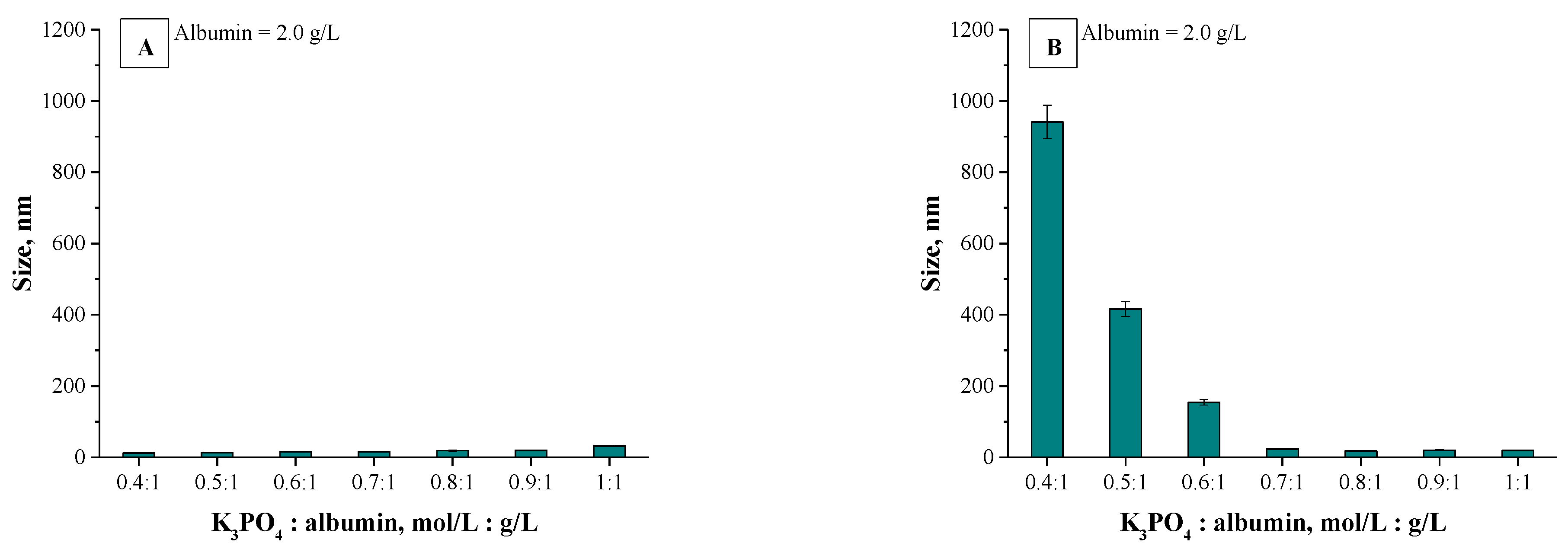
3.2.1. Measurements of Particle Sizes via DLS Method
3.2.2. Morphological Analysis Using Transmission Electron Microscopy (TEM)
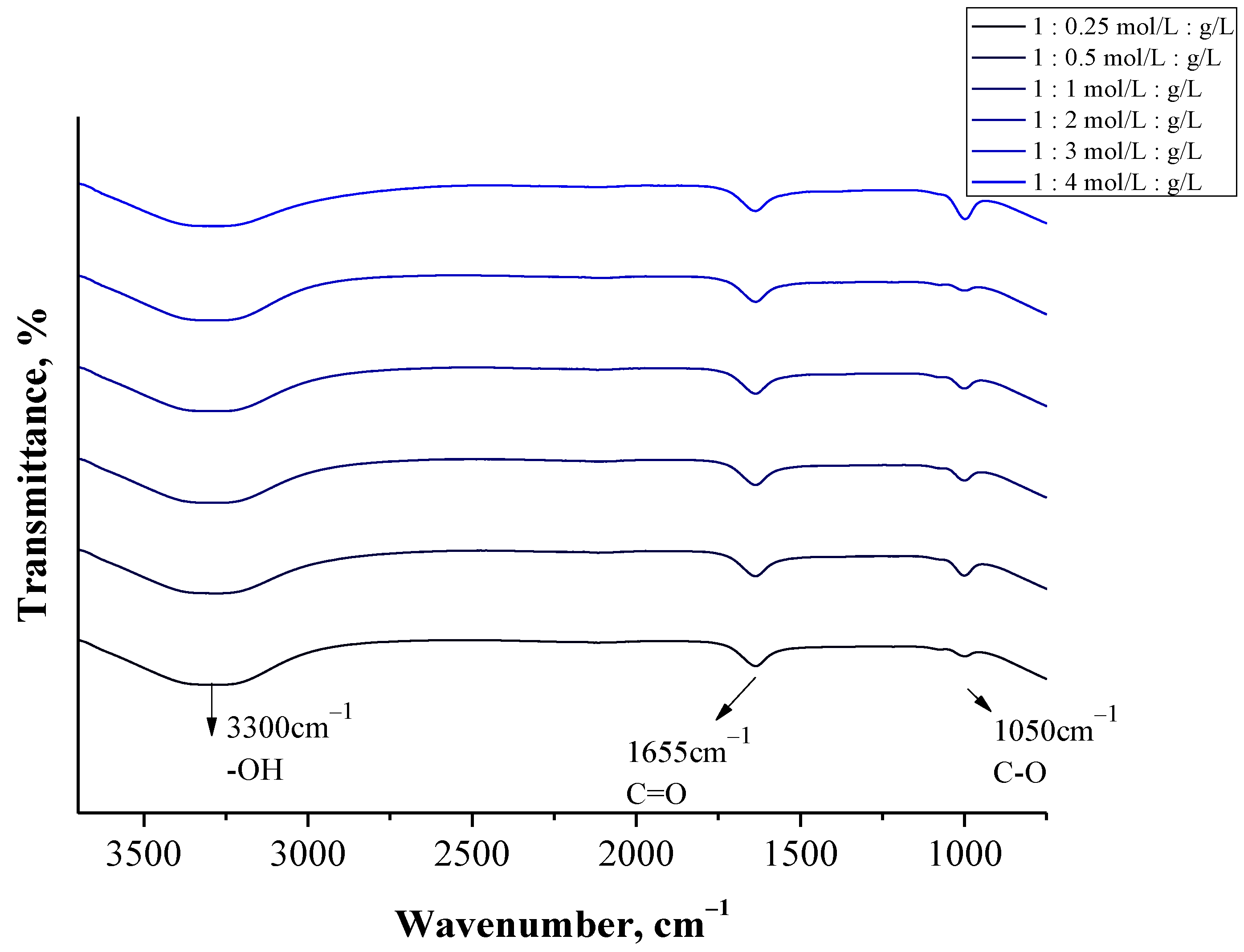
3.2.3. Analysis of Albumin Particles via FT-IR Spectroscopy
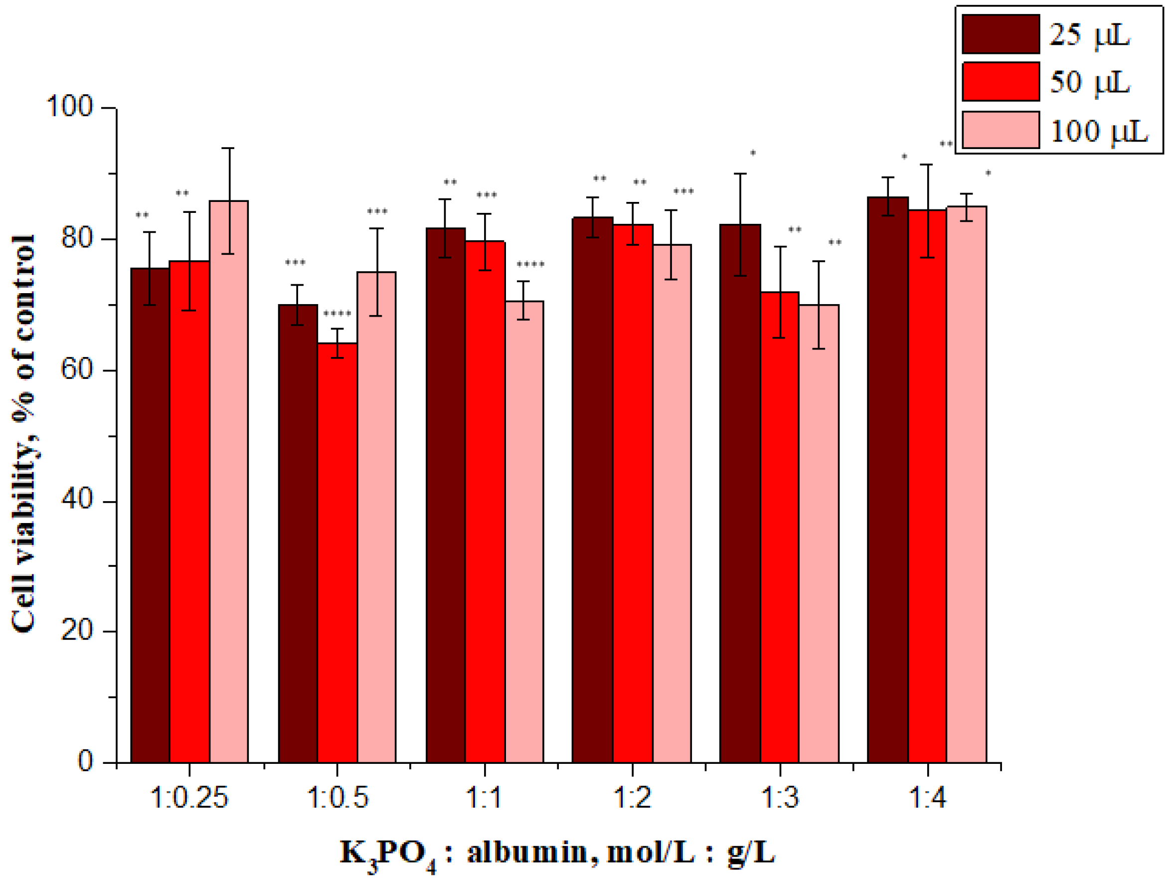
3.2.4. Results of the Investigation on the Cytotoxicity of Albumin Particles via MTT Reduction Assay
3.2.5. Analysis of the Hemolytic Activity of Albumin Particles
4. Conclusions
Supplementary Materials
Author Contributions
Funding
Institutional Review Board Statement
Informed Consent Statement
Data Availability Statement
Acknowledgments
Conflicts of Interest
References
- Yun, Y.H.; Lee, B.K.; Park, K. Controlled Drug Delivery: Historical perspective for the next generation. J. Control Release 2015, 219, 2–7. [Google Scholar] [CrossRef] [Green Version]
- Dutta, R.C. Drug Carriers in Pharmaceutical Design: Promises and Progress. Curr. Pharm. Des. 2007, 13, 761–769. [Google Scholar] [CrossRef] [PubMed]
- Tong, X.; Pan, W.; Su, T.; Zhang, M.; Dong, W.; Qi, X. Recent advances in natural polymer-based drug delivery systems. React. Funct. Polym. 2020, 148, 104501. [Google Scholar] [CrossRef]
- Liechty, W.B.; Kryscio, D.R.; Slaughter, B.V.; Peppas, N.A. Polymers for Drug Delivery Systems. Ann. Rev. Chem. Biomol. Eng. 2020, 1, 149–173. [Google Scholar] [CrossRef] [Green Version]
- Rozga, J.; Piątek, T.; Małkowski, P. Human albumin: Old, new, and emerging applications. Ann. Transplant. 2013, 18, 205–217. [Google Scholar]
- Elzoghby, A.O.; Samy, W.M.; Elgindy, N.A. Albumin-based nanoparticles as potential controlled release drug delivery systems. J. Control Release 2012, 157, 168–182. [Google Scholar] [CrossRef]
- Kratz, F. Albumin as a drug carrier: Design of prodrugs, drug conjugates and nanoparticles. J. Control Release 2008, 132, 171–183. [Google Scholar] [CrossRef]
- Hassanin, I.; Elzoghby, A. Albumin-based nanoparticles: A promising strategy to overcome cancer drug resistance. Cancer Drug Resist. 2020, 3, 930–946. [Google Scholar]
- Rahimizadeh, P.; Yang, S.; Lim, S.I. Albumin: An Emerging Opportunity in Drug Delivery. Biotechnol. Bioprocess Eng. 2020, 25, 985–995. [Google Scholar] [CrossRef]
- Sleep, D. Albumin and its application in drug delivery. Expert Opin. Drug Deliv. 2014, 12, 1–20. [Google Scholar] [CrossRef] [PubMed]
- Elsadek, B.; Kratz, F. Impact of albumin on drug delivery—New applications on the horizon. J. Control Release 2020, 157, 4–28. [Google Scholar] [CrossRef]
- Onafuye, H.; Pieper, S.; Mulac, D.; Conatl, J., Jr.; Wass, M.N.; Langer, K.; Michaelis, M. Doxorubicin-loaded human serum albumin nanoparticles overcome transporter-mediated drug resistance in drug-adapted cancer cells. Beilstein J. Nanotechnol. 2019, 10, 1707–1715. [Google Scholar] [CrossRef] [Green Version]
- Kayani, Z.; Firuzi, O.; Bordbar, A.-K. Doughnut-shaped bovine serum albumin nanoparticles loaded with doxorubicin for overcoming miltidrug-resistant cancer cells. Int. J. Biol. Macromol. 2018, 107, 1835–1843. [Google Scholar] [CrossRef] [PubMed]
- Taneja, N.; Singh, K.K. Rational design of polysorbate 80 stabilized human serum albumin nanoparticles tailored for high drug loading and entrapment of irinotecan. Int. J. Pharm. 2018, 536, 82–94. [Google Scholar] [CrossRef] [Green Version]
- Wang, D.; Liang, N.; Kawashima, Y.; Cui, F.; Yan, P.; Sun, S. Biotin-modified bovine serum albumin nanoparticles as a potential drug delivery system for paclitaxel. J. Mater. Sci. 2019, 54, 8613–8626. [Google Scholar] [CrossRef]
- Kalhor, N.F.; Saeidifar, M.; Ramshini, H.; Saboury, A.A. Interaction, cytotoxicity and sustained release assessment of a novel anti-tumour agent using bovine serum albumin nanocarrier. J. Biomol. Struct. Dyn. 2020, 38, 2546–2558. [Google Scholar] [CrossRef] [PubMed]
- Jithan, A.V.; Madhavi, K.; Prabhakar, K. Preparation and characterization of albumin nanoparticles encapsulating curcumin intended for the teatment of breast cancer. Int. J. Pharm. Investig. 2011, 1, 119–125. [Google Scholar] [CrossRef] [PubMed] [Green Version]
- Karami, E.; Behdani, M.; Kazemi-Lomedasht, F. Albumin nanoparticles as nanocarriers for drug delivery: Focusing on antibody and nanobody delivery and albumin-based drugs. J. Drug Deliv. Sci. Technol. 2020, 55, 101471. [Google Scholar] [CrossRef]
- Loureiro, A.; Azoia, N.G.; Gomes, A.C.; Cavaco-Paulo, A. Albumin-Based Nanodevices as Drug Carriers. Curr. Pharm. Des. 2016, 22, 1371–1390. [Google Scholar] [CrossRef] [PubMed]
- Lee, S.H.; Heng, D.; Ng, W.K.; Chan, H.-K.; Tan, R.B.H. Nano spray drying: A novel method for preparing protein nanoparticles for protein therapy. Int. J. Pharm. 2011, 403, 192–200. [Google Scholar] [CrossRef]
- Fu, Q.; Sun, J.; Zhang, W.; Sui, X.; Yan, Z.; He, Z. Nanoparticle Albumin-Bound (NAB) Technology is a Promising Method for Anti-Cancer Drug Delivery. Recent Pat. Anticancer Drug Discov. 2009, 4, 262–272. [Google Scholar] [CrossRef]
- Hong, S.; Choi, D.W.; Kim, H.N.; Park, C.G.; Lee, W.; Park, H.H. Protein-Based Nanoparticles as Drug Delivery Systems. Pharmaceutics 2020, 12, 604. [Google Scholar] [CrossRef]
- Weber, C.; Coester, C.; Kreuter, J.; Langer, K. Desolvation process and surface characterization of protein nanoparticles. Int. J. Pharm. 2000, 194, 91–102. [Google Scholar] [CrossRef]
- Das, R.P.; Gandhi, V.V.; Singh, B.G.; Kunwar, A.; Kumar, N.N.; Priyadarsini, K.I. Preparation of albumin nanoparticles: Optimum size for cellular uptake of entrapped drug (Curcumin). Coll. Surf. A 2019, 567, 86–95. [Google Scholar] [CrossRef]
- Jahanban-Esfahlan, A.; Dastmalchi, S.; Davaran, S. A simple improved desolvation method for the rapid preparation of albumin nanoparticles. Int. J. Biol. Macromol. 2016, 91, 703–709. [Google Scholar] [CrossRef]
- Zhao, Z.; Li, Y.; Xie, M.-B. Silk Fibroin-Based Nanoparticles for Drug Delivery. Int. J. Mol. Sci. 2015, 16, 4880–4903. [Google Scholar] [CrossRef] [PubMed] [Green Version]
- Lammel, A.S.; Hu, X.; Park, S.-H.; Kaplan, D.L.; Scheibel, T.R. Controlling silk fibroin particle features for drug delivery. Biomaterials 2010, 31, 4583–4591. [Google Scholar] [CrossRef] [Green Version]
- Saeki, K.; Kunito, T.; Sakai, M. Effect of Tris-HCl buffer on DNA adsorption by a variety of soil constituents. Microbes Environ. 2011, 26, 88–91. [Google Scholar] [CrossRef] [Green Version]
- El-Ashram, S.; Nasr, I.A.; Suo, X. Nucleic acid protocols: Extraction and optimization. Biotechnol. Rep. 2016, 12, 33–39. [Google Scholar] [CrossRef] [Green Version]
- Novak, P.; Havlicek, V. Protein Extraction and Precipitation. In Proteomic Profilling and Analytical Chemistry; Ciborowski, P., Silberring, J., Eds.; Elsevier: New York, NY, USA, 2016; pp. 51–62. [Google Scholar]
- Dumetz, A.C.; Snellinger-O’Brien, A.M.; Kaler, E.W.; Lenhoff, A.M. Patterns of protein-protein interactions in salt solutions and implications for protein crystallization. Protein Sci. 2007, 16, 1867–1877. [Google Scholar] [CrossRef]
- Hyde, A.M.; Zultanski, S.L.; Waldman, J.H.; Zhong, Y.-L.; Shevlin, M.; Peng, F. General Principles and Strategies for Salting-Out Informed by the Hofmeister Series. Org. Process Res. Dev. 2017, 21, 1355–1370. [Google Scholar] [CrossRef] [Green Version]
- Omana, D.A.; Wu, J. Effect of different concentrations of calcium chloride and potassium chloride on egg white proteins during isoelectric precipitation of ovomucin. Poult. Sci. 2009, 88, 2224–2234. [Google Scholar] [CrossRef] [PubMed]
- Filipczak, N.; Jaromin, A.; Piwoni, A.; Mahmud, M.; Sarisozen, C.; Torchilin, V.; Gubernator, J. A Triple Co-Delivery Liposomal Carrier That Enhances Apoptosis via an Intrinsic Pathway in Melanoma Cells. Cancers 2019, 11, 1982. [Google Scholar] [CrossRef]
- Mosmann, T. Rapid colorimetric assay for cellular growth and survival: Application to proliferation and cytotoxicity assays. J. Immunol. Methods 1983, 65, 55–63. [Google Scholar] [CrossRef]
- Jaromin, A.; Korycińska, M.; Piętka-Ottlik, M.; Musiał, W.; Peczyńska-Czoch, W.; Kaczmarek, Ł.; Kozubek, A. Membrane perturbations induced by new analogs of neocryptolepine. Biol. Pharm. Bull. 2012, 35, 1432–1439. [Google Scholar] [CrossRef] [Green Version]
- Duong-Ly, K.C.; Gabelli, S.B. Salting out of proteins using ammonium sulfate precipitation. In Methods in Enzymology; Pecoraro, V.L., Ed.; Academic Press: Cambridge, MA, USA, 2014; pp. 85–94. [Google Scholar]
- Soper, A.K.; Weckstrom, K. Ion solvation and water structure in potassium halide aqueous solutions. Biophys. Chem. 2006, 124, 180–191. [Google Scholar] [CrossRef]
- Fogarty, A.C.; Laage, D. Water Dynamics in Protein Hydration Shells: The Molecular Origins of the Dynamical Perturbation. J. Phys. Chem. B 2014, 118, 7715–7729. [Google Scholar] [CrossRef] [PubMed]
- Großhans, S.; Wang, G.; Hubbuch, J. Water on hydrophobic surfaces: Mechanistic modeling of polyethylene glycol-induced protein precipitation. Bioprocess Biosyst. Eng. 2019, 42, 513–520. [Google Scholar] [CrossRef] [Green Version]
- Shaw, K.L.; Grimsley, G.R.; Yakovlev, G.I.; Makarov, A.A.; Pace, C.N. The effect of net charge on the solubility, activity, and stability of ribonuclease Sa. Protein Sci. 2001, 10, 1206–1215. [Google Scholar] [CrossRef] [Green Version]
- Allard, E.; Passirani, C.; Benoit, J.-P. Convection-enhanced delivery of nanocarriers for the treatment of brain tumors. Biomaterials 2009, 30, 2302–2318. [Google Scholar] [CrossRef]
- Bhaw-Luximon, A.; Goonoo, N.; Jhurry, D. Nanotherapeutics promises for colorectal cancer and pancreatic ductal adenocarcinoma. In Nanobiomaterials in Cancer Therapy; Grumezescu, A.M., Ed.; Elsevier: New York, NY, USA, 2016; pp. 147–201. [Google Scholar]
- Rani, K. Fourier Transform Infrared Spectroscopy (FTIR) spectral analysis of BSA nanoparticles (BSA NPs) and egg albumin nanoparticles (EA NPs). Res. J. Chem. Sci. 2016, 6, 29–36. [Google Scholar]
- Mahobia, S.; Bajpai, J.; Bajpai, A.K. An in-vitro investigation of swelling controlled delivery of insulin from egg albumin nanocarriers. IJRP 2016, 15, 695–711. [Google Scholar]
- Abrosimova, K.V.; Shulenina, O.V.; Paston, S.V. FTIR study of secondary structure of bovine serum albumin and ovalbumin. J. Phys. Conf. Ser. 2016, 769, 012016. [Google Scholar] [CrossRef]
- Jaiswal, V.D.; Dongre, P.M. Biophysical interactions between silver nanoparticle-albumin interface and curcumin. J. Pharm. Anal. 2020, 10, 164–177. [Google Scholar] [CrossRef]
- Ranjan, S.; Dasgupta, N.; Srivastava, P.; Ramalingam, C. A spectroscopic study on interaction between bovine serum albumin and titanium dioxide nanoparticle synthesized from microwave-assisted hybrid chemical approach. J. Photochem. Photobiol. B Biol. 2016, 161, 472–481. [Google Scholar] [CrossRef]
- Usoltsev, D.; Sitnikova, V.; Kajava, A.; Uspenskaya, M. Systematic FTIR Spectroscopy Study of the Secondary Structure Changes in Human Serum Albumin under Various Denaturation Conditions. Biomolecules 2019, 9, 359. [Google Scholar] [CrossRef] [PubMed] [Green Version]
- Vecchione, R.; Quagliariello, V.; Calabria, D.; Calcagno, V.; De Luca, E.; Iaffaioli, R.V.; Netti, P.A. Curcumin bioavailability from oil in water nano-emulsions: In vitro and in vivo study on the dimensional, compositional and interactional dependence. J. Control Release 2016, 233, 88–100. [Google Scholar] [CrossRef]
- Jaromin, A.; Parapini, S.; Basilico, N.; Zaremba-Czogalla, M.; Lewińska, A.; Zagórska, A.; Walczak, M.; Tyliszczak, B.; Grzeszczak, A.; Łukaszewicz, M.; et al. Azacarbazole n-3 and n-6 polyunsaturated fatty acids ethyl esters nanoemulsion with enhanced efficacy against Plasmodium falciparum. Bioact. Mater. 2020, 24, 1163–1174. [Google Scholar]
- Zaremba-Czogalla, M.; Jaromin, A.; Sidoryk, K.; Zagórska, A.; Cybulski, M.; Gubernator, J. Evaluation of the In Vitro Cytotoxic Activity of Caffeic Acid Derivatives and Liposomal Formulation against Pancreatic Cancer Cell Lines. Materials 2020, 19, 5813. [Google Scholar] [CrossRef]
- Salehiabar, M.; Nosrati, H.; Javani, E.; Aliakbarzadeh, F.; Kheiri Manjili, H.; Davaran, S.; Danafar, H. Production of biological nanoparticles from bovine serum albumin as controlled release carrier for curcumin delivery. Int. J. Biol. Macromol. 2018, 115, 83–89. [Google Scholar] [CrossRef]
- Nosrati, H.; Rakhshbahar, A.; Salehiabar, M.; Afroogh, S.; Kheiri Manjili, H.; Danafar, H.; Davaran, S. Bovine serum albumin: An efficient biomacromolecule nanocarrier for improving the therapeutic efficacy of chrysin. J. Mol. Liq. 2018, 271, 639–646. [Google Scholar] [CrossRef]
- Scutera, S.; Argenziano, M.; Sparti, R.; Bessone, F.; Bianco, G.; Bastiancich, C.; Castagnoli, C.; Stella, M.; Musso, T.; Cavalli, R. Enhanced Antimicrobial and Antibiofilm Effect of New Colistin-Loaded Human Albumin Nanoparticles. Antibiotics 2021, 10, 57. [Google Scholar] [CrossRef]
- Avalos, A.; Haza, A.; Mateo, D.; Morales, P. Interactions of manufactured silver nanoparticles of different sizes with normal human dermal fibroblasts. Int. Wound. J. 2016, 13, 101–109. [Google Scholar] [CrossRef]
- Qu, N.; Lee, R.J.; Sun, Y.; Cai, G.; Wang, J.; Wang, M.; Lu, J.; Meng, Q.; Teng, L.; Wang, D.; et al. Cabazitaxel-loaded human serum albumin nanoparticles as a therapeutic agent against prostate cancer. Int. J. Nanomed. 2016, 26, 3451–3459. [Google Scholar]
- Ghosh, P.; Singha Roy, A.; Chaudhury, S.; Jana, S.K.; Chaudhury, K.; Dasgupta, S. Preparation of albumin based nanoparticles for delivery of fisetin and evaluation of its cytotoxic activity. Int. J. Biol. Macromol. 2016, 86, 408–417. [Google Scholar] [CrossRef]


| Sample | Albumin Solvent | Salting-Out Agent | Salting-Out Agent Concentration [mol/L] | Observations |
|---|---|---|---|---|
| 1 | Tris-HCl (pH = 4.8) | CaCl2 | 1 | No changes |
| 2 | 2 | No changes | ||
| 3 | KH2PO4 | 1 | No changes | |
| 4 | 2 | No changes | ||
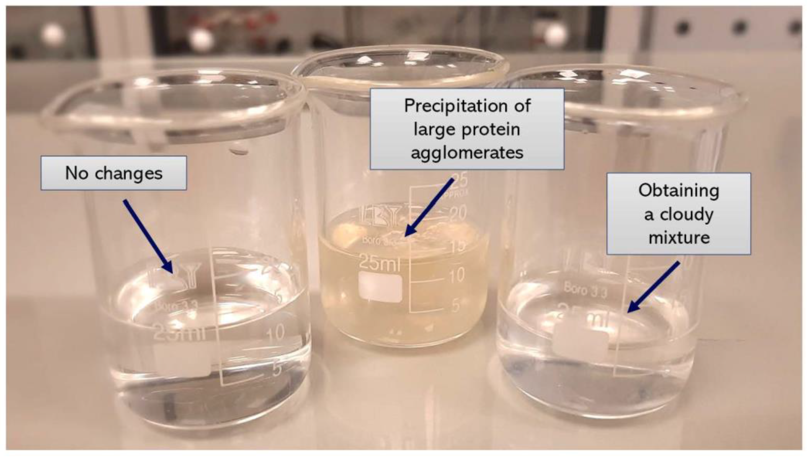
| 5 | K3PO4 | 1 | Precipitation of large protein agglomerates | |
| 6 | 2 | Precipitation of large protein agglomerates | ||
| 7 | Tris-HCl (pH = 7) | CaCl2 | 1 | No changes |
| 8 | 2 | No changes | ||
| 9 | KH2PO4 | 1 | No changes | |
| 10 | 2 | No changes | ||
| 11 | K3PO4 | 1 | Obtaining a cloudy mixture | |
| 12 | 2 | Obtaining a cloudy mixture |
| Sample | Potassium Phosphate Concentration *: Albumin Concentration RATIO (mol/L: g/L) |
| 1 | 1:0.25 |
| 2 | 1:0.5 |
| 3 | 1:1 |
| 4 | 1:2 |
| 5 | 1:3 |
| 6 | 1:4 |
| Sample | Potassium Phosphate Concentration: Albumin Concentration ** RATIO (mol/L: g/L) |
| 7 | 0.4:1 |
| 8 | 0.5:1 |
| 9 | 0.6:1 |
| 10 | 0.7:1 |
| 11 | 0.8:1 |
| 12 | 0.9:1 |
| 13 | 1:1 |
| Sample | Potassium Phosphate Concentration *: Albumin Concentration RATIO (mol/L: g/L) | PDI (Syringe System) | PDI (Burette) |
| 1 | 1:0.25 | 0.13 ± 0.021 | 0.09 ± 0.024 |
| 2 | 1:0.5 | 0.15 ± 0.015 | 0.10 ± 0.016 |
| 3 | 1:1 | 0.12 ± 0.018 | 0.13 ± 0.011 |
| 4 | 1:2 | 0.85 ± 0.022 | 0.14 ± 0.012 |
| 5 | 1:3 | 0.79 ± 0.013 | 0.08 ± 0.022 |
| 6 | 1:4 | 0.98 ± 0.017 | 0.06 ± 0.014 |
| Sample | Potassium phosphate concentration: Albumin concentration ** RATIO (mol/L: g/L) | ||
| 7 | 0.4:1 | 0.18 ± 0.018 | 0.16 ± 0.011 |
| 8 | 0.5:1 | 0.13 ± 0.022 | 0.13 ± 0.018 |
| 9 | 0.6:1 | 0.14 ± 0.011 | 0.09 ± 0.023 |
| 10 | 0.7:1 | 0.17 ± 0.017 | 0.11 ± 0.022 |
| 11 | 0.8:1 | 0.20 ± 0.023 | 0.12 ± 0.010 |
| 12 | 0.9:1 | 0.19 ± 0.014 | 0.13 ± 0.013 |
| 13 | 1:1 | 0.12 ± 0.019 | 0.13 ± 0.019 |
Publisher’s Note: MDPI stays neutral with regard to jurisdictional claims in published maps and institutional affiliations. |
© 2021 by the authors. Licensee MDPI, Basel, Switzerland. This article is an open access article distributed under the terms and conditions of the Creative Commons Attribution (CC BY) license (https://creativecommons.org/licenses/by/4.0/).
Share and Cite
Kudłacik-Kramarczyk, S.; Drabczyk, A.; Głąb, M.; Gajda, P.; Czopek, A.; Zagórska, A.; Jaromin, A.; Gubernator, J.; Makara, A.; Tyliszczak, B. The Development of the Innovative Synthesis Methodology of Albumin Nanoparticles Supported by Their Physicochemical, Cytotoxic and Hemolytic Evaluation. Materials 2021, 14, 4386. https://doi.org/10.3390/ma14164386
Kudłacik-Kramarczyk S, Drabczyk A, Głąb M, Gajda P, Czopek A, Zagórska A, Jaromin A, Gubernator J, Makara A, Tyliszczak B. The Development of the Innovative Synthesis Methodology of Albumin Nanoparticles Supported by Their Physicochemical, Cytotoxic and Hemolytic Evaluation. Materials. 2021; 14(16):4386. https://doi.org/10.3390/ma14164386
Chicago/Turabian StyleKudłacik-Kramarczyk, Sonia, Anna Drabczyk, Magdalena Głąb, Paweł Gajda, Anna Czopek, Agnieszka Zagórska, Anna Jaromin, Jerzy Gubernator, Agnieszka Makara, and Bożena Tyliszczak. 2021. "The Development of the Innovative Synthesis Methodology of Albumin Nanoparticles Supported by Their Physicochemical, Cytotoxic and Hemolytic Evaluation" Materials 14, no. 16: 4386. https://doi.org/10.3390/ma14164386
APA StyleKudłacik-Kramarczyk, S., Drabczyk, A., Głąb, M., Gajda, P., Czopek, A., Zagórska, A., Jaromin, A., Gubernator, J., Makara, A., & Tyliszczak, B. (2021). The Development of the Innovative Synthesis Methodology of Albumin Nanoparticles Supported by Their Physicochemical, Cytotoxic and Hemolytic Evaluation. Materials, 14(16), 4386. https://doi.org/10.3390/ma14164386