Review

Peptides and Peptidomimetics as Inhibitors of Enzymes Involved in Fibrillar Collagen Degradation

1
Department of Bioorganic Chemistry, Faculty of Chemistry, Wroclaw University of Science and Technology, 50-370 Wroclaw, Poland
2
Interdepartmental Research Unit of Peptide and Protein Chemistry and Biology, Department of Neurosciences, Psychology, Drug Research and Child Health-Section of Pharmaceutical Sciences and Nutraceutics, University of Florence, 50019 Sesto Fiorentino, Firenze, Italy
3
Interdepartmental Research Unit of Peptide and Protein Chemistry and Biology, Department of Chemistry “Ugo Schiff”, University of Florence, 50019 Sesto Fiorentino, Firenze, Italy
*
Authors to whom correspondence should be addressed.
Materials 2021, 14(12), 3217; https://doi.org/10.3390/ma14123217
Submission received: 5 May 2021 / Revised: 28 May 2021 / Accepted: 7 June 2021 / Published: 10 June 2021

Abstract

:
Collagen fibres degradation is a complex process involving a variety of enzymes. Fibrillar collagens, namely type I, II, and III, are the most widely spread collagens in human body, e.g., they are responsible for tissue fibrillar structure and skin elasticity. Nevertheless, the hyperactivity of fibrotic process and collagen accumulation results with joints, bone, heart, lungs, kidneys or liver fibroses. Per contra, dysfunctional collagen turnover and its increased degradation leads to wound healing disruption, skin photoaging, and loss of firmness and elasticity. In this review we described the main enzymes participating in collagen degradation pathway, paying particular attention to enzymes degrading fibrillar collagen. Therefore, collagenases (MMP-1, -8, and -13), elastases, and cathepsins, together with their peptide and peptidomimetic inhibitors, are reviewed. This information, related to the design and synthesis of new inhibitors based on peptide structure, can be relevant for future research in the fields of chemistry, biology, medicine, and cosmeceuticals.

1. Introduction

Collagen is one of the most essential proteins in the human body and a major component of the extracellular matrix (ECM) [1]. There are 28 classified collagen types, of which collagen type I forms around 85%, with up to 15% formed by type III [2,3]. Type I composes mainly the tissue fibrillar structure, while type III functions as the linchpin of type I fibres and supports skin elasticity. Collagen, in general, represents approximately one-third of all human body proteins, being the most abundant protein-like body component [4]. On the basis of molecular structure, collagen is classified into three morphological forms, namely fibrillar, non-fibrillar, and network-forming collagens [5].
Three polypeptide collagen molecules spontaneously form a helical structure, so-called tropocollagen. A single chain (α chain) has usually the length of about 1000 residues [6]. Tropocollagen is stabilized by covalent bonds and hydrogen interactions between hydroxyproline (HyP) and hydroxylysine (HyK) [2]. It was observed that tropocollagen superhelix is visibly more constrained, with the rise per residue value of 0.29 nm comparing to 0.36 nm for common proteins [7]. This feature makes it exceptionally resistant to the widespread proteolytic digestion, making collagen susceptible only to a few enzymes [8]. Remarkable regularity of repeated amino acid triad, Gly-X-Y (where X—proline, Pro; Y—hydroxyproline, HyP), provides a variety of beneficial properties—for instance, stability and resistance to distortion. Glycine replacement by larger and more branched residues provokes inaccurate folding and decreases helix resistance to external conditions, i.e., temperature [7].
The essential role played by collagen in the human body has a relevant influence on many physiological, as well as pathological states. Collagen turnover is the complex and interconnected process of collagen biosynthesis and degradation. In physiological states, the degradative pathway acts as a regulator of collagen deposition, thus preventing various fibrotic disorders. Moreover, a proper intracellular collagen degradation prevents the secretion of defective sequences [9]. Accordingly, the control of collagen homeostasis is one of the major factors in bleeding syndromes therapies [10], while hyperactivity of fibrotic process and collagen accumulation results with joints, bone, heart, lungs, kidneys or liver fibroses [4,11,12]. Per contra, dysfunctional collagen turnover and its increased degradation lead to wound healing disruption, skin photoaging, and loss of skin firmness and elasticity [13]. The latter aspects are particularly relevant in the field of aesthetic medicine and cosmetics, to which we will pay special attention.
In this review we will briefly describe the collagen turnover process in relevant cells and tissues, indicating the main degradation pathways in particular and underlining the key enzymes involved in these mechanisms. We will review peptide-based inhibitors of collagenases, elastase, and cathepsin, mentioning also some relevant non-peptide compounds. The final aim is to give a valuable starting point for future research, providing chemical and biological basis for the design and the development of new peptide-based inhibitors, potentially applicable as cosmeceuticals.

2. Collagen Turnover in Cells

In this section, we briefly describe the collagen turnover process in relevant cells, in order to clarify its biosynthesis and degradation pathways. This complex process has been discussed in details in the following reviews: Rodriguez-Feo et al. [14], Gelse et al. [15], Smith and Rennie [4], Sprangers and Everts [12], together with the review by Vannella and Wynn, depicting the wound healing process and tissue remodelling [16].
Each fibrillar-type collagen is synthesised in fibroblasts in the form of procollagen, consisting of two N- and C-terminal propeptide domains [12,15]. There is a spectrum of already known collagen synthesis stimulators, such as Transforming Growth Factor beta (TGF-β), Endothelin-1 (ET-1), Angiotensin-II (Ang II), Platelet-Derived Growth Factor B (PDGF-BB), Interleukin-1 (IL-1), Aldosterone, Interferon g (IFN-g), Basic Fibroblast Growth Factor (bFGF) and Nitric Oxide (NO) [14]. As indicated previously, proline residues play a pivotal role in the collagen structure formation. At this point, Pro undergoes the hydroxylation by prolyl-3- and -4-hydroxylases, together with lysyl-hydroxylase acting on Lys [1]. Then, fibrillar procollagens are secreted into the extracellular space. Afterwards, terminal noncollagenous fragments are removed via enzymatic cascade, thus enabling the spontaneous formation of fibrils. This process, called fibrillogenesis, is regulated by collagen type V and this assembling is favoured by covalent cross-linking between lysine side chains [1,12].
As far as degradation is concerned, the tenor of collagen disruption is strictly connected to its type. Mainly, there are two recognized categories of these pathways: extracellular and intracellular. Extracellular degradation includes the cleavage of mature collagen, while the intracellular mechanism degrades the procollagen chains, before their assembling into procollagen molecules [17]. These processes prevent the formation of defective collagen and its incorporation into the extracellular matrix.
The extracellular pathway is due to secreted proteolytic enzymes, capable to digest collagen fibres. Various enzyme classes participate in this process, i.e., matrix metalloproteases (MMPs), serine proteases, active at neutral pH, as well as cysteine, aspartate, and threonine proteases, requiring mainly acidic environment [18]. MMPs target a broad range of proteins, depending on the enzyme type. For example, stromelysins (MMP-3 and -10) act preferentially on proteoglycans, fibronectin, and laminin [18]. Other MMPs, such as gelatinases, cleaves already denaturated collagen only. ECM modulation is affected also by the so-called A Disintegrin and Metalloproteinase (ADAM) and ADAM with Thrombospondin Motifs (ADAMTS) family [18,19]. The extracellular degradation process and the ECM dynamics, have been reviewed by Lu et al. in 2011 [18].
The intracellular mechanism follows the internalization of collagen fragments, previously partially degraded in the ECM [12]. The kinetics of this process was found to be very rapid [20,21]. Time and percentage of degradation depends on the tissue type, revealing significant differences between, e.g., skin and heart. Two intracellular localizations of collagen degradation have been proposed: one within the lysosome and the second within the cisternae (in the endoplasmic reticulum or Golgi apparatus).
In this review, we will put a particular attention on the extracellular breakdown pathway, describing the most substantial enzymes involved in this process. Table 1 presents a list of enzymes participating in the degradation, with reference to the relevant collagen class. Figure 1 illustrates the influence of various MMPs on extracellular collagen breakdown. Other factors indirectly stimulating collagen disruption are the following [13]: (1) ultraviolet radiation, inducing MMPs and enhancing their proteolytic action; (2) age, due to the overactive secretion of proteases observed in elder skin, together with incorrect (or diminished) fibroblasts activity [2]; (3) sex, in view of an inhibitory effect of oestrogen on collagen synthesis [22]. Two interesting papers, describing the influence of menstrual cycle on collagen synthesis, were published in 2006 and 2007. These investigations did not show any significant change between luteal or follicular phases. However, some differences between male and female collagen synthesis and remodelling were observed [23,24,25].
In this review we will focus on enzymes degrading mainly fibrillar collagen, leading to loss of skin elasticity and skin aging in general, due to their cosmeceutical interest, i.e., collagenases (MMP-1, 8, and 13), elastase, and cathepsins, together with their inhibitors. It must be highlighted that there are two pathways of enzyme activity control—direct (inhibition) and indirect (up or downregulation of enzymes expression) [42]. The following paper will consider only inhibitors directly affecting enzymes activity.

2.1. Methods of Collagen Turnover Measurements

As stated above, there are two interrelated pathways, contributing to collagen degradation in cells: intracellular (based on lysosomal digestion) and extracellular (initially engaging collagenases and later on also other proteases) (Scheme 1) [4]. Due to their correlated activity, cellular collagen turnover measurement, including both biosynthesis and degradation, became a challenge. Nevertheless, there are a few methods directly and indirectly evaluating this factor. These discussed in our review are summarized in Scheme 2.

2.1.1. Indirect Methods

Collagen synthesis can be followed by the rate of appearance of specific peptides, deriving from procollagen: (1) PICP, C-terminal pro-peptide from collagen type I; (2) PINP, N-terminal pro-peptide from collagen type I; (3) Pro-C3, internal epitope in the N-terminal pro-peptide from collagen type I; (4) P4NP7S, internal epitope in type IV collagen 7S domain [4,25].
Degradation process can be monitored by the measurement of collagen breakdown markers, such as: (1) NTX and CTX, crosslinked telopeptides of type I collagen; (2) (deoxy)pyridinoline, pyridinium crosslinkers of type I collagen; (3) C1M, neo-epitope of MMP-2,9,13- mediated collagen type I degradation; (4) C3M, neo-epitope of MMP-9- mediated collagen type III degradation; (5) C4M, neo-epitope of MMP-2, -9, -12-mediated collagen type IV (α1 chain) degradation [4,25]. In order to estimate collagen degradation products, Nielsen et al. measured the CTX-II, CTX-I, and hydroxyproline content in protein extract from joints [43]. Therefore, they indicated “an assessment of collagen degradation epitopes as a quantitative measure of cartilage damage”.
Veidal et al. in 2011 described the applicability of ELISA assay in measuring a degradation fragment specific to the type IV collagen, namely GTPSVDHGFL [44]. This neoepitope is generated during MMP-induced degradation of collagen and was associated with liver fibrosis in animal models.
Smith and Rennie, who listed in their review several of the above-mentioned methods, put attention on the applicability of indirect measurements [4], underlining that using these methods also some procollagen can be measured. Therefore, they are semi-qualitative and cannot define a precise content of mature collagen in a probe.

2.1.2. Direct Methods

One collagen monomer contains approximately 13% of hydroxyproline residues. Starting from this observation, Hyp excretion in urine has been used as a collagen degradation indicator. In 2005, McAnulty proposed the application of radiolabelled Hyp and Pro for in vitro and in vivo determination of collagen synthesis and degradation [45]. Moreover, the activity of prolyl-4-hydroxylase can be evaluated as an index of collagen synthesis. This enzyme catalyses the proline hydroxylation, one of the initial steps in collagen post-translational processing [46]. The reaction takes place exclusively during the collagen biosynthesis, therefore can be employed for the above-mentioned purpose. Gorres and Raines in 2010 described for the first time a relatively simple and direct assay for prolyl-4-hydroxylase, based on a detection of the Pro-containing substrate turnover [47].

2.1.3. Others

Widely described methods of collagen turnover monitoring include the use of radiolabelled atoms, e.g., Pro with 3H, 14C; exposure to 18O2 during the Pro hydroxylation; heavy water D2O administration (followed by deuterium incorporation into Ala and Pro).
In 2018, Cipriani et al. described a quantitative detection of soluble collagen by western blot technique, similar to the protocol previously published by Poobalarahi et al. [27,48]. Briefly, fibroblast cell cultures were treated with trypsin in order to analyse the intracellular forms of collagen. The presence and intensity of bands corresponding to procollagen were then observed in cell lysates.

3. Inhibitors of Collagen-Degrading Enzymes

Fibrillar collagen is responsible for skin elasticity, firmness, and overall wellness and good appearance. In the field of cosmeceuticals, being on the border between traditional cosmetics and drug-like products, the claimed effect of a given cosmetic product should be connected with proven efficacy and activity of its active ingredient(s) [49]. Therefore, collagen degradation inhibitors and collagen biosynthesis enhancers are highly requested and broadly investigated in the recent years. Among other classes of active ingredients, peptides play an important role in these fields, due to their high specificity, low toxicity, relatively easy synthesis, and applicability as carriers for other molecules [50]. However, peptides in some cases get degraded very quickly on the skin surface, or are not able to penetrate sufficiently into the skin layers [51]. In the aim to enhance peptides stability and efficacy as bioactive ingredients, the use of peptidomimetics is increasingly considered as a viable alternative to unmodified peptides.
In general, two mechanisms of inhibition can be identified: covalent and non-covalent binding pathways [52] (Figure 2). The covalent mechanism is based on specific binding to the target protein, with formation of a permanent complex with the enzyme. On the other hand, non-covalent inhibition assumes the semi-permanent interaction between enzyme and inhibitor, through cyclic binding, unbinding, and rebinding [53]. Comparing both these pathways, covalent inhibition is advantageous over the non-covalent one. The presence of the inhibitor warhead allows the formation of particular interactions that leads to the development of highly specific inhibitors. Cysteine, serine, threonine or lysine residues participate in covalent bonds formation due to their nucleophilic character; hydroxyl, epoxy or carbonyl moiety in the ligand structure are usually responsible for the arrangement of these covalent adducts [54].

3.1. Collagenases

MMPs inhibitors (so-called MMPIs) have been investigated for many years, with the first publications from 1980. The first synthetically obtained inhibitors were characterized by functional groups able to coordinate a zinc(II) ion in MMPs active site [55]. Relatedly, four classes of zinc MMPIs can be designated: (1) hydroxamates (CONH-O); (2) carboxylates (COO); (3) thiolates (S); and (4) phosphinyls (PO2−) [56]. Recently, some commercially developed MMPIs were submitted to clinical trials, such as batimastat, marimastat, and others [55]. These two were then approved as drugs [57]. Despite many efforts in this field, selective inhibitors of specific MMPs have not been found yet. This is probably due to the complicated mechanism of MMPs proteolytic activity, and mutual interactions of many factors, leading together to the inhibition process [56]. This mechanism has been studied for years, among others by Moroder and his group, which reported their results in 2000 [58]. They examined the substrate specificity and preferences of collagenases and gelatinases, using synthetic peptides derived from collagen. It was clearly indicated that the inhibitory activity of both classes has different basis, but since the activity of all MMPs is zinc-dependent, compounds containing chelating groups are generally good, but weakly selective MMPIs. More detailed structural data and widely described catalytic activity of metalloproteases, are described in two papers by Bode and Maskos [59,60].
Studies focused on structure-activity relationship (SAR) of MMP-13 inhibitors were described by Roush et al., providing relevant indications for further developments [61,62]. In addition, both papers include the evaluation of amino acids derivatives, which will be further described in Section 3.1.4.

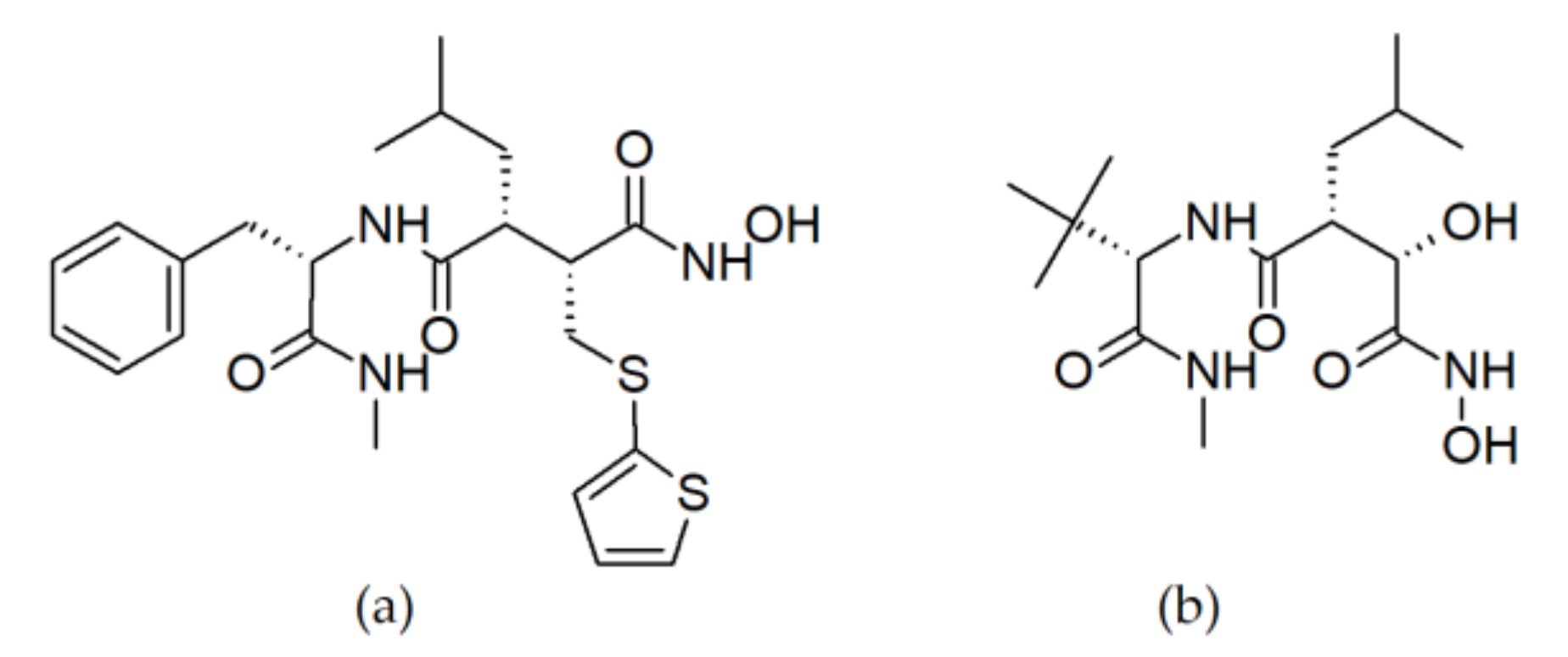
3.1.1. Peptide Hydroxamates

To the best of our knowledge, the very first paper describing the hydroxamate group (CONH-O) as an efficient inhibitor of collagenases was published in 1986 by Moore and Spilburg [63]. They studied the influence on skin fibroblasts collagenases activity of various peptide sequences, with diverse C-terminal groups (amide, carboxylate, aldehyde, and hydroxamate). Comparing the percentage of inhibition among amastatin, captopril, phosphoramidon, and zincov (standard metalloproteases inhibitors; structures are reported in Table 2) in 0.5 mM concentration, it was proven that only zincov was able to decrease the collagenases activity. Afterwards, a few sequences of hydroxamic acid derivatives were synthesised, in order to evaluate the influence of their length on inhibition efficacy. The lowest IC50 value was reported for Z-Pro-Leu-Gly-NHOH (0.04 mM), while shortening the sequence yielded less-active inhibitors, achieving IC50 = 40 mM for acetohydroxamic acid. It was deduced that collagenases distinguished the sequence including Pro due to its similarity to the cleavage site in native collagen, recognized by the enzyme. Finally, to determine the role of hydroxamate in the inhibition mechanism, Z-Pro-Leu-Gly-X derivatives were investigated (where X indicated hydroxamate, carboxylate, aldehyde, and amide modification on C-terminus). The results obtained for the peptide sequence with unmodified glycine residue showed the importance of this sequence in the inhibition mechanism. The C-terminally merged hydroxamate led to satisfactory results (CM = 5.3 mM and 15% of inhibition for carboxylate; CM = 0.115 mM and 70% of inhibition for hydroxamate derivative).
In 1992, Grobelny et al. confirmed the applicability of peptide hydroxamic acids as collagenase inhibitors [64]. HONHCOCH2CH(i-Bu)CO-Trp-NHMe sequence isomers (L,L-dipeptide 6A (R,S) and D,L-dipeptide 6B (S,S)) were designed and synthesised to evaluate the efficacy against human skin fibroblast collagenase, thermolysin and Pseudomonas aeruginosa elastase. the structural difference between 6A and 6B consists in the CH2CH(i-Bu)CO α-carbon configuration, thus 6A is in the L-, and 6B in the D-configuration. Surprisingly, these diastereomers distinguished very evidently the discrepancy between inhibitory preferences among human (collagenase) and bacterial (thermolysin and elastase) enzymes. It was confirmed that the 6A inhibits collagenase with Ki = 0.4 nM; in the case of 6B, this value was of 200 nM. On the contrary, bacterial enzymes were inhibited with comparable potency by both 6A (Ki = 20 nM for each protein) and 6B (Ki = 7 nM and 2 nM for thermolysin and elastase, respectively). To confirm the role of hydroxamate group in the inhibitory mechanism, similarly to the previous paper, hydroxamic acid moiety was replaced with carboxylate and hydrazide. The Ki values for all of the investigated enzymes increased from the nM to μM–mM range. Altogether, the configurational difference between 6A and 6B, both with hydroxamate moiety, enabled to distinguish the preferential interactions between human and bacterial enzymes.
Another good example of hydroxamates as collagenases inhibitors was presented in 1995 by Grams et al. [65]. They described X-ray structures of complexes between two peptide sequences and collagenase. This research was a follow-up of their previous work, in which the X-ray structure of collagenase with Pro-Leu-Gly-NHOH was examined [66]. Their observations led to the synthesis and evaluation of thiol inhibitor HSCH2CH(CH2Ph)CO-L-Ala-Gly-NH2 and hydroxamate inhibitor HONCOCH(iBu)CO-L-Ala-Gly-NH2. Both functional groups bind to the catalytic zinc ion, however the hydroxamate inhibitor binds differently from the geometrical preferences revealed by the substrate. The calculated Ki values are: 19 μM for Pro-Leu-Gly-NHOH; 1.2 μM for thiol-modified sequence; 33 μM for hydroxamate peptidomimetic.
Good examples of widely studied hydroxamates, batimastat (BB-94) and marimastat (BB-2516) (Figure 3), have to be mentioned in this section. Both molecules were approved as drugs after extended clinical trials, with relevant indications mostly in cancer therapies. Rasmussen et al. have published a very interesting review comparing the two substances and summarizing clinical data [67].
Batimastat binds covalently to Zn(II) ion in the MMPs active site. It exhibits a satisfactory influence on various MMPs, e.g., MMP-1, 3, 2, 9, and 7 (with IC50 values equal to 3, 50, 4, 4 and 6 nM, respectively). Mimicking the substrate of metalloproteases, acts as competitive and reversible inhibitor. For all advantages of batimastat, it has one weakness significantly diminishing its applicability—due to its poor solubility in water, has a very-low bioavailability after oral administration. On the contrary, marimastat can be administered orally with adequate results. It binds both to the zinc(II) ion and the binding site of the enzyme. Marimastat is almost as potent as batimastat, revealing reduced activity only against MMP-3 (IC50 values of 5, 6, 230, 16 and 3 nM against MMP-1, 2, 3, 7, and 9, respectively). New MMP-13 inhibitors based on hydroxamates were developed by Gooljarsingh et al. in 2007 [68]. Pyrimidine dicarboxamide, hydroxamate mimic, and acetohydroxamate were evaluated in the context of MMP-13 inhibition, resulting with nanomolar IC50 values for pyrimidine dicarboxamide and hydroxamate mimic, and millimolar values for the less potent acetohydroxamate.
In conclusion, hydroxamate-modified peptide inhibitors have been known for years, but they have been less studied in the last two decades. However, they are undoubtedly a group of compounds which played a pioneering role in collagenases inhibitors studies and that, in our opinion, could still offer pertinent starting points for the development of active ingredients relevant to the cosmeceutical sector.

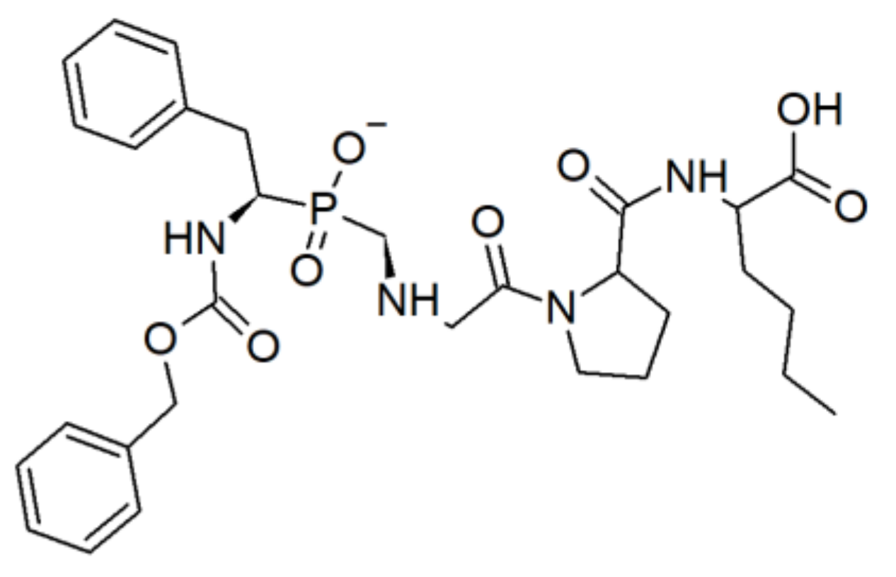
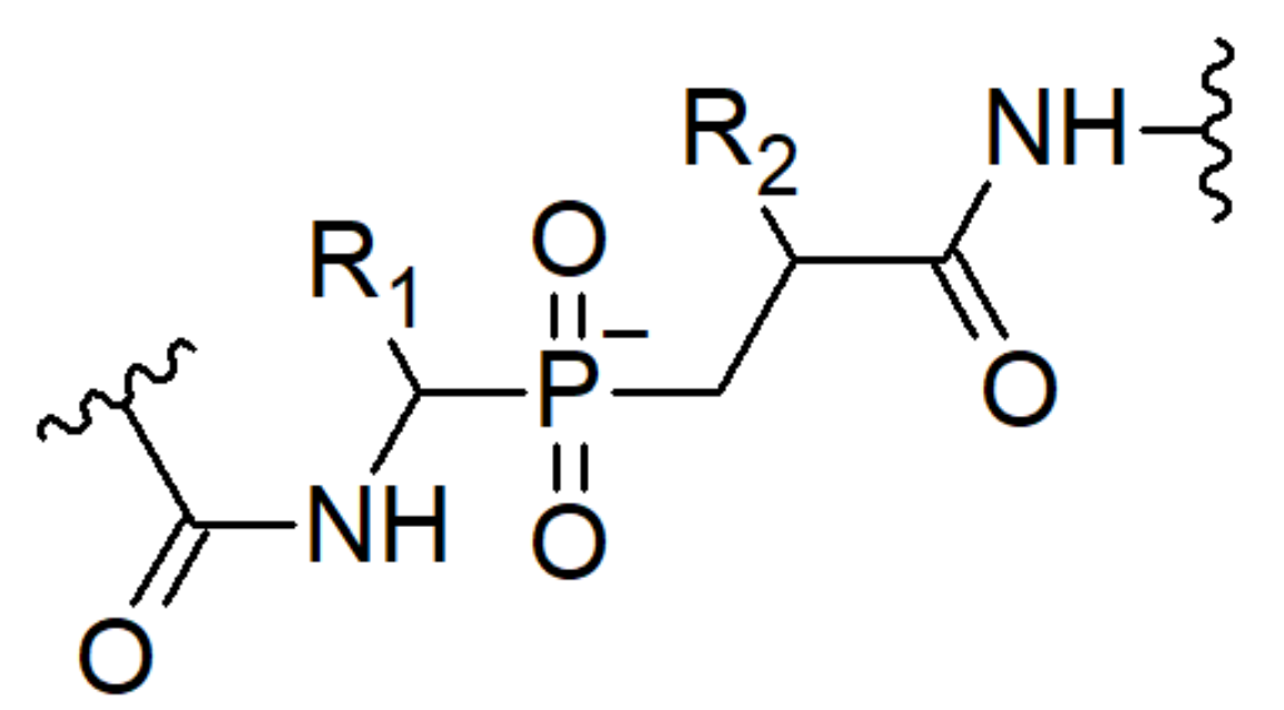
3.1.2. Phosphinic acid Derivatives

Shortly after the discovery of hydroxamate peptides acting as collagenases inhibitors, several compounds bearing the phosphinic acid moiety have been reported. In 1994 Yiotakis published two papers describing linear and cyclic peptides containing phosphinic group (PO2−), relevant for bacterial collagenases inhibition [69,70]. Substrate analogues of Corynebacterium rathayii collagenase were synthesised, replacing the scissile peptide bond by phosphinic moiety. Tetrapeptide Z-Phe-ψ(PO2CH2)-Gly-Pro-Nle (Figure 4) exhibited the collagenase inhibition with Ki value of 8 nM. Then, the influence of chain elongation was investigated. It was proven that the heptapeptide Z-Phe-Gly-Pro-Phe-ψ(PO2CH2)-Gly-Pro-Nle-OMe is an even more effective inhibitor than his shorter analogue (Ki = 0.6 nM). Further modifications of the tetrapeptide, including CH2 replacement by NH group, demonstrated insignificant decrease of Ki value from 8 nM to 6 nM.
Cyclic analogues of above-mentioned sequences were studied in tandem [70]. Generally speaking, the importance of conformationally constrained peptides was underpinned and investigated for various applications, mainly for drug-design purposes [71,72,73,74,75]. The design of new cyclic collagenase inhibitors was based on the following rules: (1) at least four interacting residues should be present; (2) there should be a bulky, hydrophobic group present in the P1 position; (3) it is required to incorporate glycine and proline in P1’ and P2’ positions, respectively; (4) P3’ position should be occupied by either linear, basic or apolar side chain residue (lysine or aminocaproic acid, Ahx). Cyclo[Gly-Pro-Phe-ψ(PO2CH2)-Gly-Pro-Ahx] together with cyclo[βAla-Pro-Phe-ψ(PO2CH2)-Gly-Pro-Ahx] were found to be relatively efficient, but not excellent inhibitors of this bacterial collagenase (Ki = 120 nM and 90 nM, respectively), comparing to the linear analogue Z-Phe-ψ(PO2CH2)-Gly-Pro-Nle (Ki = 8 nM). The role of configuration L and D was studied additionally, putting in a clear evidence that L analogues are favourable. Therefore, it can be summarized that, linear peptides in L conformation, fulfilling the above-mentioned requirements, are better candidates for collagenase inhibitors; nevertheless, also cyclic structures are promising.
Phosphinic pseudodipeptides (PPDs) were described by Bhowmick, Fields et al. in 2011 and 2012 [76,77]. All examined compounds were synthesised as building blocks in the Fmoc-containing form for further peptidomimetic development, with potential application as MMPs inhibitors. In 2011, Fmoc-protected phosphinic analogues of Gly-Val and Gly-Leu were obtained, but supplemental inhibition assays were not performed. Additionally, in 2012, another PPD Fmoc-Gly-Ile was successfully developed. All derivatives structures were based on common phosphonic peptides arrangement, reported in Figure 5.
Dive group achievements together with other scientists accomplishments in the field of phosphinic MMPs inhibitors, were summarized in the review by Yiotakis et al. in 2004 [78]. Furthermore, it was reported by Gall et al. that the phosphinic moiety incorporation led to a good inhibition of stromelysin-3 (MMP-11) as well [79]. Moreover, Lauer-Fields et al. proven a relevance of phosphinic peptides, a triple-helical transition state analogues, in MMP-2 and MMP-9 inhibition [80].

3.1.3. Unmodified Peptides

Whilst hydroxamate and phosphonic modifications represent the main direction of collagenase inhibitors design about 30 years ago, after 2000 scientists begun to examine also unmodified sequences. Mostly, these peptides derive from proteolytic digestion fragments of other proteins from natural sources, mimicking biological substrates. Recognized by the collagenase, these peptides occupy its active site and decrease the catalytic ability. Another pathway comprises indirect activity when the aforesaid fragments are acting as signal peptides. Herein we will report sequences described within past 20 years, possessing effective collagenases inhibitory properties.

Synthetic Peptides

Oono et al. in 2002 investigated an influence of human neutrophil peptide-1 (HNP-1) on MMP-1 and proα1-collagen type I expression [81]. However in this paper the HNP-1 sequence was not reported, while it can be found online as commercially available product: H-ACYCRIPACIAGERRYGTCIYQGRLWAFCC-OH (A) [82]. To explore the possibility of HNP-1 application in wound healing, these authors used one-step reverse transcription polymerase chain reaction (RT-PCR) for semiquantitative analysis of mRNA of MMP-1 and proα1 collagen (type I and III). ELISA test was then applied in order to quantify the production of MMP-1 by fibroblasts treated and not treated with HNP-1. Their experiments suggested that HNP-1 upregulates collagen synthesis and decrease collagenase activity.
Field’s research group investigations have shown a significant role of peptides as inhibitors of MMPs, not only collagenases [83]. Among others, MMP-1 activity was noticeably decreased in the presence of STX-S4-CT peptide (sequence C) [84]. Ki value was calculated for 4.5 μM. Regasepin1, a short peptide D, was found to be a good and selective inhibitor of collagenases—IC50 value for MMP-8 (collagenase-2) was 3 μM; in the case of MMP-1 and MMP-13 (collagenases-1 and -3) this value increased up to 100 μM [85]. Finally, TM8 peptide E inhibited all three collagenases well, with the Ki values of 10, 28 and 12 nM for MMP-1, -8, and -13, respectively [86].
The crystallized complexes of catalytically inactive human collagenase-3 (MMP-13) with peptides were described by Stura et al. in 2013 [87]. Two different peptides (F and G) deriving from the cleaved prodomain proMMP-13 throughout activation were bound to the enzyme active site. N-terminal modifications within these sequences were variable and showed interactions in different parts of catalytic domain. Structural details of crystals with relatively long peptide chains (up to 26 residues) can be found in Protein Data Bank library (codes 4FU4, 4FVL, and 4G0D). They refer to the complexes with the following peptides: G25GDEDDLSEEDLQFAERYLRSYYHPT50 (Figure 6, left), D30LSEEDLQFAERYLRSYYHPT50 (Figure 7), and G25GDEDDLSEEDLQFAERYLRSYYHPT50 with the full enzyme (Figure 6, right), respectively. This study does not depict typical inhibitory properties of the mentioned sequences. Nevertheless, it can be a good starting point for new compounds development due to the structural properties and interactions found in crystals.
The relevance of MMP-13 inhibitors as a useful tool for decreasing the collagen degradation was confirmed by another study of Howes et al. [89]. Their investigations have shown that MMP-1 acts differently to MMP-13 and they recognize diverse collagen cleavage sites. Moreover, MMP-13 digest preferably collagen type II over types I and III, cleaving the α chains sequentially. To check the recognition sites in collagen, these authors synthesised a library of overlapping homotrimeric peptides, including a 27 amino acids sequence from primary collagen and repeatable (GPP)5 fragments (located both on N- and C-terminus), providing a triple-helical conformation. Their studies revealed the highest affinity of MMP-13 to two sequences, so-called II-44 and II-8 (H and I, respectively). The first peptide, II-44, was found to occur in a triple helix form in 80%; for II-4 this value was 48% (at 37 °C). Calculated IC50 for both compounds shown a higher inhibitory potency for II -44 (IC50 = 8 μg/mL), while II-8 was a fourfold weaker inhibitor of MMP-13. Moreover, it was confirmed that the fragments essential for the recognition and responsible for a robust binding are GLXGQR, found twice in II-44 and not found in II-8. Therefore, it was possible to explain the slight difference observed between the obtained results.
Fields research group, previously mentioned, investigated MMP-13 triple-helical inhibitors J, K, and L, obtained using click chemistry [90]. These authors found that assembling of three different chains into heterotrimeric helices provides adequate thermal stability and results in MMP-13 and MT1-MMP inhibition in the range 100–400 nM. What is more, the heterotrimeric analogues were selective between MT1-MMP and MMP-1, on the contrary to homotrimer-based structures, favouring MMP-13.

Peptides from Natural Sources

In 2011, Park et al. published an article delineating the isolation of a peptide inhibiting MMP-1, derived from Dipturus chilensis skate skin protein [91]. The extracted protein was then digested by various enzymes, e.g., alcalase, α-chymotrypsin, trypsin, neutrase, papain, and pepsin. Obtained cocktails were then tested against collagenase-1, exhibiting an inhibitory effect of pepsin hydrolysate. Bioactive peptide was purified, and mass spectrometry confirmed the molecular weight value 961 Da of purified product B Additionally, the IC50 value for the isolated peptide was 87.0 μM.

3.1.4. Amino Acids and Peptide Derivatives

Apart the above-mentioned categories of collagenases inhibitors, there are other uncategorizable molecules. One of them is MMP-13 inhibitor (MMP13i-A) described in 2011 by Quillard and co-authors [92]. It is a nonhydroxamate molecule, significantly decreasing MMP-13 activity (Figure 8). What is particularly interesting, among all tested MMPs (1, 2, 7, 8, 9, 12, 13, and 14) only MMP-13 was efficiently inhibited (IC50 = 33.5 nM). Other metalloproteases shown no inhibition with IC50 value over 20000 nM. These observations suggested a certain selectivity and were applied during in vivo observations, including macrophage accumulation and MMP-13 activity monitoring. Mice treated for 10 weeks with MMP-13i-A, revealed reduced activity of MMP-13 without affecting macrophage content, measured by molecular imaging.
A great example of cosmeceutically relevant compounds are Mycosporine-like Amino Acids (MAA) from marine sources [93]. According to the paper by Hartmann et al., three bioactive molecules were firstly isolated from the marine red algae (Porphyra sp., Palmaria palmata) and then chromatographically purified for further use. Inhibitory properties of shinorine, porphyra, and palythine were evaluated against Chlostridium histolyticum collagenase. Obtained IC50 values are presented in the Table 3. To compare, two standard inhibitors phosphoramidon and 1,10-phenanthroline were considered additionally in the assay. Furthermore, molecular docking study confirmed the competitive mechanism of inhibition revealed by tested MAAs, with IC50 values between 100–160 μM.
In two papers from Fields, Roush et al., mentioned in the introduction of collagenases section, the authors evaluated inhibitory activity of various amino acids derivatives against MMP-13 [61,62]. They reported the two potent and selective inhibitors, 10d and (S)-17b, occupying the MMP-13 binding site and surrounding the catalytically active Zn2+ ion, without its chelation. IC50s found for these two derivatives were calculated as 3.4 nM and 2.7 nM, respectively. Then, Fuerst et al. identified the two compounds 2 and 31, exhibiting improved microsomal half-life, kinetic solubility, and permeability coefficient. Both compounds were selective among three tested MMPs (IC50 values for compound 2: 2.7 nM for MMP-13 and over 5000 nM for MMP-2, MMP-8; for compound 31: 8.5 nM, over 5000 nM, and 832 nM for MMP-13, -2, and -8, respectively).
A wide and detailed review describing inhibitors of MMPs in general, including collagenases, was published in 2020 by Raeeszadeh-Sarmazdeh et al. [94].

3.2. Elastase

In 1949 Baló and Banga published a communication in Nature, claiming that fresh pancreas extraction as well as dried pancreas powder “contains a specific enzyme, called ‘elastase’” [95]. Thus, this was the first scientific statement describing this important enzyme, which was thereafter investigated by other researchers. Tsuji and co -workers delineated the role of fibroblast elastase in wrinkle formation, providing also some indications in their inhibitors design [96]. These authors proven the importance of skin fibroblast elastase in elastic fibre degradation. At this point, it is worthy to underline that at least two elastase types are present in the skin, i.e., neutrophil and skin fibroblast elastase. While the first is a serin protease, the latter belongs to the metalloprotease family [97]. Because both of them have the ability to degrade fibrillar collagen, we will highlight the interesting inhibitors of each one.
In a paper by Imokawa group, the authors evidenced an undoubted difference between these elastases [96]. Comparing different enzyme inhibitors, inhibiting profiles for both neutrophil and fibroblast elastase were created. Among PMSF (phenylmethyl sulfonylfluoride), elastatinal, leupeptin, pepstatin A, EDTA, 1,10-phenantrolin, and phosphoramidon, the last one (already listed in Table 2) together with EDTA and, 1,10-phenantrolin (all known as metalloprotease inhibitor), significantly decreased the fibroblast elastase activity. On the other hand, neutrophil elastase remained active in these conditions, inhibited only by PMSF and elastatinal (typical serine protease inhibitors). Thus, the dissimilarity between the mechanisms of action of the two elastases was proven and explained. Additionally, the work by Azmi et al. showed a relevant inhibitory activity of elastase, tyrosinase, and MMP-1 by worm extracts, indicating its potential application in the cosmeceutical field [98]. However, due to the biological extract incorporation instead of purified compounds, precise inhibitory differences among three evaluated enzymes cannot be clearly distinguished.
For more detailed papers about biological activity and functions of human neutrophil elastase (HNE), together with examples of diverse inhibitors, we indicate a set of review papers by Ohbayashi et al., Kelly et al., Groutas et al., and a recently released one by Ahmad et al. [99,100,101,102].

3.2.1. Chloromethyl Ketones

One of the very first papers describing the inhibition of porcine pancreas elastase (PPE), human leukocyte elastase (HLE), and cathepsin G (CatG) was published in 1977 by Powers et al. [103]. The cleavage pattern of various peptides by PPE was examined, thus determining substrate structural preferences. Afterwards, a few new chloromethyl ketones Ac-Ala-Ala-Pro-AACH2Cl (where AA = Ile, Val, Thr) were synthesised. PPE was inhibited more rapidly by the peptide bearing Ile, while the Thr variant did not show significant inhibition. On the other hand, HLE was inhibited more efficiently by sequences including Ile and Val. MeO-Suc-Ala-Ala-Pro-ValCH2Cl, soluble in water, was established as a very-good inhibitor of HLE; Z-GIy-Leu-PheCH2Cl was effective against cathepsin G, but not leukocyte elastase.
In 1989, Navia and co-workers presented a work in the field of peptide chloromethyl ketones as HNE inhibitors [104]. They have noticed some side effects of peptides evaluated by Powers et al. in humans, and therefore designed new molecules with diminished toxicity. Crystal structure of methoxysuccinyl-Ala-Ala-Pro-Ala chloromethyl ketone (MSACK) is presented in Figure 9. It was proven that MSACK is able to form the cross-linkage between the His-57 and Ser-195 residues, responsible for the catalytic activity.
In vivo experiments on rats were reported in the paper by Cowan et al. [105]. Animals with advanced pulmonary vascular disease were then treated with peptidyl trifluoromethylketone serine elastase inhibitors, so-called ZD0892 and M249314. It was demonstrated that untreated rats have shown 900% loss of cellular matrix after 21 days, with further increase of 800% within next week. After M249314 administration, elastase inhibitory activity was noticeable over the first 6 hours, and then decreased but remained evident until the second dose. What is more, the mortality of sick and untreated rats has begun after 23 days, while after 28 days only one rat from each group (ZD0892 and M249314-treated for 1 week) was died. Summarizing, the survival of M249314-treated rats was 86%, whereas all untreated rats had died by 30 days of the trial.

3.2.2. Cyclic Depsipeptides

Advantages of cyclic structures were already mentioned previously in the paragraph related to collagenases. In 2001 and 2003, Matern et al. described the inhibition of PPE by scyptolin A and B, together with the crystal structure of scyptolin A bound to this enzyme [106,107]. Two cyclic depsipeptides (peptides with one or more amides replaced by the ester groups) were isolated from cyanobacterium Scytonema hofmanni PCC7110, namely scyptolin A and B. Both molecules were selective inhibitors of PPE, exhibiting IC50 values of approximately 3 mg/mL. The crystal structure has shown the rigid ring of scyptolin A occupying the elastase active site, thus preventing a hydrolytic attack and decreasing the enzyme activity (Figure 10). Due to the similarity of PPE and human elastase, the structures of scyptolin A and B appear as valuable starting points for further investigations in the field of human elastase inhibitors.
Another interesting example of cyclopeptides from natural sources, relevant as elastase inhibitors, were presented a recent paper by Keller et al. [108]. These authors described three new cyclic peptides, so-called tutuilamides A–C, structurally defined by various techniques such as NMR and chemical derivatization. Particularly, 3-amino-6-hydroxy-2-piperidone and 2-amino-2-butenoic acid, and a novel vinyl chloride residue, were found in these sequences. IC50 values obtained during the PPE inhibition assay were 1.18, 2.05 and 4.93 nM for tutuilamide A, B, and C, respectively. Moreover, trypsin inhibition was not found in the presence of the discussed cyclopeptides (IC50 > 20 000). X-ray crystallography revealed the formation of hydrogen bond between the amino acid backbone of tutuilamide A and the enzyme binding pocket, thus stabilizing the complex end explaining the inhibitory activity.

3.2.3. Unmodified Peptides

Structurally unmodified sequences can be found in the literature, too. In 1995, Toth et al. published the results of in vitro and in vivo studies, concerning lipophilic peptide inhibitors of human neutrophil elastase [109]. Four lipopeptides, namely 1be, shown relevant HNE inhibition. Obtained IC50 values were 2.9 × 10⁻⁹, 2.8 × 10⁻¹⁰, 1.8 × 10−l0, and 4.1 × 10−l0 mmol × mL−1, for 1b, 1c, 1d, and 1e, respectively. It was found that the inhibition was increasing while the lipophilicity of sequences was higher: moreover, carboxylic acid esterification in analogue 1e led to decreased inhibitory effect. All sequences were based on X-AAPV-Y fragment, with proper N- and C-terminal modifications (X and Y, respectively). All evaluated sequences are reported in the Table 4. In vivo experiments on rabbit skin proved the protection of elastic fibre against elastolysis, exhibited by the lipopeptide 1d (M) administered intradermally.
In 2000, Hilpert et al. designed and conducted a very broad investigation in order to identify and finally modify a sequence for efficient PPE inhibition [110]. Shorter chains were designed on the basis of OMTKY3 (third domain of turkey ovomucoid inhibitor), exhibiting the Ki value of 5.5 × 10⁻¹² M. Synthesis of peptides together with their cyclisation were performed on cellulose membranes, and were followed by the binding affinity measurements carried out on the membrane, too. The significance of each amino acid was determined by the exchange of every residue with each one of the other 19 coded amino acids. Thereby, over 700 different sequences were evaluated in binding to PPE, and 320 of them were then measured in the inhibition assay in microtiter plates with punched out peptide spots. Interestingly, cyclization did not provide any beneficial interactions in the active cleft and did not provoke an extended inhibition. Among all tested sequences, the shortest one exhibiting the 84% decrease of PPE activity, was peptide N. Ki values were of 6.7 × 10⁻⁷ M, 1.1 × 10⁻⁴ M, and 1.3 × 10⁻⁴ M for PPE, HLE, and BPC (bovine pancreas α-chymotrypsin), respectively. Furthermore, the described experiment including N sequence has been continued and broaden into the hybrid miniprotein design, inhibiting PPE likewise [111].
Another study on synthetic peptides as HNE inhibitors was performed by Vasconcelos et al. in 2011 [112]. Inhibitory effect on HNE and PPE of two sequences were compared to commercially available elastatinal. These sequences were based on Bowman–Birk protein-derived peptides able to inhibit HNE, summarized by McBride et al. [113]. Peptides O and P resulted to act as competitive inhibitors of HNE, with higher efficacy found for the sequence of peptide P. Probably, the increased hydrophobicity conferred by the (GA)7 tail provided relevant interactions with the enzyme, thus confirming the results reported in the previously mentioned work by Toth et al. [109]. Obtained IC50 values were at micromolar level (for the peptide 2: 8.1 and 6.3 μM; for the peptide P: 7.0 and 0.4 μM in the case of PPE and HNE, respectively).
One more example of non-modified peptide sequence, applicable as elastase inhibitor, was presented in the paper by Wan et al. in 2013 [114]. Authors identified the first Kunitz-type serine protease inhibitor, namely AvKTI (R), found in Araneus ventricosus spider. The amino acid sequence was deduced from AvKTI cDNA from GenBank and established as KDRCLLPKVTGPCKASLTRYYYDKDTKACVEFIYGGCRGNRNNFKQKDECEKACTDH (57 residues). Its activity against plasmin and elastase was proven, with Ki values of 4.89 nM and 169.07 nM, respectively, whereas IC50 values were calculated as 10.07 and 446.93 nM, respectively.

3.2.4. Peptide Derivatives and Cyclic Compounds

In this paragraph we would like to pay particular attention to the peptide and peptide-like sequences without functional groups belonging to the wider category described above, and exhibiting a relevant inhibitory activity of elastase. Therefore, macrocyclic compounds, together with unnatural amino acid-containing sequences will be described further. Exceptionally, we will also mention a few non-peptide molecules, in order to provide valuable indications for further design of new compounds.
In 2000, Nakanishi and co-workers determined the crystal structure of PPE in the complex with FR901277 inhibitor (S), a natural compound extracted from the culture filtrate of Streptomyces resistomicificus (Figure 11 and Figure 12) [115]. Previous examination of this compound against HLE and PPE, resulted with the IC50 value of 18 and 26 μM, respectively [116]. FR901277 consists of 7 amino acids, among which 4 are natural amino acids [L-Orn(1), L-Thr(2), L-Phe(5), and L-Val(7)] and 3 are unusual [dehydroxyThr(3), AA(4), and AA(6)], with the bridge between L-Thr(2) and L-Val(7). The crystal structure of PPE with FR901277 have shown the occupation of PPE binding subsites by inhibitor hydrophobic residues, with additional van der Waals contacts.
The relevance of elastase inhibition in cosmeceutical application has been already noticed in early 2000s by Tsukahara et al. [117]. These authors reported a significant role of elastase in the damage of dermal elastic fibres, leading to wrinkle formation. N-phenetylphosphonyl-leucyl-tryptophane (NPLT), an elastase inhibitor, was applied topically 5 times a week on rat skin, leading to diminished wrinkle formation and maintained skin elasticity. 0.1 ± 10.0 mM concentrations were tested, revealing the peak of efficacy at 1.0 mM concentration.
Novel, low-molecular HNE inhibitors were described by Schepetkin et al. in 2007, setting a milestone in heterocyclic elastase inhibitors field [118]. Structures based on N-benzoylpyrazoles were eventually modified and optimized, leading to a library of potent compounds (for a general structure of N-benzoylpyrazoles, please see the Figure 13) and 17 molecules were then chosen for further experiments and SAR (structure-active relationship) analysis, revealing Ki values between 6-300 nM. Molecular docking studies were performed in order to explain the differences in potency among the most active and inactive compounds. It has been noticed that the carbonyl group of the inhibitor was located in the NE binding site, thus interacting with the catalytic triad. On the other hand, inactive analogues were either sterically hindered, or showed unpreferable orientation, hence excluding the presence of relevant interactions.
Starting from 2011, Giovannoni group was working on N-benzoylindazole derivatives acting as HNE inhibitors, progressing the work of above-mentioned Quinn’s investigations [119,120]. Among all tested compounds, so-called 8a and 8b showed the most potent, competitive and pseudo-irreversible inhibition, with IC50 values of ≈0.089 and ≈0.13 μM, respectively. Accordingly, also in this study molecular modeling have highlighted the importance of the carbonyl group in the N-benzoylindazole derivatives structures, due to the favourable interactions with -OH from Ser195 in the active cleft. Going forward, other analogues were developed to increase the inhibition efficacy and overall stability. Two more compounds, 5b and 20f, resulted with the inhibition level of IC50 ≈ 7 and 10 nM, respectively (Figure 14).

3.3. Cathepsins

Among a variety of cathepsins, the most relevant classes for cosmeceutical purposes are cathepsins K, L, and S (CatK, CatL, and CatS, respectively). These endopeptidases belongs to the cysteine cathepsins family and regulate several biological functions, such as inflammatory and proteolytic processes, or ECM remodeling [121]. Due to their role in fibrillar collagen degradation, a few examples of cathepsins inhibitors will be reported here.
Brömme and Lecaille summarized the features of the ideal CatK inhibitor, pointing to the importance of low molecular mass, minimal peptide character, reversibility and selectivity [122]. Aguda et al. evidenced that CatK is capable of fibrillar collagen degradation, underlining the participance of collagen-bound glycosaminoglycans in this process [123]. Additionally, a role of CatL in type I collagen degrading pathway was proved by Parks et al. [32]. It was found that CatL requires the presence of CatK to degrade effectively tendon ECM.

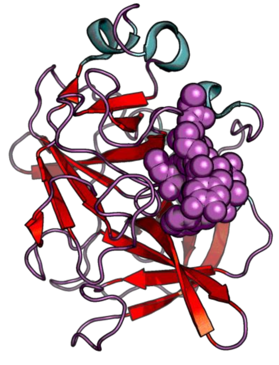
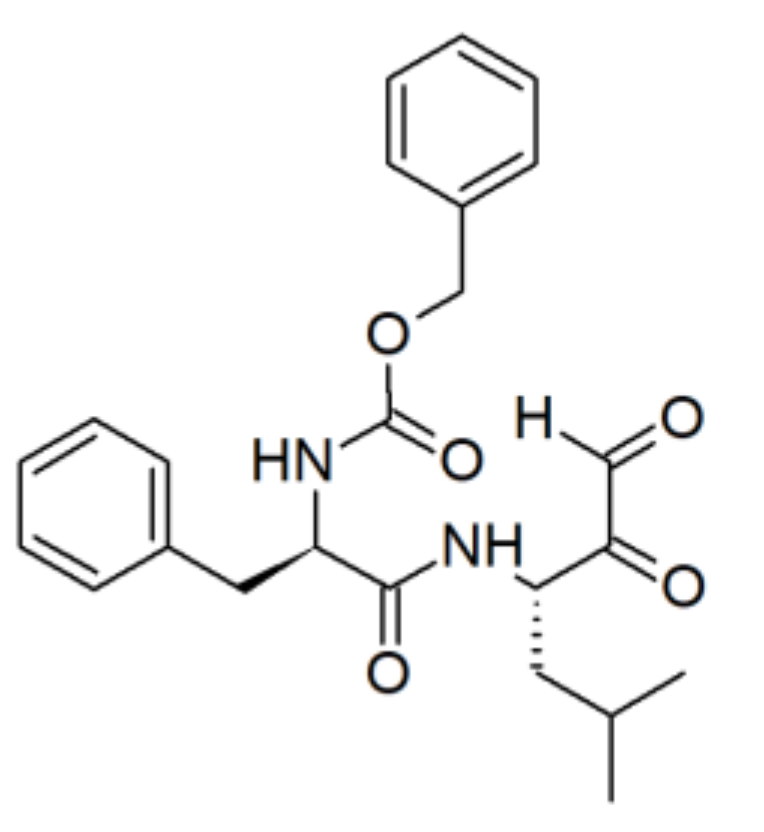
3.3.1. Peptide Aldehydes

Peptidyl aldehydes as cathepsins inhibitors are known at least since 1969, when leupeptin (Ac-Leu-Leu-Arg-CHO, T) and antipain (N-[Nα-carbonyl-Arg-Val-Arg-al]-Phe, U) were described for the first time [124,125]. Then, in 1990 Sasaki et al. synthesised eight different di- and tripeptide aldehydes [126]. Each analogue contained L-leucine followed by an aldehyde located at the C-terminus (on L-norleucine, L-methionine, or L-phenylalanine). Performed enzymatic tests shown the inhibition of cathepsin B and L, with a particular efficacy against the second one. V inhibited CatL with Ki = 0.5 nM; X inhibited CatB with Ki = l00 nM.
Building on the efficacy of the above-mentioned analogues, Votta et al. in 1997 published a study dealing with other peptide-aldehyde inhibitors [127]. Two classes of compounds were distinguished, namely time-dependent and time-independent inhibitors. Cbz-Leu-Leu-Leu-CHO (Y) was selected to be the most potent one, classified into the time-independent group. The values of Ki and IC50 were 1.4 nM and 0.02 μM, respectively. Furthermore, Y reduced extensive bone loss provoked by CatK.
Other than CatK and CatL, also CatS became known to be inhibited by peptidic aldehydes. In 2000, Walker et al., from already mentioned Brömme group, published an evaluation of dipeptide α-keto-β-aldehydes (glyoxals) as new inhibitors of cathepsin S [128]. Through the solid-phase peptide synthesis (SPPS), a spectrum of dipeptide analogues was obtained and tested against cathepsin S. One of them, Z, exhibited good selectivity (Ki = 0.185 nM for CatS and 76 nM for CatB) (the structure can be found in the Figure 15).

3.3.2. Peptidic Nitriles

Balicatib (AAE581), a basic peptide nitrile, has been developed by Novartis and was found to be a potent and selective inhibitor of human CatK (IC50 = 0.0014 μM, compared to 2.9–28 μM for other cathepsins) [129]. Administered orally, was applied in osteoporosis treatment [130]. Another example is odanacatib (MK-0822) together with MK-1256 analogue [131,132]. Odanacatib inhibits CatK, B, L, and S with IC50 values of 0.2 nM, 1034 nM, 2995 nM, and 60 nM, respectively. CatK inhibition by MK-1256 was effective and selective among other cathepsins (IC50 = 0.62 nM, >1100-fold less potent against CatL and CatS i.a.). Incorporation of balicatib and odanacatib scaffolds was then evaluated by Burtoloso et al. in 2017 [133]. These authors developed new compounds with anti-trypanosomal activity, relevant for the discovery of further anti-chagasic agents. All depicted structures, and the inhibitory values are summarized in the Table 5.
For a broad review dealing with nitrile-based peptide inhibitors of cathepsins, supported by the knowledge of this inhibitory mechanism, we highlight the paper by Frizler et al. published in 2010 [134].

3.3.3. Peptide Ketoheterocycles

(Keto)heterocyclic modifications of peptides were assessed in cathepsins inhibition at the beginning of 2000s. McGrath and colleagues synthesised a set of peptide α-ketoheterocyclic inhibitors, occupying the substrate recognition cleft of targeted cysteine protease CatK [135]. Molecular modeling supported by the crystallographic data revealed the reversible covalent bonding with Cys25 nucleophile. One of the most potent examples is the compound 4, presented in the Table 6. Ki values calculated for 4 and CatK, B, L, and S were of 0.0029 nM, 0.64 nM, 0.02 nM, and 0.0066 nM, respectively.
Quibell group in 2004 and 2005 reported the synthesis and evaluation of cis-hexahydropyrrolo[3,2-b]pyrrol-3-one, and bicyclic peptidomimetic tetrahydrofuro[3,2-b]pyrrol-3-one and hexahydrofuro[3,2-b]pyridine-3-one derivatives as cysteinyl proteases inhibitors [136,137]. In their first paper, 5,5-bicyclic ketone-containing scaffold was depicted, underlining the valuable chiral stability. Kinetic analysis shown a significantly lower association with CatK of 6,5-analogues, and subsequently lower potency. Among all synthesised compounds, 10 was found to be the most effective and exhibited sub-micromolar potency in the cell-based assay (Table 6). This idea was further pursued and in 2005 another analogue, i.e., compound 22, was described as an even more potent CatK inhibitor than those previously reported (Table 6).
Moreover, a summary of patents revealed in the field of cathepsin K inhibitors was published by Wijkmans and Gossen in 2011 [138].

4. Conclusions and Future Prospect

Collagen represents approximately one-third of all human body proteins, being the most abundant protein-like body component. The essential role played by collagen has an influence on many physiological, as well as pathological states. For example, dysfunctional collagen turnover and its increased degradation lead to wound healing disruption, skin photoaging, and loss of skin firmness and elasticity. Therefore, inhibitors of enzymes leading to the degradation of fibrillar collagen were in the spotlight for years, and still are the focus of many research projects. Healthy and younger-looking skin becomes increasingly significant targets, as they contribute to the wellness of each individual, as clearly shown by the critical growth of the skin-care products market observed recently. In this review, we describe a group of inhibitors of enzymes degrading fibrillar collagen, paying attention to peptides and peptidomimetics (Scheme 3 and Table 7). Additionally, a few valuable non-peptide molecules were mentioned, thereby providing a platform for further investigations and design of new active compounds. Prospectively, these peptides and peptidomimetics, together with their analogues, can be applicable as active ingredients in the cosmeceutical field.

Author Contributions

P.L., A.M.P., P.R. and R.L. participated in the manuscript preparation; P.L. wrote an outline and the first draft of the manuscript; A.M.P., P.R. and R.L. edited the final version. All authors have read and agreed to the published version of the manuscript.

Funding

The Authors declare the support from the Polish Ministry of Science and Higher Education for the Faculty of Chemistry of Wroclaw University of Science and Technology. PeptLab of the University of Florence is supported by the Region Toscana for the project: BIOPEPTIDI. Sviluppo di nuovi peptidi biologicamente attivi. POR CREO FESR 2007-2013 ASSE I-LINEA DI INTERVENTO 1.1.c.

Institutional Review Board Statement

Not applicable.

Informed Consent Statement

Not applicable.

Data Availability Statement

No new data were created or analysed in this study. Data sharing is not applicable to this article.

Acknowledgments

P.L. is the participant of “BioTechNan” project–Interdisciplinary Environmental Doctoral Studies KNOW in the field of Biotechnology and Nanotechnology, co-financed by the European Union. The PhD of P.L. is performed in the context of a cotutorate between the PhD Schools in Drug Research and Innovative Treatments of the University of Florence (XXXV Ciclo) and in Chemical Sciences of the Wroclaw University of Science and Technology (Supervisors: P.R. and R.L.). We kindly acknowledge the European Peptide Society for providing the Mobility Fellowship grant for P.L. in the period of Apr-Aug 2021, helpful during the manuscript preparation.

Conflicts of Interest

The authors declare no conflict of interest. The funders had no role in the design of the study; in the collection, analyses, or interpretation of data; in the writing of the manuscript, or in the decision to publish the results.

References

  1. Yue, B. Biology of the Extracellular Matrix: An Overview. J. Glaucoma 2014, 23, S20–S23. [Google Scholar] [CrossRef] [PubMed]
  2. Żelaszczyk, D.; Waszkielewicz, A.; Marona, H. Kolagen—Struktura Oraz Zastosowanie w Kosmetologii i Medycynie Estetycznej. Estetol. Med. Kosmetol. 2012, 14–20. [Google Scholar] [CrossRef]
  3. Veit, G.; Kobbe, B.; Keene, D.R.; Paulsson, M.; Koch, M.; Wagener, R. Collagen XXVIII, a Novel von Willebrand Factor A Domain-Containing Protein with Many Imperfections in the Collagenous Domain. J. Biol. Chem. 2006, 281, 3494–3504. [Google Scholar] [CrossRef] [Green Version]
  4. Smith, K.; Rennie, M.J. New Approaches and Recent Results Concerning Human-Tissue Collagen Synthesis. Curr. Opin. Clin. Nutr. Metab. Care 2007, 10, 582–590. [Google Scholar] [CrossRef] [PubMed]
  5. Burgeson, R.E.; Nimni, M.E. Collagen Types. Molecular Structure and Tissue Distribution. Clin. Orthop Relat Res. 1992, 250–272. [Google Scholar]
  6. Bhattacharjee, A.; Bansal, M. Collagen Structure: The Madras Triple Helix and the Current Scenario. IUBMB Life Int. Union Biochem. Mol. Biol. Life 2005, 57, 161–172. [Google Scholar] [CrossRef] [PubMed]
  7. Brodsky, B.; Thiagarajan, G.; Madhan, B.; Kar, K. Triple-Helical Peptides: An Approach to Collagen Conformation, Stability, and Self-Association. Biopolymers 2008, 89, 345–353. [Google Scholar] [CrossRef]
  8. Fields, G.B. Interstitial Collagen Catabolism. J. Biol. Chem. 2013, 288, 8785–8793. [Google Scholar] [CrossRef] [Green Version]
  9. Bishop, J.E.; Laurent, G.J. Collagen Turnover and Its Regulation in the Normal and Hypertrophying Heart. Eur. Heart J. 1995, 16, 38–44. [Google Scholar] [CrossRef] [PubMed]
  10. Manon-Jensen, T.; Kjeld, N.G.; Karsdal, M.A. Collagen-Mediated Hemostasis. J. Thromb Haemost. 2016, 14, 438–448. [Google Scholar] [CrossRef]
  11. Karsdal, M.A.; Genovese, F.; Madsen, E.A.; Manon-Jensen, T.; Schuppan, D. Collagen and Tissue Turnover as a Function of Age: Implications for Fibrosis. J. Hepatol. 2016, 64, 103–109. [Google Scholar] [CrossRef] [PubMed]
  12. Sprangers, S.; Everts, V. Molecular Pathways of Cell-Mediated Degradation of Fibrillar Collagen. Matrix Biol. 2019, 75–76, 190–200. [Google Scholar] [CrossRef]
  13. Kim, M.; Park, H.J. Molecular Mechanisms of Skin Aging and Rejuvenation; InTech: Seoul, Korea, 2016; pp. 57–76. [Google Scholar]
  14. Rodriguez-Feo, J.; Sluijter, J.; Kleijn, D.; Pasterkamp, G. Modulation of Collagen Turnover in Cardiovascular Disease. Curr. Pharm. Des. 2005, 11, 2501–2514. [Google Scholar] [CrossRef] [PubMed]
  15. Gelse, K. Collagens—Structure, Function, and Biosynthesis. Adv. Drug Deliv. Rev. 2003, 55, 1531–1546. [Google Scholar] [CrossRef] [Green Version]
  16. Vannella, K.M.; Wynn, T.A. Mechanisms of Organ Injury and Repair by Macrophages. Annu. Rev. Physiol. 2017, 79, 593–617. [Google Scholar] [CrossRef]
  17. Rennard, S.I.; Stier, L.E.; Crystal, R.G. Intracellular Degradation of Newly Synthesized Collagen. J. Investig. Dermatol. 1982, 79, 77–82. [Google Scholar] [CrossRef] [Green Version]
  18. Lu, P.; Takai, K.; Weaver, V.M.; Werb, Z. Extracellular Matrix Degradation and Remodeling in Development and Disease. Cold Spring Harb. Perspect. Biol. 2011, 3. [Google Scholar] [CrossRef]
  19. Zhong, S.; Khalil, R.A. A Disintegrin and Metalloproteinase (ADAM) and ADAM with Thrombospondin Motifs (ADAMTS) Family in Vascular Biology and Disease. Biochem. Pharmacol. 2019, 164, 188–204. [Google Scholar] [CrossRef]
  20. McAnulty, R.J.; Laurent, G.J. Collagen Synthesis and Degradation In Vivo. Evidence for Rapid Rates of Collagen Turnover with Extensive Degradation of Newly Synthesized Collagen in Tissues of the Adult Rat. Collagen Relat. Res. 1987, 7, 93–104. [Google Scholar] [CrossRef]
  21. Laurent, G.J. Dynamic State of Collagen: Pathways of Collagen Degradation in Vivo and Their Possible Role in Regulation of Collagen Mass. Am. J. Physiol. Cell Physiol. 1987, 252, C1–C9. [Google Scholar] [CrossRef]
  22. McClung, J.M.; Davis, J.M.; Wilson, M.A.; Goldsmith, E.C.; Carson, J.A. Estrogen Status and Skeletal Muscle Recovery from Disuse Atrophy. J. Appl. Physiol. 2006, 100, 2012–2023. [Google Scholar] [CrossRef] [Green Version]
  23. Miller, B.F.; Hansen, M.; Olesen, J.L.; Flyvbjerg, A.; Schwarz, P.; Babraj, J.A.; Smith, K.; Rennie, M.J.; Kjaer, M. No Effect of Menstrual Cycle on Myofibrillar and Connective Tissue Protein Synthesis in Contracting Skeletal Muscle. Am. J. Physiol. Endocrinol. Metab. 2006, 290, E163–E168. [Google Scholar] [CrossRef] [PubMed]
  24. Miller, B.F.; Hansen, M.; Olesen, J.L.; Schwarz, P.; Babraj, J.A.; Smith, K.; Rennie, M.J.; Kjaer, M. Tendon Collagen Synthesis at Rest and after Exercise in Women. J. Appl. Physiol. 2007, 102, 541–546. [Google Scholar] [CrossRef] [PubMed]
  25. Kehlet, S.N.; Willumsen, N.; Armbrecht, G.; Dietzel, R.; Brix, S.; Henriksen, K.; Karsdal, M.A. Age-Related Collagen Turnover of the Interstitial Matrix and Basement Membrane: Implications of Age- and Sex-Dependent Remodeling of the Extracellular Matrix. PLoS ONE 2018, 13, e0194458. [Google Scholar] [CrossRef] [Green Version]
  26. Bohn, G.; Liden, B.; Schultz, G.; Yang, Q.; Gibson, D.J. Ovine-Based Collagen Matrix Dressing: Next-Generation Collagen Dressing for Wound Care. Adv. Wound Care 2016, 5, 1–10. [Google Scholar] [CrossRef] [PubMed] [Green Version]
  27. Cipriani, C.; Pascarella, S.; Errante, F.; Menicacci, B.; Magnelli, L.; Mocali, A.; Rovero, P.; Giovannelli, L. Serpin A1 and the Modulation of Type I Collagen Turnover: Effect of the C-Terminal Peptide 409-418 (SA1-III) in Human Dermal Fibroblasts: Serpin-A1 C-Terminal Modulates Collagen Levels. Cell Biol. Int. 2018, 42, 1340–1348. [Google Scholar] [CrossRef]
  28. Jabłońska-Trypuć, A.; Matejczyk, M.; Rosochacki, S. Matrix Metalloproteinases (MMPs), the Main Extracellular Matrix (ECM) Enzymes in Collagen Degradation, as a Target for Anticancer Drugs. J. Enzym. Inhib. Med. Chem. 2016, 31, 177–183. [Google Scholar] [CrossRef] [Green Version]
  29. Williams, K.E.; Olsen, D.R. Matrix Metalloproteinase-1 Cleavage Site Recognition and Binding in Full-Length Human Type III Collagen. Matrix Biol. 2009, 28, 373–379. [Google Scholar] [CrossRef]
  30. Ruettger, A.; Schueler, S.; Mollenhauer, J.A.; Wiederanders, B. Cathepsins B, K, and L Are Regulated by a Defined Collagen Type II Peptide via Activation of Classical Protein Kinase C and P38 MAP Kinase in Articular Chondrocytes. J. Biol. Chem. 2008, 283, 1043–1051. [Google Scholar] [CrossRef] [Green Version]
  31. Klaus, V.; Schmies, F.; Reeps, C.; Trenner, M.; Geisbüsch, S.; Lohoefer, F.; Eckstein, H.-H.; Pelisek, J. Cathepsin S Is Associated with Degradation of Collagen I in Abdominal Aortic Aneurysm. Vasa 2018, 47, 285–293. [Google Scholar] [CrossRef]
  32. Parks, A.N.; Nahata, J.; Edouard, N.-E.; Temenoff, J.S.; Platt, M.O. Sequential, but Not Concurrent, Incubation of Cathepsin K and L with Type I Collagen Results in Extended Proteolysis. Sci. Rep. 2019, 9, 5399. [Google Scholar] [CrossRef]
  33. Kafienah, W.; Buttle, J.D.; Burnett, D.; Hollander, P.A. Cleavage of Native Type I Collagen by Human Neutrophil Elastase. Biochem. J. 1998, 330, 897–902. [Google Scholar] [CrossRef] [Green Version]
  34. Bertini, I.; Fragai, M.; Luchinat, C.; Melikian, M.; Toccafondi, M.; Lauer, J.L.; Fields, G.B. Structural Basis for Matrix Metalloproteinase 1-Catalyzed Collagenolysis. J. Am. Chem. Soc. 2012, 134, 2100–2110. [Google Scholar] [CrossRef] [Green Version]
  35. Song, F. Matrix Metalloproteinase Dependent and Independent Collagen Degradation. Front. Biosci. 2006, 11, 3100. [Google Scholar] [CrossRef] [PubMed] [Green Version]
  36. Moilanen, M.; Sorsa, T.; Stenman, M.; Nyberg, P.; Lindy, O.; Vesterinen, J.; Paju, A.; Konttinen, Y.T.; Stenman, U.-H.; Salo, T. Tumor-Associated Trypsinogen-2 (Trypsinogen-2) Activates Procollagenases (MMP-1, -8, -13) and Stromelysin-1 (MMP-3) and Degrades Type I Collagen. Biochemistry 2003, 42, 5414–5420. [Google Scholar] [CrossRef] [PubMed]
  37. Kafienah, W.; Brömme, D.; Buttle, D.J.; Croucher, L.J.; Hollander, A.P. Human Cathepsin K Cleaves Native Type I and II Collagens at the N-Terminal End of the Triple Helix. Biochem. J. 1998, 331, 727–732. [Google Scholar] [CrossRef] [PubMed] [Green Version]
  38. Zhu, Y.; Liu, X.; Sköld, C.M.; Wang, H.; Kohyama, T.; Wen, F.-Q.; Ertl, R.F.; Rennard, S.I. Collaborative Interactions between Neutrophil Elastase and Metalloproteinases in Extracellular Matrix Degradation in Three-Dimensional Collagen Gels. Respir. Res. 2001, 2, 300. [Google Scholar] [CrossRef] [PubMed]
  39. Zhu, Y.K.; Liu, X.D.; Sköld, C.M.; Umino, T.; Wang, H.J.; Spurzem, J.R.; Kohyama, T.; Ertl, R.F.; Rennard, S.I. Synergistic Neutrophil Elastase-Cytokine Interaction Degrades Collagen in Three-Dimensional Culture. Am. J. Physiol. Lung Cell. Mol. Physiol. 2001, 281, L868–L878. [Google Scholar] [CrossRef] [PubMed]
  40. Stenman, M.; Ainola, M.; Valmu, L.; Bjartell, A.; Ma, G.; Stenman, U.-H.; Sorsa, T.; Luukkainen, R.; Konttinen, Y.T. Trypsin-2 Degrades Human Type II Collagen and Is Expressed and Activated in Mesenchymally Transformed Rheumatoid Arthritis Synovitis Tissue. Am. J. Pathol. 2005, 167, 1119–1124. [Google Scholar] [CrossRef] [Green Version]
  41. van Deemter, M.; Kuijer, R.; Harm Pas, H.; Jacoba van der Worp, R.; Hooymans, J.M.M.; Los, L.I. Trypsin-Mediated Enzymatic Degradation of Type II Collagen in the Human Vitreous. Mol. Vis. 2013, 19, 1591–1599. [Google Scholar]
  42. Pascarella, S.; Tiberi, C.; Sabatino, G.; Nuti, F.; Papini, A.M.; Giovannelli, L.; Rovero, P. Serpin A1 C-Terminal Peptides as Collagen Turnover Modulators. ChemMedChem 2016, 11, 1850–1855. [Google Scholar] [CrossRef]
  43. Nielsen, R.H.; Stoop, R.; Leeming, D.J.; Stolina, M.; Qvist, P.; Christiansen, C.; Karsdal, M.A. Evaluation of Cartilage Damage by Measuring Collagen Degradation Products in Joint Extracts in a Traumatic Model of Osteoarthritis. Biomarkers 2008, 13, 79–87. [Google Scholar] [CrossRef]
  44. Veidal, S.S.; Karsdal, M.A.; Nawrocki, A.; Larsen, M.R.; Dai, Y.; Zheng, Q.; Hägglund, P.; Vainer, B.; Skjøt-Arkil, H.; Leeming, D.J. Assessment of Proteolytic Degradation of the Basement Membrane: A Fragment of Type IV Collagen as a Biochemical Marker for Liver Fibrosis. Fibrogenesis Tissue Repair 2011, 4, 22. [Google Scholar] [CrossRef] [Green Version]
  45. McAnulty, R.J. Methods for Measuring Hydroxyproline and Estimating in Vivo Rates of Collagen Synthesis and Degradation. Methods Mol. Med. 2005, 117, 189–207. [Google Scholar] [CrossRef] [PubMed]
  46. Gorres, K.L.; Raines, R.T. Prolyl 4-Hydroxylase. Crit. Rev. Biochem. Mol. Biol. 2010, 45, 106–124. [Google Scholar] [CrossRef]
  47. Gorres, K.L.; Raines, R.T. Direct and Continuous Assay for Prolyl 4-Hydroxylase. Anal. Biochem. 2009, 386, 181–185. [Google Scholar] [CrossRef] [PubMed] [Green Version]
  48. Poobalarahi, F.; Baicu, C.F.; Bradshaw, A.D. Cardiac Myofibroblasts Differentiated in 3D Culture Exhibit Distinct Changes in Collagen I Production, Processing, and Matrix Deposition. Am. J. Physiol. Heart Circ. Physiol. 2006, 291, H2924–H2932. [Google Scholar] [CrossRef] [Green Version]
  49. Errante, F.; Ledwoń, P.; Latajka, R.; Rovero, P.; Papini, A.M. Cosmeceutical Peptides in the Framework of Sustainable Wellness Economy. Front. Chem. 2020, 8, 572923. [Google Scholar] [CrossRef]
  50. Draelos, Z.D. The Cosmeceutical Realm. Clin. Dermatol. 2008, 26, 627–632. [Google Scholar] [CrossRef]
  51. Ledwoń, P.; Errante, F.; Papini, A.M.; Rovero, P.; Latajka, R. Peptides as Active Ingredients: A Challenge for Cosmeceutical Industry. Chem. Biodivers. 2021, 18, e2000833. [Google Scholar] [CrossRef] [PubMed]
  52. Copeland, R.A. Enzymes: A Practical Introduction to Structure, Mechanism, and Data Analysis, 2nd ed.; Wiley: New York, NY, USA, 2000; ISBN 978-0-471-35929-6. [Google Scholar]
  53. Aljoundi, A.; Bjij, I.; El Rashedy, A.; Soliman, M.E.S. Covalent Versus Non-Covalent Enzyme Inhibition: Which Route Should We Take? A Justification of the Good and Bad from Molecular Modelling Perspective. Protein J. 2020, 39, 97–105. [Google Scholar] [CrossRef]
  54. Adeniyi, A.A.; Muthusamy, R.; Soliman, M.E. New Drug Design with Covalent Modifiers. Expert Opin. Drug Discov. 2016, 11, 79–90. [Google Scholar] [CrossRef] [PubMed]
  55. Brown, P.D. Ongoing Trials with Matrix Metalloproteinase Inhibitors. Expert Opin. Investig. Drugs 2000, 9, 2167–2177. [Google Scholar] [CrossRef] [PubMed]
  56. Cuniasse, P.; Devel, L.; Makaritis, A.; Beau, F.; Georgiadis, D.; Matziari, M.; Yiotakis, A.; Dive, V. Future Challenges Facing the Development of Specific Active-Site-Directed Synthetic Inhibitors of MMPs. Biochimie 2005, 87, 393–402. [Google Scholar] [CrossRef] [PubMed]
  57. Rasmussen, H.S.; McCann, P.P. Matrix Metalloproteinase Inhibition as a Novel Anticancer Strategy: A Review with Special Focus on Batimastat and Marimastat. Pharmacol. Ther. 1997, 75, 69–75. [Google Scholar] [CrossRef]
  58. Ottl, J.; Gabriel, D.; Murphy, G.; Knäuper, V.; Tominaga, Y.; Nagase, H.; Kröger, M.; Tschesche, H.; Bode, W.; Moroder, L. Recognition and Catabolism of Synthetic Heterotrimeric Collagen Peptides by Matrix Metalloproteinases. Chem. Biol. 2000, 7, 119–132. [Google Scholar] [CrossRef] [Green Version]
  59. Bode, W.; Maskos, K. Structural Basis of the Matrix Metalloproteinases and Their Physiological Inhibitors, the Tissue Inhibitors of Metalloproteinases. Biol. Chem. 2003, 384. [Google Scholar] [CrossRef]
  60. Maskos, K. Crystal Structures of MMPs in Complex with Physiological and Pharmacological Inhibitors. Biochimie 2005, 87, 249–263. [Google Scholar] [CrossRef]
  61. Choi, J.Y.; Fuerst, R.; Knapinska, A.M.; Taylor, A.B.; Smith, L.; Cao, X.; Hart, P.J.; Fields, G.B.; Roush, W.R. Structure-Based Design and Synthesis of Potent and Selective Matrix Metalloproteinase 13 Inhibitors. J. Med. Chem. 2017, 60, 5816–5825. [Google Scholar] [CrossRef]
  62. Fuerst, R.; Yong Choi, J.; Knapinska, A.M.; Smith, L.; Cameron, M.D.; Ruiz, C.; Fields, G.B.; Roush, W.R. Development of Matrix Metalloproteinase-13 Inhibitors—A Structure-Activity/Structure-Property Relationship Study. Bioorg. Med. Chem. 2018, 26, 4984–4995. [Google Scholar] [CrossRef]
  63. Moore, W.M.; Spilburg, C.A. Peptide Hydroxamic Acids Inhibit Skin Collagenase. Biochem. Biophys. Res. Commun. 1986, 136, 390–395. [Google Scholar] [CrossRef]
  64. Grobelny, D.; Poncz, L.; Galardy, R.E. Inhibition of Human Skin Fibroblast Collagenase, Thermolysin, and Pseudomonas Aeruginosa Elastase by Peptide Hydroxamic Acids. Biochemistry 1992, 31, 7152–7154. [Google Scholar] [CrossRef] [PubMed]
  65. Grams, F.; Reinemer, P.; Powers, J.C.; Kleine, T.; Pieper, M.; Tschesche, H.; Huber, R.; Bode, W. X-Ray Structures of Human Neutrophil Collagenase Complexed with Peptide Hydroxamate and Peptide Thiol Inhibitors. Implications for Substrate Binding and Rational Drug Design. Eur. J. Biochem. 1995, 228, 830–841. [Google Scholar] [CrossRef] [PubMed]
  66. Bode, W.; Reinemer, P.; Huber, R.; Kleine, T.; Schnierer, S.; Tschesche, H. The X-Ray Crystal Structure of the Catalytic Domain of Human Neutrophil Collagenase Inhibited by a Substrate Analogue Reveals the Essentials for Catalysis and Specificity. EMBO J. 1994, 13, 1263–1269. [Google Scholar] [CrossRef] [PubMed]
  67. Rasmussen, H.S. Batimastat and Marimastat in Cancer. In Antiangiogenic Agents in Cancer Therapy; Teicher, B.A., Ed.; Humana Press: Totowa, NJ, USA, 1999; pp. 399–405. ISBN 978-1-4757-4518-4. [Google Scholar]
  68. Gooljarsingh, L.T.; Lakdawala, A.; Coppo, F.; Luo, L.; Fields, G.B.; Tummino, P.J.; Gontarek, R.R. Characterization of an Exosite Binding Inhibitor of Matrix Metalloproteinase 13. Protein Sci. 2007, 17, 66–71. [Google Scholar] [CrossRef] [PubMed] [Green Version]
  69. Yiotakis, A.; Lecoq, A.; Nicolaou, A.; Labadie, J.; Dive, V. Phosphinic Peptide Analogues as Potent Inhibitors of Corynebacterium Rathayii Bacterial Collagenase. Biochem. J. 1994, 303, 323–327. [Google Scholar] [CrossRef] [PubMed] [Green Version]
  70. Yiotakis, A.; Lecoq, A.; Vassiliou, S.; Raynal, I.; Cuniasse, P.; Dive, V. Cyclic Peptides with a Phosphinic Bond as Potent Inhibitors of a Zinc Bacterial Collagenase. J. Med. Chem. 1994, 37, 2713–2720. [Google Scholar] [CrossRef] [PubMed]
  71. Cantel, S.; Le Chevalier Isaad, A.; Scrima, M.; Levy, J.J.; DiMarchi, R.D.; Rovero, P.; Halperin, J.A.; D’Ursi, A.M.; Papini, A.M.; Chorev, M. Synthesis and Conformational Analysis of a Cyclic Peptide Obtained via i to i +4 Intramolecular Side-Chain to Side-Chain Azide−Alkyne 1,3-Dipolar Cycloaddition. J. Org. Chem. 2008, 73, 5663–5674. [Google Scholar] [CrossRef]
  72. Kracker, O.; Góra, J.; Krzciuk-Gula, J.; Marion, A.; Neumann, B.; Stammler, H.-G.; Nieß, A.; Antes, I.; Latajka, R.; Sewald, N. 1,5-Disubstituted 1,2,3-Triazole-Containing Peptidotriazolamers: Design Principles for a Class of Versatile Peptidomimetics. Chem. Eur. J. 2018, 24, 953–961. [Google Scholar] [CrossRef]
  73. Schröder, D.C.; Kracker, O.; Fröhr, T.; Góra, J.; Jewginski, M.; Nieß, A.; Antes, I.; Latajka, R.; Marion, A.; Sewald, N. 1,4-Disubstituted 1H-1,2,3-Triazole Containing Peptidotriazolamers: A New Class of Peptidomimetics With Interesting Foldamer Properties. Front. Chem. 2019, 7, 155. [Google Scholar] [CrossRef]
  74. D’Ercole, A.; Sabatino, G.; Pacini, L.; Impresari, E.; Capecchi, I.; Papini, A.M.; Rovero, P. On-resin Microwave-assisted Copper-catalyzed Azide-alkyne Cycloaddition of H1-relaxin B Single Chain ‘Stapled’ Analogues. Pept. Sci. 2020, 112. [Google Scholar] [CrossRef]
  75. Staśkiewicz, A.; Ledwoń, P.; Rovero, P.; Papini, A.M.; Latajka, R. Triazole-Modified Peptidomimetics: An Opportunity for Drug Discovery and Development. Front. Chem. 2021, 9, 674705. [Google Scholar] [CrossRef]
  76. Bhowmick, M.; Sappidi, R.R.; Fields, G.B.; Lepore, S.D. Efficient Synthesis of Fmoc-Protected Phosphinic Pseudodipeptides: Building Blocks for the Synthesis of Matrix Metalloproteinase Inhibitors. Biopolymers 2011, 96, 1–3. [Google Scholar] [CrossRef] [Green Version]
  77. Bhowmick, M.; Fields, G.B. Synthesis of Fmoc-Gly-Ile Phosphinic Pseudodipeptide: Residue Specific Conditions for Construction of Matrix Metalloproteinase Inhibitor Building Blocks. Int. J. Pept. Res. 2012, 18, 335–339. [Google Scholar] [CrossRef] [PubMed]
  78. Yiotakis, A.; Georgiadis, D.; Matziari, M.; Makaritis, A.; Dive, V. Phosphinic Peptides: Synthetic Approaches and Biochemical Evaluation as Zn-Metalloprotease Inhibitors. Curr. Org. Chem. 2004, 8, 1135–1158. [Google Scholar] [CrossRef]
  79. Gall, A.-L.; Ruff, M.; Kannan, R.; Cuniasse, P.; Yiotakis, A.; Dive, V.; Rio, M.-C.; Basset, P.; Moras, D. Crystal Structure of the Stromelysin-3 (MMP-11) Catalytic Domain Complexed with a Phosphinic Inhibitor Mimicking the Transition-State11Edited by R. Huber. J. Mol. Biol. 2001, 307, 577–586. [Google Scholar] [CrossRef] [PubMed]
  80. Lauer-Fields, J.; Brew, K.; Whitehead, J.K.; Li, S.; Hammer, R.P.; Fields, G.B. Triple-Helical Transition State Analogues: A New Class of Selective Matrix Metalloproteinase Inhibitors. J. Am. Chem. Soc. 2007, 129, 10408–10417. [Google Scholar] [CrossRef] [Green Version]
  81. Oono, T.; Shirafuji, Y.; Huh, W.-K.; Akiyama, H.; Iwatsuki, K. Effects of Human Neutrophil Peptide-1 on the Expression of Interstitial Collagenase and Type I Collagen in Human Dermal Fibroblasts. Arch. Derm. Res. 2002, 294, 185–189. [Google Scholar] [CrossRef] [PubMed]
  82. Defensin HNP-1 Human D2043 (Merck/Sigma-Aldrich Catalogue Website). Available online: https://www.sigmaaldrich.com/catalog/product/sigma/d2043 (accessed on 21 April 2021).
  83. Ndinguri, M.W.; Bhowmick, M.; Tokmina-Roszyk, D.; Robichaud, T.K.; Fields, G.B. Peptide-Based Selective Inhibitors of Matrix Metalloproteinase-Mediated Activities. Molecules 2012, 17, 14230–14248. [Google Scholar] [CrossRef] [Green Version]
  84. Lauer-Fields, J.L.; Cudic, M.; Wei, S.; Mari, F.; Fields, G.B.; Brew, K. Engineered Sarafotoxins as Tissue Inhibitor of Metalloproteinases-like Matrix Metalloproteinase Inhibitors. J. Biol. Chem. 2007, 282, 26948–26955. [Google Scholar] [CrossRef] [Green Version]
  85. Hu, J.; Van den Steen, P.E.; Dillen, C.; Opdenakker, G. Targeting Neutrophil Collagenase/Matrix Metalloproteinase-8 and Gelatinase B/Matrix Metalloproteinase-9 with a Peptidomimetic Inhibitor Protects against Endotoxin Shock. Biochem. Pharm. 2005, 70, 535–544. [Google Scholar] [CrossRef]
  86. Bahudhanapati, H.; Zhang, Y.; Sidhu, S.S.; Brew, K. Phage Display of Tissue Inhibitor of Metalloproteinases-2 (TIMP-2). J. Biol. Chem. 2011, 286, 31761–31770. [Google Scholar] [CrossRef] [Green Version]
  87. Stura, E.A.; Visse, R.; Cuniasse, P.; Dive, V.; Nagase, H. Crystal Structure of Full-length Human Collagenase 3 (MMP-13) with Peptides in the Active Site Defines Exosites in the Catalytic Domain. FASEB J. 2013, 27, 4395–4405. [Google Scholar] [CrossRef] [Green Version]
  88. The PyMOL Molecular Graphics System; Version 2.2.3; Schrödinger, LLC.: New York, NY, USA, 2018.
  89. Howes, J.-M.; Bihan, D.; Slatter, D.A.; Hamaia, S.W.; Packman, L.C.; Knauper, V.; Visse, R.; Farndale, R.W. The Recognition of Collagen and Triple-Helical Toolkit Peptides by MMP-13: Sequence Specificity for Binding and Cleavage. J. Biol. Chem. 2014, 289, 24091–24101. [Google Scholar] [CrossRef] [Green Version]
  90. Bhowmick, M.; Stawikowska, R.; Tokmina-Roszyk, D.; Fields, G.B. Matrix Metalloproteinase Inhibition by Heterotrimeric Triple-Helical Peptide Transition State Analogues. Chembiochem 2015, 16, 1084–1092. [Google Scholar] [CrossRef]
  91. Park, S.-H.; Lee, J.-K.; Jeon, J.-K.; Byun, H.-G. Characterization of a Collagenase-1 Inhibitory Peptide Purified from Skate Dipturus Chilensis Skin. Kor. J. Fish. Aquat. Sci. 2011, 44, 456–463. [Google Scholar] [CrossRef] [Green Version]
  92. Quillard, T.; Tesmenitsky, Y.; Croce, K.; Travers, R.; Shvartz, E.; Koskinas, K.C.; Sukhova, G.K.; Aikawa, E.; Aikawa, M.; Libby, P. Selective Inhibition of Matrix Metalloproteinase-13 Increases Collagen Content of Established Mouse Atherosclerosis. Arter. Thromb. Vasc. Biol. 2011, 31, 2464–2472. [Google Scholar] [CrossRef] [Green Version]
  93. Hartmann, A.; Gostner, J.; Fuchs, J.; Chaita, E.; Aligiannis, N.; Skaltsounis, L.; Ganzera, M. Inhibition of Collagenase by Mycosporine-like Amino Acids from Marine Sources. Planta Med. 2015, 81, 813–820. [Google Scholar] [CrossRef] [Green Version]
  94. Raeeszadeh-Sarmazdeh, M.; Do, L.; Hritz, B. Metalloproteinases and Their Inhibitors: Potential for the Development of New Therapeutics. Cells 2020, 9, 1313. [Google Scholar] [CrossRef] [PubMed]
  95. Baló, J.; Banga, I. Elastase and Elastase-Inhibitor. Nature 1949, 164, 491. [Google Scholar] [CrossRef] [PubMed]
  96. Tsuji, N.; Moriwaki, S.; Suzuki, Y.; Takema, Y.; Imokawa, G. The Role of Elastases Secreted by Fibroblasts in Wrinkle Formation: Implication through Selective Inhibition of Elastase Activity. Photochem. Photobiol. 2001, 74, 283–290. [Google Scholar] [CrossRef]
  97. Godeau, G.; Hornebeck, W. Morphometric Analysis of the Degradation of Human Skin Elastic Fibres by Human Leukocyte Elastase (EC 3-4-21-37) and Human Skin Fibroblast Elastase (EC 3-4-24). Pathol. Biol. 1988, 36, 1133–1138. [Google Scholar]
  98. Azmi, N.; Hashim, P.; Hashim, D.M.; Halimoon, N.; Majid, N.M.N. Anti–Elastase, Anti–Tyrosinase and Matrix Metalloproteinase–1 Inhibitory Activity of Earthworm Extracts as Potential New Anti–Aging Agent. Asian Pac. J. Trop. Biomed. 2014, 4, S348–S352. [Google Scholar] [CrossRef] [Green Version]
  99. Ohbayashi, H. Current Synthetic Inhibitors of Human Neutrophil Elastase in 2005. Expert Opin. Ther. Pat. 2005, 15, 759–771. [Google Scholar] [CrossRef]
  100. Kelly, E.; Greene, C.M.; McElvaney, N.G. Targeting Neutrophil Elastase in Cystic Fibrosis. Expert Opin. Ther. Targets 2008, 12, 145–157. [Google Scholar] [CrossRef]
  101. Groutas, W.C.; Dou, D.; Alliston, K.R. Neutrophil Elastase Inhibitors. Expert Opin. Ther. Pat. 2011, 21, 339–354. [Google Scholar] [CrossRef] [PubMed] [Green Version]
  102. Ahmad, S.; Saleem, M.; Riaz, N.; Lee, Y.S.; Diri, R.; Noor, A.; Almasri, D.; Bagalagel, A.; Elsebai, M.F. The Natural Polypeptides as Significant Elastase Inhibitors. Front. Pharmacol. 2020, 11, 688. [Google Scholar] [CrossRef] [PubMed]
  103. Powers, J.C.; Gupton, B.F.; Harley, A.D.; Nishino, N.; Whitley, R.J. Specificity of Porcine Pancreatic Elastase, Human Leukocyte Elastase and Cathepsin G Inhibition with Peptide Chloromethyl Ketones. Biochim. Biophys. Acta BBA Enzymol. 1977, 485, 156–166. [Google Scholar] [CrossRef]
  104. Navia, M.A.; McKeever, B.M.; Springer, J.P.; Lin, T.Y.; Williams, H.R.; Fluder, E.M.; Dorn, C.P.; Hoogsteen, K. Structure of Human Neutrophil Elastase in Complex with a Peptide Chloromethyl Ketone Inhibitor at 1.84-A Resolution. Proc. Natl. Acad. Sci. USA 1989, 86, 7–11. [Google Scholar] [CrossRef] [Green Version]
  105. Cowan, K.N.; Heilbut, A.; Humpl, T.; Lam, C.; Ito, S.; Rabinovitch, M. Complete Reversal of Fatal Pulmonary Hypertension in Rats by a Serine Elastase Inhibitor. Nat. Med. 2000, 6, 698–702. [Google Scholar] [CrossRef] [PubMed]
  106. Matern, U.; Oberer, L.; Falchetto, R.A.; Erhard, M.; König, W.A.; Herdman, M.; Weckesser, J. Scyptolin A and B, Cyclic Depsipeptides from Axenic Cultures of Scytonema Hofmanni PCC 7110. Phytochemistry 2001, 58, 1087–1095. [Google Scholar] [CrossRef]
  107. Matern, U.; Schleberger, C.; Jelakovic, S.; Weckesser, J.; Schulz, G.E. Binding Structure of Elastase Inhibitor Scyptolin A. Chem. Biol. 2003, 10, 997–1001. [Google Scholar] [CrossRef] [PubMed] [Green Version]
  108. Keller, L.; Canuto, K.M.; Liu, C.; Suzuki, B.M.; Almaliti, J.; Sikandar, A.; Naman, C.B.; Glukhov, E.; Luo, D.; Duggan, B.M.; et al. Tutuilamides A-C: Vinyl-Chloride-Containing Cyclodepsipeptides from Marine Cyanobacteria with Potent Elastase Inhibitory Properties. ACS Chem. Biol. 2020, 15, 751–757. [Google Scholar] [CrossRef] [PubMed]
  109. Toth, I.; Christodoulou, M.; Bankowsky, K.; Flinn, N.; Gibbons, W.A.; Godeau, G.; Moczar, E.; Hornebeck, W. Design of Potent Lipophilic-Peptide Inhibitors of Human Neutrophil Elastase: In Vitro and in Vivo Studies. Int. J. Pharm. 1995, 125, 117–122. [Google Scholar] [CrossRef]
  110. Hilpert, K.; Hansen, G.; Wessner, H.; Schneider-Mergener, J.; Hohne, W. Characterizing and Optimizing Protease/Peptide Inhibitor Interactions, a New Application for Spot Synthesis. J. Biochem. 2000, 128, 1051–1057. [Google Scholar] [CrossRef]
  111. Hilpert, K.; Wessner, H.; Schneider-Mergener, J.; Welfle, K.; Misselwitz, R.; Welfle, H.; Hocke, A.C.; Hippenstiel, S.; Höhne, W. Design and Characterization of a Hybrid Miniprotein That Specifically Inhibits Porcine Pancreatic Elastase. J. Biol. Chem. 2003, 278, 24986–24993. [Google Scholar] [CrossRef] [Green Version]
  112. Vasconcelos, A.; Azoia, N.G.; Carvalho, A.C.; Gomes, A.C.; Güebitz, G.; Cavaco-Paulo, A. Tailoring Elastase Inhibition with Synthetic Peptides. Eur. J. Pharm. 2011, 666, 53–60. [Google Scholar] [CrossRef] [Green Version]
  113. McBride, J.D.; Watson, E.M.; Brauer, A.B.E.; Jaulent, A.M.; Leatherbarrow, R.J. Peptide Mimics of the Bowman-Birk Inhibitor Reactive Site Loop. Biopolymers 2002, 66, 79–92. [Google Scholar] [CrossRef]
  114. Wan, H.; Lee, K.S.; Kim, B.Y.; Zou, F.M.; Yoon, H.J.; Je, Y.H.; Li, J.; Jin, B.R. A Spider-Derived Kunitz-Type Serine Protease Inhibitor That Acts as a Plasmin Inhibitor and an Elastase Inhibitor. PLoS ONE 2013, 8, e53343. [Google Scholar] [CrossRef]
  115. Nakanishi, I.; Kinoshita, T.; Sato, A.; Tada, T. Structure of Porcine Pancreatic Elastase Complexed with FR901277, a Novel Macrocyclic Inhibitor of Elastases, at 1.6 A Resolution. Biopolymers 2000, 53, 434–445. [Google Scholar] [CrossRef]
  116. Fujita, T.; Hatanaka, H.; Hayashi, K.; Shigematsu, N.; Takase, S.; Okamoto, M.; Okuhara, M.; Shimatani, K.; Satoh, A. FR901451, a Novel Inhibitor of Human Leukocyte Elastase from Flexibacter Sp. I. Producing Organism, Fermentation, Isolation, Physico-Chemical and Biological Properties. J. Antibiot. 1994, 47, 1359–1364. [Google Scholar] [CrossRef] [Green Version]
  117. Tsukahara, K.; Takema, Y.; Moriwaki, S.; Tsuji, N.; Suzuki, Y.; Fujimura, T.; Imokawa, G. Selective Inhibition of Skin Fibroblast Elastase Elicits a Concentration-Dependent Prevention of Ultraviolet B-Induced Wrinkle Formation. J. Investig. Dermatol. 2001, 117, 671–677. [Google Scholar] [CrossRef] [PubMed]
  118. Schepetkin, I.A.; Khlebnikov, A.I.; Quinn, M.T. N-Benzoylpyrazoles Are Novel Small-Molecule Inhibitors of Human Neutrophil Elastase. J. Med. Chem. 2007, 50, 4928–4938. [Google Scholar] [CrossRef] [PubMed]
  119. Crocetti, L.; Giovannoni, M.P.; Schepetkin, I.A.; Quinn, M.T.; Khlebnikov, A.I.; Cilibrizzi, A.; Piaz, V.D.; Graziano, A.; Vergelli, C. Design, Synthesis and Evaluation of N-Benzoylindazole Derivatives and Analogues as Inhibitors of Human Neutrophil Elastase. Bioorganic Med. Chem. 2011, 19, 4460–4472. [Google Scholar] [CrossRef] [PubMed]
  120. Crocetti, L.; Schepetkin, I.A.; Cilibrizzi, A.; Graziano, A.; Vergelli, C.; Giomi, D.; Khlebnikov, A.I.; Quinn, M.T.; Giovannoni, M.P. Optimization of N-Benzoylindazole Derivatives as Inhibitors of Human Neutrophil Elastase. J. Med. Chem. 2013, 56, 6259–6272. [Google Scholar] [CrossRef] [PubMed] [Green Version]
  121. Vidak, E.; Javoršek, U.; Vizovišek, M.; Turk, B. Cysteine Cathepsins and Their Extracellular Roles: Shaping the Microenvironment. Cells 2019, 8, 264. [Google Scholar] [CrossRef] [Green Version]
  122. Brömme, D.; Lecaille, F. Cathepsin K Inhibitors for Osteoporosis and Potential Off-Target Effects. Expert Opin. Investig. Drugs 2009, 18, 585–600. [Google Scholar] [CrossRef] [PubMed] [Green Version]
  123. Aguda, A.H.; Panwar, P.; Du, X.; Nguyen, N.T.; Brayer, G.D.; Brömme, D. Structural Basis of Collagen Fiber Degradation by Cathepsin K. Proc. Natl. Acad. Sci. USA 2014, 111, 17474–17479. [Google Scholar] [CrossRef] [PubMed] [Green Version]
  124. Aoyagi, T.; Takeuchi, T.; Matsuzaki, A.; Kawamura, K.; Kondo, S. Leupeptins, New Protease Inhibitors from Actinomycetes. J. Antibiot. 1969, 22, 283–286. [Google Scholar] [CrossRef] [Green Version]
  125. Suda, H.; Aoyagi, T.; Hamada, M.; Takeuchi, T.; Umezawa, H. Antipain, a New Protease Inhibitor Isolated from Actinomycetes. J. Antibiot. 1972, 25, 263–266. [Google Scholar] [CrossRef] [Green Version]
  126. Sasaki, T.; Kishi, M.; Saito, M.; Tanaka, T.; Higuchi, N.; Kominami, E.; Katunuma, N.; Murachi, T. Inhibitory Effect of Di- and Tripeptidyl Aldehydes on Calpains and Cathepsins. J. Enzym. Inhib. 1990, 3, 195–201. [Google Scholar] [CrossRef] [PubMed]
  127. Votta, B.J.; Levy, M.A.; Badger, A.; Bradbeer, J.; Dodds, R.A.; James, I.E.; Thompson, S.; Bossard, M.J.; Carr, T.; Connor, J.R.; et al. Peptide Aldehyde Inhibitors of Cathepsin K Inhibit Bone Resorption Both in Vitro and in Vivo. J. Bone Miner. Res. 1997, 12, 1396–1406. [Google Scholar] [CrossRef]
  128. Walker, B.; Lynas, J.F.; Meighan, M.A.; Brömme, D. Evaluation of Dipeptide α-Keto-β-Aldehydes as New Inhibitors of Cathepsin S. Biochem. Biophys. Res. Commun. 2000, 275, 401–405. [Google Scholar] [CrossRef]
  129. Palmer, J.T.; Bryant, C.; Wang, D.-X.; Davis, D.E.; Setti, E.L.; Rydzewski, R.M.; Venkatraman, S.; Tian, Z.-Q.; Burrill, L.C.; Mendonca, R.V.; et al. Design and Synthesis of Tri-Ring P Benzamide-Containing Aminonitriles as Potent, Selective, Orally Effective Inhibitors of Cathepsin K. J. Med. Chem. 2005, 48, 7520–7534. [Google Scholar] [CrossRef]
  130. Jerome, C.; Missbach, M.; Gamse, R. Balicatib, a Cathepsin K Inhibitor, Stimulates Periosteal Bone Formation in Monkeys. Osteoporos Int. 2011, 22, 3001–3011. [Google Scholar] [CrossRef] [PubMed]
  131. Gauthier, J.Y.; Chauret, N.; Cromlish, W.; Desmarais, S.; Duong, L.T.; Falgueyret, J.-P.; Kimmel, D.B.; Lamontagne, S.; Léger, S.; LeRiche, T.; et al. The Discovery of Odanacatib (MK-0822), a Selective Inhibitor of Cathepsin K. Bioorg. Med. Chem. Lett. 2008, 18, 923–928. [Google Scholar] [CrossRef] [PubMed]
  132. Robichaud, J.; Black, W.C.; Thérien, M.; Paquet, J.; Oballa, R.M.; Bayly, C.I.; McKay, D.J.; Wang, Q.; Isabel, E.; Léger, S.; et al. Identification of a Nonbasic, Nitrile-Containing Cathepsin K Inhibitor (MK-1256) That Is Efficacious in a Monkey Model of Osteoporosis. J. Med. Chem. 2008, 51, 6410–6420. [Google Scholar] [CrossRef]
  133. Burtoloso, A.C.B.; de Albuquerque, S.; Furber, M.; Gomes, J.C.; Gonçalez, C.; Kenny, P.W.; Leitão, A.; Montanari, C.A.; Quilles, J.C.; Ribeiro, J.F.R.; et al. Anti-Trypanosomal Activity of Non-Peptidic Nitrile-Based Cysteine Protease Inhibitors. PLoS Negl. Trop. Dis. 2017, 11, e0005343. [Google Scholar] [CrossRef]
  134. Frizler, M.; Stirnberg, M.; Sisay, M.; Gutschow, M. Development of Nitrile-Based Peptidic Inhibitors of Cysteine Cathepsins. Curr. Top. Med. Chem. 2010, 10, 294–322. [Google Scholar] [CrossRef]
  135. McGrath, M.E.; Sprengeler, P.A.; Hill, C.M.; Martichonok, V.; Cheung, H.; Somoza, J.R.; Palmer, J.T.; Janc, J.W. Peptide Ketobenzoxazole Inhibitors Bound to Cathepsin K. Biochemistry 2003, 42, 15018–15028. [Google Scholar] [CrossRef] [PubMed]
  136. Quibell, M.; Benn, A.; Flinn, N.; Monk, T.; Ramjee, M.; Wang, Y.; Watts, J. Bicyclic Peptidomimetic Tetrahydrofuro[3,2-b]Pyrrol-3-One and Hexahydrofuro[3,2-b]Pyridine-3-One Based Scaffolds: Synthesis and Cysteinyl Proteinase Inhibition. Bioorg. Med. Chem. 2004, 12, 5689–5710. [Google Scholar] [CrossRef] [PubMed]
  137. Quibell, M.; Benn, A.; Flinn, N.; Monk, T.; Ramjee, M.; Ray, P.; Wang, Y.; Watts, J. Synthesis and Evaluation of Cis-Hexahydropyrrolo[3,2-b]Pyrrol-3-One Peptidomimetic Inhibitors of CAC1 Cysteinyl Proteinases. Bioorg. Med. Chem. 2005, 13, 609–625. [Google Scholar] [CrossRef] [PubMed]
  138. Wijkmans, J.; Gossen, J. Inhibitors of Cathepsin K: A Patent Review (2004–2010). Expert Opin. Ther. Pat. 2011, 21, 1611–1629. [Google Scholar] [CrossRef] [PubMed]
Figure 1. Collagen degradation pathway involving various MMPs [26].
Materials 14 03217 g001
Scheme 1. Collagen degradation pathways in cells. The focus of this review, enzymes participating in extracellular degradation, was highlighted in blue.
Materials 14 03217 sch001
Scheme 2. Methods of collagen turnover measurements discussed in this review. All abbreviations are explained in the text above.
Materials 14 03217 sch002
Figure 2. Illustration of two binding pathways in major inhibition mechanisms: covalent (left) and non-covalent (right) [53].
Materials 14 03217 g002
Figure 3. Chemical structures of: (a) batimastat; (b) marimastat.
Materials 14 03217 g003
Figure 4. Phosphinic peptide Z-Phe-ψ(PO2CH2)-Gly-Pro-Nle, a potent inhibitor of collagenase, reported by Yiotakis et al. [69].
Materials 14 03217 g004
Figure 5. Common structural motif in phosphinopeptides [76].
Materials 14 03217 g005
Figure 6. Human collagenase 3 (MMP-13) in two complexes with peptide F (PDB codes: 4FU4, 4G0D) [87]. The inhibitor molecules are presented as spheres. All figures of crystal structures were prepared in PyMOL [88].
Materials 14 03217 g006
Figure 7. Human collagenase 3 (MMP-13) in the complex with peptide G (PDB code: 4FVL) [87].
Materials 14 03217 g007
Figure 8. Chemical structure of MMP-13 inhibitor (MMP13i-A) [92].
Materials 14 03217 g008
Figure 9. Structure of human neutrophil elastase in complex with MSACK (PDB code: 1HNE) [104]. The inhibitor molecule is presented as spheres.
Materials 14 03217 g009
Figure 10. Binding structure of elastase inhibitor Scyptolin A (PDB code: 1OKX) [107]. The inhibitor molecules are presented as spheres.
Materials 14 03217 g010
Figure 11. Crystal structure of porcine pancreatic elastase in complex with S (PDB code: 1QR3) [115]. The inhibitor molecule is presented as spheres.
Materials 14 03217 g011
Figure 12. Crystal structure of porcine pancreatic elastase in complex with S (PDB code: 1QR3) [115]. The proximity of His-57 and Ser-195 residues (green) and the inhibitor molecule (pink) is visible.
Materials 14 03217 g012
Figure 13. General chemical structure of N-benzoylpyrazoles.
Materials 14 03217 g013
Figure 14. N-benzoylindazole derivatives presented in the papers by Crocetti et al. [119,120].
Materials 14 03217 g014
Figure 15. Cbz-Phe-Leu-COCHO (Z), one of the most relevant peptide aldehydes evaluated in the work by Walker et al., exhibiting CatS inhibition [128].
Materials 14 03217 g015
Scheme 3. Scheme presenting all classes of inhibitors discussed in this review.
Materials 14 03217 sch003
Table 1. Major enzymes involved in the extracellular degradation pathway of collagen [8,12,14,27,28,29,30,31,32,33,34,35,36,37,38,39,40,41].
Enzyme FamilyEnzyme Common NameEnzyme Classification or AbbreviationDegraded Collagen TypeType of Preferentially Degraded Fibrillar Collagen Type 1 Preferential Cleavage Site3
Matrix metalloproteinasesCollagenase-1MMP-1I, II, III, VII, VIII, XI and IIIPro-neutral amino acid- Materials 14 03217 i001-Gly-Pro
Collagenase-2MMP-8I–III, V, VII, VIII, XI
Collagenase-3MMP-13II, IIIII, III
Gelatinase AMMP-2IV–VI, Xn.a. Pro-Gln-Gly- Materials 14 03217 i002-Ile/Leu-Ala-Gly-Gln
Gelatinase BMMP-9IV, V, VII, X, XIVn.a.
Stromelysin-1MMP-3IIIIII (indirectly; by the MMP-1 activation)Ser- Materials 14 03217 i003-Met
Stromelysin-2MMP-10III–VIII (indirectly; by the MMP-1 activation)
MatrilysinMMP-7IV–Xn.a.Ser- Materials 14 03217 i004-Leu
Ala- Materials 14 03217 i005-Leu
Tyr- Materials 14 03217 i006-Leu
MetalloelastaseMMP-12I, IVn.a.Pro-X-X- Materials 14 03217 i007-Leu
Matrilysin-2MMP-26IVn.a.Ser- Materials 14 03217 i008-Leu
MT-MMP-12MMP-14I, II, IIIn.a.Ser- Materials 14 03217 i009-Leu
MT-MMP-3MMP-16IIIn.a.n.a.
Serine proteasesElastase-I, IIII (independently and enhancing MMPs activity)X- Materials 14 03217 i010-Gly/Ala/Ser
Trypsin-2-I, II, IIII (independently and enhancing MMPs activity)Lys/Arg- Materials 14 03217 i011-X
Cysteine proteasesCathepsin KCatKI, III, IIn.a.
Cathepsin BCatBIIn.a.n.a. (carboxypeptidase)
Cathepsin LCatLI, IIIArg- Materials 14 03217 i012-X
Cathepsin SCatSIIn.a.
1 n.a.: no data available. 2 MT-MMP: membrane-bound MMP. 3 Materials 14 03217 i013: cleaved bond.
Table 2. Selectivity of human collagenases inhibition, among available inhibitors of metalloproteases (by Moore and Spilburg) [63].
Trade NameIUPAC or PubChem NameStructure% of Inhibition
Amastatin(2S)-2-[(2S)-2-[(2S)-2-[(2S,3R)-3-Amino-2-hydroxy-5-methylhexanoyl]amino]-3-methylbutanoyl]amino]-3-methylbutanoyl]amino]butanedioic acid Materials 14 03217 i0140
Captopril(2S)-1-[(2S)-2-methyl-3-sulfanylpropanoyl]pyrrolidine-2-carboxylic acid Materials 14 03217 i0150
Phosphoramidon(2S)-2-[(2S)-2-[hydroxy-[(2S,3R,4R,5R,6S)-3,4,5-trihydroxy-6-methyloxan-2-yl]oxyphosphoryl]amino]-4-methylpentanoyl]amino]-3-(1H-indol-3-yl)propanoic acid
N-[N-[(6-deoxy-α-L-mannoopyranosyl)oxy]hydroxyphosphinyl]-L-leucyl]-L-tryptophan
Materials 14 03217 i0160
Zincov2-(N-hydroxycarboxamido)-4-methylpentanoyl-alanyl-glycinamide Materials 14 03217 i01727
Table 3. Mycosporine-like amino acids (MAA) from marine sources and two standard inhibitors, tested in the collagenase inhibition assay described by Hartmann et al. [93].
NameIC50 Value
Shinorine104.0 ± 3.7
Porphyra105.9 ± 2.3
Palythine158.9 ± 3.2
Phosphoramidone 118.8 ± 1.6
1,10-phenanthroline 1238.1 ± 3.4
1 Standard inhibitor.
Table 4. Lipopeptides reported in Toth et al. [109] All structures are based on X-AAPV-Y fragment, with proper N- and C-terminal modifications (X and Y, respectively).
NameStructure
1aCH3(CH2)7CH=CH(CH2)7CO-AAPV-OH
1b(CH3)3COCO-DL-NH[CH(CH2)11CH3]-AAPV-OH
1c(CH3)3COCO{DL-NH[CH(CH2)11CH3]CO}2-AAPV-OH
1d(CH3)3COCO{DL-NH[CH(CH2)11CH3]CO}3-AAPV-OH
1e(CH3)3COCO{DL-NH[CH(CH2)11CH3]CO}3-AAPV-OCH3
Table 5. Structures and IC50 values obtained for peptidic nitriles, selective inhibitors of cathepsin K.
CompoundStructureIC50 Value for CatK Reference
Balicatib (AAE581) Materials 14 03217 i0180.0014 μM[129]
Odanacatib (MK 0822) Materials 14 03217 i0190.2 nM[131]
MK-1256 Materials 14 03217 i0200.62 nM[132]
Table 6. Structures and Ki values in nM obtained for heterocyclic peptidomimetics, synthesised and evaluated against various cathepsins. Results for CatK, L, and S are evidenced.
CompoundStructureCathepsin K
Ki [nM]
Cathepsin L
Ki [nM]
Cathepsin S
Ki [nM]
Reference
4 Materials 14 03217 i0210.00290.020.0066[135]
10 Materials 14 03217 i02287.4 +/− 0.8>25,000>41,000[136]
22 Materials 14 03217 i02310.1 +/− 6.7>3500>4500[137]
Table 7. Summary of unmodified peptide sequences exhibiting collagenases and elastases inhibition, together with peptide aldehydes inhibiting cathepsins, evaluated in this review.
Peptide SymbolEnzyme ClassSequence of an InhibitorReference
ACollagenasesACYCRIPACIAGERRYGTCIYQGRLWAFCC[81]
BNLDVLEVF[91]
CCSCSDMTDKECLYFCMSEMS[84]
DPRCBCGE[85]
ECDCAPV[86]
FGGDEDDLSEEDLQFAERYLRSYYHPT
DLSEEDLQFAERYLRSYYHPT
[87]
G
HGLAGQRGIVGLOGQRGERGFOGLOGRS
GLOGERGRTGPAGAAGARGNDGQOGPA
[89]
I
J Materials 14 03217 i024[90]
K
L
MElastase(CH3)3COCO{DL-NH[CH(CH2)11CH3]CO}3-AAPV-OH[109]
NPMTLEYR[110]
OMGWCTASVPPQCYG
MGWCTASVPPQCYG(GA)7
[112]
P
RKDRCLLPKVTGPCKASLTRYYYDKDTKACVEFIYGGCRGNRNNFKQKDECEKACTDH[114]
SOT-dehydroxyT-AA1-F-AA2-V 2[115,116]
TCathepsinsAc-LLR-CHO[124]
UN-[Nα-carbonyl-RVR(aldehyde)]-F[125]
VAc-LLnorL-CHO
Ac-LLM-CHO
[126]
X
YCbz-LLL-CHO[127]
ZCbz-FL-COCHO[128]
1 Materials 14 03217 i025 – indicates the sites of click ligation; O – Hyp. 2 AA1, AA2—non-abbreviated, unnatural amino acids. For cyclic and detailed structures, please see the references.
Publisher’s Note: MDPI stays neutral with regard to jurisdictional claims in published maps and institutional affiliations.

