
Promising Green Technology in Obtaining Functional Plant Preparations: Combined Enzyme-Assisted Supercritical Fluid Extraction of Flavonoids Isolation from Medicago Sativa Leaves


 , and
, and 
Abstract
1. Introduction
2. Materials and Methods
2.1. Chemicals and Reagents
2.2. Plant Material
2.3. Extraction Procedure
2.4. Determination of Total Flavonoids Content (TFC)
2.5. The Selection of Conditions for Enzymatic Hydrolysis
2.6. HPLC-ESI-MS/MS Analysis of Polyphenolic Compounds
2.7. Antioxidant Activity of the Extracts
2.7.1. DPPH Method
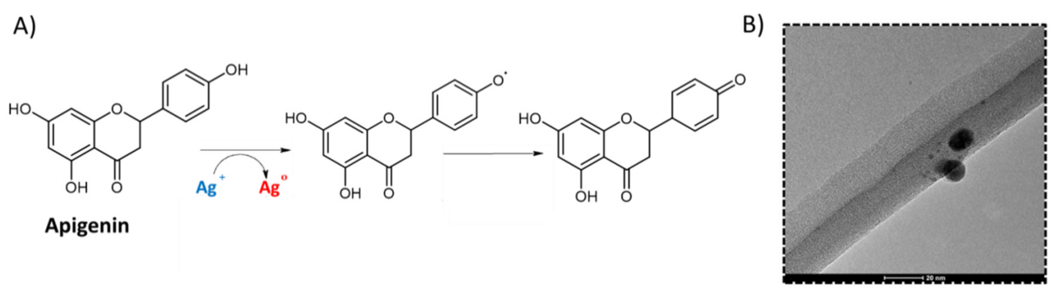
2.7.2. AgNP Method
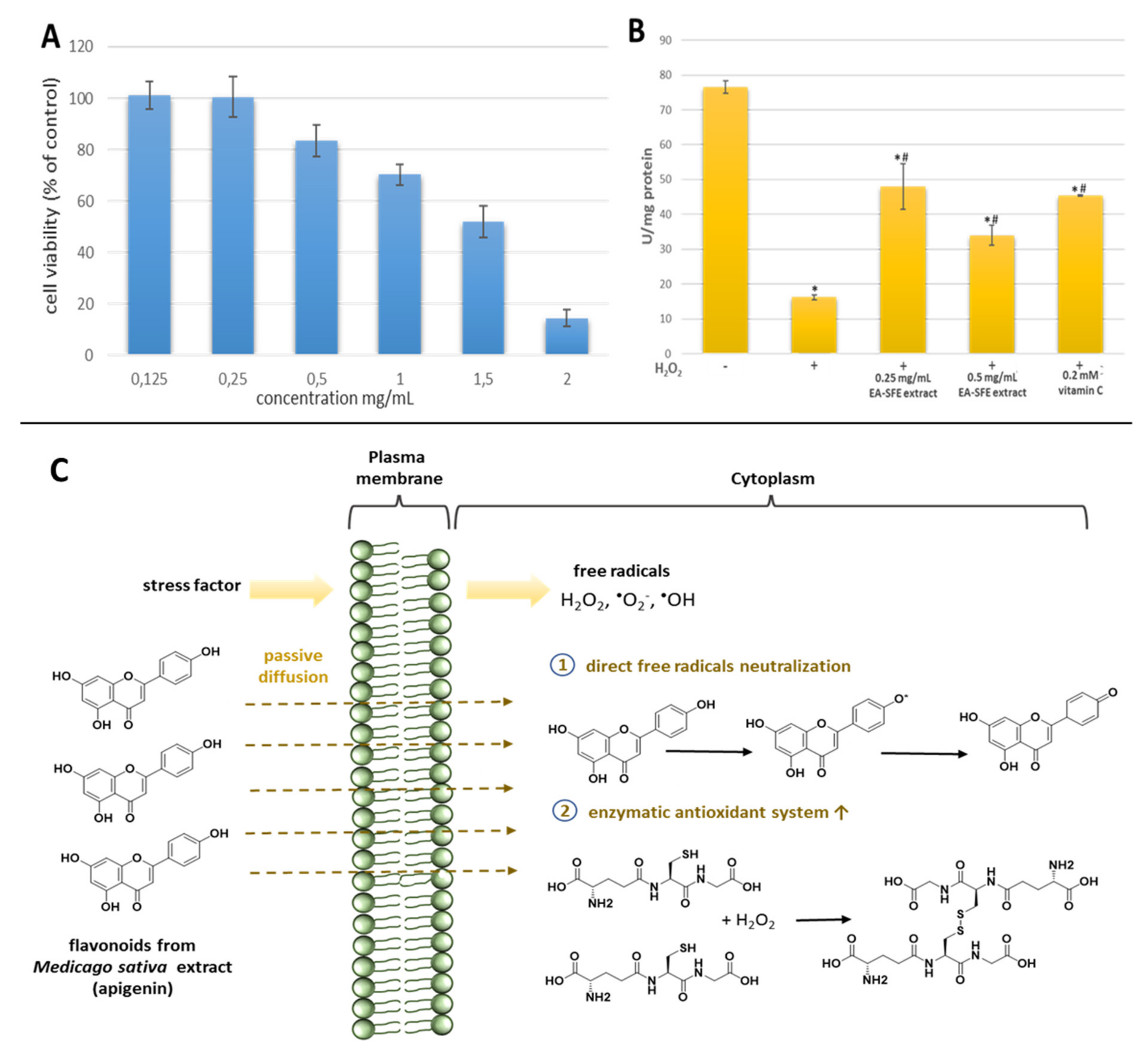
2.7.3. Impact of EA-SFE M. Sativa L. Extract on Antioxidant Enzyme Activity GSH-Px
3. Results and Discussion
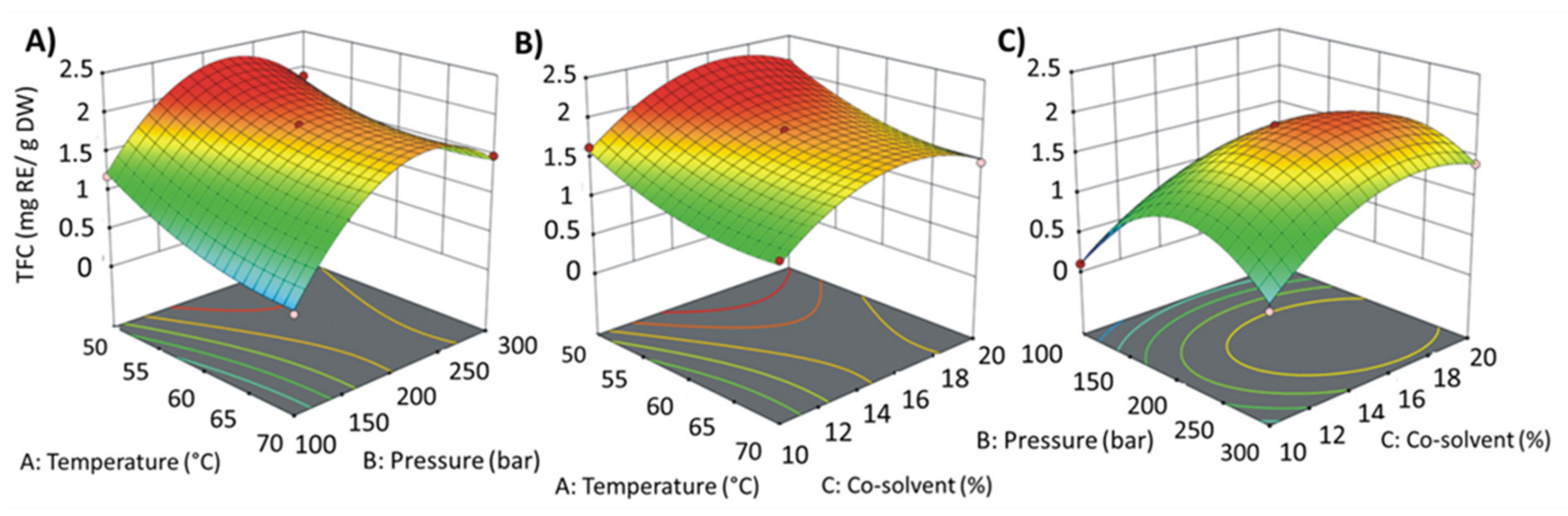
3.1. The Selection of Supercritical Fluid Extraction Conditions
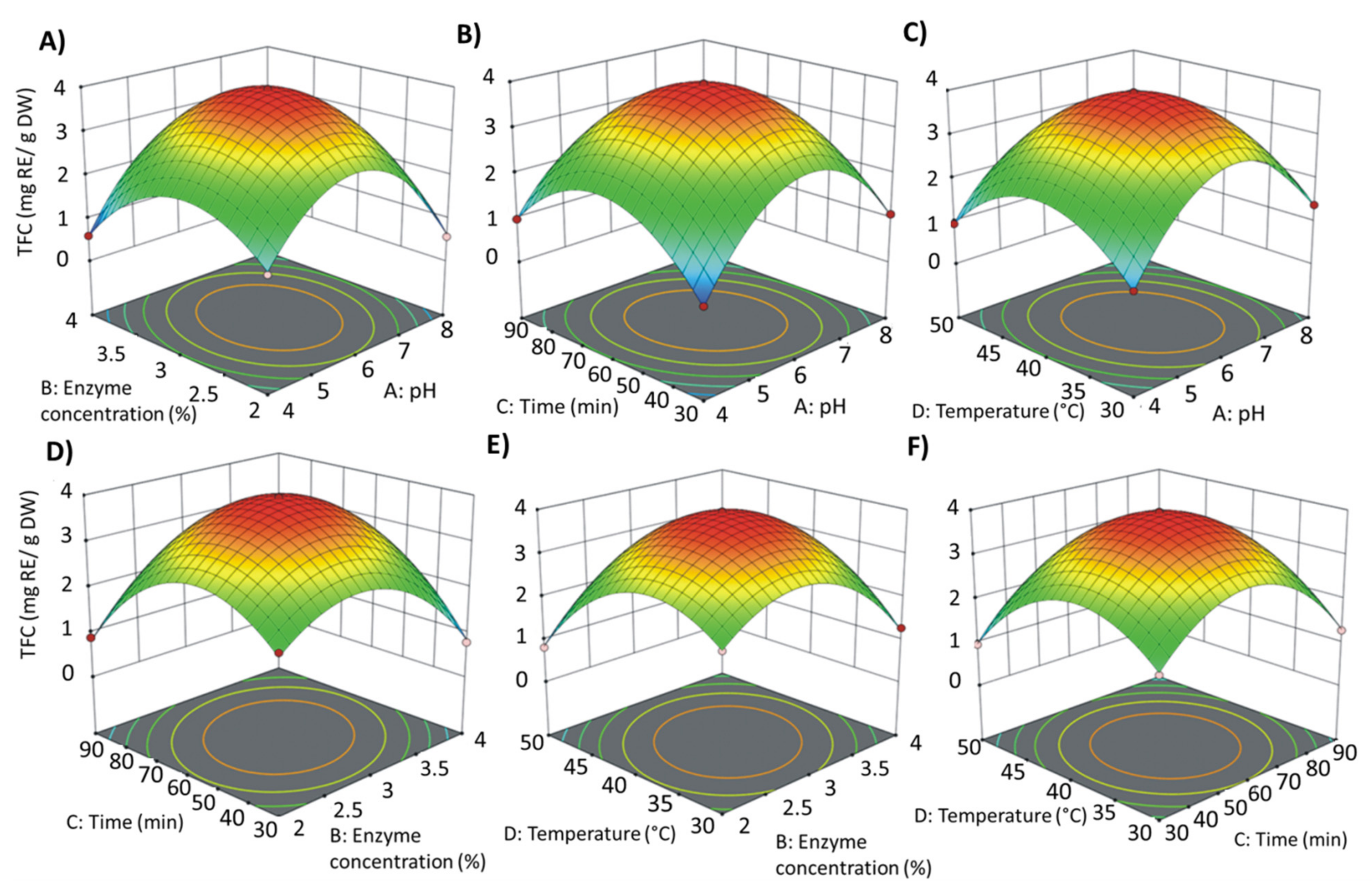
3.2. The Effect of Enzymatic Hydrolysis Conditions on the Total Flavonoid Content (TFC) in M. sativa Leaves Extracts
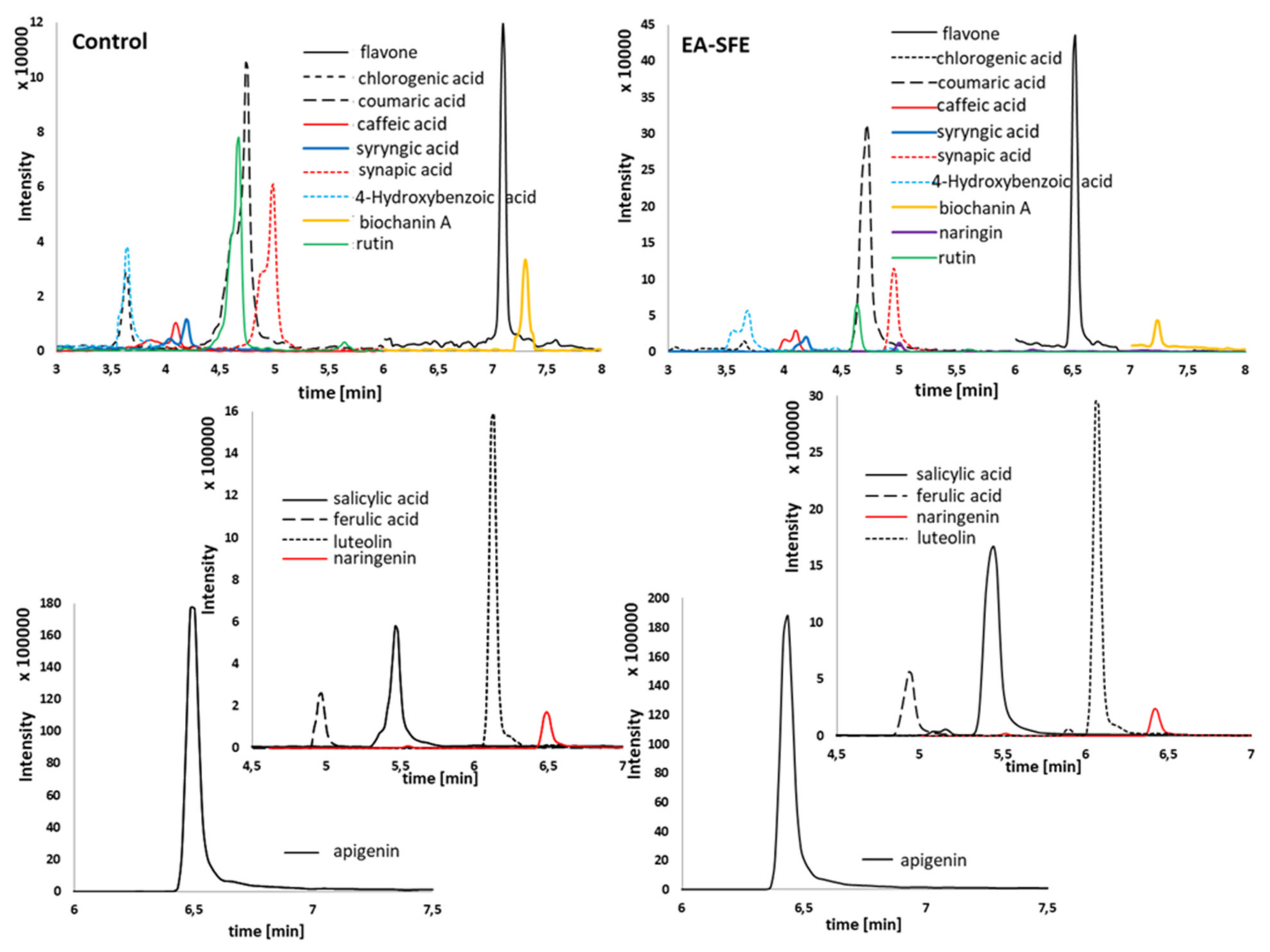
3.3. Chemical Analysis of Obtained Extracts by HPLC-ESI-MS/MS
3.4. Antioxidant Activity of the Extracts
4. Conclusions
Supplementary Materials
Author Contributions
Funding
Institutional Review Board Statement
Informed Consent Statement
Data Availability Statement
Acknowledgments
Conflicts of Interest
References
- Wink, M. Plant breeding: Importance of plant secondary metabolites for protection against pathogens and herbivores. Theor. Appl. Genet. 1988, 75, 225–233. [Google Scholar] [CrossRef]
- Wink, M. Introduction: Functions and Biotechnology of Plant Secondary Metabolites. In Annual Plant Reviews, 2nd ed.; Wiley-Blackwell: Hoboken, NJ, USA, 2010; Volume 39, pp. 1–16. [Google Scholar]
- Balandrin, M.F.; Klocke, J.A.; Wurtele, E.S.; Bollinger, W.H. Natural plant chemicals: Sources of industrial and medicinal materials. Science 1985, 228, 1154–1160. [Google Scholar] [CrossRef]
- Rice-Evans, C.; Miller, N.J.; Paganga, G. Structure-antioxidant activity relationships of flavonoids and phenolic acids. Free Radical Biol. Med. 1996, 20, 933–956. [Google Scholar] [CrossRef]
- Raffa, D.; Maggio, B.; Raimondi, M.V.; Plescia, F.; Daidon, G. Recent discoveries of anticancer flavonoids. Eur. J. Med. Chem. 2017, 142, 213–228. [Google Scholar] [CrossRef]
- Rafińska, K.; Pomastowski, P.; Wrona, O.; Górecki, R.; Buszewski, B. Medicago sativa as a source of secondary metabolites for agriculture and pharmaceutical industry. Phytoch. Lett. 2017, 20, 520–539. [Google Scholar] [CrossRef]
- Krakowska, A.; Rafińska, K.; Walczak, J.; Kowalkowski, T.; Buszewski, B. Comparison of various extraction techniques of Medicago sativa: Yield, antioxidant activity and content of phytochemical constituent. J. AOAC Int. 2017, 100, 1–13. [Google Scholar] [CrossRef]
- Krakowska, A.; Rafińska, K.; Walczak, J.; Buszewski, B. Enzyme-assisted optimized supercritical fluid extraction to improve Medicago sativa polyphenolics isolation. Ind. Crop. Prod. 2018, 124, 931–940. [Google Scholar] [CrossRef]
- Gaweł, E. Chemical composition of lucerne leaf extract (EFL) and its applications as a phytobiotic in human nutrition. Acta Sci. Pol. Technol. Aliment. 2012, 11, 303–310. [Google Scholar] [PubMed]
- Fu, Y.J.; Liu, W.; Zu, Y.-G.; Tong, M.-H.; Li, S.-M.; Yan, M.-M.; Efferth, T.; Luo, H. Enzyme assisted extraction of luteolin and apigenin from pigeonpea [Cajanuscajan (L.) Millsp.] leaves. Food Chem. 2008, 111, 508–512. [Google Scholar] [CrossRef]
- Mushtaq, M.; Sultana, B.; Akram, S.; Anwar, F.; Adnan, A.; Rizvi, S.S.H. Enzyme-assisted supercritical fluid extraction: An alternative and green technology for non-extractable polyphenols. Anal. Bioanal. Chem. 2017, 409, 3645–3655. [Google Scholar] [CrossRef] [PubMed]
- Cho, J.H.; Saurabh, B.; Tae-Jin, O.; Jong, H.J. Enzymatic Extraction of Pilocarpine from Pilocarpus jaborandi. Korean J. Microbiol. Biotechnol. 2013, 41, 236–241. [Google Scholar]
- Cavero, S.; García-Risco, M.R.; Marín, F.R.; Jaime, L.; Santoyo, S.; Señoráns, F.J.; Reglero, G.; Ibañez, E. Supercritical fluid extraction of antioxidant compounds from oregano. J. Supercrit. Fluid. 2006, 38, 62–69. [Google Scholar] [CrossRef]
- Sonia, A.O.; Santos, S.A.O.; Silva, C.A.; Neto, C.P.; Silvestre, A.J.D. Supercritical fluid extraction of phenolic compound from Eucalyptus globulus Labill bark. J. Supercrit. Fluid. 2012, 71, 71–79. [Google Scholar]
- Azmir, J.; Zaidul, I.S.M.; Rahman, M.M.; Sharif, K.M.; Mohamed, A.; Sahena, F.; Jahurul, M.H.A.; Ghafoor, K.; Norulaini, N.A.N.; Omar, A.K.M. Techniques for extraction of bioactive compounds from plant materials: A review. J. Food Eng. 2013, 117, 426–436. [Google Scholar] [CrossRef]
- Lang, Q.; Wai, C.M. Supercritical fluid extraction in herbal and natural product studies—A practical review. Talanta 2001, 53, 771–782. [Google Scholar] [CrossRef]
- Chemat, F.; Rombaut, N.; Meullemiestre, A.; Turk, M.; Perino, S.; Fabiano-Tixier, A.S.; Abert-Vian, M. Review of green food processing techniques. Preservation, transformation, and extraction. Innov. Food Sci. Emerg. Technol. 2017, 41, 357–377. [Google Scholar] [CrossRef]
- Radenkovs, V.; Püssa, T.; Juhnevica-Radenkova, K.; Kviesis, J.; Salar, F.J.; Moreno, D.A.; Drudze, I. Wild apple (Malus spp.) by-products as a source of phenolic compounds and vitamin C for food applications. Food Biosci. 2020, 38, 100744. [Google Scholar] [CrossRef]
- Tepić, A.N.; Dimić, G.R.; Vujičić, B.L.; Kevrešan, Ž. S.; Varga, M.; Šumić, Z.M. Quality of commercial ground paprika and its oleoresins. Acta Period. Technol. 2008, 39, 1–212. [Google Scholar]
- Taylor, L.T. Supercritical fluid chromatography for the 21st century. J. Supercrit. Fluids 2009, 47, 566–573. [Google Scholar] [CrossRef]
- Herreroa, M.; Mendiola, J.A.; Cifuentes, A.; Ibánez, E. Supercritical fluid extraction: Recent advances and applications. J. Chromatogr. A 2010, 1217, 2495–2511. [Google Scholar] [CrossRef]
- Rafińska, K.; Pomastowski, P.; Rudnicka, J.; Krakowska, A.; Maruśka, A.; Narkute, M.; Buszewski, B. Effect of solvent and extraction technique on composition and biological activity of Lepidium sativum extracts. Food Chem. 2019, 289, 16–25. [Google Scholar] [CrossRef]
- Buszewski, B.; Szultka, M. Past, Present, and Future of Solid Phase Extraction: A Review. Crit. Rev. Anal. Chem. 2012, 42, 198–213. [Google Scholar] [CrossRef]
- Bezerra, I.C.F.; Ramos, R.T.M.; Ferreira, M.R.A.; Soares, L.A.L. Optimization Strategy for Extraction of Active Polyphenols from Leaves of Eugenia uniflora Linn. Food Anal. Method 2020, 13, 735–750. [Google Scholar] [CrossRef]
- Zeković, Z.; Cvetanović, A.; Pavlić, B.; Švarc-Gajić, J.; Radojković, M. Optimization of the Polyphenolics Extraction from Chamomile Ligulate Flowers Using Response Surface Methodology. Int. J. Plant Prod. 2014, 4, 43–50. [Google Scholar]
- Buszewski, B.; Rafińska, K.; Cvetanović, A.; Walczak, J.; Krakowska, A.; Rudnicka, J.; Zeković, Z. Phytochemical analysis and biological activity of Lupinus luteus seeds extracts obtained by supercritical fluid extraction. Phytochem. Lett. 2019, 30, 338–348. [Google Scholar] [CrossRef]
- Li, B.B.; Smith, B.; Hossain, M.M. Extraction of phenolics from citrus peels: II. Enzyme assisted extraction method. Sep. Purif. Technol. 2006, 48, 189–196. [Google Scholar] [CrossRef]
- Gómez-García, R.; Martínez-Ávila, G.C.G.; Aguilar, C.N. Enzyme assisted extraction of antioxidative phenolics from grape (Vitis vinifera L.) residues. 3 Biotech 2012, 2, 297–300. [Google Scholar] [CrossRef]
- Wang, L.; Wu, Y.; Liu, Y.; Wu, Z. Complex enzyme-assisted extraction releases antioxidative phenolic compositions from guava leaves. Molecules 2017, 22, 1648. [Google Scholar] [CrossRef]
- Ghosh, D.; Biswas, P.K. Enzyme-aided extraction of carotenoids from pumpkin tissues. Indian Chem. Eng. 2015, 4506, 1–11. [Google Scholar] [CrossRef]
- Dal Magro, L.; Dalagnol, L.M.G.; Manfroi, V.; Hertz, P.F.; Klein, M.P.; Rodrigues, R.C. Synergistic effects of Pectinex Ultra Clear and Lallzyme Beta on yield and bioactive compounds extraction of Concord grape juice. LWT Food Sci. Technol. 2016, 72, 157–165. [Google Scholar] [CrossRef]
- Qin, Y.; Yuan, Q.; Zhang, Y.; Li, J.; Zhu, X.; Zhao, L.; Wen, J.; Liu, J.; Zhao, L.; Zhao, J. Enzyme-Assisted Extraction Optimization, Characterization and Antioxidant Activity of Polysaccharides from Sea Cucumber Phyllophorus proteus. Molecules 2018, 23, 590. [Google Scholar] [CrossRef]
- Li, S.; Han, D.; Row, K.H. Optimization of enzymatic extraction of polysaccharides from some marine algae by response surface methodology. Korean J. Chem. Eng. 2012, 29, 650–656. [Google Scholar] [CrossRef]
- Krakowska-Sieprawska, A.; Rafińska, K.; Walczak-Skierska, J.; Buszewski, B. The influence of plant material enzymatic hydrolysis and extraction conditions on the polyphenolic profiles and antioxidant activity of extracts: A green and efficient approach. Molecules 2020, 25, 2074. [Google Scholar] [CrossRef] [PubMed]
- Rouphael, Y.; Bernardi, J.; Cardarelli, M.; Bernardo, L.; Kane, D.; Colla, G.; Lucini, L. Phenolic compounds and sesquiterpene lactones profile in leaves of nineteen artichoke cultivars. J. Agr. Food Chem. 2016, 64, 8540–8548. [Google Scholar] [CrossRef]
- Szydłowska-Czerniak, A.; Tułodziecka, A.; Szłyk, E. A silver nanoparticle-based method for determination of antioxidant capacity of rapeseed and its products. Analyst 2012, 137, 3750–3759. [Google Scholar] [CrossRef] [PubMed]
- Wrona, O.; Rafińska, K.; Możeński, C.; Buszewski, B. Supercritical fluid extraction of bioactive compounds from plant materials. J. AOAC Int. 2017, 100, 1624–1635. [Google Scholar] [CrossRef]
- Juhnevica-Radenkova, K.; Kviesis, J.; Moreno, D.A.; Seglina, D.; Vallejo, F.; Valdovska, A.; Radenkovs, V. Highly-Efficient Release of Ferulic Acid from Agro-Industrial By-Products via Enzymatic Hydrolysis with Cellulose-Degrading Enzymes: Part I–The Superiority of Hydrolytic Enzymes Versus Conventional Hydrolysis. Foods 2021, 10, 782. [Google Scholar] [CrossRef] [PubMed]
- Khaw, K.Y.; Parat, M.O.; Shaw, P.N.; Falconer, J.R. Solvent supercritical Fluid technologies to extract bioactive compounds from natural sources: A review. Molecules 2017, 22, 1186. [Google Scholar] [CrossRef]
- Chemat, F.; Vian, M.A.; Cravotto, G. Green Extraction of Natural Products: Concept and Principles. Int. J. Mol. Sci. 2012, 13, 8615–8627. [Google Scholar] [CrossRef] [PubMed]
- Wrona, O.; Rafińska, K.; Walczak-Skierska, J.; Możeński, C.; Buszewski, B. Extraction and determination of polar bioactive compounds from alfalfa (Medicago sativa L.) using supercritical techniques. Molecules 2019, 24, 4608. [Google Scholar] [CrossRef]
- Rodríguez De Luna, S.L.; Ramírez-Garza, R.E.; Serna Saldívar, S.O. Environmentally friendly methods for flavonoid extraction from plant material: Impact of their operating conditions on yield and antioxidant properties. Sci. World J. 2020, 2020, 1–38. [Google Scholar] [CrossRef] [PubMed]
- Goyeneche, R.; Di Scala, K.; Ramirez, C.L.; Fanovich, M.A. Recovery of bioactive compounds from beetroot leaves by supercritical CO2 extraction as a promising bioresource. J. Supercrit. Fluids 2020, 155, 104658. [Google Scholar] [CrossRef]
- Rosenthal, A.; Pyle, D.L.; Niranjan, K. Aqueous and enzymatic processes for edible oil extraction. Enzyme Microb. Technol. 1996, 19, 402–420. [Google Scholar] [CrossRef]
- Bezzera, M.A.; Santelli, R.E.; Oliveira, E.P.; Villar, L.S.; Escaleira, L.A. Response surface methodology (RSM) as a tool for optimization in analytical chemistry. Talanta 2008, 76, 965–977. [Google Scholar] [CrossRef] [PubMed]
- Wijesinghe, W.A.; Jeon, Y.J. Enzyme-assistant extraction (EAE) of bioactive components: A useful approach for recovery of industrially important metabolites from seaweeds: A review. Fitoterapia 2012, 83, 6–12. [Google Scholar] [CrossRef] [PubMed]
- Marathe, S.J.; Jadhav, S.B.; Bankar, S.B.; Dubey, K.K.; Singhal, R.S. Improvements in the extraction of bioactive compounds by enzymes. Curr. Opin. Food Sci. 2019, 25, 62–72. [Google Scholar] [CrossRef]
- Robinson, P.K. Enzymes: Principles and biotechnological applications. Essays Biochem. 2015, 59, 1–41. [Google Scholar] [CrossRef]
- Nadar, S.S.; Rao, P.; Rathod, V.K. Enzyme assisted extraction of biomolecules as an approach to novel extraction technology: A review. Food Res. Int. 2018, 108, 309–330. [Google Scholar] [CrossRef]
- Szultka, M.; Papaj, K.; Rusin, A.; Szeja, W.; Buszewski, B. Determination of flavonoids and their metabolites by chromatographic techniques. Trend. Anal. Chem. 2013, 47, 47–67. [Google Scholar] [CrossRef]
- Williams, R.J.; Spencer, J.P.E.; Rice-Evans, C. Flavonoids: Antioxidants or signaling molecules? Free Radical Bio. Med. 2004, 36, 838–849. [Google Scholar] [CrossRef] [PubMed]
- Procházková, D.; Boušová, I.; Wilhelmová, N. Antioxidant and prooxidant properties of flavonoids. Fitoterapia 2011, 82, 513–523. [Google Scholar] [CrossRef]
- Kutchan, T.M. A role for intra- and intercellular translocation in natural product biosynthesis. Curr. Opin. Plant Biol. 2005, 8, 292–300. [Google Scholar] [CrossRef]
- Mushtaq, M.; Sultana, B.; Bhatti, H.N.; Asghar, M. RSM based optimized enzyme-assisted extraction of antioxidant phenolics from underutilized watermelon (Citrullus lanatus Thunb.) rind. J. Food Sci. Tech. 2015, 52, 5048–5056. [Google Scholar] [CrossRef] [PubMed]
- Bittencourt, G.M.; Firmiano, D.M.; Fachini, R.P.; Lacaz-Ruiz, R.; Fernandes, A.M.; Oliveira, A.L. Application of green technology for the acquisition of extracts of araçá (Psidium grandifolium Mart. ex DC.) using supercritical CO2 and pressurized ethanol: Characterization and analysis of activity. J. Food Sci. 2019, 84, 1297–1307. [Google Scholar] [CrossRef] [PubMed]
- Espín, J.C.; Soler-Rivas, C.; Wichers, H.J. Characterization of the total free radical scavenger capacity of vegetable oils and oil fractions using 2,2-diphenyl-1-picrylhydrazyl radical. J. Agr. Food Chem. 2000, 48, 648–656. [Google Scholar] [CrossRef]
- Buszewski, B.; Rafińska, K.; Pomastowski, P.; Walczak, J.; Rogowska, A. Novel aspects of silver nanoparticles functionalization. Colloids Surf. A 2016, 506, 170–178. [Google Scholar] [CrossRef]
- Ismail, H.F.; Hashim, Z.; Soon, W.T.; Rahman, N.S.A.; Zainudin, A.N.; Majid, F.A.A. Comparative study of herbal plants on the phenolic and flavonoid content, antioxidant activities and toxicity on cells and zebrafish embryo. J. Tradit. Complement. Med. 2017, 7, 452–465. [Google Scholar] [CrossRef] [PubMed]


| Source | Sum of Squares | Degrees of Freedom | Mean of Square | F-Value | p-Value |
|---|---|---|---|---|---|
| Selection of SFE Conditions | |||||
| TFC | |||||
| Model | 5.77 | 9 | 0.6416 | 92.68 | <0.0001 (significant) |
| Residual | 0.0346 | 5 | 0.0069 | ||
| Lack of fit | 0.0328 | 3 | 0.0109 | 11.74 | 0.0795 (not significant) |
| Pure error | 0.0019 | 2 | 0.0009 | ||
| Total | 5.81 | 14 | |||
| * Y (TFC) = 1.85 − 0.29X1 + 0.40X2 + 0.26X3 + 0.10X1X2 − 0.03X1X3 + 0.15X2X3 + 0.19X12 − 0.79X220.45X32 | |||||
| Selection of Enzymatic Hydrolysis Conditions | |||||
| TFC | |||||
| Model | 26.13 | 14 | 1.87 | 515.06 | <0.0001 (significant) |
| Residual | 0.0435 | 12 | 0.0036 | ||
| Lack of fit | 0.0430 | 10 | 0.0043 | 18.80 | 0.0515 (not significant) |
| Pure error | 0.0005 | 2 | 0.0002 | ||
| Total | 26.17 | 26 | |||
| ** Y (TFC) = 3.94 − 0.013X1 − 0.040X2 − 0.06X3 − 0.23X4 + 0.33X1X2 − 0.23X1X3 − 0.22X1X4 + 0.51X2X3 + 0.45X2X4 + 0.19X3X4 − 1.78X12 − 1.23X22 − 1.38X32 − 1.23X42 | |||||
| Optimal Conditions of the SFE | ||||||||
|---|---|---|---|---|---|---|---|---|
| Response Variable | Temperature (°C) | Pressure (bar) | Cosolvent (%) | Predicted Value | Experimental Value | Confidence Interval | ||
| Desirability-1.0 | −95% | 95% | ||||||
| TFC (mg RE/g DW) | 50 | 216 | 19.4 | 2.28 | 2.15 | 2.11 | 2.45 | |
| Optimal Conditions of the Enzymatic Treatment | ||||||||
| Response Variable | pH | Enzyme Concentration (%) | Time(min) | Temperature (°C) | Predicted Value | Experimental Value | Confidence Interval | |
| Desirability-1.0 | −95% | 95% | ||||||
| TFC (mg RE/g DW) | 6.00 | 2.96 | 58.92 | 38.96 | 3.96 | 3.89 | 3.88 | 4.03 |
| HPLC-MS/MS Determination | |||||
|---|---|---|---|---|---|
| Compound | tR (min) | MRM | Concentration (µg/g) | ||
| Maceration | SFE (Control) | EA-SFE (Optimal Conditions) | |||
| Phenolic acids | |||||
| Gallic acid + | 2.138 | 169–124 | ND a | ND | ND |
| Salicylic acid + | 3.961 | 137–93 | 6.79 ± 0.62 | 26.50 ± 2.94 | 100.48 ± 2.53 |
| p-Coumaric acid + | 3.450 | 163–119 | 0.83 ± 0.34 | 1.34 ± 0.33 | 3.78 ± 0.10 |
| Chlorogenic acid + | 2.681 | 353–191 | ND | 0.32 ± 0.14 | 0.13 ± 0.04 |
| Caffeic acid + | 3.036 | 179–135 | 0.59 ± 0.22 | 0.92 ± 0.05 | 2.44 ± 0.11 |
| Syringic acid + | 3.118 | 197–95 | 3.34 ± 0.99 | 4.73 ± 1.12 | 14.74 ± 2.77 |
| Ferulic acid + | 3.566 | 193–134 | 56.36 ± 9.43 | 126.93 ± 6.99 | 277.41 ± 7.87 |
| Protocatechuic acid + | 2.482 | 153–108 | 11.72 ± 2.78 | 2.82 ± 0.12 | 5.73 ± 1.89 |
| Sinapic acid + | 3.553 | 223–121 | 3.03 ± 1.65 | 7.88 ± 3.57 | 9.28 ± 0.39 |
| 4-Hydroxybenzoic acid + | 2.890 | 137–65 | 4.48 ± 1.07 | 6.17 ± 2.22 | 20.80 ± 2.76 |
| ⅀ | 87.14 ± 17.10 | 177.61 ± 17.48 | 434.78 ± 18.46 | ||
| Flavonoids | |||||
| Flavone ++ | 5.452 | 223–121 | 0.01 ± 0.00 | 0.23 ± 0.02 | 0.16 ± 0.01 |
| Fisetin + | 3.928 | 285–121 | ND | ND | ND |
| Kaempferol + | 4.811 | 285–255 | ND | ND | ND |
| Apigenin + | 4.730 | 269–117 | 63.09 ± 1.33 | 87.48 ± 4.32 | 95.53 ± 1.19 |
| Luteolin + | 4.359 | 285–133 | 9.12 ± 0.48 | 5.15 ± 0.23 | 9.86 ± 1.09 |
| Rutin + | 3.447 | 609–300 | 0.62 ± 0.12 | 0.92 ± 0.13 | 0.49 ± 0.17 |
| Quercetin + | 4.382 | 301–227 | 0.02 ± 0.00 | 0.61 ± 0.04 | 1.02 ± 0.23 |
| Naringin + | 3.528 | 579–271 | ND | ND | 0.15 ± 0.01 |
| Naringenin + | 4.744 | 271–119 | 1.39 ± 0.70 | 2.09 ± 0.13 | 2.53 ± 0.10 |
| Esculin + | 2.492 | 339–177 | ND | 0.01 ± 0.00 | 0.01 ± 0.00 |
| Esculetin + | 3.053 | 177–89 | 0.41 ± 0.06 | 0.37 ± 0.11 | 1.10 ± 0.13 |
| Biochanin A + | 5.666 | 283–211 | 0.06 ± 0.01 | 0.21 ± 0.04 | 0.21 ± 0.01 |
| Catechin + | 3.012 | 289–123 | ND | ND | ND |
| ⅀ | 74.72 ± 2.70 | 97.07 ± 5.02 | 111.06 ± 2.94 | ||
| ⅀ | 161.86 ± 19.80 | 274.68 ± 22.50 | 545.84 ± 21.40 | ||
| Antioxidant activity | |||||
| AgNP method (µmol AP/g DW) | 22.91 ± 0.81 | 20.50 ± 0.81 | 27.36 ± 0.75 | ||
| DPPH method (µmol TEAC/g DW) | 0.85 ± 0.01 | 0.97 ± 0.02 | 1.71 ± 0.08 | ||
Publisher’s Note: MDPI stays neutral with regard to jurisdictional claims in published maps and institutional affiliations. |
© 2021 by the authors. Licensee MDPI, Basel, Switzerland. This article is an open access article distributed under the terms and conditions of the Creative Commons Attribution (CC BY) license (https://creativecommons.org/licenses/by/4.0/).
Share and Cite
Krakowska-Sieprawska, A.; Rafińska, K.; Walczak-Skierska, J.; Kiełbasa, A.; Buszewski, B. Promising Green Technology in Obtaining Functional Plant Preparations: Combined Enzyme-Assisted Supercritical Fluid Extraction of Flavonoids Isolation from Medicago Sativa Leaves. Materials 2021, 14, 2724. https://doi.org/10.3390/ma14112724
Krakowska-Sieprawska A, Rafińska K, Walczak-Skierska J, Kiełbasa A, Buszewski B. Promising Green Technology in Obtaining Functional Plant Preparations: Combined Enzyme-Assisted Supercritical Fluid Extraction of Flavonoids Isolation from Medicago Sativa Leaves. Materials. 2021; 14(11):2724. https://doi.org/10.3390/ma14112724
Chicago/Turabian StyleKrakowska-Sieprawska, Aneta, Katarzyna Rafińska, Justyna Walczak-Skierska, Anna Kiełbasa, and Bogusław Buszewski. 2021. "Promising Green Technology in Obtaining Functional Plant Preparations: Combined Enzyme-Assisted Supercritical Fluid Extraction of Flavonoids Isolation from Medicago Sativa Leaves" Materials 14, no. 11: 2724. https://doi.org/10.3390/ma14112724
APA StyleKrakowska-Sieprawska, A., Rafińska, K., Walczak-Skierska, J., Kiełbasa, A., & Buszewski, B. (2021). Promising Green Technology in Obtaining Functional Plant Preparations: Combined Enzyme-Assisted Supercritical Fluid Extraction of Flavonoids Isolation from Medicago Sativa Leaves. Materials, 14(11), 2724. https://doi.org/10.3390/ma14112724