Article

Structural Dependent Eu3+ Luminescence, Photoelectric and Hysteresis Effects in Porous Strontium Titanate

1
Department of Micro- and Nanoelectronics, Belarusian State University of Informatics and Radioelectronics, P. Browka Str. 6, 220013 Minsk, Belarus
2
Department of Condensed Matter Physics, National Research Nuclear University MEPhI, Kashirskoe Shosse 31, 115409 Moscow, Russia
3
Department of Micro and Nano Electronics, Ryazan State Radio Engineering University, Gagarin Str. 59, 390005 Ryazan, Russia
4
Faculty of Electronics, Saint Petersburg Electrotechnical University “LETI”, Professor Popov Str. 5, 197376 Saint Petersburg, Russia
5
Department of Engineering, University of Applied Sciences Brandenburg, Magdeburger Str. 50, 14770 Brandenburg an der Havel, Germany
6
Scientific-Practical Materials Research Centre of NASB, P. Browka Str. 19, 220072 Minsk, Belarus
*
Author to whom correspondence should be addressed.
Materials 2020, 13(24), 5767; https://doi.org/10.3390/ma13245767
Submission received: 18 November 2020 / Revised: 9 December 2020 / Accepted: 14 December 2020 / Published: 17 December 2020
(This article belongs to the Section Porous Materials)

Abstract

:
Eu3+ doped porous nanostructured SrTiO3 films and powder fabricated by sol-gel route without using any precursor template are characterized by different morphology and phase composition. The films and the power show red and yellow luminescence with the most intensive photoluminescence (PL) bands at 612 nm and 588 nm, respectively. Raman, secondary ion mass spectrometry (SIMS), and X-ray diffraction (XRD) analysis of undoped nanostructured porous SrTiO3 films showed the presence of TiO2, SrO, and SrTiO3 phases and their components. The undoped porous SrTiO3 films are photosensitive and demonstrate resistive switching. The capacitance-voltage hysteresis loops with the width of about 6 V in the frequency range of 2 kHz—2 MHz were observed.

1. Introduction

Materials containing Eu3+ ions are promising as red phosphors in optoelectronic devices, e.g., LEDs and lasers, due to their excellent photoluminescence properties [1,2,3]. Optical radiation in the europium emission band at 612 nm is perceived by the human eye as red light, which makes these materials particularly important. The luminescence spectrum can differ in different crystal matrices. Material composition, crystal structure, and presence of defects have a significant influence.
Oxide compounds with a perovskite structure, such as strontium titanate (SrTiO3), are widely used in nonlinear optics [4], electro-optical modulators [5], photocatalysis [6,7], thin-film capacitors [8,9], and memory devices [10,11]. It is a good host matrix for Eu3+ due to its radiation resistance and thermal stability.
Properties of SrTiO3 depend not only on the chemical composition, but also on the structure and morphology [12,13]. As a result of the modification of titanium and layered metal oxide systems, fabricated oxide coatings have different compositions, surface morphology, and properties that are determined by the structural parameters, in particular, nanosized grains [14,15], which is also right for more complex oxide systems. A decrease in the grain size of SrTiO3 at the nanoscale leads to distinctive properties as compared to the bulk material [16,17]. In recent years, several attempts were made to synthesize porous perovskites SrTiO3 and BaTiO3 [18,19].
Nanostructured perovskites have great potential for applications. Nanostructured SrTiO3 is a promising material for water purification from heavy metals [19] and photocatalysis [20]. Grain size and porosity affect the electrophysical parameters of perovskites. For example, porous barium titanate has a lower dielectric constant [21], and the Curie temperature increased up to 350 °C as compared to the bulk material [18]. Hysteresis of capacitance-voltage (C-V) characteristics of bulk monocrystralline SrTiO3 with Ni and Au electrodes was investigated before and after irradiation in [22,23], where C-V hysteresis was considered from the point of the drift of nonequilibrium charge carriers in the crystal. The hysteresis effects were observed not only in dielectric properties but also in magnetization loops [24,25]. Thus, the study of nanostructured porous perovskites is of particular interest for obtaining new properties of materials and creating novel structures based on them.
The synthesis of such nanostructured composites is possible by chemical methods, which include the sol-gel technology. This technology has a low cost, allows variation of the grain size, phase composition, concentration of dopants. The use of titanium alkoxides is the most promising for the synthesis of nanostructured SrTiO3, since they easily hydrolyze, forming hydrated oxides. By varying the temperature of dehydration, one can control the dispersion and morphology, the phase composition and the physical properties of the resulting xerogels. The use of metal alkoxides in the synthesis of multicomponent oxides ensures a high chemical homogeneity of the product, significantly reducing the temperature of oxide phases formation [26,27].
One of the synthesis techniques includes using templates, which control formation periodical porous SrTiO3 structure and repeatable combs form [28]. Another approach is based on using alcoxides and influence of water on the morphology of xerogels [18,29,30,31]. This approach allows for the fabrication of porous perovskites with grains larger than 100 nm. Nevertheless, the formation of porous perovskites with much smaller grains is a problem to be solved.
In this paper we present structural, luminescent and dielectric properties of porous undoped and Eu-doped SrTiO3 films with grains as small as 30 nm fabricated on monocrystalline silicon substrates by the sol-gel technique without any templates and compare them with those of identically synthesized bulk materials.

2. Materials and Methods

Porous SrTiO3 films doped with Eu, undoped ones and SrTiO3:Eu powder were synthesized using water containing sols. The sols were synthesized in the following way. First titanium isopropoxide was dissolved in a mixture of ethylene glycol monomethyl ether and nitric acid to prevent gelation initiated by titanium isopropoxide. Then, strontium nitrate was dissolved in the distilled water, followed by the addition of ethylene glycol monomethyl ether. Finally, both solutions were mixed to obtain the sols. To fabricate europium doped sols europium nitrate was added.
The sols were deposited by spinning at the rate of 2700 rpm for 30 s on monocrystalline (111) silicon wafers. The samples were then dried at 200 °C for 10 min and next film was identically deposited and dried. The deposition was repeated 5–10 times in order to fabricate the film of an appropriate thickness. Final calcination was performed at 800 °C for 40 min in air. Porous SrTiO3:Eux (x = 0.053) films containing 5 layers and undoped SrTiO3 films containing 10 layers were fabricated on silicon substrates. Powder materials were synthesized by the same heat processing of the precursors in a ceramic crucible.
Silicon substrate covered by TiOx/Pt layers with Pt playing a role of an isolated from the substrate electric contact was used for fabrication of the heterostructure Si/TiOx/Pt/SrTiO3/Ni for electrical measurements. The top round nickel electrode of about 330 µm in diameter was formed by ion-beam evaporation technique [9].
The morphology of the experimental samples was examined with scanning electron microscope S-4800 (Hitachi, Tokyo, Japan). The photoluminescence spectra were recorded at room temperature using a laser spectroscopic complex based on a Solar TII MS 7504i spectrograph-monochromator (Solar TII, Minsk, Belarus) in which a 1 kW xenon lamp was used for excitation. The photoluminescence light was detected by a digital camera (Proscan, Minsk, Belarus) with a cooled silicon CCD matrix. To isolate monochromatic lines from a wide spectrum of the lamp, we used a Solar TII DM 160 double monochromator (Solar TII, Minsk, Belarus). The photoluminescence was excited by the monochromatic light with the wavelength of 345 nm.
XRD (BOUREVESTNIK JSC, Saint Petersburg, Russia) studies of the films were carried out with a DRON-3 diffractometer using monochromatized CuKα radiation.
Raman spectra were measured with 3D Scanning Laser Confocal Raman Microscope Confotec NR500 (SOL Instruments, Minsk, Belarus) using 473 nm laser radiation for excitation. The measurements were performed at room temperature.
The depth distributions of the elements were investigated by secondary-ion mass spectrometry (SIMS) (TOF.SIMS 5, IONTOF, Munster, Germany). Secondary positive ions were produced by the bombardment of the samples with 30 keV Bi+ ions (1 pA). In situ etching of the samples was performed by sputtering with a focused 2 keV Cs+ ion beam (100 nA) raster scanned across a sample surface of 150 µm2.
Capacitance-voltage (C-V) and current-voltage (I-V) characteristics were recorded at room temperature using an Agilent E4980A Precision LCR meter (Agilent Technologies, Bayan Lepas, Pulau Pinang, Malaysia) integrated with Keithley 6517B (Keithley Instruments, Solon, Ohio USA) voltage source and digital V7-23 voltmeter, V7-27A ammeter and stabilized TEC-23 power supply, respectively. The C-V measurements were performed in the frequency range of 2 kHz–2 MHz. All measurements were done in automatic mode, implemented using the LabVIEW engineering graphic programming environment [32].

3. Results and Discussion

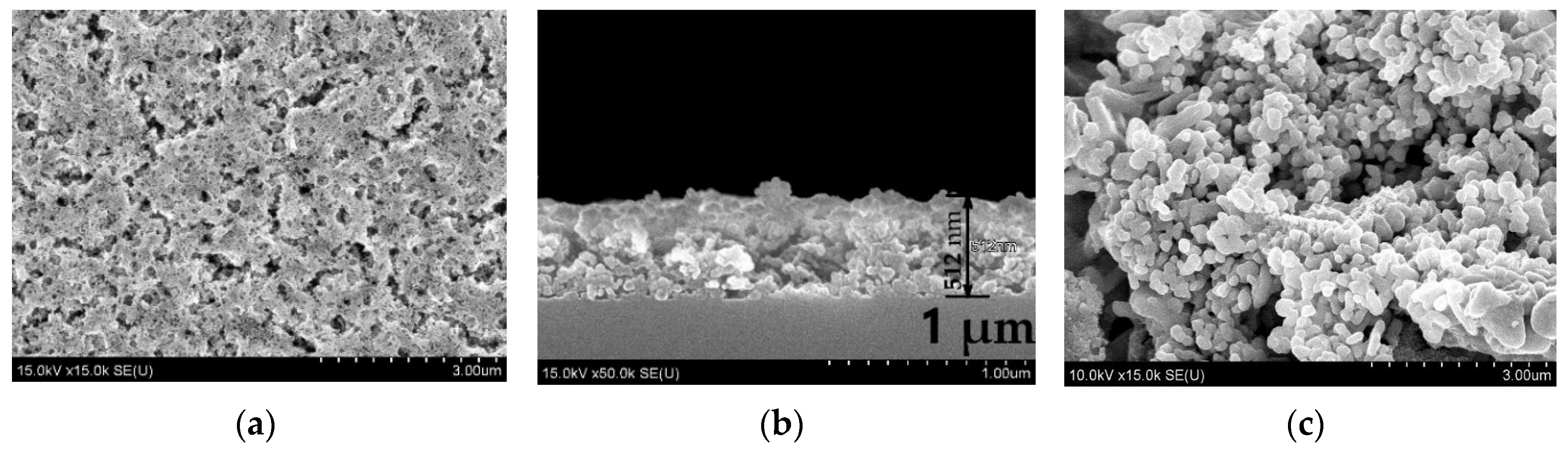
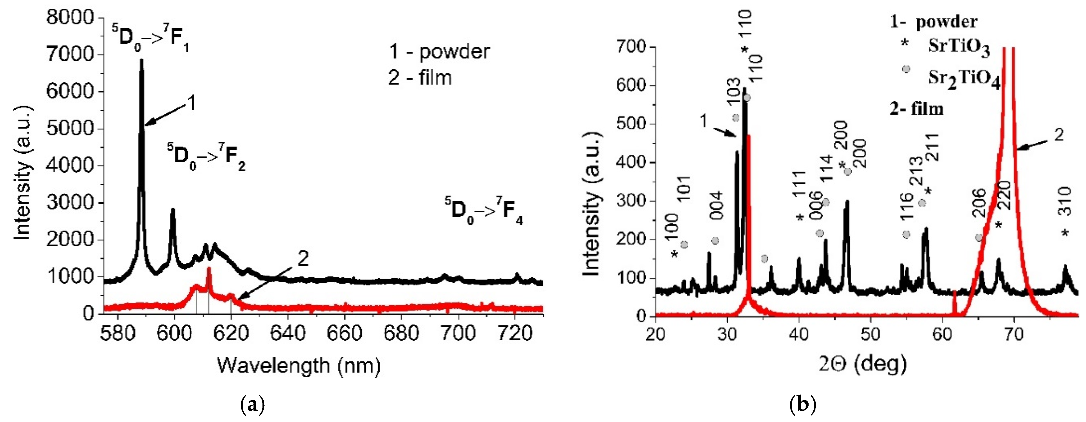
3.1. Porous Films and Powder Containing Strontium Titanate and Europium Ions

The SEM images of the five-layer SrTiO3:Eu3+ film with the thickness of about 500 nm and identically synthesized powder are given in Figure 1. The films clearly reveal high porosity and highly developed surface. Grains in the powder are larger than in the film.
PL spectra of the films presented in Figure 2a demonstrate the bands at 607, 612 and 619 nm corresponding to electrical dipole transition 5D07F2.
Due to the high solubility of europium ions and the ability to deploy in defective areas of the crystal, the incorporation of these ions can contribute to the phase formation in the material [33,34]. The intense PL band corresponding to the 5D07F2 electrical dipole transitions of Eu3+ indicates that europium occupies the position in the SrTiO3 crystallites that does not coincide with the symmetry center, most likely in defect regions [35].
PL spectra of the synthesized powder contain the bands at 588, 599, 607, 611, 626, 695, 700, 720 and 726 nm corresponding to the 5D07F1, 5D07F2, 5D07F4 transitions (Figure 2a). In this case the most intensive PL band is observed for magnetic dipole transition 5D07F1 at 588 nm. It indicates that Eu3+ ions occupy the positions with the symmetry center [35].
The XRD analysis of the powder presented in Figure 2b shows the diffraction peaks typical for SrTiO3 (PDF 01-089-4934) and Sr2TiO4 (PDF 00-039-1471). There are no signals belonging to the europium oxide. In addition to the identified peaks, there are ones that can be attributed to unreacted initial oxides and related solid solutions. The observed formation of Sr2TiO4 phase can be a result of Eu substitution by Sr leading to the enrichment of the powder with Sr.
XRD pattern of the five-layer SrTiO3:Eu3+ film contains the intense peak associated with the silicon substrate (Figure 2b). Apparently, the SrTiO3:Eu3+ film has a diffuse line in the region of 32.2 deg (influx to the left of the first sharp peak).
The powder synthesized from the same sol as the porous film has much larger grains as compared with the porous film and is characterized by the obvious phase formation. As a result, the PL spectra of the powder and the film differ. The presence of defects and disordering of the crystal structure certainly affects the luminescence spectrum of europium. The most intensive peak at 588 nm corresponding to the magnetic dipole transition 5D07F1 confirms that Eu3+ occupies the position with the symmetry center to be consistent with the XRD data. The typical doping concentration for lanthanides in solids is about 1 atomic %. In this case, it is not mandatory that trivalent lanthanide ion substitute only trivalent ion in host matrix, and there are no other local states for lanthanides. Luminescence of trivalent lanthanide ions with typical transitions between the corresponding terms was observed in inorganic perovskites: Tb3+ in YAlO3 [36], Er3+ in LiNbO3 corresponds to a [37], Eu3+ in BaTiO3 [38], as well as Eu3+ in sol-gel derived amorphous yttrium alumina composites [39]. Europium ion is the dopant that can exhibit divalent or trivalent valence state [40,41]. The luminescence spectra correspond to PL bands of Eu3+, as the PL spectra of Eu2+ lies in the region of about 380–500 nm [40]. In the case when Eu3+ occupies the position of strontium while remaining trivalent, the charge is compensated by the field of local environment, for example, titanium ions [42]. Replacement of divalent ions with trivalent leads to distortions of the crystal lattice of the structure [43,44]. Since the degree of doping of the image with europium is low; doping of the sample does not introduce significant distortions of the crystalline environment of europium ion, which corresponds to the presence of a narrow intense magnetic transition 5D07F1 (588 nm) in the spectrum. The electric dipole transition 5D07F2, observed at 599–626 nm, is allowed only when Eu3+ is located at a noncentrosymmetric crystallographic site. Otherwise considering low solid-solubility limit of Eu3+ in the perovskite matrix, such PL spectra is probably the result of the simultaneous exchange substitution of Sr2+ and Ti4+ positions by Eu3+ ions [42].

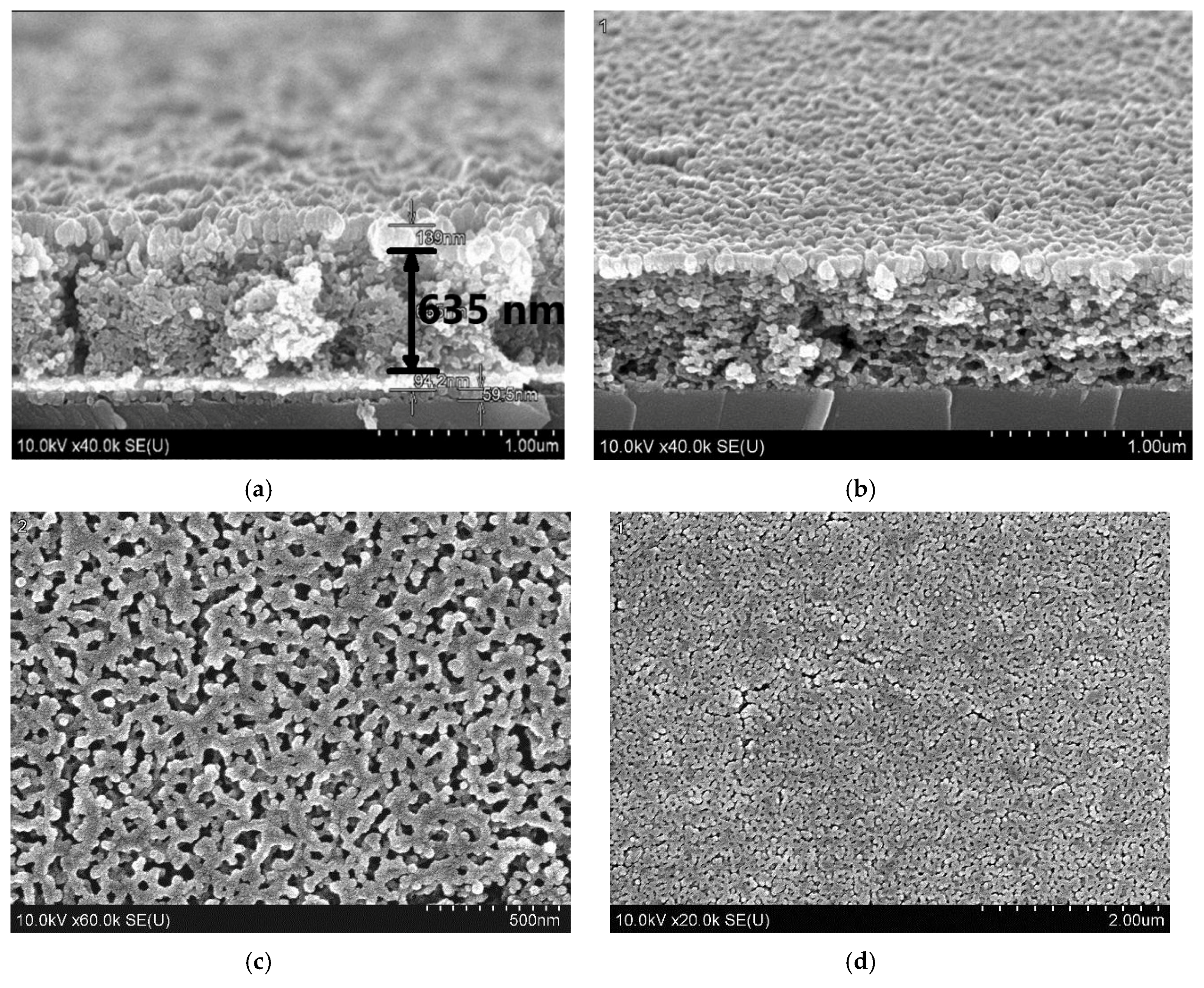
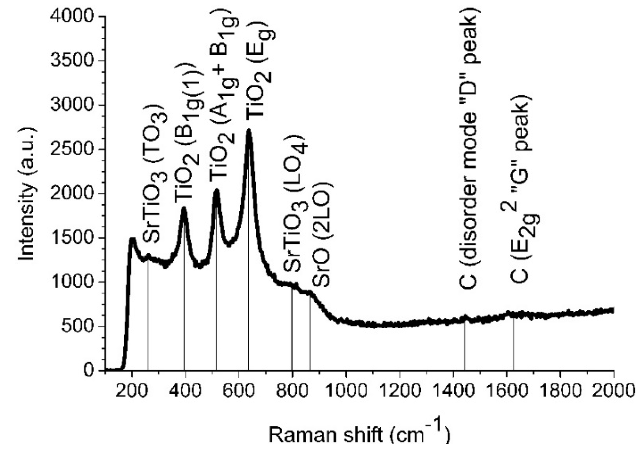
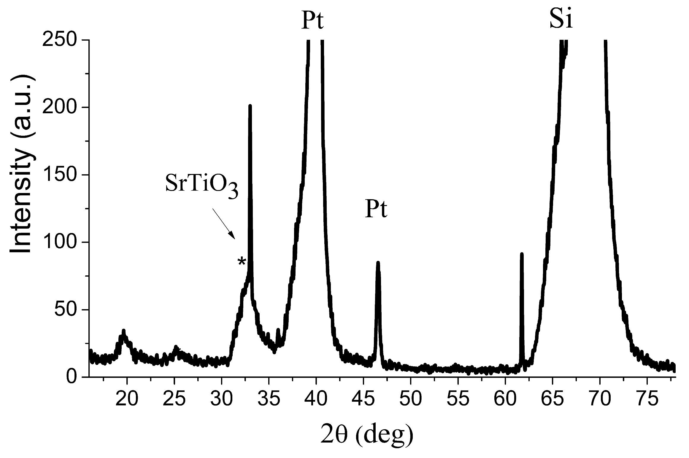
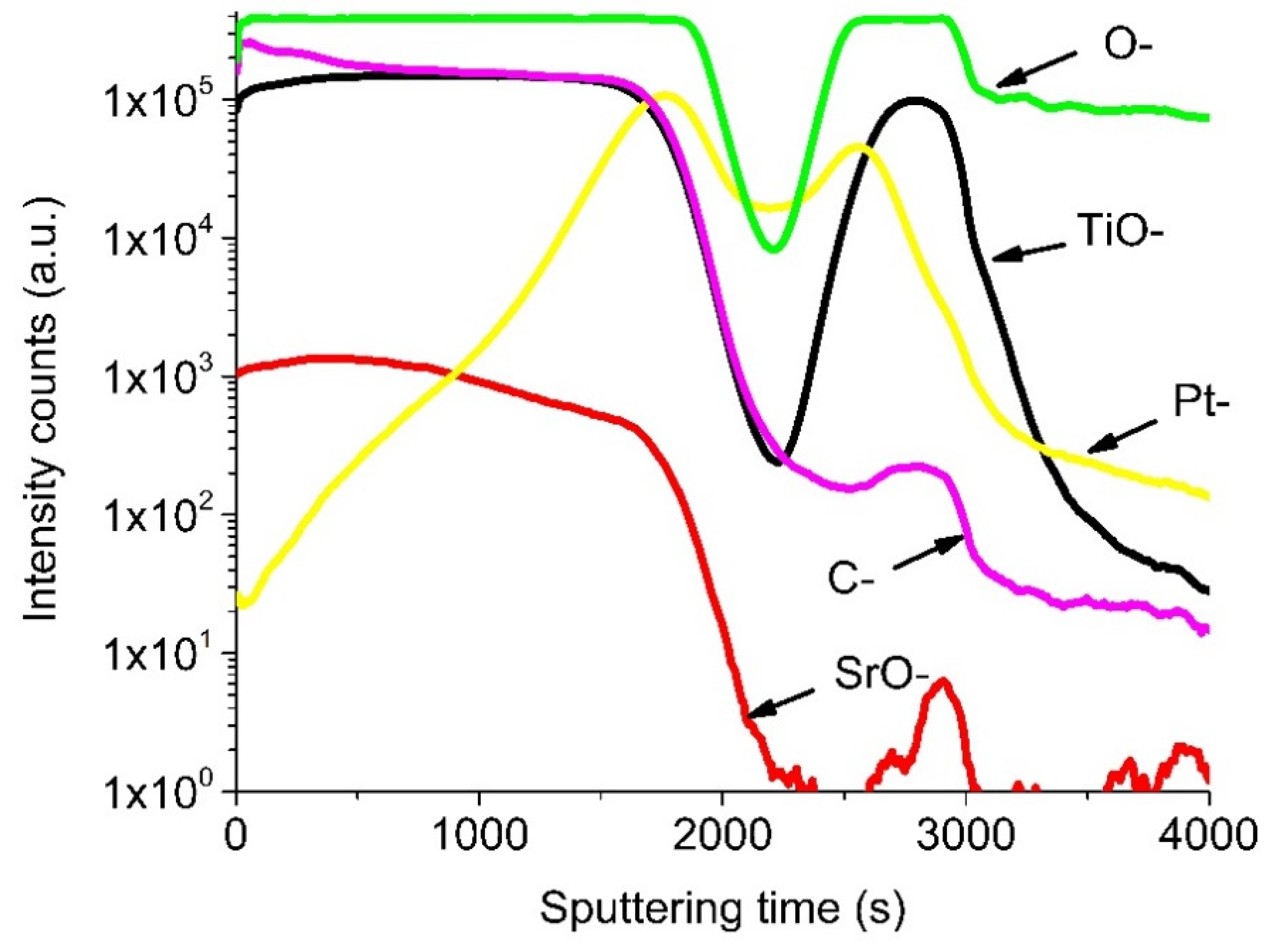
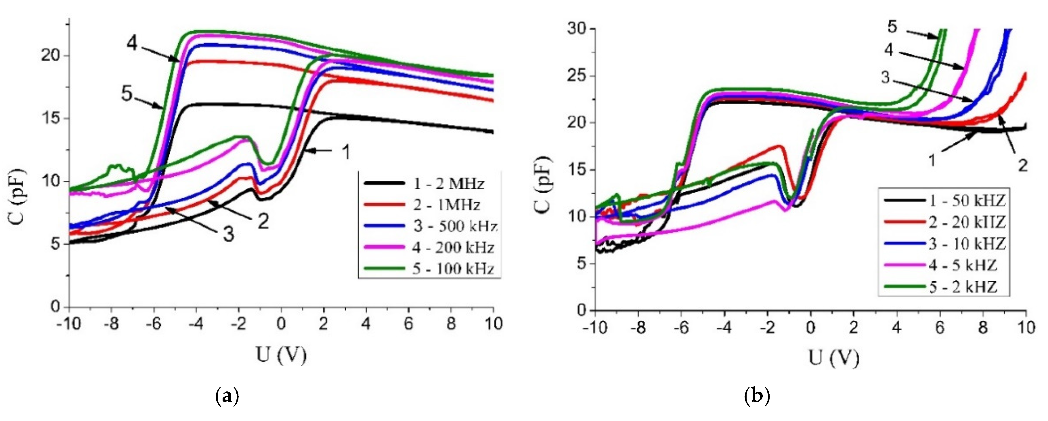
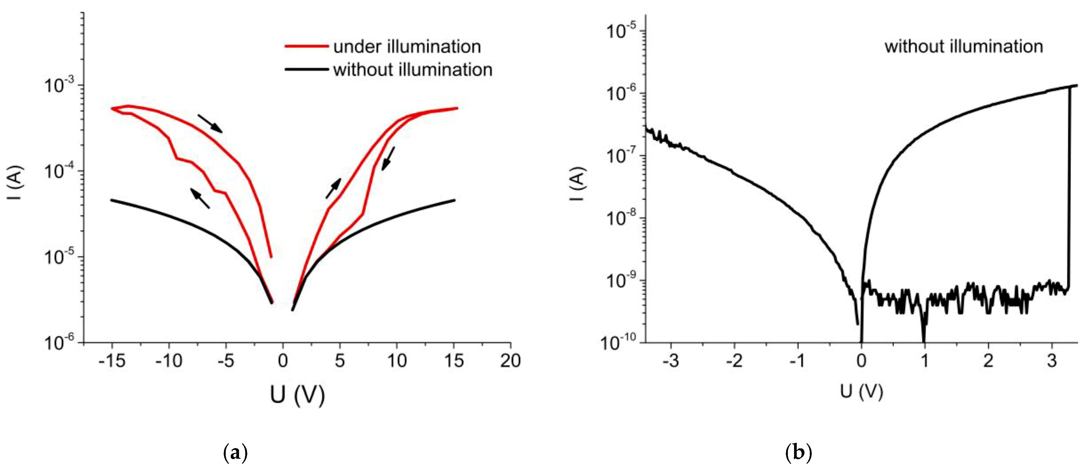
3.2. Heterostructures Si/SrTiO3/Ni and Si/TiOx/Pt/SrTiO3/Ni

Figure 3 shows cross-sections and plane views of Si/TiOx/Pt/SrTiO3/Ni and Si/SrTiO3/Ni heterostructures. The structures contain ten-layer porous SrTiO3 films with the total thickness of about 630 nm and the average grain size of about 30 nm.
A Raman spectrum of the Si/TiOx/Pt/SrTiO3 heterostructure is presented in Figure 4. Several bands corresponding to TiO2 and SrTiO3 phases are observed.
Three bands at 395, 515 and 635 cm−1 correspond to TiO2 anatase crystalline phase. The band at 395 cm−1 corresponds to B1g vibration mode, the band at 635 cm−1 corresponds to Eg and the band at 515 cm−1 is a doublet of A1g and B1g modes [45]. The bands at 261 and 797 cm−1 are an indication of cubic SrTiO3 phase in the film. The band at 261 cm−1 is associated with first-order transversal TO3 phonon mode and the band at 797 cm−1 is a longitudinal LO4 phonon mode [46,47,48]. The activation of TO3 and LO4 bands indicates that structural distortions extended over a distance of the order of the phonon wavelength in the synthesized material [46]. Other SrTiO3 bands may be veiled by strong bands of TiO2. The single band at 865 cm−1 can be associated with SrO 2LO mode [49]. Two lines located at 1443 and 1625 cm−1 are related to carbon presenting in the structure [50]. A summary of the band identification is given in Table 1.
Porous films with small grains go through difficulties with the formation of the SrTiO3 phase in conditions of limiting grain growth because it is accompanied by the development of defects. They make TiO2 to dominate over SrTiO3.
The XRD pattern of Si/TiOx/Pt/SrTiO3 heterostructure contains intense peaks associated with silicon substrate and platinum layer (Figure 5). The five-layer SrTiO3:Eu3+ film XRD pattern of Si/TiOx/Pt/SrTiO3 heterostructure contains a diffuse line in the region of 32.2 deg associated with SrTiO3. They also contain unidentified lines that can be attributed to oxides and oxide composites, including components of the precursors.
Figure 6 shows SIMS depth distribution profiles of the components in the porous nanostructured SrTiO3 film formed on Pt/TiOx/Si substrate.
The thickness of the SrTiO3 film estimated from the SIMS data is about 450 nm. It is smaller than the one determined by SEM which is explainable noting that the sputtering rate of porous structures is larger than of dense films [51]. Qualitatively, the SIMS data are in a good agreement with the results of XRD and Raman spectroscopy analysis. TiO2, SrO, and SrTiO3 are formed homogeneously in the nanostructured porous films.
The SIMS depth profiles of the components in the Si/TiOx/Pt/SrTiO3 heterostructure illustrate the presence of several layers. The layer corresponding to the sputtering time of 1800 s is enriched with SrO we associate with the porous SrTiO3. It contains some carbon that is a residue of the incomplete elimination of the carbon containing isopropoxy groups and organic groups of the solvent [51,52]. The next layers corresponding to the sputtering time in the ranges of 1800–2500 and 2500–3000 s are associated with the Pt and TiOx, respectively. Silicon ions of the substrate were not detected under these experimental conditions.
Figure 7 shows C-V characteristics of the Si/SrTiO3/Ni heterostructure after several measurement cycles at different frequencies. Initially, the structure was in the state with the maximum capacity followed by the high frequency bypass performed from 0 V counterclockwise. The voltage polarity corresponds to the sign of the electric potential on one top electrode relative to the other top electrode. The charge of the capacitor basically occurs only by applying a positive voltage. The capacity of this structure with two upper electrodes varies from 6 pF to 22 pF. The width of the hysteresis is ~6 V in the frequency range of the test measurement signal from 2 kHz to 2 MHz. The width of the hysteresis loop is about 6 V.
The nonlinearity of the C–V characteristics is caused by the presence of electrically active deep energy levels in the band gap, created by various defects. As the capacity growth is observed only with one positive polarity, electrons are trapped by the defect states [22]. The nonlinear dependence of the capacitance on voltage applied can be associated with the formation of potential wells in the surface layer of nanostructured grains due to the porosity.
The frequency dependence of the hysteresis loop area indicates the presence of fast and slow energy states involved in the recharging of electrical capacitance. A sharp increase of the capacitance at a positive bias voltage with a decrease in the frequency of the test signal to 2 kHz indicates the electrical charge accumulation on slow energy states. A shift of the C-V characteristics towards negative bias voltages by approximately the same value indicates the presence of a fixed positive charge in the SrTiO3 film or near the interfaces. The nature of the traps and energy states is still not clear and may be the goal of a separate study by electric thermally stimulated methods.
In-plane I-V measurements between two top Ni electrodes of the structure Si/SrTiO3/Ni show that illumination of the samples with the light of a halogen lamp at the intensity of 39 mW/cm2 leads to significant changes in the forward and reverse branches of the I-V curves. This is illustrated in Figure 8a. Under illumination, the curves are characterized by hysteresis for both forward and reverse biasing, which was also early observed in [53]. The transversal electrical measurements on the Si/TiOx/Pt/SrTiO3/Ni structure shows resistance switching even without illumination (Figure 8b) when the bias was applied between top Ni and bottom Pt electrodes.

4. Conclusions

We described the sol-gel approach to the fabrication of porous Eu doped SrTiO3 multilayer film structures. The technique does not require any organic template precursors in the sol for making the material porous. Both porous nanostructured films and bulk powders demonstrate the most intensive PL bands at 612 nm and 588 nm, respectively. Thus, this material can be used as correspondingly yellow and red phosphors in optoelectronic devices depending on the structural state of Eu3+ ions.
XRD, Raman spectroscopy, and SIMS analysis are in a good agreement with each other and confirm the formation of TiO2, SrO, and SrTiO3 phases and their components in the porous SrTiO3 material. The thin film structures, including porous SrTiO3, exhibit hysteresis of C-V and I-V characteristics, which is of interest for nonvolatile memory elements.

Author Contributions

Data curation, E.C. and Y.R.; formal analysis, V.L., A.E., E.C., V.B., N.M. and Y.R.; investigation, M.R. and N.G.; resources, M.R., A.T. and A.G.; supervision, N.G.; writing—original draft, M.R.; writing—review & editing, V.B. and N.M. All authors have read and agreed to the published version of the manuscript.

Funding

This research was funded by the grant T19-MLDG of State Committee on Science and Technology of the Republic of Belarus. Nikolay Gaponenko and Victor Borisenko acknowledge the partial financial support of the “Improving of the Competitiveness” Program of the National Research Nuclear University MEPhI—Moscow Engineering Physics Institute. The part of work was supported by the Russian Foundation for Basic Research under the project № 19-07-00600 A.

Conflicts of Interest

The authors declare no conflict of interest.

References

  1. Gonçalves, R.F.; Moura, A.P.; Godinho, M.J.; Longo, E.; Machado, M.A.C.; De Castro, D.A.; SiuLid, M.; Marques, A.P.A. Crystal growth and photoluminescence of the europium-doped strontium titanate prepared by a microwave hydrothermal method. Ceram. Int. 2015, 41, 3549–3554. [Google Scholar] [CrossRef]
  2. Rudenko, M.V.; Raichynok, T.F.; Radush, Y.V.; Podhorodecki, A.; Ilkov, V.K. Luminescence of porous nanostructured strontium titanate films doped with Eu3+ ions. Int. J. Nanosci. 2019, 18, 1940075. [Google Scholar] [CrossRef]
  3. Rudenko, M.V.; Raichenok, T.F.; Mukhin, N.V.; Gaponenko, N.V. Synthesis and photoluminescence of strontium titanate xerogels doped with terbium, ytterbium and europium. NATO Sci. Peace Secur. Ser. B Phys. Biophys. 2018, 435–437. [Google Scholar] [CrossRef]
  4. Rubano, A.; Scigaj, M.; Sánchez, F.; Herranz, G.; Paparo, D. Optical second harmonic generation from LaAlO3/SrTiO3 interfaces with different in-plane anisotropies. J. Phys. Condens. Matter 2020, 32, 135001. [Google Scholar] [CrossRef] [PubMed]
  5. Ortmann, J.E.; Duncan, M.A.; Demkov, A.A. Designing near-infrared electro-optical devices from the SrTiO3 /LaAlO3 materials system. Opt. Mater. Express 2019, 9, 2982. [Google Scholar] [CrossRef]
  6. Luo, J.; Maggard, P.A. Hydrothermal synthesis and photocatalytic activities of SrTiO3-Coated Fe2O3 and BiFeO3. Adv Mater. 2006, 18, 514. [Google Scholar] [CrossRef]
  7. Xie, J.; Lei, K.; Wang, H.; Wang, C.; Liu, B.; Zhang, L.; Bai, P. Strontium titanate with inverse opal structure as the photocatalysts. J. Mater. Sci. Mater. Electron. 2020, 31, 2691–2698. [Google Scholar] [CrossRef]
  8. Schrott, A.G.; Misewich, J.A. Ferroelectric field-effect transistor with a SrRuxTi1−xO3 channel. Appl. Phys. Lett. 2003, 82, 4770. [Google Scholar] [CrossRef]
  9. Sohrabi Anaraki, H.; Gaponenko, N.V.; Rudenko, M.V.; Guk, A.F.; Zavadskij, S.M.; Golosov, D.A.; Kolosnitsyn, B.S.; Kolos, V.V.; Pyatlitskij, A.N.; Turtsevich, A.S. On the sol-gel synthesis of strontium-titanate thin films and the prospects of their use in electronics. Semiconductors 2014, 48, 1685–1687. [Google Scholar] [CrossRef]
  10. Ma, C.; Luo, Z.; Huang, W.; Zhao, L.; Chen, Q.; Lin, Y.; Liu, X.; Chen, Z.; Liu, C.; Sun, H.; et al. Sub-nanosecond memristor based on ferroelectric tunnel junction. Nat. Commun. 2020, 11, 1439. [Google Scholar] [CrossRef] [Green Version]
  11. Funck, C.; Bäumer, C.; Wiefels, S.; Hennen, T.; Waser, R.; Hoffmann-Eifert, S.; Dittmann, R.; Menzel, S. Comprehensive model for the electronic transport in Pt/SrTiO3 analog memristive devices. Phys. Rev. B 2020, 102. [Google Scholar] [CrossRef]
  12. Rüdiger, A.; Schneller, T.; Roelofs, A.; Tiedke, S.; Schmitz, T.; Waser, R. Nanosize ferroelectric oxides–tracking down the superparaelectric limit. Appl. Phys. A 2005, 80, 1247–1255. [Google Scholar] [CrossRef]
  13. Wu, X.; Wu, D.; Liu, X. Negative pressure effects in SrTiO3 nanoparticles investigated by Raman spectroscopy. Solid State Comm. 2008, 145, 255–258. [Google Scholar] [CrossRef]
  14. Fomin, A.A.; Fomina, M.A.; Koshuro, V.A.; Rodionov, I.V.; Voiko, A.V.; Zakharevich, A.M.; Aman, A.; Oseev, A.; Hirsch, S.; Majcherek, S. Micro- and nanostructure of a titanium surface electric-spark-doped with tantalum and modified by high-frequency currents. Tech. Phys. Lett. 2016, 42, 932–935. [Google Scholar] [CrossRef]
  15. Fomin, A.A.; Steinhauer, A.B.; Rodionov, I.V.; Fomina, M.A.; Zakharevich, A.M.; Skaptsov, A.A.; Gribov, A.N.; Karsakova, Y.D. Properties of titanium dioxide coatings produced by induction-thermal oxidation of VT1-00 alloy. J. Frict. Wear 2014, 35, 32–39. [Google Scholar] [CrossRef]
  16. Zhang, W.; Yin, Z.; Zhang, M. Study of photoluminescence and electronic states in nanophase strontium titanate. Appl. Phys. A 2000, 70, 93–96. [Google Scholar] [CrossRef]
  17. Kwun, S.; Song, T. Nano-size effects on the quantum paraelectric SrTiO3 fine particles. Ferroelectrics 1997, 197, 125–130. [Google Scholar] [CrossRef]
  18. Suzuki, N.; Osada, M.; Billah, M.; Abdullah Alothman, Z.; Bando, Y.; Yamauchi, Y.; Shahriar, A.; Hossain, M. Origin of thermally stable ferroelectricity in a porous barium titanate thin film synthesized through block copolymer templating. APL Mater. 2017, 5, 076111. [Google Scholar] [CrossRef] [Green Version]
  19. Zhang, D.; Zhang, Y.L. Preparation of porous nano-strontium titanate and its application in removal of heavy metals from environmental water. AMR 2011, 194–196, 765–768. [Google Scholar] [CrossRef]
  20. Feng, L.-L.; Zou, X.; Zhao, J.; Zhou, L.-J.; Wang, D.-J.; Zhang, X.; Li, G.-D. Nanoporous Sr-rich strontium titanate: A stable and superior photocatalyst for H2 evolution. Chem. Commun. 2013, 49, 9788–9790. [Google Scholar] [CrossRef]
  21. Sharma Pramod, K.; Varadan, V.V.; Varadan, V.K. Porous behavior and dielectric properties of barium strontium titanate synthesized by sol−gel method in the presence of triethanolamine. Chem. Mater. 2000, 12, 2590–2596. [Google Scholar] [CrossRef]
  22. Tikhov, S.V.; Gorshkov, O.N.; Pavlov, D.A.; Antonov, I.N.; Bobrov, A.I.; Kasatkin, A.P.; Koryazhkina, M.N.; Shenina, M.E. Capacitors with nonlinear characteristics based on stabilized zirconia with built-in gold nanoparticles. Tech. Phys. Lett. 2014, 40, 369–371. [Google Scholar] [CrossRef]
  23. Dedyk, A.I.; Semenov, A.A.; Pavlova, Y.V.; Belyavskii, P.Y.; Nikitin, A.A.; Pakhomov, O.V.; Myl’nikov, I.L. Photoelectrical properties of strontium titanate. Tech. Phys. 2015, 60, 624–627. [Google Scholar] [CrossRef]
  24. Youssef, A.M.; Farag, H.K.; El-Kheshen, A.; Hammad, F.F. Synthesis of nano-structured strontium titanate by sol-gel and solid state routes. Silicon 2018, 10, 1225–1230. [Google Scholar] [CrossRef]
  25. Tolstykh, N.A.; Korotkova, T.N.; Jaafari, F.D.; Kashirin, M.A.; Fedotova, Y.A.; Yemelyanov, N.A.; Korotkov, L.N.; Kasyuk, Y.V. Dielectric and magnetic properties of nanocrystal barium titanate, strontium titanate, and a blended nanocomposite based on them. Bull. Russ. Acad. Sci. Phys. 2019, 83, 1086–1090. [Google Scholar] [CrossRef]
  26. Sohrabi Anaraki, H.; Gaponenko, N.V.; Rudenko, M.V.; Kolos, V.V.; Petlitskii, A.N.; Turtsevich, A.S. Thin-film capacitor based on the strontium titanate formed by the sol-gel technique. Russ. Microelectron. 2015, 44, 425–429. [Google Scholar] [CrossRef]
  27. Rudenko, M.V.; Kortov, V.S.; Gaponenko, N.V.; Mudryi, A.V.; Zvonarev, S.V. Photo- and cathode luminescence of strontium titanate xerogel films doped with terbium ions. J. Surf. Investig. X-ray Synchrotron Neutron Tech. 2015, 9, 1012–1015. [Google Scholar] [CrossRef]
  28. Mizera, A.; Drożdż, E.; Łańcucki, Ł. Synthesis of highly porous SrTiO3 materials. Acta Phys. Pol. A 2018, 133, 873–875. [Google Scholar] [CrossRef]
  29. Lee, J.-T.; Wey, M.-Y. PVA/Pt/N-TiO2/SrTiO3 porous films with adjustable pore size for hydrogen production under simulated sunlight. J. Colloid Interface Sci. 2020, 573, 158–164. [Google Scholar] [CrossRef]
  30. Li, S.; Li, M.; Tao, A.; Song, M.; Wang, B.; Niu, J.; Yu, F.; Wu, Y. Synthesis of a bicontinuous structured SrTiO3 porous film with significant photocatalytic activity by controlling phase separation process. J. Sol-Gel Sci. Technol. 2020, 94, 288–297. [Google Scholar] [CrossRef]
  31. Ujiie, K.; Kojima, T.; Ota, K.; Phuenhinlad, P.; Pleuksachat, S.; Meethong, N.; Itoi, T.; Uekawa, N. Preparation of spherical and porous strontium titanate particles by hot water and hydrothermal conversion of hydrous titania. Ceram. Int. 2020, 46, 6146–6153. [Google Scholar] [CrossRef]
  32. Litvinov, V.G.; Ermachikhin, A.V.; Kusakin, D.S.; Vishnyakov, N.V.; Gudzev, V.V.; Karabanov, A.S.; Karabanov, S.M.; Vikhrov, S.P. Investigation of deep-level defects lateral distribution in active layers of multicrystalline silicon solar cells. MRS Adv. 2017, 2, 3141–3146. [Google Scholar] [CrossRef]
  33. Shyamal, S.; Hajra, P.; Paramita, M.; Harahari, B.; Aparajita, S.; Sariket, D.; Satpati, A.; Malashchonak, M.; Mazanik, A.; Korolik, O.; et al. Eu modified Cu2O thin films: Significant enhancement in efficiency of photoelectrochemical processes through suppression of charge caarier recombination. Chem. Eng. J. 2018, 335, 676–684. [Google Scholar] [CrossRef]
  34. Zulueta, Y.A.; Lim, T.C.; Dawson, J.A. Defect clustering in rare-earth-doped BaTiO3 and SrTiO3 and its influence on dopand incorporation. J. Phys. Chem. C. 2017, 121, 23642–23648. [Google Scholar] [CrossRef]
  35. Binnemans, K. Interpretation of europium(III) spectra. Coord. Chem. Rev. 2015, 295, 1–45. [Google Scholar] [CrossRef] [Green Version]
  36. Podhorodecki, A.; Banski, M.; Misiewicz, J.; Serafińczuk, J.; Gaponenko, N.V. Influence of annealing on excitation of terbium luminescence in YAlO3 films deposited onto porous anodic alumina. J. Electrochem. Soc. 2010, 157, H628–H632. [Google Scholar] [CrossRef]
  37. Kenyon, A.J. Recent developments in rare-earth doped materials for optoelectronics. Progr. Quantum Electron. 2002, 26, 225–284. [Google Scholar] [CrossRef]
  38. Strek, W.; Hreniak, D.; Boulon, G.; Guyot, Y.; Pązik, R. Optical behavior of Eu3+-doped BaTiO3 nano-crystallites prepared by sol–gel method. Opt. Mater. 2003, 24, 15–22. [Google Scholar] [CrossRef]
  39. Villegas Brito, J.C.; Gaponenko, N.V.; Sukalin, K.S.; Raichenok, T.F.; Tikhomirov, S.A.; Yankovskaya, V.A.; Kargin, N.I. Luminescence of Eu3+ in Yttrium–Alumina Films on Fused Silica Substrates. J. Appl. Spectrosc. 2017, 84, 674–678. [Google Scholar] [CrossRef]
  40. Yang, Y.; Wang, B.; Cormack, A.; Zych, E.; Seo, H.J.; Wu, Y. Theoretical analysis and experiment on Eu reduction in alumina optical materials. Opt. Mater. Express 2016, 6, 2404. [Google Scholar] [CrossRef]
  41. Ahadi, K.; Gui, Z.; Porter, Z.; Lynn, J.W.; Xu, Z.; Wilson, S.D.; Janotti, A.; Stemmer, S. Carrier density control of magnetism and Berry phases in doped EuTiO3. APL Mater. 2018, 6, 56105. [Google Scholar] [CrossRef] [Green Version]
  42. Jiang, C.; Fang, L.; Shen, M.; Zheng, F.; Wu, X. Effects of Eu substituting positions and concentrations on luminescent, dielectric, and magnetic properties of SrTiO3 ceramics. Appl. Phys. Lett. 2009, 94, 71110. [Google Scholar] [CrossRef]
  43. García, C.R.; Oliva, J.; Romero, M.T.; Ochoa-Valiente, R.; Trujillo, L.A.G. Effect of Eu3+ Concentration on the Luminescent Properties of SrTiO3 Phosphors Prepared by Pressure-Assisted Combustion Synthesis. Adv. Mater. Sci. Eng. 2015, 2015, 1–7. [Google Scholar] [CrossRef] [Green Version]
  44. Chand, S.; Chopra, A.; Singh, I. Enhanced Red Emission from SrTiO3:Eu3+ [Li+, Na+, K+] Nano-Phosphors Prepared by Combustion Synthesis. JCCS 2017, 7, 1283–1289. [Google Scholar] [CrossRef]
  45. Balachandran, U.; Eror, N.G. Raman spectra of titanium dioxide. J. Solid State Chem. 1982, 42, 276–282. [Google Scholar] [CrossRef]
  46. Moreira, M.L.; Longo, V.M.; Avansi, W., Jr.; Ferrer, M.M.; Andrés, J.; Mastelaro, V.R.; Varela, J.A.; Longo, É. Quantum mechanics insight into the microwave nucleation of SrTiO3 nanospheres. J. Phys. Chem. C 2012, 116, 24792–24808. [Google Scholar] [CrossRef]
  47. Yuzyuk, Y.I. Raman scattering spectra of ceramics, films, and superlattices of ferroelectric perovskites: A review. Phys. Solid State 2012, 54, 1026–1059. [Google Scholar] [CrossRef]
  48. Da Silva, L.F.; Avansi, W.; Andres, J.; Ribeiro, C.; Moreira, M.L.; Longo, E.; Mastelaro, V.R. Long-range and short-range structures of cube-like shape SrTiO3 powders: Microwave-assisted hydrothermal synthesis and photocatalytic activity. Phys. Chem. Chem. Phys. 2013, 15, 12386. [Google Scholar] [CrossRef]
  49. Rieder, K.H.; Migoni, R.; Renker, B. Lattice dynamics of strontium oxide. Phys. Rev. B Condens. Matter 1975, 12, 3374. [Google Scholar] [CrossRef]
  50. Dennison, J.R.; Holtz, M.; Swain, G. Raman spectroscopy of carbon materials. Spectroscopy 1996, 11, 38–45. [Google Scholar]
  51. Dorofeev, A.M.; Gaponenko, N.V.; Bondarenko, V.P.; Bachilo, E.E.; Kazuchits, N.M.; Leshok, A.A.; Troyanova, G.N.; Vorosov, N.N.; Borisenko, V.E. Erbium luminescence in porous silicon doped from spin-on films. J. Appl. Phys. 1995, 77, 2679. [Google Scholar] [CrossRef]
  52. Gaponenko, N.V.; Gnaser, H.; Becker, P.; Grozhik, V.A. Carbon depth distribution in spin-on silicon dioxide films. Thin Solid Films 1995, 261, 186–191. [Google Scholar] [CrossRef]
  53. Rudenko, M.V.; Kholov, P.A.; Gaponenko, N.V.; Mukhin, N.V.; Ivanov, V.A.; Stas’kov, N.I. Photocurrent hysteresis of sol-gel derived strontium titanate films on silicon. Int. J. Nanosci. 2019, 18, 1940090. [Google Scholar] [CrossRef]
Figure 1. SEM images of the five-layer SrTiO3:Eu3+ film (a,b) and SrTiO3:Eu3+ powder (c).
Materials 13 05767 g001
Figure 2. PL spectra of the SrTiO3:Eu3+ powder and five-layer SrTiO3:Eu3+ film (a) and diffraction patterns of the SrTiO3:Eu3+ powder and the SrTiO3 film (b).
Materials 13 05767 g002
Figure 3. SEM images of Si/TiOx/Pt/SrTiO3/Ni (a), Si/SrTiO3/Ni (b) heterostructures, the surfaces of SrTiO3 (c) and Ni electrode (d).
Materials 13 05767 g003
Figure 4. Raman shift spectra of Si/TiOx/Pt/SrTiO3/Ni heterostructure.
Materials 13 05767 g004
Figure 5. XRD spectra of Si/TiOx/Pt/SrTiO3/Ni heterostructure.
Materials 13 05767 g005
Figure 6. SIMS depth distribution of the precursor components in the Si/TiOx/Pt/SrTiO3 heterostructure.
Materials 13 05767 g006
Figure 7. Capacitance-voltage characteristics of the Si/SrTiO3/Ni heterostructure recorded in the frequency ranges of 100 kHz-2 MHz (a) and 2 kHz-50 kHz (b).
Materials 13 05767 g007
Figure 8. In-plane (a) and transversal (b) I-V characteristics of Si/SrTiO3/Ni (a) and Si/TiOx/Pt/SrTiO3/Ni (b) containing ten layers of SrTiO3 under and without illumination.
Materials 13 05767 g008
Table 1. Raman spectra bands.
Peak, cm−1MaterialModeReference
261SrTiO3 (cubic)TO3 (O–Sr–O bending)[46,47,48]
395TiO2 (Anatase)B1g[45]
515TiO2 (Anatase)A1g + B1g[45]
635TiO2 (Anatase)Eg[45]
797SrTiO3 (cubic)LO4 (Ti–O stretching)[46,47,48]
865SrO2LO[49]
1443CarbonDisorder[50]
1625CarbonE2g[50]
Publisher’s Note: MDPI stays neutral with regard to jurisdictional claims in published maps and institutional affiliations.

