Article

Broadening the Photoluminescence Excitation Spectral Bandwidth of YVO4:Eu3+ Nanoparticles via a Novel Core-Shell and Hybridization Approach

1
School of Materials Science and Engineering, Nanchang University, Nanchang 330031, China
2
Hunan Engineering Laboratory for Control and Optimization of PV Systems, Hunan Vocational Institute of Technology, Xiangtan 411104, China
3
The Center of Collaboration and Innovation, Jiangxi University of Technology, Nanchang 330098, China
*
Authors to whom correspondence should be addressed.
Jianhua Huang and Lu Tang contributed equally to this article.
Materials 2019, 12(23), 3830; https://doi.org/10.3390/ma12233830
Submission received: 30 October 2019 / Revised: 17 November 2019 / Accepted: 19 November 2019 / Published: 21 November 2019
(This article belongs to the Special Issue Advances in Luminescent Engineered Nanomaterials)

Abstract

:
For many optoelectronic applications, it is desirable for the lanthanide-doped phosphors to have broad excitation spectrum. The excitation mechanism of the lanthanide-doped YVO4, a high quantum efficient lasing material, primarily originates from the energy transfer process from the host VO43− complexes to the lanthanide ions, which has an excitation spectral bandwidth range of 230–330 nm. For applications in silicon solar cells, such phosphors can convert ultraviolet light to visible light for more efficient power generation, but this spectral range is still not broad enough to cover the entire ultraviolet spectrum of solar light. In this work, a novel core-shell and inorganic–organic hybridization strategy has been employed to fabricate Eu3+-doped YVO4 nanoparticles to broaden their photoluminescence excitation spectral bandwidth to the range of 230–415 nm, covering the entire ultraviolet spectrum of solar light and enabling their potential applications in silicon solar cells.

1. Introduction

Lanthanide ion-doped YVO4 (YVO4:Ln3+), such as YVO4:Nd3+, has been widely used for fabricating lasers [1,2]. Recently, luminescent YVO4:Ln3+ nanomaterials, such as YVO4:Eu3+ [3,4], YVO4:Eu3+,Bi3+ [5,6], YVO4:Yb3+ [7], YVO4:Yb3+,Bi3+ [8] etc., have received extensive attention for enhancing the conversion efficiency of solar cells. Solar cells, especially the silicon solar cells, usually have rather low quantum efficiency in the ultraviolet (UV) spectrum, but, in the visible/near infrared spectrum, however, their quantum efficiency is quite high. YVO4:Ln3+ nanomaterials are excited by UV lights of solar irradiance, and down-convert them to visible or near infrared lights. Through such down-conversion, the conversion efficiency of solar cells is consequently enhanced as a result of more efficient utilization of UV photons of solar irradiance [9,10].
The strong photoluminescence of YVO4:Ln3+ relies on the efficient energy transfer process from the host VO43- complexes to the Ln3+ [11], in which the VO43- is well excited by the UV lights. Such an energy transfer process effectively overcomes the drawback of low absorption cross sections of parity forbidden 4f–4f transitions of Ln3+ ions. The excitation spectrum of VO43− peaks at about 325 nm, and has a spectral range from 260 nm to 330 nm at the half maximum and a band edge at 350 nm [12]. This, however, is too narrow to cover the UV spectrum (250–400 nm) of the solar lights on earth surface for applications in solar cells. To extend its excitation spectrum, Bi3+ ions have been co-doped into YVO4:Ln3+ [5,6,8,13,14]. It was found that the peaking wavelength of its excitation spectrum is shifted to the longer wavelength at 342 nm and the band edge is extended to about 360 nm [8]. Up to now, however, the excitation bandwidth of YVO4:Ln3+ is still not wide enough to cover the UV spectrum of the solar irradiance. Previous work on LaF3:Eu3+ [15] has shown that its excitation spectral bandwidth can be effectively broadened by using a mixed photoluminescence sensitization from different types of organic ligands.
In this work, a novel strategy was employed to remarkably broaden the excitation spectrum of YVO4:Eu3+ nanoparticles through a dual–channel excitation approach, and the entire UV spectrum of the solar irradiance can be effectively covered. This was implemented by combining the two strong excitation channels, the efficient VO43−→Eu3+ energy transfer after annealing and the photoluminescence sensitization by organic ligand after forming an inorganic–organic hybrid nanoparticle [16,17].

2. Experimental

The experimental procedure is illustrated in Figure 1, and it is detailed in the following paragraphs.

2.1. Materials

Yttrium nitrate (Y(NO3)3·6H2O, 99.99 %), europium nitrate (Eu(NO3)3·6H2O, 99.99 %), sodium vanadate (Na3VO4·12H2O, AR), polyvinyl pyrrolidone (PVP K30, AR), ethylene glycol (EG, AR), anhydrous ethanol (AR), and 2-thenoyltrifluoroacetone (TTA, 98.0%) were used in this work. All were bought from Sinopharm Chemical Reagent Co., Ltd. (Shanghai, China). Deionized water was used throughout this work.

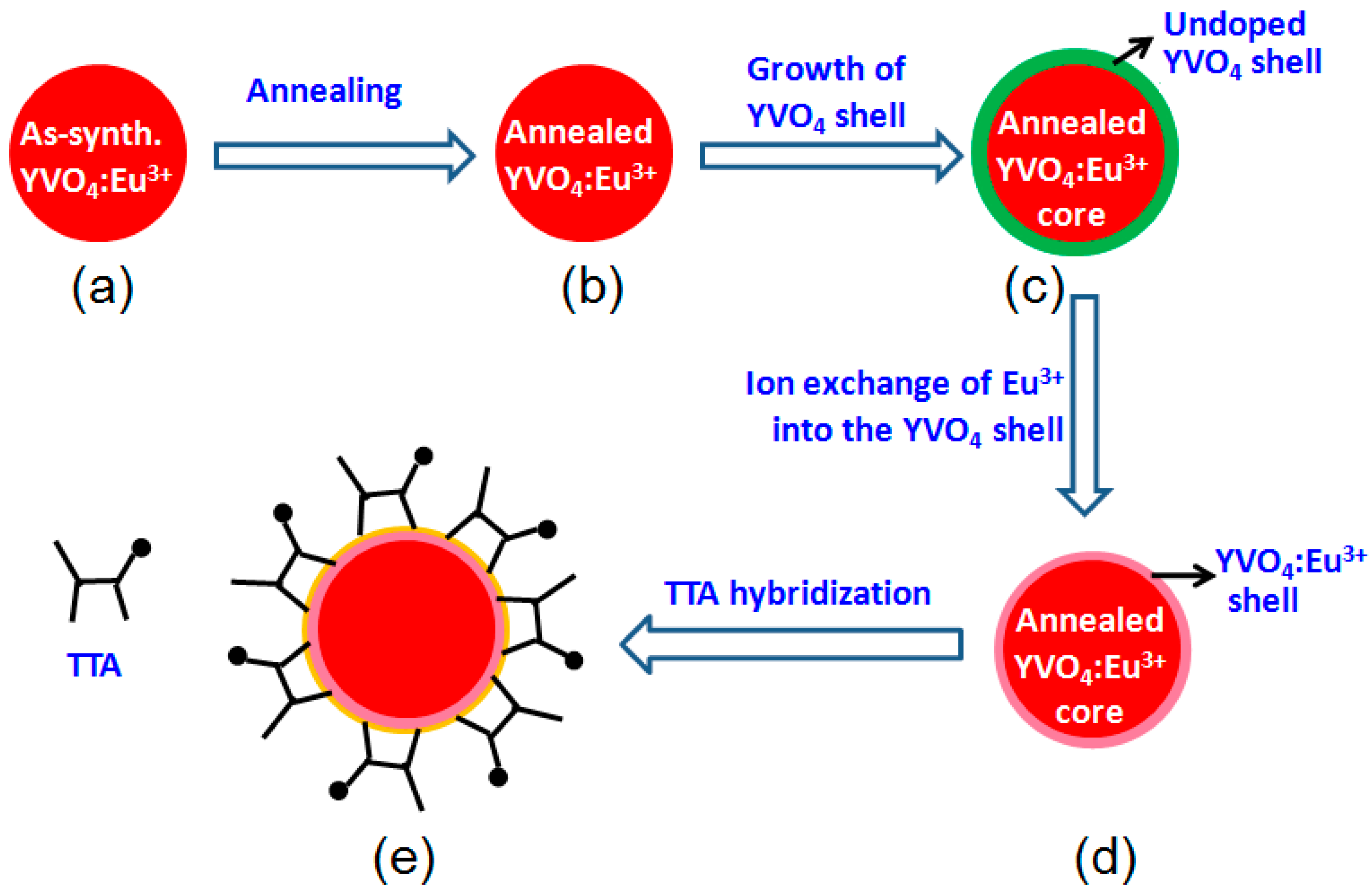
2.2. Synthesis and Annealing of YVO4:Eu3+ Nanoparticles

The YVO4:Eu3+ nanoparticles were synthesized using the solvothermal method [18]. First, 0.75 mmol Na3VO4·12H2O and 0.05 g PVP were dissolved in 8 mL ethylene glycol and 1 mL deionized water. Then, 9 mL of ethylene glycol solution containing 1 mmol Y(NO3)3·6H2O and 0.05 mmol Eu(NO3)3·6H2O were added to the above solution with vigorous magnetic stirring for 5 min. Then, 17 mL of de-ionized water was added to the above mixture solution, with stirring for another 5 min. The mixture solution was poured into a 50 mL Teflon-lined stainless steel autoclave and heated at 180 °C for 6 h to obtain YVO4:Eu3+ nanoparticles, and then naturally cooled to room temperature. The YVO4:Eu3+ nanoparticles were collected by centrifugation, washed with anhydrous ethanol for 3 times, and dried at 80 °C in air.
As shown later in this paper, the as-synthesized YVO4:Eu3+ nanoparticles possess only weak luminescence, which can be greatly enhanced by annealing. In this work, an annealing process at 800 °C for 2 h in air was conducted for the as-synthesized YVO4:Eu3+ nanoparticles for strong photoluminescence (Figure 1). It was found that their particle size only increased to some extent after the annealing, but they remained to be in nanometer scale. However, if the annealing temperature is over 800 °C, the YVO4:Eu3+ nanoparticles quickly grow to a much larger size. Similar behavior has been reported by others [19,20].

2.3. Synthesis of YVO4:Eu3+@YVO4 Core-Shell Nanoparticles

The annealed YVO4:Eu3+ nanoparticles (Section 2.2) were used as a core to grow a pristine YVO4 shell for forming the YVO4:Eu3+@YVO4 core–shell nanoparticles (Figure 1). The pristine YVO4 shell, which is homo-nanoparticled to the core, is expected to passivate the surface non-radiative traps or defects of the annealed YVO4:Eu3+ nanoparticles [21,22,23]. First, 0.75 mmol Na3VO4·12H2O and 0.05 g PVP were dissolved in 8 mL ethylene glycol and 1 mL deionized water. Second, 1 mmol of the annealed YVO4:Eu3+ nanoparticles were re-dispersed into 9 mL ethylene glycol under continuous ultrasonication for 30 min. The above two solutions were mixed, and appropriate amount of Y(NO3)3·6H2O was added to the mixture solution with vigorous magnetic stirring for 5 min. Third, 17 mL of de-ionized water was added to the above mixture solution under magnetic stirring for another 5 min. The mixture solution was poured into a 50 mL Teflon-lined stainless steel autoclave and heated at 200 °C for 2 h, and then naturally cooled down to room temperature. The resultant YVO4:Eu3+@YVO4 core–shell nanoparticles were collected by centrifugation, washed with anhydrous ethanol for 3 times, and dried at 80 °C in air. The molar ratios of the core to the shell (core/shell ratio) were 1:0.2, 1:0.25, 1:0.5, and 1:1. In this work, it was found when the core/shell ratio was 1:0.25, the YVO4:Eu3+@YVO4 core–shell nanoparticles had the highest photoluminescence intensity. Thus, the core/shell ratio of 1:0.25 was chosen for the following experimental steps.

2.4. Synthesis of YVO4:Eu3+@ YVO4:Eu3+ Nanoparticles

As shown in Figure 1, an ion exchange method was used to transform the as-synthesized YVO4:Eu3+@YVO4 core–shell nanoparticles (Section 2.3) to YVO4:Eu3+@YVO4:Eu3+ core–shell nanoparticles by following the procedures described previously [18]. Appropriate amount of Eu(NO3)3·6H2O was dissolved in 15 mL of anhydrous ethanol. The YVO4:Eu3+@YVO4 nanoparticles were added to above solution with ultrasonic processing for 30 min and then vigorous magnetic stirring for 1 h, the mixture solution was let to stand for 24 h. The resultant product was collected by centrifugation, washed with anhydrous ethanol for 3 times, and finally dried in air at 80 °C.

2.5. Synthesis of YVO4:Eu3+@YVO4:Eu3+–TTA Nanoparticles

At last, the YVO4:Eu3+@YVO4:Eu3+–TTA inorganic–organic hybrid nanoparticles were synthesized by the previously described procedures [17,18]. It was found in this work that both the as-synthesized YVO4:Eu3+ and the annealed YVO4:Eu3+ cannot form YVO4:Eu3+–TTA inorganic–organic hybrid structures. Only the ion exchanged YVO4:Eu3+ (Section 2.4) can readily chelate with TTA to form YVO4:Eu3+–TTA inorganic–organic hybrid structures [18]. The YVO4:Eu3+@YVO4:Eu3+ nanoparticles (Section 2.4) were added to 10 mL anhydrous ethanol with ultrasonic processing for 30 min. An appropriate amount of TTA was dissolved in 10 mL of anhydrous ethanol with ultrasonic processing for 15 min and then slowly dripped into the YVO4:Eu3+@YVO4:Eu3+ ethanol solution. The mixture solution was stirred for 1 h and then was left at rest for 12 h. The resultant product was collected by centrifugation, washed with anhydrous ethanol for 3 times and finally dried in air at 80 °C to obtain the YVO4:Eu3+@YVO4:Eu3+–TTA inorganic–organic hybrid nanoparticles. The amounts of TTA used in this step were 0.05, 0.1, 0.15, and 0.2 mmol.

2.6. Characterization

X-ray diffraction (XRD, Bruker D8 Focus, Karlsruhe, Germany) and transmission electron microscopy (TEM, JEM 2010, Japan) were used to investigate the crystal structures and microstructures of the nanoparticles. Their luminescence excitation and emission spectra were recorded with a Hitachi F-4600 fluorescence spectrophotometer, and their chemical bonding information was characterized by Fourier transform infrared (FTIR) spectroscopy (Nicolet 380, Thermo Fisher, USA). Their UV–Vis absorption spectra were monitored with a TU-1950 UV–Vis spectrophotometer. All measurements were conducted in air at room temperature.

3. Results and Discussion

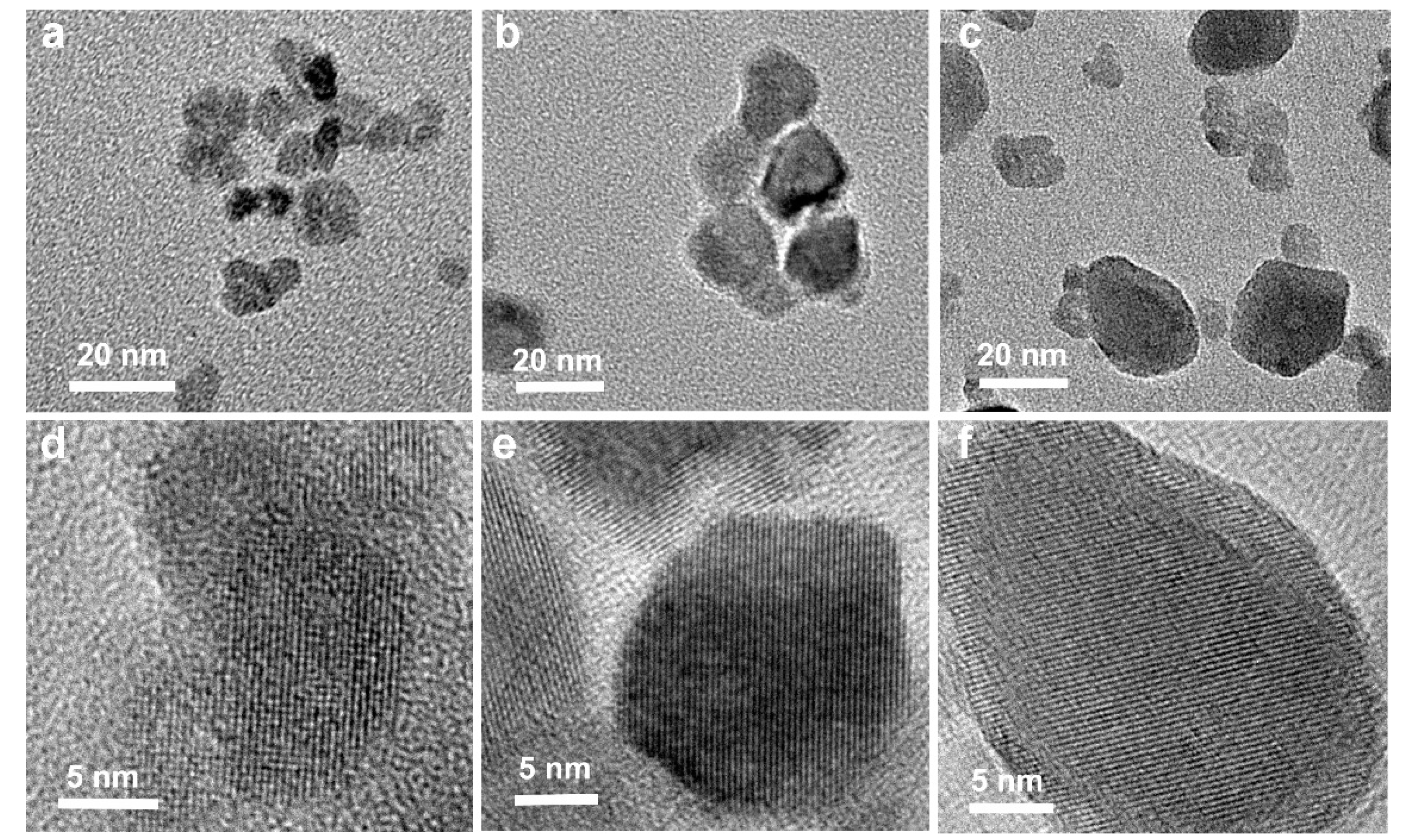
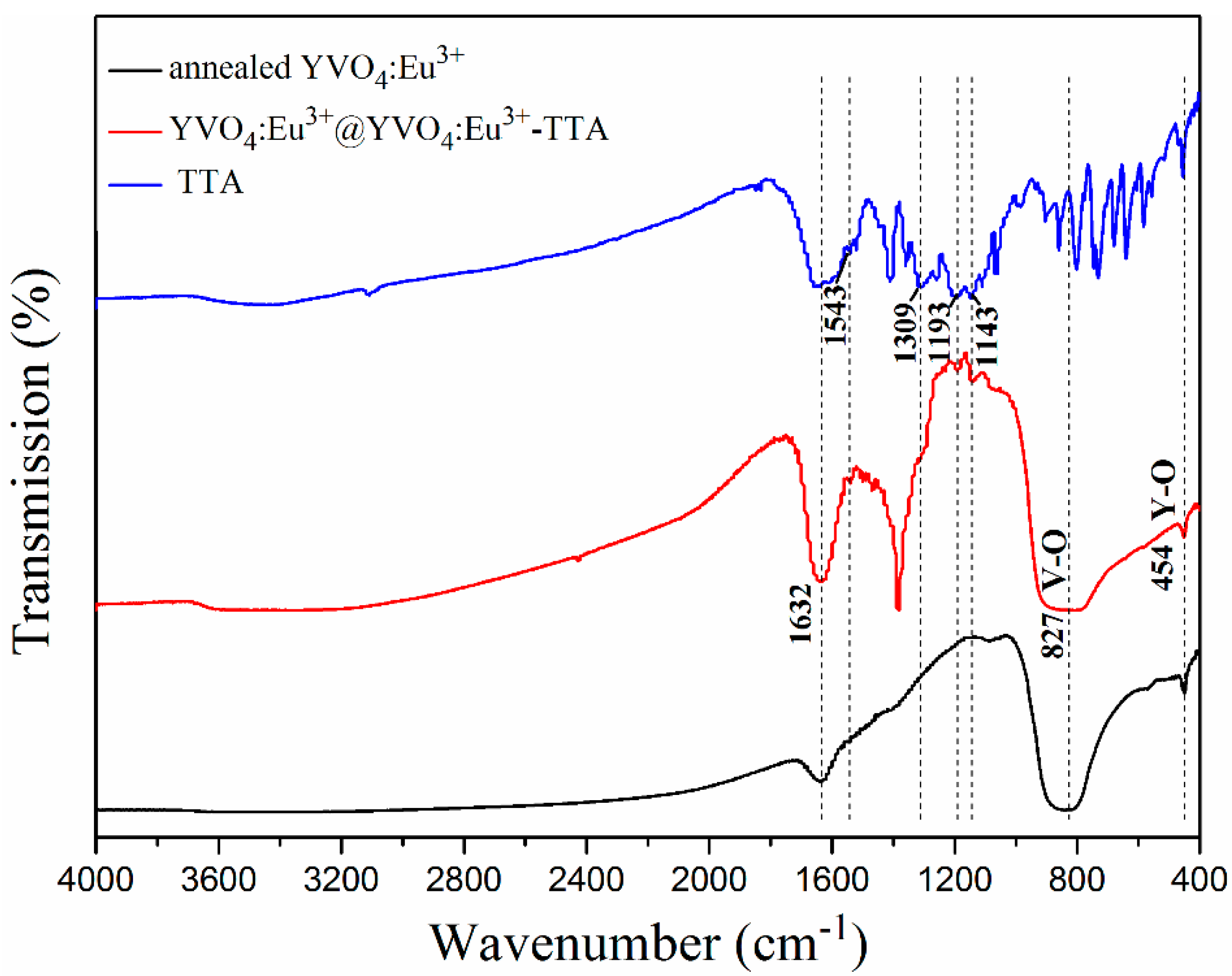
3.1. Microstructural Characteristics

Figure 2 shows the X-ray diffraction (XRD, Bruker D8 Focus) patterns of the as-synthesized YVO4:Eu3+, the annealed YVO4:Eu3+ and the YVO4:Eu3+@YVO4:Eu3+–TTA hybrid nanoparticles. All of the diffraction peaks of samples are identical to the JCPDS 17-0341 of the tetragonal YVO4, and no impurity phases were present. The strong and narrow XRD peaks of the annealed YVO4:Eu3+ nanoparticles in Figure 2 suggested that the annealing process greatly enhanced their crystallization. It is also noted in Figure 2 that, after the core-shell process and hybridization with TTA, the YVO4:Eu3+ nanoparticles still had the same XRD patterns.
The microstructures of the as-synthesized and unannealed YVO4:Eu3+ nanoparticles, the annealed YVO4:Eu3+ nanoparticles and the YVO4:Eu3+@YVO4:Eu3+ core-shell nanoparticles are shown in Figure 3. The average particle sizes of the as-synthesized YVO4:Eu3+, the annealed YVO4:Eu3+, and the YVO4:Eu3+@YVO4:Eu3+ core-shell nanoparticles were about 15, 28, and 36 nm, respectively. As shown in Figure 3d–f, all of the nanoparticles were single crystals, and this may suggest that the YVO4 shell could be epitaxially grown on the annealed YVO4:Eu3+ core.
The Fourier transform infrared (FTIR, Nicolet 380) spectra of TTA, the annealed YVO4:Eu3+ and YVO4:Eu3+@YVO4:Eu3+–TTA hybrid nanoparticles are shown in Figure 4. The strong absorption band located at 1638 cm−1 is both from the H–O–H bending vibration of adsorbed water [24] and the ketonic group and thiophene of TTA [25]. Four new peaks at 1543 cm−1, 1309 cm−1, 1193 cm−1, and 1143 cm−1 for the YVO4:Eu3+@YVO4:Eu3+–TTA hybrid nanoparticles are attributed to the characteristic peaks of TTA [26]. This indicates that TTA successfully hybridized with the YVO4:Eu3+@YVO4:Eu3+ nanoparticles. The strong absorption peak at 827 cm−1 is due to the absorption of V–O bonds [27], while the weak peak at 454 cm−1 is due to the absorption of Y(Eu)-O bonds [28,29].

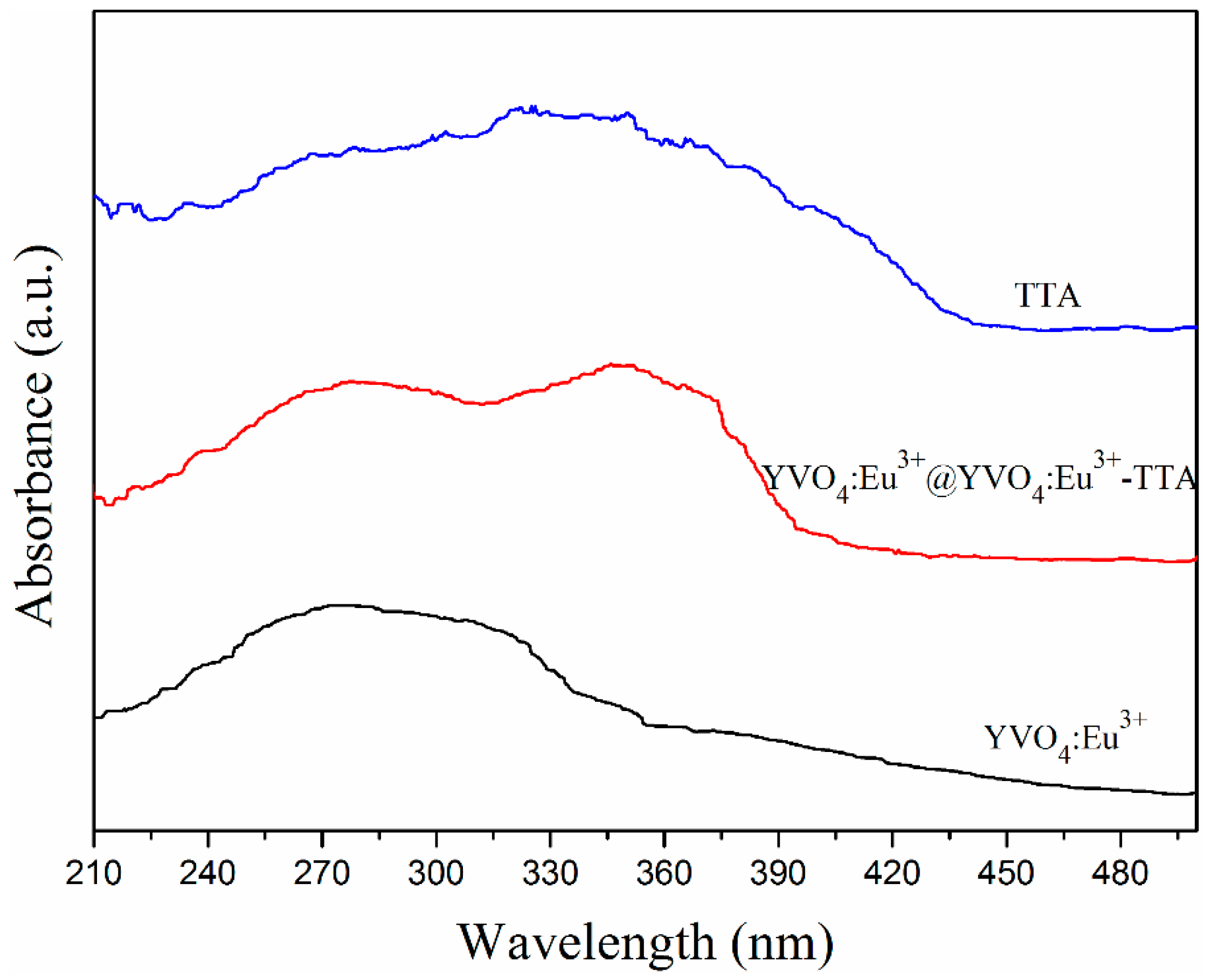
3.2. UV–Vis Absorption Spectra

The UV–Vis (TU-1950) absorption spectra of the annealed YVO4:Eu3+ nanoparticles, TTA, and YVO4:Eu3+@YVO4:Eu3+–TTA hybrid nanoparticles are shown in Figure 5. The broad absorption band (230–330 nm) of the annealed YVO4:Eu3+ nanoparticles was due to the VO43- groups in the host lattice of YVO4 nanoparticle [30]. It also can be seen in Figure 5 that TTA had a broad absorption band of 240–420 nm. Consequently, YVO4:Eu3+@YVO4:Eu3+–TTA nanoparticles possessed two broad absorption bands in the ultraviolet range, which is due to the presence of VO43 groups and TTA ligands.

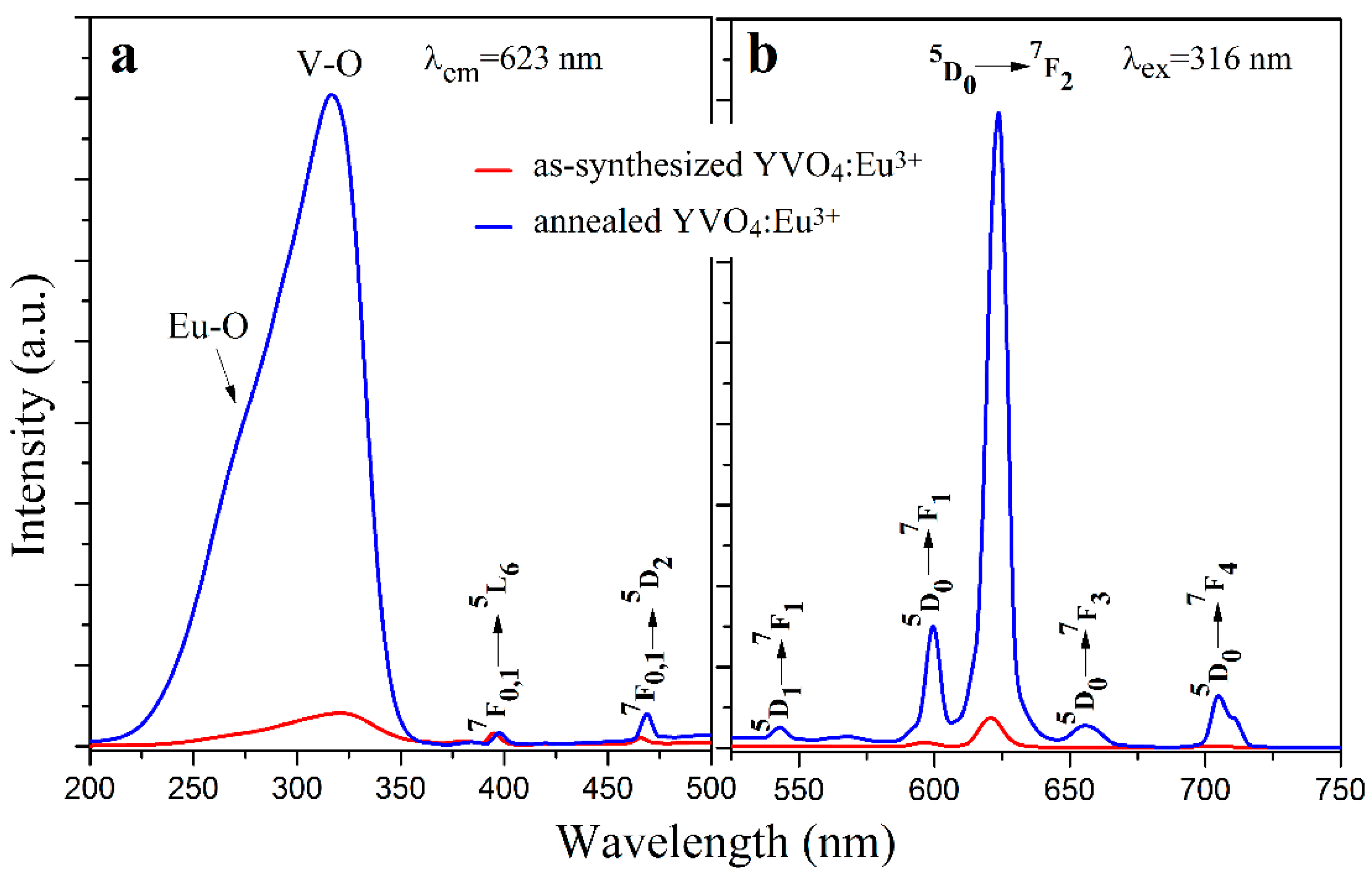
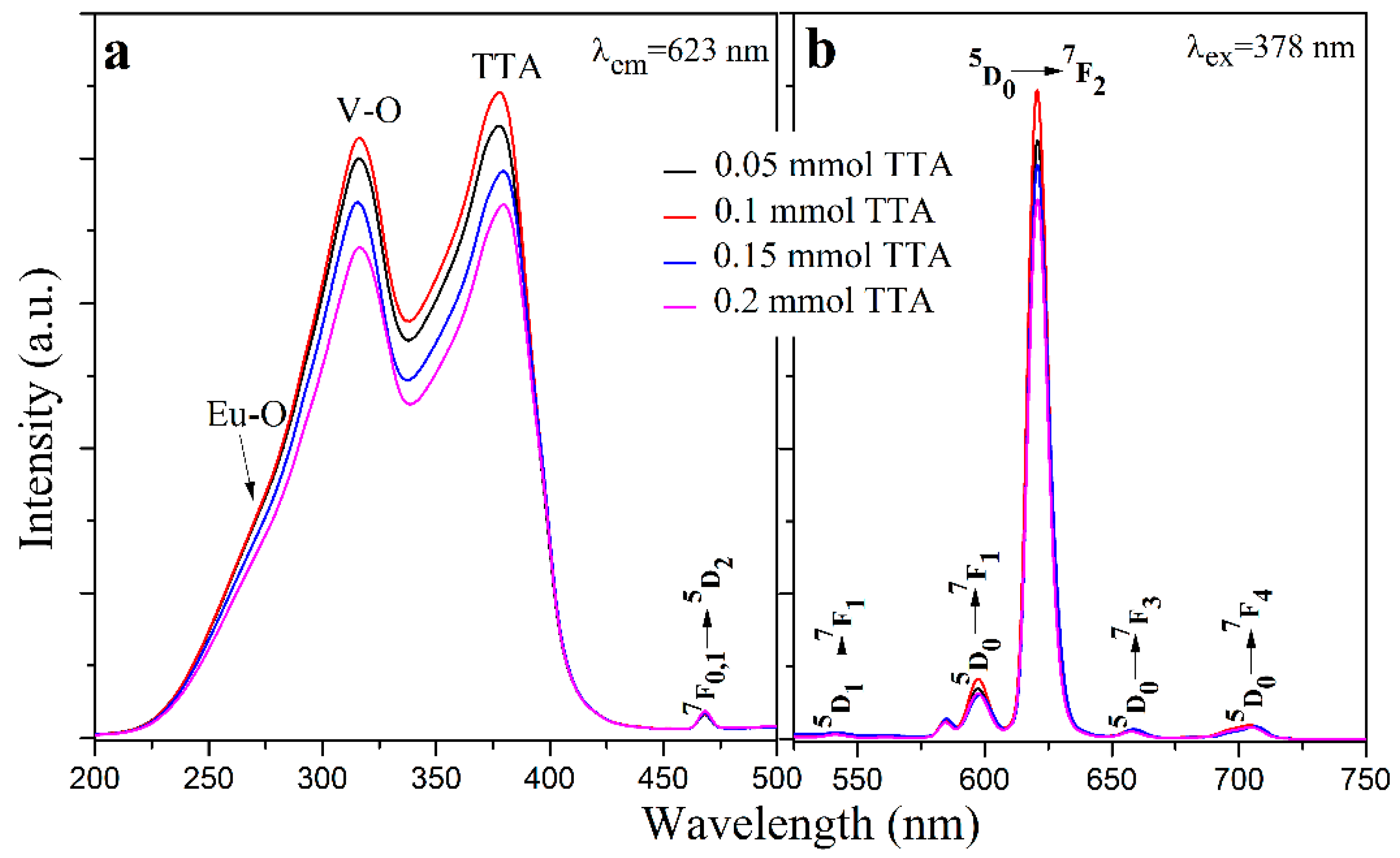
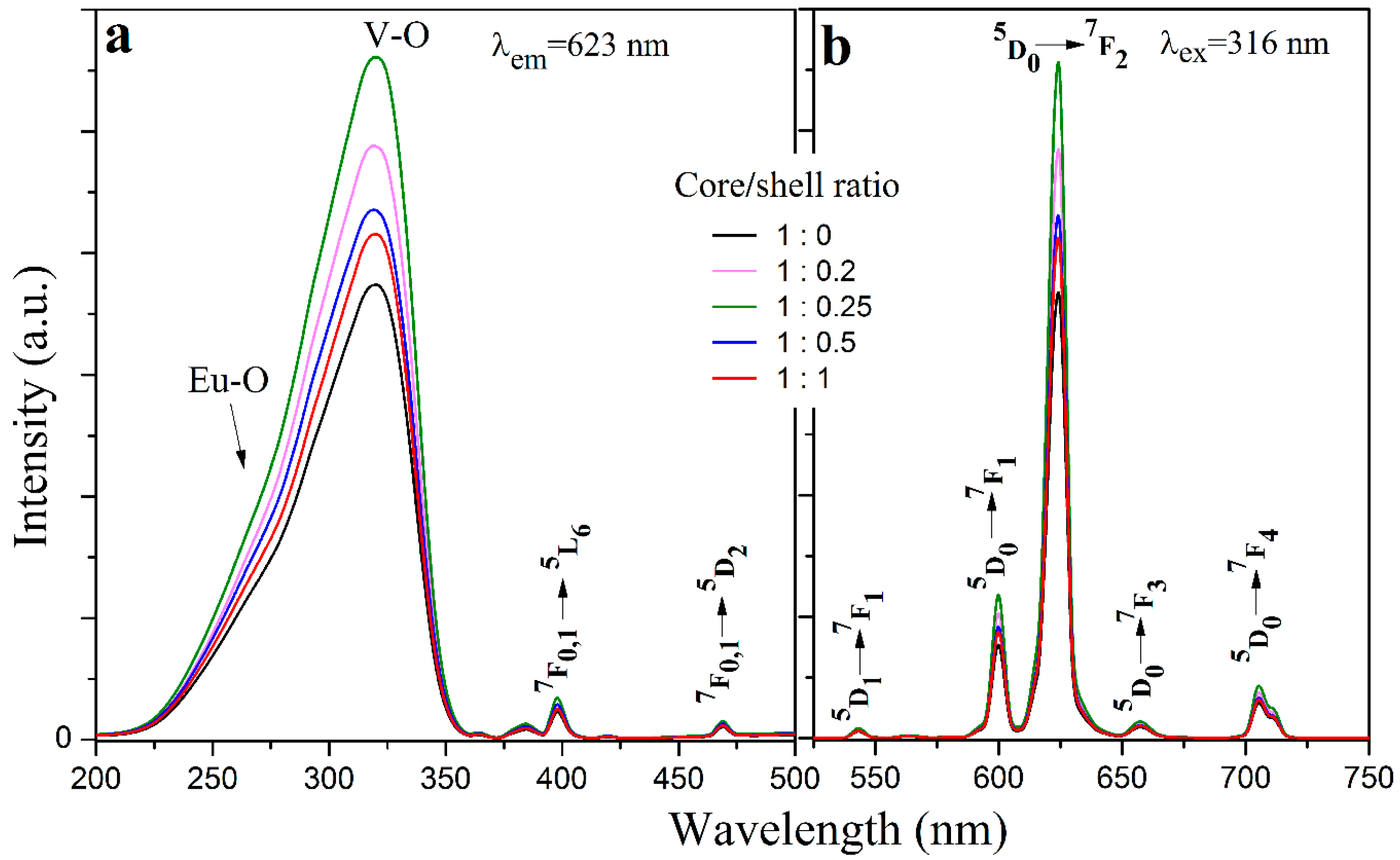
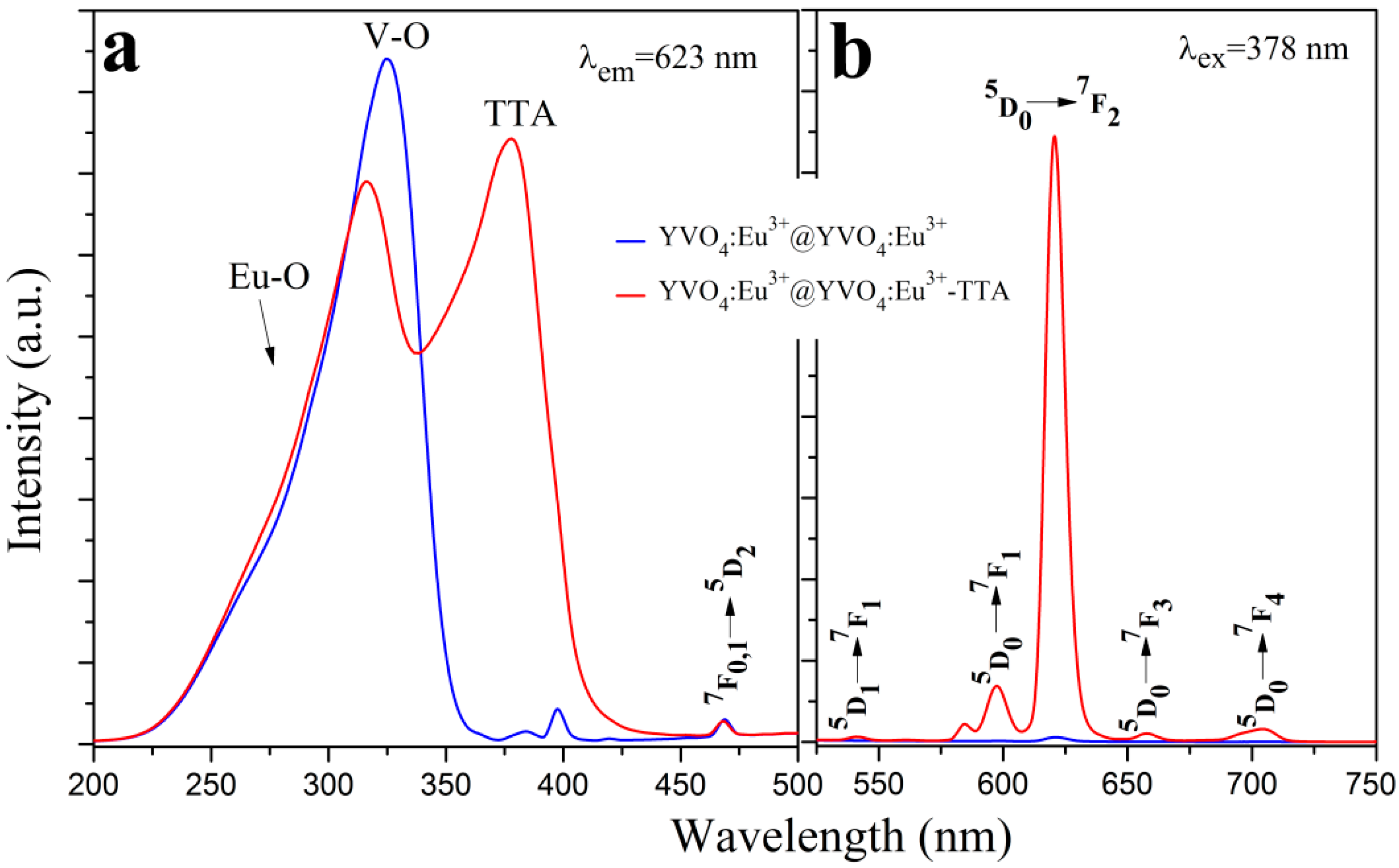
3.3. Photoluminescence Properties

The photoluminescence excitation and emission spectra (Hitachi F-4600) of the as-synthesized (unannealed) YVO4:Eu3+ nanoparticles and the annealed YVO4:Eu3+ nanoparticles are shown in Figure 6. In Figure 6a, the strongest excitation band peaking at about 316 nm was a result of the charge transfer absorption of VO43- groups [31]. The minor hump-like peak at about 260 nm was due to the charge transfer band of Eu-O. The other weak excitation peaks at 397 nm (7F0,15L6) and 469 nm (7F0,15D2) were from Eu3+ ions [32]. In Figure 6b, all of the photoluminescence emissions were from the f–f transitions of Eu3+, including 542 nm (5D17F1), 598 nm (5D07F1), 623 nm (5D07F2), 655 nm (5D07F3), and 705 nm (5D07F4) [33].
After annealing, a remarkable enhancement in their photoluminescence was resulted. This is consistent with the previously reported results [19,21,34,35]. An increase of about 22 times in the emission of the YVO4:Eu3+ nanoparticles at 623 nm was observed after the annealing process. As shown in Section 3.1, the average particle size of the YVO4:Eu3+ nanoparticles increased from about 15 nm to 28 nm after annealing, but this size difference may not sufficiently explain such a remarkable increase of their photoluminescence. The XRD patterns in Figure 2 indicates that the crystallinity of the as-synthesized YVO4:Eu3+ nanoparticles was greatly improved by the annealing process. Furthermore, the number of crystal defects in the YVO4:Eu3+ nanoparticles as well as the non-radiative traps should also simultaneously decrease. These factors are mainly responsible for the notable enhancement of photoluminescence of the YVO4:Eu3+ nanoparticles by annealing.
Figure 7 shows the photoluminescence excitation and emission spectra of the YVO4:Eu3+@YVO4 core–shell nanoparticles having different core/shell ratios. Such a homo-nanoparticled shell is believed to be able to effectively passivate the non-radiative traps or defects on the surface of the core [19,20]. The photoluminescence spectra in Figure 7 are typical for the Eu3+-doped YVO4 phosphors. After the core-shell process, the YVO4:Eu3+@YVO4 nanoparticles had stronger photoluminescence than the unshelled YVO4:Eu3+ nanoparticles (core/shell ratio = 1:0 in Figure 7), indicating the passivation effect of the YVO4 shell. The YVO4:Eu3+@YVO4 core–shell nanoparticles with the core/shell ratio of 1:0.25 had the highest photoluminescence property (Figure 7).
Figure 8 shows the photoluminescence spectra of the YVO4:Eu3+@YVO4:Eu3+–TTA inorganic–organic hybrid nanoparticles with different TTA amounts. Figure 9 compares the photoluminescence spectra of the YVO4:Eu3+@YVO4:Eu3+ nanoparticles and the YVO4:Eu3+@YVO4:Eu3+–TTA inorganic–organic hybrid nanoparticles. Compared with the excitation spectra of the as-synthesized and the annealed YVO4:Eu3+ nanoparticles (Figure 6a), the YVO4:Eu3+@YVO4:Eu3+–TTA inorganic–organic hybrid nanoparticles had a new excitation peak at about 378 nm (Figure 8a), which is a result of the TTA ligands. Consequently, the excitation spectral bandwidth of YVO4:Eu3+ was notably broadened from the range of 230–330 nm (Figure 6a) to the range of 230–415 nm (Figure 8a).
At the excitation wavelength of 378 nm, all of the emission peaks (Figure 8b) were from the Eu3+ ions, including 542 nm (5D17F1), 598 nm (5D07F1), 623 nm (5D07F2), 655 nm (5D07F3), and 705 nm (5D07F4) [31]. This suggests that the YVO4:Eu3+@YVO4:Eu3+–TTA inorganic–organic hybrid nanoparticles are efficiently sensitized by TTA ligands. The YVO4:Eu3+@YVO4:Eu3+–TTA inorganic–organic hybrid nanoparticles had the strongest photoluminescence when 0.1 mmol of TTA was used for hybridization.

4. Conclusions

In summary, YVO4 shells were grown on annealed YVO4:Eu3+ nanoparticle cores, and they were ion exchanged with Eu3+ ions, followed by hybridizing with TTA to successfully form the YVO4:Eu3+@YVO4:Eu3+–TTA inorganic–organic hybrid nanoparticles. It has been found that annealing process considerably enhances the photoluminescence of the as-synthesized YVO4:Eu3+ nanoparticles, and the presence of YVO4 shells further improves the photoluminescence of the annealed YVO4:Eu3+ nanoparticles. When the core/shell ratio was 1:0.25, the photoluminescence of the annealed YVO4:Eu3+ nanoparticles was the strongest. The inorganic–organic hybridization process greatly broadens the photoluminescence excitation spectral bandwidth range of the YVO4:Eu3+ nanoparticles. After hybridization with TTA, the YVO4:Eu3+@YVO4:Eu3+–TTA inorganic–organic hybrid nanoparticles broadened the excitation spectral bandwidth range from 230–330 nm to 230–415 nm, which covers the entire ultraviolet spectrum of the solar light and enables their potential applications in silicon solar cells.

Author Contributions

Conceptualization, G.D.; methodology, N.C.; software, J.H.; validation, J.H. and L.T.; formal analysis, L.T.; investigation, J.H. and L.T.; resources, N.C.; data curation, L.T.; writing—original draft preparation, L.T.; writing—review and editing, J.H.; visualization, L.T.; supervision, G.D.; project administration, N.C.; funding acquisition, G.D.

Funding

This research was funded by the National Natural Science Foundation of China (grant number 21571095) and the Scientific Research Projects of Hunan Education Department (grant number 18C1442).

Conflicts of Interest

The authors declare no conflict of interest.

References

  1. Feugnet, G.; Bussac, C.; Larat, C.; Schwarz, M.; Pocholle, J.P. High-efficiency TEM(00) Nd:YVO(4) laser longitudinally pumped by a high-power array. Opt. Lett. 1995, 20, 157–159. [Google Scholar] [CrossRef]
  2. Croitoru, G.; Pavel, N. Passive Q-switching by Cr4+:YAG saturable absorber of buried depressed-cladding waveguides obtained in Nd-doped media by femtosecond laser beam writing. Materials 2018, 11, 1689. [Google Scholar]
  3. Chander, N.; Khan, A.F.; Chandrasekhar, P.S.; Thouti, E.; Swami, S.K.; Dutta, V.; Komarala, V.K. Reduced ultraviolet light induced degradation and enhanced light harvesting using YVO4:Eu3+ down-shifting nano-phosphor layer in organometal halide perovskite solar cells. Appl. Phys. Lett. 2014, 105, 033904. [Google Scholar] [CrossRef]
  4. Shokouhimehr, M.; Rafiaei, S.M. Combustion synthesized YVO4:Eu3+ phosphors: Effect of fuels on nanostructure and luminescence properties. Ceram. Int. 2017, 43, 11469–11473. [Google Scholar] [CrossRef]
  5. Huang, C.K.; Chen, Y.C.; Hung, W.B.; Chen, T.M.; Sun, K.W.; Chang, W.-L. Enhanced light harvesting of Si solar cells via luminescent down-shifting using YVO4:Bi3+,Eu3+ nanophosphors. Prog. Photovolt. Res. Appl. 2013, 21, 1507–1513. [Google Scholar] [CrossRef]
  6. Xu, W.; Song, H.; Yan, D.; Zhu, H.; Wang, Y.; Xu, S.; Bai, X.; Dong, B.; Liu, Y. YVO4:Eu3+,Bi3+ UV to visible conversion nano-films used for organic photovoltaic solar cells. J. Mater. Chem. 2011, 21, 12331–12336. [Google Scholar] [CrossRef]
  7. Peng, Y.; Liu, J.; Zhang, K.; Luo, H.; Li, J.; Xu, B.; Han, L.; Li, X.; Yu, X. Near-infrared luminescent and antireflective in SiO2/YVO4:Yb3+ bilayer films for c-Si solar cells. Appl. Phys. Lett. 2011, 99, 121110. [Google Scholar] [CrossRef]
  8. Huang, X.Y.; Wang, J.X.; Yu, D.C.; Ye, S.; Zhang, Q.Y.; Sun, X.W. Spectral conversion for solar cell efficiency enhancement using YVO4: Bi3+, Ln3+ (Ln = Dy, Er, Ho, Eu, Sm, and Yb) phosphors. J. Appl. Phys. 2011, 109, 113526. [Google Scholar] [CrossRef]
  9. Ho, W.-J.; You, B.-J.; Liu, J.-J.; Bai, W.-B.; Syu, H.-J.; Lin, C.-F. Photovoltaic performance enhancement of silicon solar cells based on combined ratios of three species of europium-doped phosphors. Materials 2018, 11, 845. [Google Scholar] [CrossRef]
  10. Ho, W.-J.; Deng, Y.-J.; Liu, J.-J.; Feng, S.-K.; Lin, J.-C. Photovoltaic performance characterization of textured silicon solar cells using luminescent down-shifting Eu-doped phosphor particles of various dimensions. Materials 2017, 10, 21. [Google Scholar] [CrossRef]
  11. Riwotzki, K.; Haase, M. Colloidal YVO4:Eu and YP0.95V0.05O4:Eu nanoparticles: Luminescence and energy transfer processes. J. Phys. Chem. B 2001, 105, 12709–12713. [Google Scholar] [CrossRef]
  12. Liao, Y.; Chen, N.; Du, G. Strong luminescence enhancement of YVO4:Eu3+,Ba2+ phosphors prepared by a solvothermal method. J. Alloys Compd. 2013, 561, 214–219. [Google Scholar] [CrossRef]
  13. Takeshi, S.; Ogato, H.; Isobe, T.; Wawayama, T.; Niikura, S. Effects of Citrate Additive on Transparency and Photostability Properties of YVO4:Bi3+,Eu3+ Nanophosphor. J. Electrochem. Soc. 2010, 157, J74–J80. [Google Scholar] [CrossRef]
  14. Chen, D.; Yu, Y.; Huang, P.; Lin, H.; Shan, Z.; Zeng, L.; Yang, A.; Wang, Y. Color-tunable luminescence for Bi3+/Ln3+:YVO4 (Ln = Eu, Sm, Dy, Ho) nanophosphors excitable by near-ultraviolet light. Phys. Chem. Chem. Phys. 2010, 12, 7775–7778. [Google Scholar] [CrossRef] [PubMed]
  15. Wang, X.; Yang, Y.; Du, G.; Chen, N.; Zhang, A. Broadband UV excited LaF3:Eu3+ based inorganic–organic mixed hybrid nanoparticles for strong luminescence. J. Nanosci. Nanotechnol. 2018, 18, 1876–1881. [Google Scholar] [CrossRef] [PubMed]
  16. Carlos, L.D.; Ferreira, R.A.S.; de Zea Bermudez, V.; Ribeiro, S.J.L. Lanthanide-containing light-emitting organic–inorganic hybrids: A Bet on the Future. Adv. Mater. 2009, 21, 509–534. [Google Scholar] [CrossRef]
  17. He, Y.; Chen, N.; Du, G. Synthesis of LaOF:Eu3+ Nanoparticles with strong luminescence enhanced by organic ligands. J. Am. Ceram. Soc. 2014, 97, 1931–1936. [Google Scholar] [CrossRef]
  18. Tang, L.; Gui, W.; Ding, K.; Chen, N.; Du, G. Ion exchanged YVO4:Eu3+ nanoparticles and their strong luminescence enhanced by energy transfer of thenoyltrifluoroacetone ligands. J. Alloys Compd. 2014, 590, 277–282. [Google Scholar] [CrossRef]
  19. Mialon, G.; Gohin, M.; Gacoin, T.; Boilot, J.-P. High temperature strategy for oxide nanoparticle synthesis. ACS Nano 2008, 2, 2505–2512. [Google Scholar] [CrossRef]
  20. Lin, Q.; Xu, Y.; Fu, E.; Baber, S.; Bao, Z.; Yu, L.; Deng, S.; Kundu, J.; Hollingsworth, J.; Bauer, E.; et al. Polymer-assisted chemical solution approach to YVO4:Eu nanoparticle networks. J. Mater. Chem. 2012, 22, 5835–5839. [Google Scholar] [CrossRef]
  21. Filho, P.C.D.; Gacoin, T.; Boilot, J.-P.; Walton, R.I.; Serra, O.A. Synthesis and luminescent properties of REVO4−REPO4 (RE = Y, Eu, Gd, Er, Tm, or Yb) heteronanoparticles: A promising class of phosphors for excitation from NIR to VUV. J. Phys. Chem. C 2015, 119, 24062–24074. [Google Scholar] [CrossRef]
  22. Zhang, F.; Che, R.; Li, X.; Yao, C.; Yang, J.; Shen, D.; Hu, P.; Li, W.; Zhao, D. Direct imaging the upconversion nanoparticle core/shell structure at the subnanometer level: Shell thickness dependence in upconverting optical properties. Nano Lett. 2012, 12, 2852–2858. [Google Scholar] [CrossRef] [PubMed]
  23. Yi, G.-S.; Chow, G.-M. Water-soluble NaYF4:Yb,Er(Tm)/NaYF4/polymer core/shell/shell nanoparticles with significant enhancement of upconversion fluorescence. Chem. Mater. 2007, 19, 341–343. [Google Scholar] [CrossRef]
  24. Velasco, D.S.; de Moura, A.P.; Medina, A.N.; Baesso, M.L.; Rubira, A.F.; Cremona, M.; Bento, A.C. Preparation, characterization, and spectroscopic properties of PC/PMMA doped blends: Study of the effect of rare-earth doping on luminescence, quenching rate, and lifetime enhancement. J. Phys. Chem. B 2010, 114, 5657–5660. [Google Scholar] [CrossRef]
  25. Meng, Q.G.; Boutinaud, P.; Franville, A.-C.; Zhang, H.J.; Mahiou, R. Preparation and characterization of luminescent cubic MCM-48 impregnated with an Eu3+ β-diketonate complex. Microporous Mesoporous Mater. 2003, 65, 127–136. [Google Scholar] [CrossRef]
  26. Guan, J.B.; Chen, B.; Sun, Y.Y.; Liang, H.; Zhang, Q.J. Effects of synergetic ligands on the thermal and radiative properties of Eu(TTA)3nL-doped poly(methyl methacrylate). J. Non-Cryst. Solids 2005, 351, 849–855. [Google Scholar] [CrossRef]
  27. Hirano, S.; Yogo, T.; Kikuta, K.; Sakamoto, W.; Koganei, H. Synthesis of Nd:YVO4 Thin Films by a Sol-Gel Method. J. Am. Ceram. Soc. 1996, 79, 3041–3044. [Google Scholar] [CrossRef]
  28. Wu, X.C.; Tao, Y.R.; Song, C.Y.; Mao, C.J.; Dong, L.; Zhu, J.J. Morphological control and luminescent properties of YVO4:Eu nanoparticles. J. Phys. Chem. B 2006, 110, 15791–15796. [Google Scholar] [CrossRef]
  29. Wang, H.; Yu, M.; Lin, C.K.; Lin, J. Core–shell structured SiO2@YVO4:Dy3+/Sm3+ phosphor particles:Sol–gel preparation and characterization. J. Colloid Interface Sci. 2006, 300, 176–182. [Google Scholar] [CrossRef]
  30. Wang, Y.; Qin, W.P.; Zhang, J.S.; Cao, C.Y.; Lü, S.Z.; Ren, X.G. Photoluminescence of colloidal YVO4:Eu/SiO2 core/shell nanoparticles. Opt. Commun. 2009, 282, 1148–1153. [Google Scholar] [CrossRef]
  31. Hsu, C.; Powell, R.C. Energy transfer in europium doped yttrium vanadate crystals. J. Lumin. 1975, 10, 273–293. [Google Scholar] [CrossRef]
  32. Meyssamy, H.; Riwotzki, K.; Kornowski, A.; Naused, S.; Haase, M. Wet-chemical synthesis of doped colloidal nanomaterials: Particles and fibers of LaPO4:Eu, LaPO4:Ce, and LaPO4:Ce,Tb. J. Cryst. Growth 1999, 11, 840–844. [Google Scholar] [CrossRef]
  33. Jia, G.; Song, Y.H.; Yang, M.; Liu, K.; Zheng, Y.H.; You, H.P. Facile synthesis and luminescence properties of octahedral YVO4:Eu3+ microcrystals. J. Cryst. Growth 2009, 311, 4213–4218. [Google Scholar] [CrossRef]
  34. Liao, Y.; Zhen, Y.; Chen, N.; Du, G. Effect of Sr2+ doping on the luminescence properties of YVO4:Eu3+,Sr2+ particles prepared by a solvothermal method. J. Sol-Gel Sci. Technol. 2013, 65, 353–358. [Google Scholar] [CrossRef]
  35. Luwang, M.N.; Ningthoujam, R.S.; Srivastava, S.K.; Vatsa, R.K. Preparation of white light emitting YVO4: Ln3+ and silica-coated YVO4:Ln3+ (Ln3+ = Eu3+, Dy3+, Tm3+) nanoparticles by CTAB/n-butanol/hexane/water microemulsion route: Energy transfer and site symmetry studies. J. Mater. Chem. 2011, 21, 5326–5337. [Google Scholar] [CrossRef]
Figure 1. Illustration of preparation steps: (a) as-synthesized YVO4:Eu3+, (b) annealed YVO4:Eu3+, (c) core-shell YVO4:Eu3+@YVO4, (d) core-shell YVO4:Eu3+@YVO4:Eu3+, (e) hybrid YVO4:Eu3+@YVO4:Eu3+–TTA.
Materials 12 03830 g001
Figure 2. X-ray diffraction (XRD) patterns of the as-synthesized YVO4:Eu3+, the annealed YVO4:Eu3+ and the YVO4:Eu3+@YVO4:Eu3+–TTA inorganic–organic hybrid nanoparticles.
Materials 12 03830 g002
Figure 3. Transmission electron microscopy (TEM) (ac) and HRTEM (df) images of the as-synthesized (unannealed) YVO4:Eu3+ nanoparticles (a,d), the annealed YVO4:Eu3+ nanoparticles (b,e) and the YVO4:Eu3+@YVO4:Eu3+ core-shell nanoparticles (c,f).
Materials 12 03830 g003
Figure 4. The Fourier transform infrared (FTIR) spectra of 2-thenoyltrifluoroacetone (TTA), the annealed YVO4:Eu3+ nanoparticles and YVO4:Eu3+@YVO4:Eu3+–TTA hybrid nanoparticles.
Materials 12 03830 g004
Figure 5. UV-Vis absorption spectra of the annealed YVO4:Eu3+ nanoparticles, TTA and YVO4:Eu3+@ YVO4:Eu3+–TTA hybrid nanoparticles.
Materials 12 03830 g005
Figure 6. The excitation (a) and emission (b) spectra of the as-synthesized (unannealed) and the annealed YVO4:Eu3+ nanoparticles.
Materials 12 03830 g006
Figure 7. The excitation (a) and emission (b) spectra of the YVO4:Eu3+@YVO4 core-shell nanoparticles with different core/shell ratios.
Materials 12 03830 g007
Figure 8. The photoluminescence excitation (a) and emission (b) spectra of YVO4:Eu3+@YVO4:Eu3+–TTA inorganic–organic hybrid nanoparticles prepared with different TTA amounts.
Materials 12 03830 g008
Figure 9. Comparison of the photoluminescence excitation (a) and emission (b) spectra of the YVO4:Eu3+@YVO4:Eu3+ nanoparticles and the YVO4:Eu3+@YVO4:Eu3+–TTA inorganic–organic hybrid nanoparticles.
Materials 12 03830 g009

