Article

Dual Doping of Silicon and Manganese in Hydroxyapatites: Physicochemical Properties and Preliminary Biological Studies

by
Katarzyna Szurkowska
1,
Agata Drobniewska
2 and
Joanna Kolmas
1,*
1
Department of Analytical Chemistry and Biomaterials, Analytical Group, Faculty of Pharmacy with Laboratory Medicine Division, Medical University of Warsaw, ul. Banacha 1, 02-097 Warsaw, Poland
2
Department of Environmental Health Sciences, Faculty of Pharmacy with Laboratory Medicine Division, Medical University of Warsaw, ul. Banacha 1, 02-097 Warsaw, Poland
*
Author to whom correspondence should be addressed.
Materials 2019, 12(16), 2566; https://doi.org/10.3390/ma12162566
Submission received: 4 July 2019 / Revised: 8 August 2019 / Accepted: 9 August 2019 / Published: 12 August 2019
(This article belongs to the Special Issue Novel Biomaterials for Orthopaedic/Musculoskeletal Tissue Engineering)

Abstract

:
Silicated hydroxyapatite powders enriched with small amounts of manganese (Mn2+) cations were synthesized via two different methods: precipitation in aqueous solution and the solid-state method. The source of Mn2+ ions was manganese acetate, while silicon was incorporated using two different reagents: silicon acetate and sodium metasilicate. Powder X-ray diffraction (PXRD) analysis showed that the powders obtained via the precipitation method consisted of single-phase nanocrystalline hydroxyapatite. In contrast, samples obtained via the solid-state method were heterogenous and contaminated with other phases, (i.e., calcium oxide, calcium hydroxide, and silicocarnotite) arising during thermal treatment. The transmission electron microscope (TEM) images showed powders obtained via the precipitation method were nanosized and elongated, while solid-state synthesis produced spherical microcrystals. The phase identification was complemented by Fourier transform infrared spectroscopy (FTIR). An in-depth analysis via solid-state nuclear magnetic resonance (ssNMR) was carried out, using phosphorus 31P single-pulse Bloch decay (BD) (31P BD) and cross-polarization (CP) experiments from protons to silicon-29 nuclei (1H → 29Si CP). The elemental measurements carried out using wavelength-dispersive X-ray fluorescence (WD-XRF) showed that the efficiency of introducing manganese and silicon ions was between 45% and 95%, depending on the synthesis method and the reagents. Preliminary biological tests on the bacteria Allivibrio fisheri (Microtox®) and the protozoan Spirostomum ambiguum (Spirotox) showed no toxic effect in any of the samples. The obtained materials may find potential application in regenerative medicine, bone implantology, and orthopedics as bone substitutes or implant coatings.

1. Introduction

Among the calcium phosphates, hydroxyapatite (HA) (Ca10(PO4)6(OH)2) has received considerable attention as a bone substitute material for orthopaedic and dental applications due to its strong affinity toward human mineralized tissues (bone and teeth) [1,2,3,4]. Biological apatite is nanocrystalline and contains various ions as impurities, mainly magnesium (Mg2+) and carbonates (CO32−), with other trace elements in various amounts (Zn2+, K+, Na+, Mn2+, SiO44−, Cl, and F) [5,6,7]. It should be noted that synthetic hydroxyapatites are highly susceptible to ionic substitution. Therefore, the introduction of various ions into the crystal structure of hydroxyapatite allows for obtaining material similar to bones and teeth. This is based on the concepts of biomimetics, where the greater the similarity existing between an implant and a tissue, the better they are expected to interact with each other [8,9]. It is important to note that ionic substitutions can change some physicochemical properties, such as the crystallographic lattice parameters, crystal morphology, and the degree of crystallinity. Substituting oxy-ions, (i.e., SiO44−, CO32−) and cations with various charges for the orthophosphates or structural hydroxyl groups and calcium ions, respectively, may lead to destabilization of the crystal lattice and a higher dissolution rate. Moreover, even a trace addition of some elements can significantly influence the thermal stability, solubility, or bioactivity of biomaterial, in vitro as well as in vivo [5,6,7].
Both manganese and silicon are classified as trace elements, with an average bone content of 1.7–3 ppm and 100–150 ppm, respectively [10,11]. The divalent manganese ion has the ability to activate integrins, a family of receptors that facilitate cellular adhesion [12]. Stimulation of bone cell adhesion, viability, and proliferation has a beneficial effect on the interaction of the implant with the host bone tissue [13,14]. Manganese is also a cofactor in enzymes, such as glycosyltransferases, involved in the remodelling of the extracellular matrix present in bone and cartilage [15]. Studies have shown that manganese supplementation in ovariectomized rats inhibits bone loss [16]. A key role for manganese in maintaining normal bone mass has been suggested, indicating its deficiency as a likely cause of osteoporosis [17,18]. Other mechanisms of manganese’s beneficial effects on bone tissue metabolism include osteocalcin stimulation, increased alkaline phosphatase activity and collagen type I production [13,14,19]. Syntheses of hydroxyapatites substituted with various amounts of manganese were carried out, from trace amounts (a few ppm) to 11.9 wt% [14,16,19,20,21,22,23,24]. However, it is important to note that in vitro studies on osteoblast cells showed significantly better results for lower manganese contents [16,19], while a Mn content of 1 wt% was regarded as high and caused drastically lower cell viability [24].
In contrast to manganese, silicon has a well-established position as a bioactivity-improving additive for bone biomaterials [11,25,26]. Carlisle’s landmark studies have defined the appropriate level of silicon required for the proper development and mineralization of osseous tissue [27,28]. The mechanism of silicon action is based on the positive impact of orthosilicic acid on the synthesis of type-I collagen [29,30]. Moreover, in the presence of orthosilicic acid in the medium, osteoblast-like MG-63 cells exhibit increased alkaline phosphatase activity and osteocalcin synthesis [29,30]. Furthermore, exposure to silicon has a stimulatory effect on the differentiation, proliferation, and activity of osteoblasts [31,32,33,34]. Thus, partial replacement of the hydroxyapatite phosphate group by silicate ions should further improve the biological activity of the bioceramic. Numerous syntheses of Si-substituted hydroxyapatite (Si-HA) were carried out, introducing up to 5 wt% of Si, which corresponds to the introduction of 1.7 moles of Si per mole of HA [35,36,37,38,39,40,41,42,43,44,45,46,47]. According to the research, to ensure phase uniformity of the obtained material along with optimal bioactivity, substitution should not exceed 1 mole of Si per mole of HA [36,37]. The beneficial effect of silicon substitution in HA has been demonstrated in numerous in vitro and in vivo experiments. High in vitro activity was confirmed by apatite precipitation when the samples were soaked in simulated body fluid (SBF), as well as in studies on osteoblasts, osteoclasts, and osteosarcoma cell cultures [37,38,39,40,41]. In vivo studies have shown faster bone remodelling in Si-HA samples, which additionally testifies to the osteoconductive properties of Si-HA [42,43,44,45,46,47].
The aim of the current study was to synthesize novel hydroxyapatite material, co-substituted with manganese (II) and silicate ions. Based on the previously quoted results [16,19,36,37], substitutions of 0.017 wt% of manganese and 1.98 wt% of silicon were planned for the current research. Synthesis was carried in two different ways: via precipitation and solid-state method. Moreover, two silicate sources were used, to compare the effectiveness of substitution for various synthesis conditions. This work focused on the structure and physico-chemical properties of the dual-doped hydroxyapatites, which were characterized using transmission electron microscopy (TEM), powder X-ray diffractometry (PXRD), Fourier transform infrared spectroscopy (FTIR,) solid-state nuclear magnetic resonance (ssNMR), and wavelength dispersive X-ray fluorescence (WD-XRF). Preliminary acute toxicity tests were also conducted.

2. Materials and Methods

2.1. Preparation of Samples

Synthesis of the materials was performed using two different methods (the precipitation method and the solid-state method) with two different sources of silicon (Na2SiO3 or Si(CH3COO)4). The planned samples, with nominal composition Ca9.997Mn0.003(PO4)5.3(SiO4)0.7(OH)1.3 and intended (Ca + Mn)/(P + Si) molar ratio of 1.67, are summarized in Table 1. Regardless of the silicon source, Si was introduced into hydroxyapatite in the form of orthosilicate SiO44− ions. In the case of metasilicate ions, hydrolysis occurred under the reaction conditions.
Precipitation synthesis was carried out at room temperature. Stoichiometric amounts of Ca(NO3)2∙4H2O and (CH3COO)2Mn were dissolved in 400 mL of distilled water in a flask under constant stirring. A Si-containing solution (Na2SiO3 or Si(CH3COO)4 dissolved in 50 mL of distilled water) and a P-containing solution ((NH4)2HPO4 dissolved in 50 mL of distilled water) were added dropwise to the Ca-Mn precursor solution using separate burettes. The pH was adjusted to approximately 10 using concentrated ammonia solution. The resultant precipitate was allowed to rest for 24 h for ageing. The precipitates were then filtered through a 0.8 µm pore size membrane filter under reduced pressure and rinsed several times with distilled water until the filtrate reached pH 7. This step was aimed at washing away residual reagents and soluble reaction products. The obtained precipitates were dried at 130 °C for 24 h and then ground in an agate mortar for physico-chemical and biological characterization.
For the solid-state synthesis, CaCO3, (CH3COO)2Mn, (NH4)2HPO4, and Na2SiO3 or Si(CH3COO)4 were used. The weighed substrates were ground in a ball mill and were pressed into tablets using a hydraulic press (pressure force 10 tons). The key step in the solid-state synthesis was sintering the tablets in the muffle furnace (Czylok FCF2,5SH, Jastrzębie-Zdrój, Poland) using the following temperature program: heating to 400 °C for 8 h, heating to 700 °C for 8 h, heating to 1000 °C for 8 h, then allowed to cool. To prevent the oxidation of Mn2+ ions, heating was carried out in an argon atmosphere. After the thermal treatment, the tablets were crushed in an agate mortar and the obtained powder was then subjected to further analysis.

2.2. Sample Characterization

The synthesized materials were analyzed via X-ray powder diffractometry (PXRD) using a Bruker D8 DISCOVER diffractometer (Bruker, Karlsruhe, Germany) with a Cu anticathode (λ = 1.54 Å). The scans were run from 20° to 70°, with a step size of 0.024°, a step time of 4 s, and a locked, coupled (theta–theta) geometry. Stoichiometric HA (inorganic crystal structure database ICSD #00-009-0432) was used as the reference pattern. On the basis of the diffraction patterns, the parameters of the unit cell (a and c) were estimated. The Scherrer equation (1) was used to calculate the crystal sizes of the samples synthesized by the precipitation method [48]:
d = 0.94 λ β cos θ ,
where
  • d—crystallite size (nm)
  • λ—radiation wavelength (nm)
  • β—the peak full width at half maximum intensity (radians)
  • θ—the diffraction angle of the corresponding reflex (°).
The reflections at approximately 25.8° and 39.9° were chosen for the calculation.
A JEOL JEM-1400 transmission electron microscope (TEM, Jeol LTD, Tokyo, Japan) with an accelerating voltage of 80 kV was used to observe the morphology of the obtained powders. In order to prepare the samples for TEM analysis, they were suspended in ethanol and then dropped on a copper grid covered with Formvar.
Elemental analysis of the synthesized apatites was carried out via the WD-XRF method in the solid state, using tablets made of samples carefully triturated with microcrystalline cellulose. In order to determine the percentage weight of manganese and silicon in the samples, a standard curve of the signal-to-element concentration was made. The standard curve for each element consisted of five measurement points, while the blank sample was a tablet made of pure microcrystalline cellulose. To ensure uniform distribution of the standard in the tablet mass, the cellulose was mixed with a calculated amount of the standard solution, which was later evaporated. The cellulose was compressed into a tablet using a hydraulic press. Each measurement was carried out in triplicate using a WD-XRF ARL ADVANT’X spectrometer (Thermo Electron Corp., Waltham, MA, USA). The (Ca + Mn)/(P + Si) ratio of the samples was determined using energy-dispersive X-ray spectroscopic microanalysis (EDS INCA Energy TEM, Oxford Instruments, Abingdon, UK; Ca and P contents measured from six spots and then averaged).
The chemical composition of the samples was investigated using Fourier transform infrared spectroscopy (FTIR, Perkin Elmer, Waltham, MA, US). The samples were ground with KBr in an agate mortar and then analyzed from 4000 to 400 cm−1 using a Perkin Elmer Spectrum 1000 spectrometer. Spectra were obtained within 30 repetitions (scans) with a resolution of 2 cm−1.
Solid-state NMR experiments on 31P and 29Si nuclei were performed using a Bruker WB 400 spectrometer (Bruker, Karlsruhe, Germany). The following experiments were carried out: 31P single-pulse Bloch decay (31P BD) and cross-polarization from protons to silicon-29 nuclei (1H → 29Si CP). The samples were spun at 7 and 5 kHz for the 31P and 29Si experiments, respectively, at the magic angle spinning (MAS), using ZrO2 rotors. The 31P and 29Si chemical shifts were referenced to external 85% orthophosphoric acid and tetramethylosilane (TMS), respectively.
Preliminary toxicity tests were performed using two tests: Microtox® and Spirotox. Two different suspensions were prepared, i.e., 1 and 2 mg/mL. The Microtox® test was based on the lyophilized luminescent bacteria Allivibrio fischeri. The suspended samples were incubated for 15 min with the bacteria in disposable glass cuvettes and then the light output of the samples was measured in the Microtox® M500 analyser (Osprey Scientific, Edmonton, AB, Canada). All samples were run in duplicate. For the control, 2% NaCl was used.
The protozoan Spirostomum ambiguum was used in the Spirotox test, which was performed in polystyrene multiwell plates according to the standard protocol [49]. In each well, 10 organisms were placed together with the suspended materials. The samples were incubated in the dark at 25 °C for 24 and 48 h. Then, a dissection microscope was used to analyze the lethal response or deformation of the S. ambiguum cell. The sample was tested in triplicate in each acute toxicity test (Microtox® and Spirotox); however, the tests were repeated (performed twice). Tyrode’s solution was used as a control.

3. Results and Discussion

3.1. PXRD, TEM, and Elemental Analysis Results

The PXRD patterns of the powders synthesized by the precipitation method (Mn,SiO3-Haw, and Mn,SiO4-HAw) revealed the presence of poorly crystallized hydroxyapatite (see Figure 1). The reflections were very wide and weakly resolved, and some of them were too broad to be detected. Therefore, it may be suggested that the materials were nanocrystalline. The calculations based on the Scherrer equation allowed the crystal size to be estimated for the Mn,SiO4-HAw and Mn,SiO3-HAw nanocrystalline samples. The lengths of crystallites were 11 ± 2 nm and 13 ± 3 nm, while the widths were 6 ± 2 nm and 7 ± 2 nm, respectively.
In contrast, in the PXRD diffractograms of the samples prepared using the solid-state method (Mn,SiO3-HAd and Mn,SiO4-HAd), narrow and well-resolved reflections were observed (see Figure 1). It is worth noting that the solid-state method of synthesis led to extra reflections on the diffractogram patterns of both samples. In the Mn,SiO3-HAd pattern, the additional reflections at ca 32.45°, 37.45°, and 33.42° may be attributed to calcium oxide and calcium hydroxide, respectively [50,51]. The results of Rietveld refinement showed that the sample Mn,SiO3-HAd contained 3 wt% of CaO and 2 wt% of Ca(OH)2. On the other hand, the Mn,SiO4-HAd pattern presented additional reflections at approximately 34.7°, 30.2°, 29.6°, 26.9°, and 34.4° that originated mainly from silicocarnotite (Ca5(PO4)2SiO4) [51,52]. It should be noted that during calcination, in both cases, the obtained hydroxyapatites partially decomposed. This is usually promoted by non-stoichiometry and the presence of impurities or additives, i.e., substituted ions. Andreev et al. [51] suggested that silicocarnotite is a material produced during high-temperature calcination of calcium phosphates with silicon sources. According to the Rietveld analysis, the Mn,SiO4-HAd sample contained 5 wt% of silicocarnotite (JCPDS #40-393).
The parameters (a and c) of the apatitic crystal unit cell of the obtained samples (see Table 1) were found to be slightly different from those of stoichiometric HA (a = 9.432 Å and c = 6.881 Å), but it is difficult to find any regularities. According to the literature, the introduction of silicon ions should cause a decrease in the parameter a and an increase in the parameter c, while manganese substitution should reduce both parameters [5,6,7]. There was a distinct decrease in parameter a and an increase in parameter c in both of the solid-state-synthesized samples. On the other hand, the unit cell parameters of the samples obtained via the precipitation method did not change significantly.
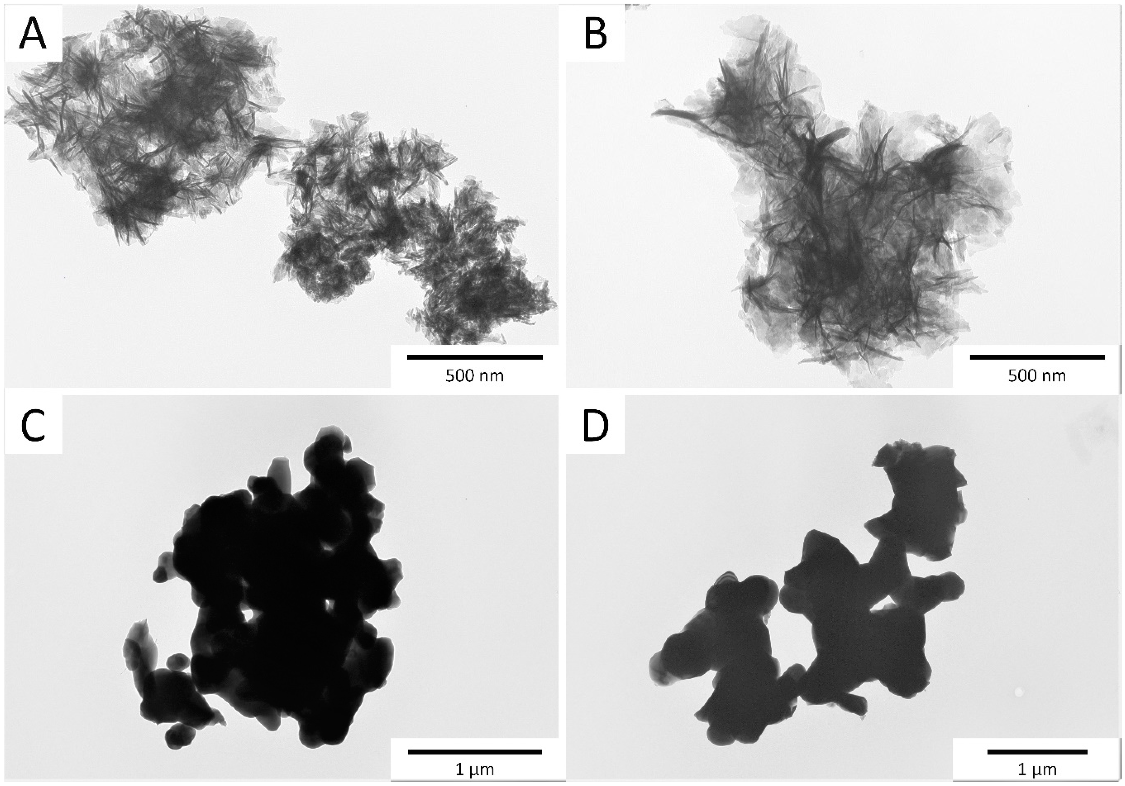
The TEM micrographs of the samples obtained via the precipitation method (Mn,SiO4-HAw and Mn,SiO3-HAw) are shown in Figure 2. The powders were nanosized, and the crystals were elongated and needle-shaped. This is in clear accordance with the literature where both the addition of manganese and silicate ions caused the formation of needle-like crystals [5,6,7]. Moreover, the crystals exhibited a high tendency to agglomerate, and therefore it was difficult to measure crystal sizes. However, we observed that the Mn,SiO4-HAw sample was characterized by smaller crystals compared to the Mn,SiO3-Haw sample. As shown in Figure 2, the crystals were heterogenous and differed significantly in size, while the calculation using the Scherrer equation shows the average crystal size. Synthesis in the solid state caused the formation of large crystal agglomerates, with sizes of up to approximately 0.5 μm. During the long heat treatment, the crystals formed a spherically shaped mould. As in the case of precipitation synthesis, the sample obtained from silicon acetate (Mn,SiO4-HAd) appeared to have smaller crystals. However, the difference was not as pronounced as in the precipitation method.
The Mn and Si content in each of the samples prepared using the two methods are presented in Table 1. In the Mn,SiO3-HAw and Mn,SiO4-HAw samples, manganese concentrations reached 50.6% and 64.7% of the nominal values, respectively. For the samples synthesized via the solid-state method, the Mn content was slightly lower (44.7%–47.1% of the nominal concentration). It is worth noting that substitution with Mn2+ ions was not trivial and ran with low efficiency. Recent works on the synthesis of hydroxyapatites enriched in manganese indicate that the substitution takes place within a limited range (up to 10 mol%) [22,24]. In addition, the thermal treatment of Mn-HA causes the decomposition of material at a fairly low temperature (600 °C) [21,24]. In contrast, the silicon substitution efficiency in Mn,SiO3-HAd and Mn,SiO4-HAd was significantly higher (91.9% and 94.9%, respectively) than in the samples produced using precipitation (61.5% and 76.4% of the nominal value for Mn,SiO3-HAw and Mn,SiO4-Haw, respectively). According to the literature, silicate ions can be located both inside the crystals (in the crystal’s core) and on the surface in the so-called hydrated surface layer. During the synthesis using the precipitation method, nanocrystals with an expanded surface layer were obtained [48]. Repeated washing of the obtained precipitates may lead to a partial loss of silicates, and thus lower substitution efficiency. In addition, silicates may “compete” with carbonates in the substitution of orthophosphates in the apatitic lattice. During the solid-state synthesis, carbonate ions contamination was easily removed by thermal decomposition. It is also worth mentioning that simultaneous substitution with silicate and manganese ions may be hindered due to the differences in ionic radii of Ca2+/Mn2+ (100 vs. 67 pm) and PO43−/SiO44− (238 vs. 240 pm). Verification of such a hypothesis requires further research (especially PXRD detailed analysis).

3.2. FTIR Spectroscopy

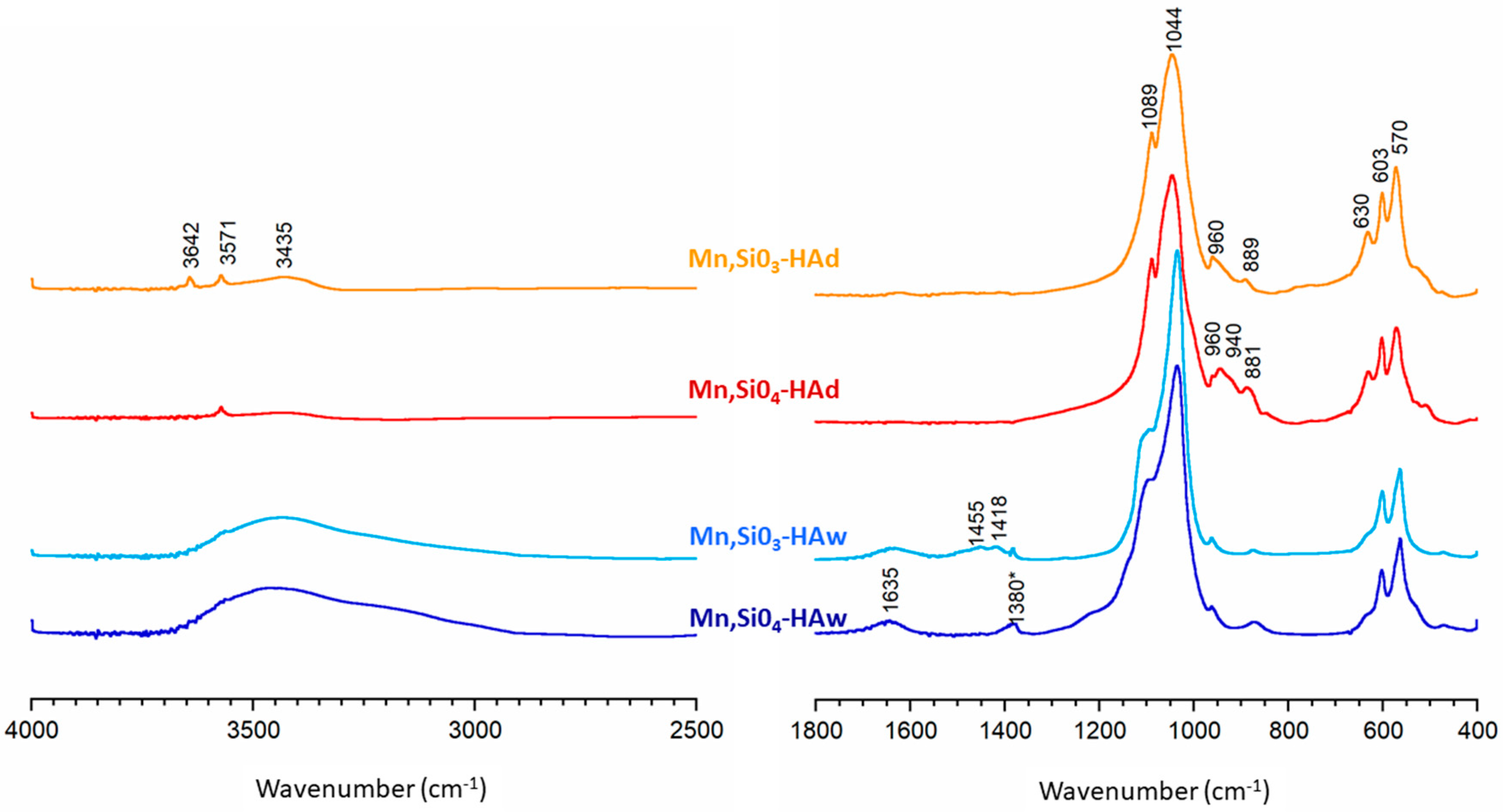
The FTIR spectra of the synthesized samples are shown in Figure 3. The absorption stretching bands in the region 1200–960 cm−1 may be assigned to the typical orthophosphate vibrations for hydroxyapatites. Phosphate bending bands (ν4) were observed at 570 and 603 cm−1 [53,54,55,56,57]. A weak band of structural hydroxyl groups at 3570 cm−1 was clearly detectable in the Mn,SiO3-HAd and Mn,SiO4-HAd spectra, while in the Mn,SiO3-HAw and Mn,SiO4-HAw spectra, it was partially obscured by a broad band in the 3700–2500 cm−1 region. This broad band and the band at 1640 cm−1 were due to moisture in the samples synthesized via the precipitation method, and they corresponded to the stretching and bending vibrations of water, respectively. As expected, in the spectra of the samples obtained via the solid-state method, the bands from water were very weak, whereas the band located at 630 cm−1, corresponding to librational vibration of structural hydroxyl groups, was easily detectable [53,54,55,56,57]. In addition, weak bands at 1455 and 1418 cm−1 observed in the Mn,SiO3-HAw spectrum, was attributed to carbonates (types A + B and B, respectively) [54].
It should be noted that during the synthesis, carbonates may be easily substituted for hydroxyl groups (type A) and orthophosphates (type B) [5,6,7]. The presence of carbonate bands was consistent with the lowest silicon content in this sample, since carbonates and orthosilicate ions compete for the same sites in the apatite structure. Moreover, in the spectrum of the Mn,SiO3-HAd sample, a narrow and weak band at 3642 cm−1 was probably due to the OH groups of calcium hydroxide, which, according to the PXRD pattern, represented a secondary phase present after synthesis via the solid-state method. Spectra for both the samples obtained using the solid-state method contained additional bands in the 940–840 cm−1 region. These were consistent with the literature and can be attributed to silicates [35,36,37,58,59]. The band located at approximately 880–890 cm−1, visible in all samples, can be assigned to Si-O vibration modes of SiO44− tetrahedra groups, while a band at 940 cm−1 in the Mn,SiO4-HAd sample corresponds to vibrations of the Si-OH bond. Indirect evidence for the substitution of orthosilicate ions into hydroxyapatite was observed as low intensity hydroxyl bands at approximately 3570 cm−1 and 630 cm−1. According to the reaction mechanism proposed by Gibson et al. [35], substitution of the trivalent orthophosphate group by a tetravalent orthosilicate group results in the loss of a hydroxyl group in order to maintain the charge balance. It is worth noting that the complex group of silicate bands is diminutive in the spectra of the Mn,SiO4-HAw and Mn,SiO3-HAw samples. This can be explained by the lower silicon content and the lower degree of crystallinity of the precipitated samples. In addition, the silicate bands may be obscured by signals from phosphates and carbonates occurring at similar frequencies [54,56].

3.3. 31P and 29Si MAS NMR Spectroscopy

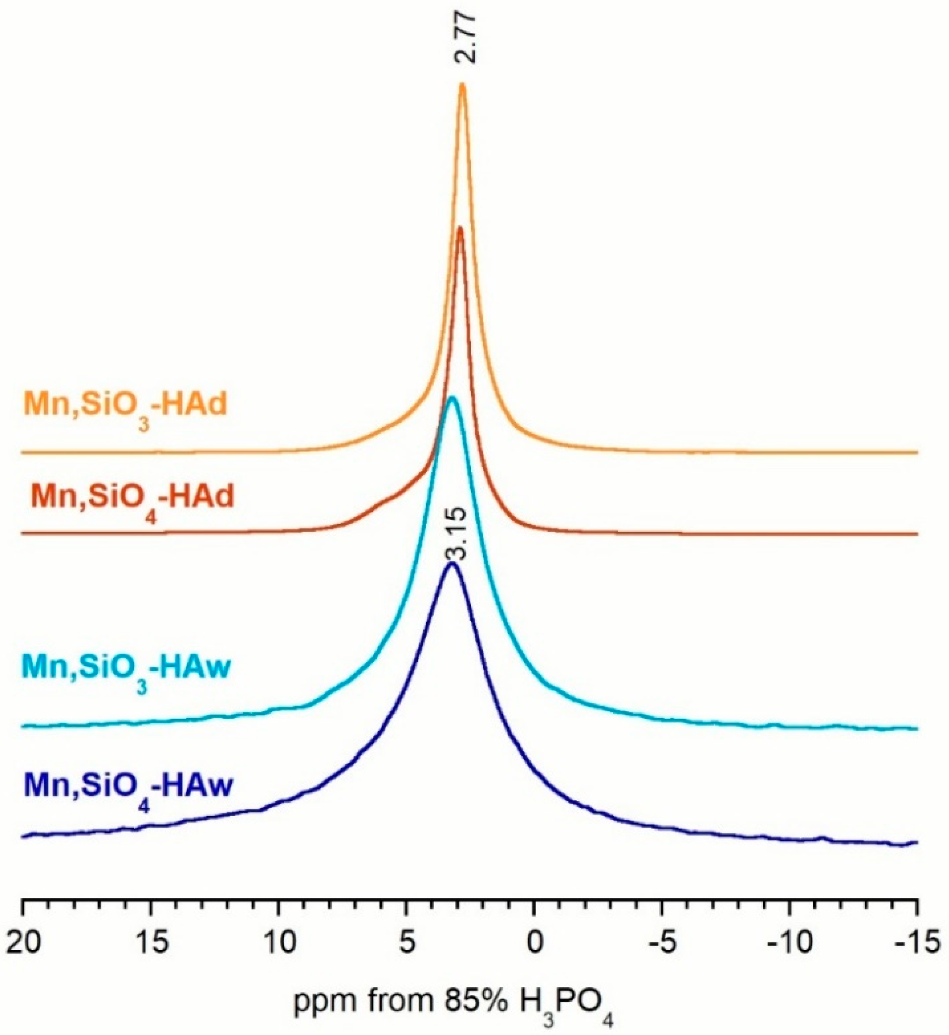
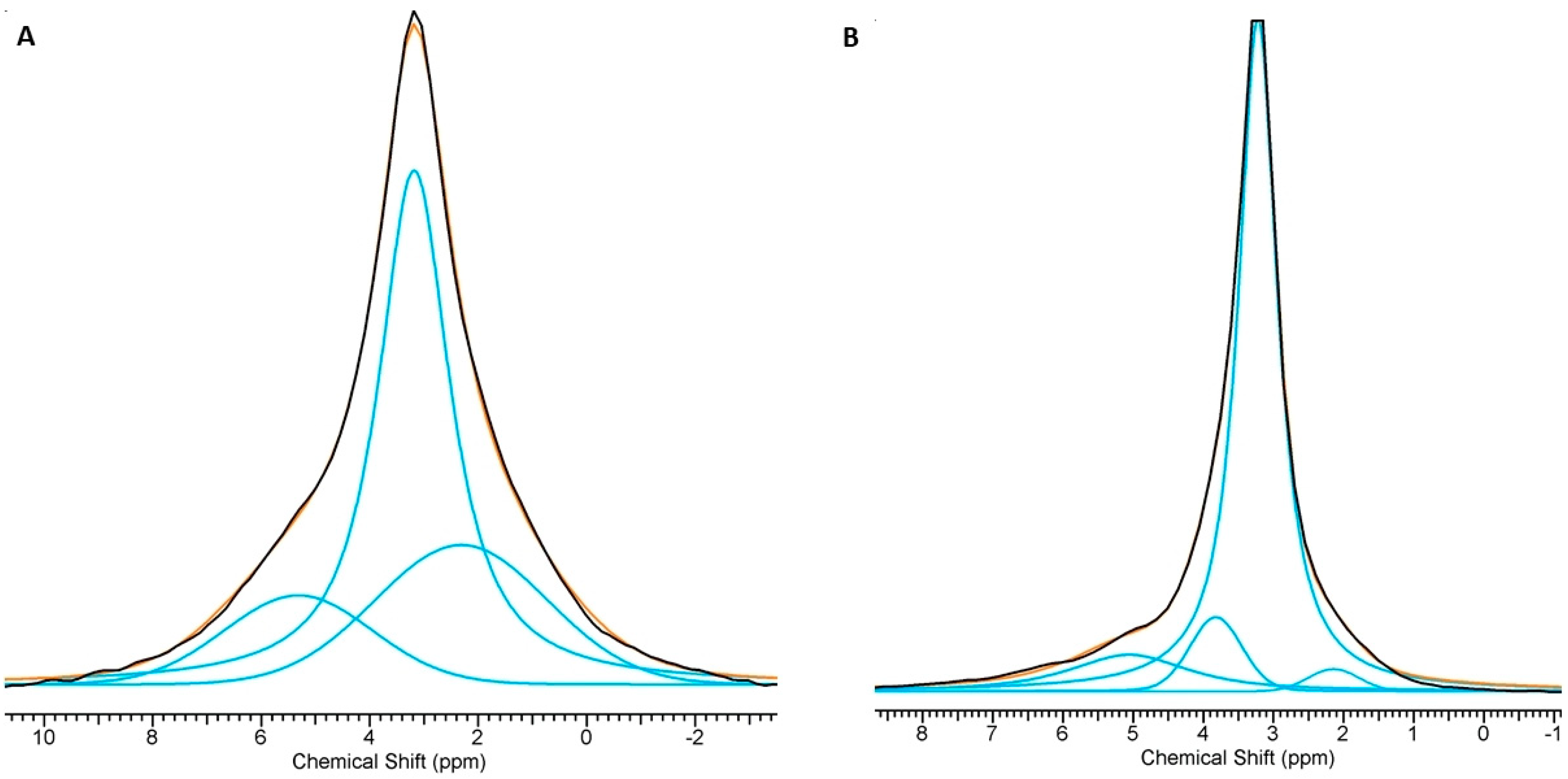
The phosphorus-31 resonance spectra of all the powders acquired using the BD technique are shown in Figure 4. As expected, all the spectra exhibited a quite narrow signal at about 2.75–3.15 ppm, which according to the literature, can be attributed to orthophosphate groups located in well-ordered apatitic crystals [60,61,62]. It should be noted that the lines obtained from the powders synthesized using precipitation were significantly wider than those obtained from the solid-state-synthesized samples. Moreover, all obtained spectra are slightly asymmetric, which suggests the presence of additional signals. Deconvolution revealed additional lines for all the spectra (see Figure 5 for examples and Table 2). In the Mn,SiO4-HAw and Mn,SiO3-HAw samples, apart from the main signal, two additional signals were detected: one at ≈2.30 ppm and another at ≈5.30 ppm. According to the literature, these additional lines come from the disordered structure of the hydrated surface layer [60,62]. The signals at ≈2.14–2.33 ppm and ≈5.10–5.35 ppm detected in all the spectra may be assigned to protonated (≡POxH) and unprotonated (≡POx) surface groups (where ≡ stands for a link to the crystal surface), respectively [60,61,62]. Due to overlap of the 31P NMR lines, it is not straightforward to determine the contribution of 31P nuclei from the disordered phase to the total 31P reservoir. However, based on the deconvolution of the spectra, it can be estimated that in the Mn,SiO3-HAw and Mn,SiO4-Haw samples, 42.5%–45.5% of the phosphorus-31 nuclei come from 31P nuclei from the disordered phase.
As presented in Figure 5 and Table 2, the Mn,SiO4-HAd and Mn,SiO3-HAd spectra revealed three additional lines. Apart from the main signal at 3.15–3.22 ppm and the weak and broad lines (from protonated (≡POxH) surface groups and from unprotonated (≡POx) surface groups), an additional line at ≈3.85–4.12 ppm was easily detected. Its contribution is not very significant (7.5% and 2.1% for Mn,SiO4-HAd and Mn,SiO3-HAd samples, respectively). It can probably be assigned to the silicocarnotite phase [51]. The PXRD pattern of Mn,SiO3-HAd did not contain reflections from silicocarnotite, but this may be explained by the small amount of this crystalline phase. Its presence in the Mn,SiO3-HAd sample was confirmed using a 31P BD NMR experiment.
It should be noted that the resonance lines originating from the phosphorus-31 nuclei in the disordered phase (especially in the hydrated surface layer) were relatively weak (up to 16.4%), suggesting that the –POH groups at the surface were relatively sparse. This is in accordance with the presumption that the hydrated surface layer of apatitic crystals synthesized under high-temperature conditions (via the solid-state method) is exiguous.
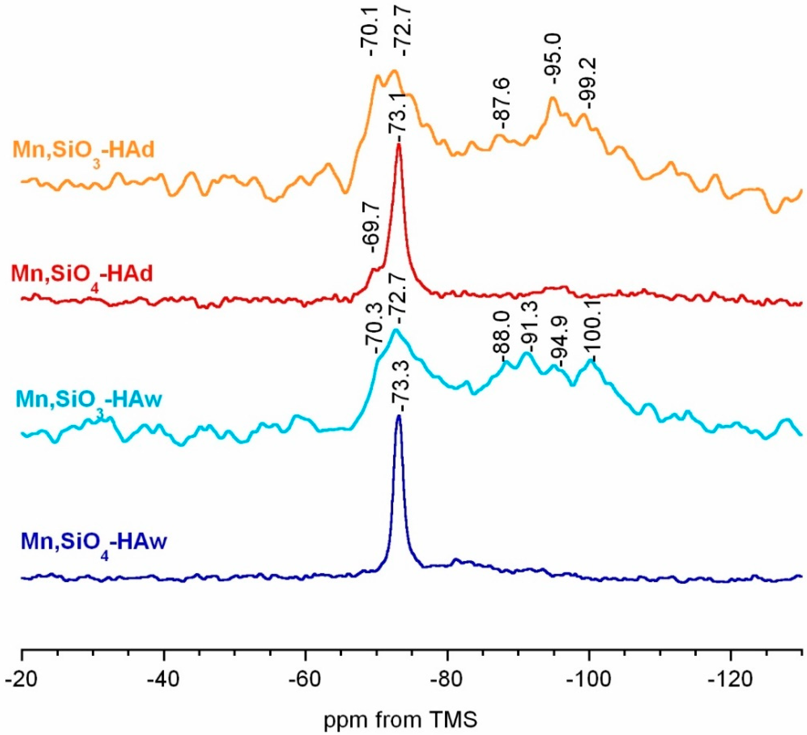
The standard 1H → 29Si CP MAS spectra are presented in Figure 6. Because of the low content of Si in the materials, the spectra presented significant noise, despite the long accumulation time. In all the spectra, a resonance line at approximately −72.7 to −73.3 ppm was predominant and can be assigned to a Q0 structure of silicates located within the crystal core [63,64,65,66]. It should be noted that in the Mn,SiO4-HAw spectrum, only one signal (−73.3 ppm) could be detected. However, in the Mn,SiO3-HAd, Mn,SiO3-Haw, and Mn,SiO4-HAd spectra, the resonance in this region was very broad and a second line (at approximately −69.3 to −70.3 ppm) was clearly visible. It could possibly be associated with the Q0 silicates located at the crystal surface. Surprisingly, in the spectra of the Mn,SiO3-HAw and Mn,SiO3-HAd samples, multiple broad and poorly resolved lines occurred in the region −88.0 to −100.1 ppm. According to the literature, these correspond to Q3/Q4 and are characteristic of non-crystalline silica species [36,64]. Therefore, we can conclude that the introduction of silicates into the apatitic crystal lattice seems to be more limited with sodium metasilicate as a source than with silicon acetate.

3.4. Preliminary Biological Tests

In order to evaluate the acute toxicity, the luminescent bacterium A. fisheri (Microtox®) and protozoan S. ambiguum (Spirotox) tests were used. Although manganese and silicon are essential trace elements, it was planned to check the toxicity of the Mn and Si content in the nanocrystals (Mn,SiO4-HAw and Mn,SiO3-HAw) and microcrystals (see Table 3). The obtained samples were observed to be non-toxic in both tests. These preliminary studies showed that the silicon and manganese contents of 1.22–1.88 wt% and 0.0076–0.0110 wt%, respectively, in HAs were completely neutral, and the obtained powders can be treated as promising materials for future biological investigations, i.e., cytotoxicity and biocompatibility.

4. Conclusions

Novel manganese and silicate co-substituted apatite materials were synthesized in this study. The study was aimed at comparing the physico-chemical properties of samples depending on the reagent used and the synthesis method. Samples were characterized using PXRD, FTIR, and ssNMR spectroscopy, as well as TEM and WD-XRF. Preliminary biological in vitro tests were also performed. Significant conclusions from these examinations are presented below.
  • Samples obtained using precipitation were nanocrystalline and monophasic. Solid-state samples were microcrystalline and contained secondary phases (impurities), i.e., calcium oxide, calcium hydroxide, and silicocarnotite, which were produced during the sintering process.
  • Manganese and silicon ions were successfully incorporated, as confirmed using WD-XRF and spectroscopic measurements. The substitution efficiency was between 45% and 95%, depending on the synthesis method and the reagents.
  • A higher efficiency of Mn2+ substitution occurred in the case of precipitation synthesis, while the substitution of silicon was favoured in the solid-state synthesis. In both cases, the yield was better when using silicon acetate instead of sodium metasilicate as a silicon source.
  • The introduction of ions did not significantly affect the crystallinity and unit cell parameters. The degree of crystallinity, as well as the size and shape of the crystals, depended mainly on the synthesis method.
  • According to the Microtox® and Spirotox tests, none of the samples was considered to be toxic. Such promising results may constitute the starting point for further biological research.

Author Contributions

K.S. participated in the synthesis and physicochemical analysis of the samples and wrote the manuscript. A.D. was responsible for the biological tests. J.K. provided the concept of the work, interpreted the results, and helped with the NMR analysis. All authors have contributed to and approved the manuscript.

Funding

This work was supported by a research grant (Project NCN UMO-2016/22/E/ST5/00564) from the National Science Centre, Poland, and a research grant (Project FW23/PM1/18) from Medical University of Warsaw.

Acknowledgments

The TEM studies were performed in the Laboratory of Electron Microscopy, Nencki Institute of Experimental Biology, Warsaw, Poland. We used equipment installed as part of a project sponsored by EU Structural Funds: Centre of Advanced Technology BIM; equipment was purchased for the Laboratory of Biological and Medical Imaging.

Conflicts of Interest

The authors declare no conflict of interest.

References

  1. Vallet-Regi, M.; Gonzales-Calbet, J.M. Calcium phosphates as substitution of bone tissues. Prog. Solid State Chem. 2004, 32, 1–31. [Google Scholar] [CrossRef]
  2. Zhou, H.; Lee, J. Nanoscale hydroxyapatite particles for bone tissue engineering. Acta Biomater. 2011, 7, 2769–2781. [Google Scholar] [CrossRef] [PubMed]
  3. Suchanek, W.; Yoshimura, M. Processing and properties of hydroxyapatite-based biomaterials for use as hard tissue replacement implants. J. Mater. Res. 1998, 13, 94–117. [Google Scholar] [CrossRef]
  4. Kolmas, J.; Krukowski, S.; Laskus, A.; Jurkitewicz, M. Synthetic hydroxyapatite in pharmaceutical applications. Ceram. Int. 2016, 42, 2472–2487. [Google Scholar] [CrossRef]
  5. Shepherd, J.H.; Shepherd, D.V.; Best, S.M. Substituted hydroxyapatites for bone repair. J. Mater. Sci. Mater. Med. 2012, 23, 2335–2347. [Google Scholar] [CrossRef] [PubMed]
  6. Ratnayake, J.T.B.; Mucalo, M.; Dias, G.J. Substituted hydroxyapatites for bone regeneration: A review of current trends. J. Biomed. Mater. Res. B Appl. Biomater. 2017, 105, 1285–1299. [Google Scholar] [CrossRef] [PubMed]
  7. Supova, M. Substituted hydroxyapatite for biomedical applications: A review. Ceram. Int. 2015, 41, 9203–9231. [Google Scholar] [CrossRef]
  8. Nassif, N.; Martineau, F.; Syzgantseva, O.; Gobeaux, F.; Willinger, M.; Coradin, T.; Cassaignon, S.; Azaïs, T.; Giraud-Guille, M.M. In vivo inspired conditions to synthesize biomimetic hydroxyapatite. Chem. Mater. 2010, 22, 3653–3663. [Google Scholar] [CrossRef]
  9. Roveri, N.; Iafisco, M. Evolving application of biomimetic nanostructured hydroxyapatite. Nanotechnol. Sci. Appl. 2010, 3, 107–125. [Google Scholar] [CrossRef]
  10. Fore, H.; Morton, R.A. Manganese in Bone. Biochem. J. 1952, 51, 598–600. [Google Scholar] [CrossRef]
  11. Jugdaohsingh, R. Silicon and bone health. J. Nutr. Health Aging 2007, 11, 99–110. [Google Scholar]
  12. Armulik, A.; Svineng, G.; Wennerberg, K.; Fassler, R.; Johansson, S. Expression of integrin subunit β1B in integrin β1-deficient GD25 cells does not interfere with αVβ3 functions. Exp. Cell Res. 2000, 254, 55–63. [Google Scholar] [CrossRef]
  13. Bracci, B.; Torricelli, T.; Panzavolta, S.; Boanini, E.; Giardino, R.; Bigi, A. Effect of Mg2+, Sr2+, and Mn2+ on the chemico-physical and in vitro biological properties of calcium phosphate biomimetic coatings. J. Inorg. Biochem. 2009, 103, 1666–1674. [Google Scholar] [CrossRef]
  14. Huang, Y.; Ding, Q.; Han, S.; Yan, Y.; Pang, X. Characterisation, corrosion resistance and in vitro bioactivity of manganese-doped hydroxyapatite films electrodeposited on titanium. J. Mater. Sci. Mater. Med. 2013, 24, 1853–1864. [Google Scholar] [CrossRef]
  15. Saltman, P.D.; Strause, L.G. The role of trace minerals in osteoporosis. J. Am. Coll. Nutr. 1993, 12, 384–389. [Google Scholar] [CrossRef]
  16. Torres, P.M.C.; Vieira, S.I.; Cerqueira, A.R.; Pina, S.; da Cruz Silva, O.A.B.; Abrantes, J.C.C.; Ferreira, J.M.F. Effects of Mn-doping on the structure and biological properties of β-tricalcium phosphate. J. Inorg. Biochem. 2014, 136, 57–66. [Google Scholar] [CrossRef]
  17. Rico, H.; Gomez-Raso, N.; Revilla, M.; Hernandez, E.R.; Seco, C.; Paez, E.; Crespo, E. Effects on bone loss of manganese alone or with copper supplement in ovariectomized rats. A morphometric and densitomeric study. Eur J. Obstet. Gynecol. Reprod. Biol. 2000, 90, 97–101. [Google Scholar] [CrossRef]
  18. Bae, Y.J.; Kim, M.H. Manganese supplementation improves mineral density of the spine and femur and serum osteocalcin in rats. Biol. Trace Elem. Res. 2008, 124, 28–34. [Google Scholar] [CrossRef]
  19. Medvecky, L.; Stulajterova, R.; Parilak, L.; Trpcevska, J.; Durisin, J.; Barinov, S.M. Influence of manganese on stability and particle growth of hydroxyapatite in simulated body fluid. Colloids Surf. A Physicochem. Eng. Asp. 2006, 281, 221–229. [Google Scholar] [CrossRef]
  20. Bigi, A.; Bracci, B.; Cuisinier, F.; Elkaim, R.; Fini, M.; Mayer, I.; Mihailescu, I.N.; Socol, G.; Sturba, L.; Torricelli, P. Human osteoblast response to pulsed laser deposited calcium phosphate coatings. Biomaterials 2005, 26, 2381–2389. [Google Scholar] [CrossRef]
  21. Kolmas, J.; Jabłoński, M.; Ślósarczyk, A.; Kołodziejski, W. Solid-state NMR study of Mn2+ for Ca2+ substitution in thermally processed hydroxyapatites. J. Am. Ceram. Soc. 2015, 98, 1265–1274. [Google Scholar] [CrossRef]
  22. Mayer, I.; Peto, G.; Karacs, A.; Molnar, G.; Popov, I. Divalent Mn in calcium hydroxyapatite by pulse laser deposition. J. Inorg. Biochem. 2010, 104, 1107–1111. [Google Scholar] [CrossRef]
  23. Sopyan, I.; Ramesh, S.; Nawawi, N.A.; Tampieri, A.; Sprio, S. Effects of manganese doping on properties of sol-gel derived biphasic calcium phosphate ceramics. Ceram. Int. 2011, 37, 3703–3715. [Google Scholar] [CrossRef]
  24. Paluszkiewicz, C.; Ślósarczyk, A.; Pijocha, D.; Sitarz, M.; Bućko, M.; Zima, A.; Chróścicka, A.; Lewandowska-Szumieł, M. Synthesis, structural properties and thermal stability of Mn-doped hydroxyapatite. J. Mol. Struct. 2010, 976, 301–309. [Google Scholar] [CrossRef]
  25. Henstock, J.R.; Canham, L.T.; Anderson, S.I. Silicon: The evolution of its use in biomaterials. Acta Biomater. 2015, 11, 17–26. [Google Scholar] [CrossRef]
  26. Szurkowska, K.; Kolmas, J. Hydroxyapatites enriched in silicon – Bioceramic materials for biomedical and pharmaceutical applications. Prog. Nat. Sci. Mater. 2017, 27, 401–409. [Google Scholar] [CrossRef]
  27. Carlisle, E.M. Silicon: A possible factor in bone calcification. Science 1970, 167, 279–280. [Google Scholar] [CrossRef]
  28. Carlisle, E.M. Silicon: An essential element for the chick. Science 1972, 178, 619–621. [Google Scholar] [CrossRef]
  29. Reffitt, D.M.; Ogston, N.; Jugdaohsingh, R.; Cheung, H.F.J.; Evans, B.A.J.; Thompson, R.P.H.; Powell, J.J.; Hampson, G.N. Orthosilicic acid stimulates collagen type 1 synthesis and osteoblastic differentiation in human osteoblast-like cells in vitro. Bone 2003, 32, 127–135. [Google Scholar] [CrossRef]
  30. Spector, T.D.; Calomme, M.R.; Anderson, S.H.; Clement, G.; Bevan, L.; Demeester, N.; Swaminathan, R.; Jugdaohsingh, R.; Vanden Berghe, D.A.; Powell, J.J. Choline-stabilized orthosilicic acid supplementation as an adjunct to calcium/vitamin D3 stimulates markers of bone formation in osteopenic females: A randomized, placebo-controlled trial. BMC Musculoskelet. Disord. 2008, 9, 85. [Google Scholar] [CrossRef]
  31. Zhou, S.; Ireland, D.; Brooks, R.A.; Rushton, N.; Best, S. The effects of silicate ions on human osteoblast adhesion, proliferation, and differentiation. J. Biomed. Mater. Res. B Appl. Biomater. 2009, 90, 123–130. [Google Scholar]
  32. Zhao, X.; Wang, T.; Qian, S.; Liu, X.; Sun, J.; Li, B. Silicon-doped titanium dioxide nanotubes promoted bone formation on titanium implants. Int J. Mol. Sci. 2016, 17, 292. [Google Scholar] [CrossRef]
  33. Wang, Q.; Hu, H.; Qiao, Y.; Zhang, Z.; Sun, J. Enhanced performance of osteoblasts by silicon incorporated porous TiO2 coating. J. Mater. Sci. Technol. 2012, 28, 109–117. [Google Scholar] [CrossRef]
  34. Zhou, X.; Moussa, F.M.; Mankoci, S.; Ustriyana, P.; Zhang, N.; Abdelmagid, S.; Molenda, J.; Murphy, W.L.; Safadi, F.F.; Sahai, N. Orthosilicic acid, Si(OH)4, stimulates osteoblast differentiation in vitro by upregulating miR-146a to antagonize NF-κB activation. Acta Biomater. 2016, 39, 192–202. [Google Scholar] [CrossRef]
  35. Gibson, I.R.; Best, S.M.; Bonfield, W. Chemical characterization of silicon-substituted hydroxyapatite. J. Biomed. Mater. Res. 1999, 44, 422–428. [Google Scholar] [CrossRef]
  36. Marchat, D.; Zymelka, M.; Coelho, C.; Gremillard, L.; Joly-Pottuz, L.; Babonneau, F.; Esnouf, C.; Chevalier, J.; Bernache-Assollant, D. Accurate characterization of pure silicon-substituted hydroxyapatite powders synthesized by a new precipitation route. Acta Biomater. 2013, 9, 6992–7004. [Google Scholar] [CrossRef] [Green Version]
  37. Palard, M.; Champion, E.; Foucaud, S. Synthesis of silicated hydroxyapatite Ca10(PO4)6−x(SiO4)x(OH)2−x. J. Solid State Chem. 2008, 181, 1950–1960. [Google Scholar] [CrossRef]
  38. Aminian, A.; Solati-Hashjin, M.; Samadikuchaksaraei, A.; Bakhshi, F.; Gorjipour, F.; Farzadi, A.; Moztarzadeh, F.; Schmucker, M. Synthesis of silicon-substituted hydroxyapatite by a hydrothermal method with two different phosphorus sources. Ceram. Int. 2011, 37, 1219–1229. [Google Scholar] [CrossRef]
  39. Botelho, C.M.; Brooks, R.A.; Best, S.M.; Lopes, M.A.; Santos, J.D.; Rushton, N.; Bonfield, W. Human osteoblast response to silicon-substituted hydroxyapatite. J. Biomed. Mater. Res. A 2006, 79, 723–730. [Google Scholar] [CrossRef]
  40. Honda, M.; Kikushima, K.; Kawanobe, Y.; Konishim, T.; Mizumoto, M.; Aizawa, M. Enhanced early osteogenic differentiation by silicon-substituted hydroxyapatite ceramics fabricated via ultrasonic spray pyrolysis route. J. Mater. Sci. Mater. Med. 2012, 23, 2923–2932. [Google Scholar] [CrossRef]
  41. Thian, E.S.; Huang, J.; Best, S.M.; Barber, Z.H.; Brooks, R.A.; Rushton, N.; Bonfield, W. The response of osteoblasts to nanocrystalline silicon-substituted hydroxyapatite thin films. Biomaterials 2006, 27, 2692–2698. [Google Scholar] [CrossRef]
  42. Patel, N.; Best, S.M.; Bonfield, W.; Gibson, I.R.; Hing, K.A.; Damien, E.; Revell, P.A. A comparative study on the in vivo behavior of hydroxyapatite and silicon substituted hydroxyapatite granules. J. Mater. Sci. Mater. Med. 2002, 13, 1199–1206. [Google Scholar] [CrossRef]
  43. Patel, N.; Brooks, R.A.; Clarke, M.T.; Lee, P.M.T.; Rushton, N.; Gibson, I.R.; Best, S.M.; Bonfield, W. In vivo assessment of hydroxyapatite and silicate-substituted hydroxyapatite granules using an ovine defect model. J. Mater. Sci. Mater. Med. 2005, 16, 429–440. [Google Scholar] [CrossRef]
  44. Porter, A.E.; Patel, N.; Skepper, J.N.; Best, S.M.; Bonfield, W. Comparison of in vivo dissolution processes in hydroxyapatite and silicon-substituted hydroxyapatite bioceramics. Biomaterials 2003, 24, 4609–4620. [Google Scholar] [CrossRef]
  45. Porter, A.E.; Patel, N.; Skepper, J.N.; Best, S.M.; Bonfield, W. Effect of sintered silicate-substituted hydroxyapatite on remodelling processes at the bone–implant interface. Biomaterials 2004, 25, 3303–3314. [Google Scholar] [CrossRef]
  46. Hing, K.A.; Revell, P.A.; Smith, N.; Buckland, T. Effect of silicon level on rate, quality and progression of bone healing within silicate-substituted porous hydroxyapatite scaffolds. Biomaterials 2006, 27, 5014–5026. [Google Scholar] [CrossRef]
  47. Zhang, E.; Zou, C. Porous titanium and silicon-substituted hydroxyapatite biomodification prepared by a biomimetic process: Characterization and in vivo evaluation. Acta Biomater. 2009, 5, 1732–1741. [Google Scholar] [CrossRef]
  48. Szurkowska, K.; Zgadzaj, A.; Kuras, M.; Kolmas, J. Novel hybrid material based on Mg2+ and SiO44- co-substituted nano-hydroxyapatite, alginate and chondroitin sulphate for potential use in biomaterials engineering. Ceram. Int. 2018, 44, 18551–18559. [Google Scholar] [CrossRef]
  49. Nałęcz-Jawecki, G. Spirotox—Spirostomum ambiguum acute toxicity test—10 years of experience. Environ. Toxicol. 2004, 19, 359–364. [Google Scholar] [CrossRef]
  50. Lafon, J.P.; Champion, E.; Bernache-Assollant, D.; Gibert, R.; Danna, A.M. Thermal decomposition of carbonated calcium phosphate apatites. J. Therm. Anal. Calorim. 2003, 72, 1127–1134. [Google Scholar] [CrossRef]
  51. Andreev, A.S.; Bulina, N.V.; Chaikina, M.V.; Prosanov, I.Y.; Terskikh, V.V.; Lapina, O.B. Solid-state NMR and computational insights into the crystal structure of silicocarnotite-based bioceramic materials synthesized mechanochemically. Solid State Nucl. Magn. Reson. 2017, 84, 151–157. [Google Scholar] [CrossRef]
  52. Hayakawa, S.; Kanaya, T.; Tsuru, K.; Shirosaki, Y.; Osaka, A.; Fujii, E.; Kawabata, K.; Gasqueres, G.; Bonhomme, C.; Babonneau, F.; et al. Heterogeneous structure and in vitro degradation behavior of wet-chemically derived nanocrystalline silicon-containing hydroxyapatite particles. Acta Biomater. 2013, 9, 4856–4867. [Google Scholar] [CrossRef]
  53. Chang, M.C.; Tanaka, J. FT-IR study for hydroxyapatite/collagen nanocomposite cross-linked by glutaraldehyde. Biomaterials 2002, 23, 4811–4818. [Google Scholar] [CrossRef]
  54. Merry, J.C.; Gibson, I.R.; Best, S.M.; Bonfield, W. Synthesis and characterization of carbonate hydroxyapatite. J. Mater. Sci. Mater. Med. 1998, 9, 779–783. [Google Scholar] [CrossRef]
  55. Ye, H.; Liu, X.Y.; Hong, H. Characterization of sintered titanium/hydroxyapatite biocomposite using FTIR spectroscopy. J. Mater. Sci. Mater. Med. 2009, 20, 843–850. [Google Scholar] [CrossRef]
  56. Shaltout, A.A.; Allam, M.A.; Moharram, M.A. FTIR spectroscopic, thermal and XRD characterization of hydroxyapatite from new natural sources. Spectrochim. Acta A Mol. Biol. Spectrosc. 2011, 83, 56–60. [Google Scholar] [CrossRef]
  57. Cengiz, B.; Gokce, Y.; Yildiz, N.; Aktas, Z.; Calimli, A. Synthesis and characterization of hydroxyapatite nanoparticles. Colloids Surf. A Physicochem. Eng. Asp. 2008, 322, 29–33. [Google Scholar] [CrossRef]
  58. Tian, T.; Jiang, D.; Zhang, J.; Lin, Q. Synthesis of Si-substituted hydroxyapatite by a wet mechanochemical method. Mater. Sci. Eng. C 2008, 28, 57–63. [Google Scholar] [CrossRef]
  59. Padmanabhan, S.K.; Haq, E.U.; Licciulli, A. Rapid synthesis and characterization of silicon substituted nano hydroxyapatite using microwave irradiation. Curr. Appl. Phys. 2004, 14, 87–92. [Google Scholar] [CrossRef]
  60. Jager, C.; Welzel, T.; Meyer-Zaika, W.; Epple, M. A solid-state NMR investigation of the structure of nanocrystalline hydroxyapatite. Magn. Reson. Chem. 2006, 44, 573–580. [Google Scholar] [CrossRef]
  61. Pourpoint, F.; Gervais, C.; Bonhomme-Coury, L.; Azaïs, T.; Coelho, C.; Mauri, F.; Alonso, B.; Babonneau, F.; Bonhomme, C. Calcium phosphates and hydroxyapatite: Solid-state NMR experiments and first-principles calculations. Appl. Magn. Reson. 2007, 32, 435–457. [Google Scholar] [CrossRef]
  62. Panda, R.N.; Hsieh, M.F.; Chung, R.J.; Chin, T.S. FTIR, XRD, SEM and solid state NMR investigations of carbonate-containing hydroxyapatite nano-particles synthesized by hydroxide-gel technique. J. Phys. Chem. Solids 2003, 64, 193–199. [Google Scholar] [CrossRef]
  63. Magi, M.; Lippmaa, E.; Samoson, A.; Engelhardt, G.; Grimmer, A.R. Solid-state high-resolution silicon-29 chemical shifts in silicates. J. Phys. Chem. 1984, 88, 1518–1522. [Google Scholar] [CrossRef]
  64. Gasqueres, G.; Bonhomme, C.; Maquet, J.; Babonneau, F.; Hayakawa, S.; Kanayab, T.; Osaka, A. Revisiting silicate substituted hydroxyapatite by solid-state NMR. Magn. Reson. Chem. 2008, 46, 342–346. [Google Scholar] [CrossRef]
  65. Gomes, S.; Renaudin, G.; Mesbah, A.; Jallot, E.; Bonhomme, C.; Babonneau, F.; Nedelec, J.M. Thorough analysis of silicon substitution in biphasic calcium phosphate bioceramics: A multi-technique study. Acta Biomater. 2010, 6, 3264–3274. [Google Scholar] [CrossRef]
  66. Schneider, J.; Mastelaro, V.R.; Panepucci, H.; Zanotto, E.D. 29Si MAS-NMR studies of Qn structural units in metasilicate glasses and their nucleating ability. J. Non-Cryst. Solids 2000, 273, 8–18. [Google Scholar] [CrossRef]
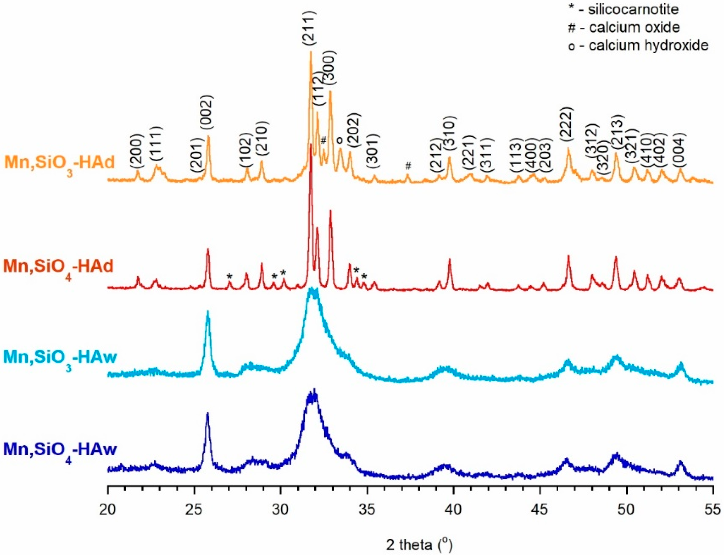
Figure 1. PXRD patterns of the analyzed samples.
Materials 12 02566 g001
Figure 2. TEM images of the analyzed samples: (A) Mn,SiO4-HAw, (B) Mn,SiO3-HAw, (C) Mn,SiO4-Had, and (D) Mn,SiO3-HAd.
Materials 12 02566 g002
Figure 3. FTIR transmission spectra of the analyzed samples.
Materials 12 02566 g003
Figure 4. 31P BD NMR spectra of the analyzed samples.
Materials 12 02566 g004
Figure 5. Representative deconvolution of the 31P BD NMR spectra: (A) Mn,SiO4-Haw and (B) Mn,SiO4-HAd.
Materials 12 02566 g005
Figure 6. 29Si CP MAS NMR spectra of the analyzed samples.
Materials 12 02566 g006
Table 1. Various parameters of the obtained samples.
Mn,SiO3-HAwMn,SiO4-HAwMn,SiO3-HAdMn,SiO4-HAd
Synthesis methodPrecipitationSolid-state
Silicon sourceNa2SiO3Si(CH3COO)4Na2SiO3Si(CH3COO)4
Parameter a (Å) a9.4249.4339.4189.415
Parameter c (Å) a6.8826.8756.8866.896
Mn content (wt%)0.0086 ± 0.00040.0110 ± 0.00040.0076 ± 0.00030.0080 ± 0.0004
Si content (wt%)1.217 ± 0.0051.512 ± 0.0031.819 ± 0.0031.879 ± 0.004
Mn substitution efficiency (%)50.5964.7144.7147.06
Si substitution efficiency (%)61.4876.3491.8894.90
(Ca + Mn)/(P + Si) molar ratio1.54 ± 0.051.59 ± 0.041.61 ± 0.031.62 ± 0.05
a Error was ±0.3%.
Table 2. NMR deconvolution results for the 31P BD NMR experiments (FWHM—full width at half maximum).
AssignmentsChemical Shift (ppm)FWHM (Hz)Percentage of Total Area (%)
Mn,SiO4-HAw≡POxH2.3160627.4
main3.1825657.4
≡POx5.3152315.1
Mn,SiO3-HAw≡POxH2.2564022.3
main3.2224854.5
≡POx5.2855123.2
Mn,SiO4-HAd≡POxH2.141382.4
main3.2210877.0
≡POx5.1032912.1
silicocarnotite3.851427.5
Mn,SiO3-HAd≡POxH2.331454.2
main3.1511178.5
≡POx5.3531615.2
silicocarnotite4.121562.1
Table 3. Toxicity of the synthesized powders in Microtox® and Spirotox tests.
SampleMicrotox® (15 min-PE) 1Spirotox 2
1.0 mg/mL2.0 mg/mL1.0 mg/mL2.0 mg/mL
Mn,SiO4-HAw00NT NT
Mn,SiO3-HAw00NTNT
Mn,SiO4-HAd00NTNT
Mn,SiO3-HAd00NTNT
1 Percent of toxic effect after 15 min of incubation; 2 NT—not toxic.