Modification of the Ceramic Implant Surfaces from Zirconia by the Magnetron Sputtering of Different Calcium Phosphate Targets: A Comparative Study

 ,
,  , , and
Abstract
1. Introduction
2. Materials and Methods
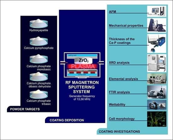
2.1. Materials
2.2. Methods
2.1.1. Coating Deposition
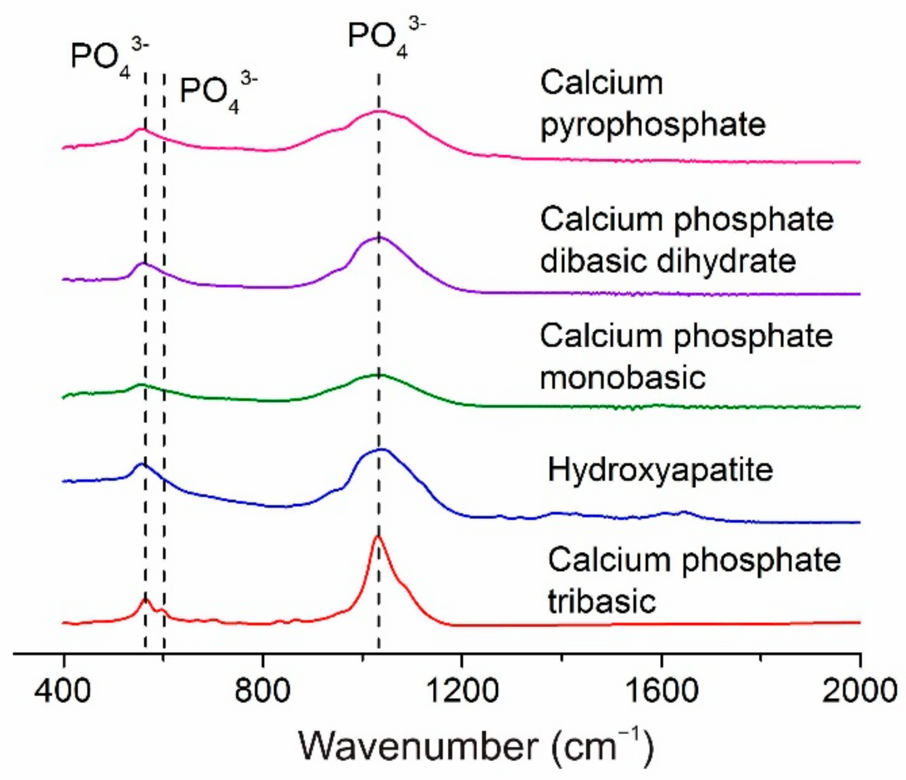
2.1.2. Coating Investigations
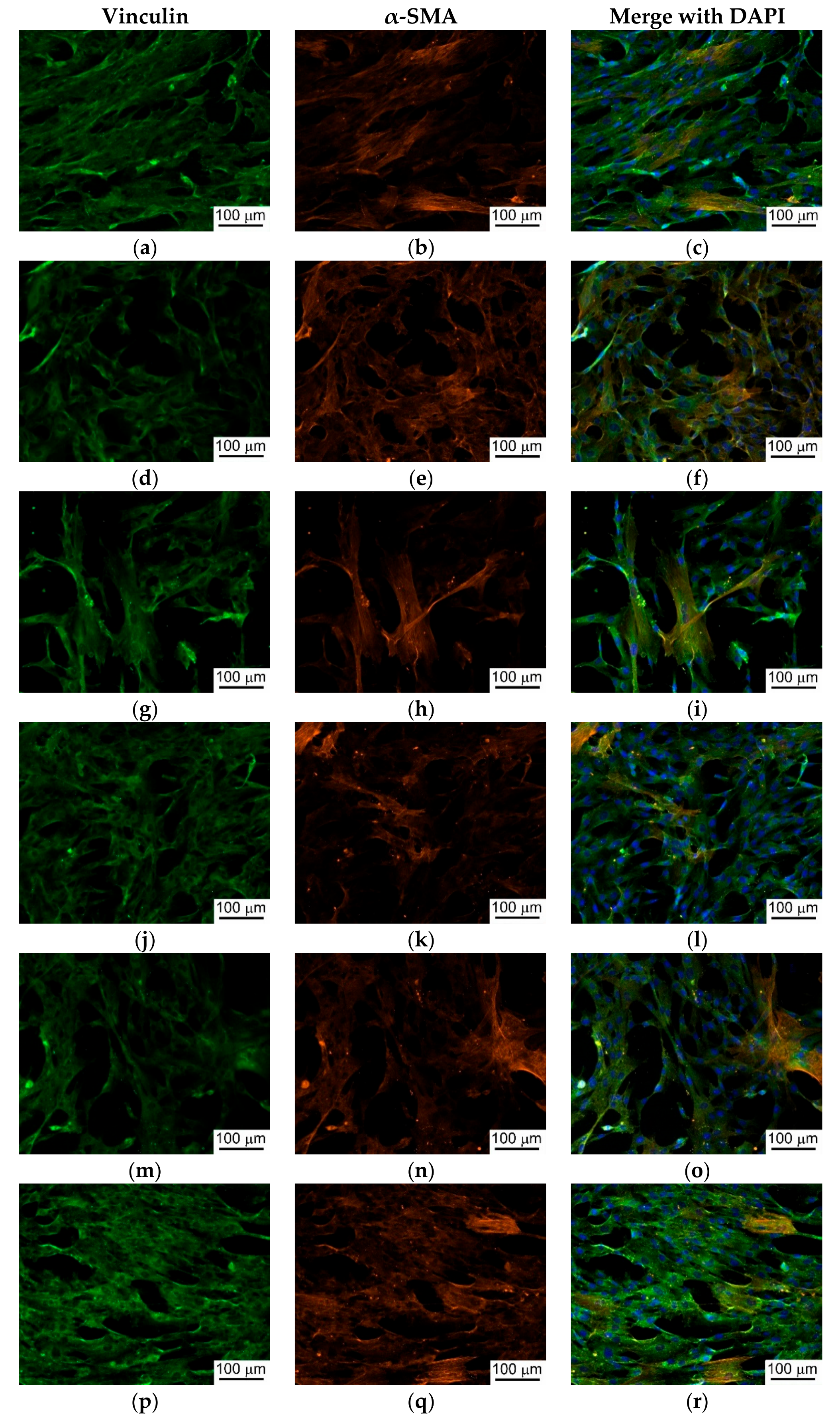
3. Results and Discussion
4. Conclusions
Author Contributions
Funding
Acknowledgments
Conflicts of Interest
References
- Bauer, S.; Schmuki, P.; von der Mark, K.; Park, J. Engineering biocompatible implant surfaces. Prog. Mater. Sci. 2013, 58, 261–326. [Google Scholar] [CrossRef]
- Hou, N.; Perinpanayagam, H.; Mozumder, M.; Zhu, J. Novel Development of Biocompatible Coatings for Bone Implants. Coatings 2015, 5, 737–757. [Google Scholar] [CrossRef]
- Treccani, L.; Yvonne Klein, T.; Meder, F.; Pardun, K.; Rezwan, K. Functionalized ceramics for biomedical, biotechnological and environmental applications. Acta Biomater. 2013, 9, 7115–7150. [Google Scholar] [CrossRef] [PubMed]
- Roy, M.; Bandyopadhyay, A.; Bose, S. Ceramics in Bone Grafts and Coated Implants. In Materials for Bone Disorders; Elsevier: Amsterdam, The Netherlands, 2017; pp. 265–314. [Google Scholar]
- Fornabaio, M.; Reveron, H.; Adolfsson, E.; Montanaro, L.; Chevalier, J.; Palmero, P. Design and development of dental ceramics. In Advances in Ceramic Biomaterials; Elsevier: Amsterdam, The Netherlands, 2017; pp. 355–389. [Google Scholar]
- Scarano, A.; Di Carlo, F.; Quaranta, M.; Piattelli, A. Bone Response to Zirconia Ceramic Implants: An Experimental Study in Rabbits. J. Oral Implantol. 2003, 29, 8–12. [Google Scholar] [CrossRef]
- Bächle, M.; Butz, F.; Hübner, U.; Bakalinis, E.; Kohal, R.J. Behavior of CAL72 osteoblast-like cells cultured on zirconia ceramics with different surface topographies. Clin. Oral Implants Res. 2007, 18, 53–59. [Google Scholar] [CrossRef] [PubMed]
- Bosshardt, D.D.; Chappuis, V.; Buser, D. Osseointegration of titanium, titanium alloy and zirconia dental implants: Current knowledge and open questions. Periodontol. 2000 2017, 73, 22–40. [Google Scholar] [CrossRef] [PubMed]
- Pieralli, S.; Kohal, R.-J.; Lopez Hernandez, E.; Doerken, S.; Spies, B.C. Osseointegration of zirconia dental implants in animal investigations: A systematic review and meta-analysis. Dent. Mater. 2017, 34, 171–182. [Google Scholar] [CrossRef] [PubMed]
- Reveron, H.; Fornabaio, M.; Palmero, P.; Fürderer, T.; Adolfsson, E.; Lughi, V.; Bonifacio, A.; Sergo, V.; Montanaro, L.; Chevalier, J. Towards long lasting zirconia-based composites for dental implants: Transformation induced plasticity and its consequence on ceramic reliability. Acta Biomater. 2017, 48, 423–432. [Google Scholar] [CrossRef] [PubMed]
- Siddiqi, A.; Khan, A.S.; Zafar, S. Thirty Years of Translational Research in Zirconia Dental Implants: A Systematic Review of the Literature. J. Oral Implantol. 2017, 43, 314–325. [Google Scholar] [CrossRef] [PubMed]
- Sorrentino, R.; Cochis, A.; Azzimonti, B.; Caravaca, C.; Chevalier, J.; Kuntz, M.; Porporati, A.A.; Streicher, R.M.; Rimondini, L. Reduced bacterial adhesion on ceramics used for arthroplasty applications. J. Eur. Ceram. Soc. 2018, 38, 963–970. [Google Scholar] [CrossRef]
- Klapperich, C.; Graham, J.; Pruitt, L.; Ries, M.D. Failure of a metal-on-metal total hip arthroplasty from progressive osteolysis. J. Arthroplasty 1999, 14, 877–881. [Google Scholar] [CrossRef]
- Kaplan, M.; Park, J.; Young Kim, S.; Ozturk, A. Production and properties of tooth-colored yttria stabilized zirconia ceramics for dental applications. Ceram. Int. 2018, 44, 2413–2418. [Google Scholar] [CrossRef]
- Schriwer, C.; Skjold, A.; Gjerdet, N.R.; Øilo, M. Monolithic zirconia dental crowns. Internal fit, margin quality, fracture mode and load at fracture. Dent. Mater. 2017, 33, 1012–1020. [Google Scholar] [CrossRef] [PubMed]
- Osman, R.B.; van der Veen, A.J.; Huiberts, D.; Wismeijer, D.; Alharbi, N. 3D-printing zirconia implants; a dream or a reality? An in-vitro study evaluating the dimensional accuracy, surface topography and mechanical properties of printed zirconia implant and discs. J. Mech. Behav. Biomed. Mater. 2017, 75, 521–528. [Google Scholar] [CrossRef] [PubMed]
- Abd El-Ghany, O.S.; Sherief, A.H. Zirconia based ceramics, some clinical and biological aspects: Review. Futur. Dent. J. 2016, 2, 55–64. [Google Scholar] [CrossRef]
- Altmann, B.; Rabel, K.; Kohal, R.J.; Proksch, S.; Tomakidi, P.; Adolfsson, E.; Bernsmann, F.; Palmero, P.; Fürderer, T.; Steinberg, T. Cellular transcriptional response to zirconia-based implant materials. Dent. Mater. 2017, 33, 241–255. [Google Scholar] [CrossRef] [PubMed]
- Siddiqi, A.; Duncan, W.J.; De Silva, R.K.; Zafar, S. One-Piece Zirconia Ceramic versus Titanium Implants in the Jaw and Femur of a Sheep Model: A Pilot Study. BioMed Res. Int. 2016, 2016, 6792972. [Google Scholar] [CrossRef] [PubMed]
- Pieralli, S.; Kohal, R.J.; Jung, R.E.; Vach, K.; Spies, B.C. Clinical Outcomes of Zirconia Dental Implants: A Systematic Review. J. Dent. Res. 2017, 96, 38–46. [Google Scholar] [CrossRef] [PubMed]
- Kohal, R.J.; Att, W.; Bächle, M.; Butz, F. Ceramic abutments and ceramic oral implants. An update. Periodontology 2000 2008, 47, 224–243. [Google Scholar] [CrossRef] [PubMed]
- Ananth, H.; Kundapur, V.; Mohammed, H.S.; Anand, M.; Amarnath, G.S.; Mankar, S. A review on biomaterials in dental implantology. Int. J. Biomed. Sci. 2015, 11, 113–120. [Google Scholar] [PubMed]
- Manicone, P.F.; Rossi Iommetti, P.; Raffaelli, L. An overview of zirconia ceramics: Basic properties and clinical applications. J. Dent. 2007, 35, 819–826. [Google Scholar] [CrossRef] [PubMed]
- Daou, E.E. The Zirconia Ceramic: Strengths and Weaknesses. Open Dent. J. 2014, 8, 33–42. [Google Scholar] [CrossRef] [PubMed]
- Hanawa, T. Biofunctionalization of titanium for dental implant. Jpn. Dent. Sci. Rev. 2010, 46, 93–101. [Google Scholar] [CrossRef]
- Zhang, B.; Myers, D.; Wallace, G.; Brandt, M.; Choong, P. Bioactive Coatings for Orthopaedic Implants—Recent Trends in Development of Implant Coatings. Int. J. Mol. Sci. 2014, 15, 11878–11921. [Google Scholar] [CrossRef] [PubMed]
- Gurgel, B.C.D.V.; Gonçalves, P.F.; Pimentel, S.P.; Nociti, F.H.; Sallum, E.A.; Sallum, A.W.; Casati, M.Z. An Oxidized Implant Surface May Improve Bone-to-Implant Contact in Pristine Bone and Bone Defects Treated With Guided Bone Regeneration: An Experimental Study in Dogs. J. Periodontol. 2008, 79, 1225–1231. [Google Scholar] [CrossRef] [PubMed]
- Wang, G.; Li, J.; Lv, K.; Zhang, W.; Ding, X.; Yang, G.; Liu, X.; Jiang, X. Surface thermal oxidation on titanium implants to enhance osteogenic activity and in vivo osseointegration. Sci. Rep. 2016, 6. [Google Scholar] [CrossRef] [PubMed]
- Lopez-Heredia, M.A.; Weiss, P.; Layrolle, P. An electrodeposition method of calcium phosphate coatings on titanium alloy. J. Mater. Sci. Mater. Med. 2007, 18, 381–390. [Google Scholar] [CrossRef] [PubMed]
- Klein, C.P.A.T.; Wolke, J.G.C.; De Blieck-Hogervorst, J.M.A.; de Groot, K. Calcium phosphate plasma-sprayed coatings and their stability: An in vivo study. J. Biomed. Mater. Res. 1994, 28, 909–917. [Google Scholar] [CrossRef] [PubMed]
- Lu, T.; Qiao, Y.; Liu, X. Surface modification of biomaterials using plasma immersion ion implantation and deposition. Interface Focus 2012, 2, 325–336. [Google Scholar] [CrossRef] [PubMed]
- Ilyin, A.A.; Skvortsova, S.V.; Petrov, L.M.; Chernyshova, Y.V.; Lukina, E.A. Effect of vacuum ion-plasma treatment on the electrochemical corrosion characteristics of titanium-alloy implants. Russ. Metall. 2007, 2007, 423–427. [Google Scholar] [CrossRef]
- Karazisis, D.; Petronis, S.; Agheli, H.; Emanuelsson, L.; Norlindh, B.; Johansson, A.; Rasmusson, L.; Thomsen, P.; Omar, O. The influence of controlled surface nanotopography on the early biological events of osseointegration. Acta Biomater. 2017, 53, 559–571. [Google Scholar] [CrossRef] [PubMed]
- Mediaswanti, K.; Wen, C.; Ivanova, E.P.; Berndt, C.C.; Wang, J. Sputtered Hydroxyapatite Nanocoatings on Novel Titanium Alloys for Biomedical Applications. In Titanium Alloys-Advances in Properties Control; InTech: London, UK, 2013. [Google Scholar]
- Liu, L.; Bhatia, R.; Webster, T. Atomic layer deposition of nano-TiO2 thin films with enhanced biocompatibility and antimicrobial activity for orthopedic implants. Int. J. Nanomed. 2017, 12, 8711–8723. [Google Scholar] [CrossRef] [PubMed]
- Yousaf, S.; Alhnan, M.A.; Abdallah, A.; Abdallah, B.; Khan, I.; Ahmed, W. Nanocoatings in medicine. In Emerging Nanotechnologies for Manufacturing; Elsevier: Amsterdam, The Netherlands, 2015; pp. 418–443. [Google Scholar]
- Sugita, Y.; Ishizaki, K.; Iwasa, F.; Ueno, T.; Minamikawa, H.; Yamada, M.; Suzuki, T.; Ogawa, T. Effects of pico-to-nanometer-thin TiO2 coating on the biological properties of microroughened titanium. Biomaterials 2011, 32, 8374–8384. [Google Scholar] [CrossRef] [PubMed]
- Loiselle, A.E.; Wei, L.; Faryad, M.; Paul, E.M.; Lewis, G.S.; Gao, J.; Lakhtakia, A.; Donahue, H.J. Specific Biomimetic Hydroxyapatite Nanotopographies Enhance Osteoblastic Differentiation and Bone Graft Osteointegration. Tissue Eng. Part A 2013, 19, 1704–1712. [Google Scholar] [CrossRef] [PubMed]
- Chae, G.-J.; Jung, U.-W.; Jung, S.-M.; Lee, I.-S.; Cho, K.-S.; Kim, C.-K.; Choi, S.-H. Healing of surgically created circumferential gap around Nano-coating surface dental implants in dogs. Surf. Interface Anal. 2008, 40, 184–187. [Google Scholar] [CrossRef]
- Jimbo, R.; Sotres, J.; Johansson, C.; Breding, K.; Currie, F.; Wennerberg, A. The biological response to three different nanostructures applied on smooth implant surfaces. Clin. Oral Implants Res. 2012, 23, 706–712. [Google Scholar] [CrossRef] [PubMed]
- Aksakal, B.; Kom, M.; Tosun, H.B.; Demirel, M. Influence of micro- and nano-hydroxyapatite coatings on the osteointegration of metallic (Ti6Al4V) and bioabsorbable interference screws: An in vivo study. Eur. J. Orthop. Surg. Traumatol. 2014, 24, 813–819. [Google Scholar] [CrossRef] [PubMed]
- Rial, L.; Rodal, P.; López-Álvarez, M.; Borrajo, J.P.; Solla, E.L.; Serra, J.; González, P.; León, B. Bioceramic Coatings on Biomorphic SiC by Electrophoretic Deposition. Mater. Sci. Forum 2008, 587–588, 86–90. [Google Scholar] [CrossRef]
- Baino, F.; Vitale-Brovarone, C. Feasibility of glass–ceramic coatings on alumina prosthetic implants by airbrush spraying method. Ceram. Int. 2015, 41, 2150–2159. [Google Scholar] [CrossRef]
- Dorozhkin, S.V.; Epple, M. Biological and Medical Significance of Calcium Phosphates. Angew. Chem. Int. Ed. 2002, 41, 3130–3146. [Google Scholar] [CrossRef]
- Costa, D.O.; Allo, B.A.; Klassen, R.; Hutter, J.L.; Dixon, S.J.; Rizkalla, A.S. Control of Surface Topography in Biomimetic Calcium Phosphate Coatings. Langmuir 2012, 28, 3871–3880. [Google Scholar] [CrossRef] [PubMed]
- Tverdokhlebov, S.I.; Bolbasov, E.N.; Shesterikov, E.V.; Malchikhina, A.I.; Novikov, V.A.; Anissimov, Y.G. Research of the surface properties of the thermoplastic copolymer of vinilidene fluoride and tetrafluoroethylene modified with radio-frequency magnetron sputtering for medical application. Appl. Surf. Sci. 2012, 263, 187–194. [Google Scholar] [CrossRef]
- Shi, J.Z.; Chen, C.Z.; Yu, H.J.; Zhang, S.J. Application of magnetron sputtering for producing bioactive ceramic coatings on implant materials. Bull. Mater. Sci. 2008, 31, 877–884. [Google Scholar] [CrossRef]
- Narushima, T.; Ueda, K.; Goto, T.; Masumoto, H.; Katsube, T.; Kawamura, H.; Ouchi, C.; Iguchi, Y. Preparation of Calcium Phosphate Films by Radiofrequency Magnetron Sputtering. Mater. Trans. 2005, 46, 2246–2252. [Google Scholar] [CrossRef]
- Vandijk, K.; Schaeken, H.; Wolke, J.; Jansen, J. Influence of annealing temperature on RF magnetron sputtered calcium phosphate coatings. Biomaterials 1996, 17, 405–410. [Google Scholar] [CrossRef]
- Dmitrieva, R.I.; Minullina, I.R.; Bilibina, A.A.; Tarasova, O.V.; Anisimov, S.V.; Zaritskey, A.Y. Bone marrow-and subcutaneous adipose tissue-derived mesenchymal stem cells: Differences and similarities. Cell Cycle 2012, 11, 377–383. [Google Scholar] [CrossRef] [PubMed]
- Tverdokhlebov, S.I.; Bolbasov, E.N.; Shesterikov, E.V.; Antonova, L.V.; Golovkin, A.S.; Matveeva, V.G.; Petlin, D.G.; Anissimov, Y.G. Modification of polylactic acid surface using RF plasma discharge with sputter deposition of a hydroxyapatite target for increased biocompatibility. Appl. Surf. Sci. 2015, 329, 32–39. [Google Scholar] [CrossRef]
- Cofino, B.; Fogarassy, P.; Millet, P.; Lodini, A. Thermal residual stresses near the interface between plasma-sprayed hydroxyapatite coating and titanium substrate: Finite element analysis and synchrotron radiation measurements. J. Biomed. Mater. Res. 2004, 70, 20–27. [Google Scholar] [CrossRef] [PubMed]
- Hutchinson, J.W.; Jensen, H.M. Delamination of thin films. Engineering 1996, 3, 45. [Google Scholar]
- Kurdej, M. Thèse Présentée Pour l’ Obtention du Grade de Docteur de l’ UTC. 2013. Available online: https://tel.archives-ouvertes.fr/tel-01143875 (accessed on 11 October 2018).
- Gnedenkov, S.V.; Sinebryukhov, S.L.; Puz’, A.V.; Egorkin, V.S.; Kostiv, R.E. In vivo study of osteogenerating properties of calcium-phosphate coating on titanium alloy Ti–6Al–4V. Biomed. Mater. Eng. 2017, 27, 551–560. [Google Scholar] [CrossRef] [PubMed]
- Pichugin, V.F.; Eshenko, E.V.; Surmenev, R.A.; Shesterikov, E.V.; Tverdokhlebov, S.I.; Ryabtseva, M.A.; Sokhoreva, V.V.; Khlusov, I.A. Application of high-frequency magnetron sputtering to deposit thin calcium-phosphate biocompatible coatings on a titanium surface. J. Surf. Investig. X-ray Synchrotron Neutron Tech. 2007, 1, 679–682. [Google Scholar] [CrossRef]
- Raynaud, S.; Champion, E.; Bernache-Assollant, D.; Thomas, P. Calcium phosphate apatites with variable Ca/P atomic ratio I. Synthesis, characterisation and thermal stability of powders. Biomaterials 2002, 23, 1065–1072. [Google Scholar] [CrossRef]
- Mobasherpour, I.; Heshajin, M.S.; Kazemzadeh, A.; Zakeri, M. Synthesis of nanocrystalline hydroxyapatite by using precipitation method. J. Alloys Compd. 2007, 430, 330–333. [Google Scholar] [CrossRef]
- Fan, C.-W.; Lee, S.-C. Surface Free Energy Effects in Sputter-Deposited WNx Films. Mater. Trans. 2007, 48, 2449–2453. [Google Scholar] [CrossRef]
- Kubiak, K.J.; Wilson, M.C.T.; Mathia, T.G.; Carval, P. Wettability versus roughness of engineering surfaces. Wear 2011, 271, 523–528. [Google Scholar] [CrossRef]
- Moita, A.S.H.; Moreira, A.L.N.; Moita, A.S. Influence of Surface Properties on the Dynamic Behavior of Impacting Droplets. In Proceedings of the 9th International Conference on Liquid Atomization and Spray Systems, Sorrento, Italy, 13–17 July 2003. [Google Scholar]
- Barshilia, H.C.; Mohan, D.K.; Selvakumar, N.; Rajam, K.S. Effect of substrate roughness on the apparent surface free energy of sputter deposited superhydrophobic polytetrafluoroethylene thin films. Appl. Phys. Lett. 2009, 95, 33116. [Google Scholar] [CrossRef]
- Zhao, G.; Schwartz, Z.; Wieland, M.; Rupp, F.; Geis-Gerstorfer, J.; Cochran, D.L.; Boyan, B.D. High surface energy enhances cell response to titanium substrate microstructure. J. Biomed. Mater. Res. Part A 2005, 74, 49–58. [Google Scholar] [CrossRef] [PubMed]
- Kubies, D.; Himmlová, L.; Riedel, T.; Chánová, E.; Balík, K.; Douděrová, M.; Bártová, J.; Pešáková, V. The interaction of osteoblasts with bone-implant materials: 1. The effect of physicochemical surface properties of implant materials. Physiol. Res. 2011, 60, 95–111. [Google Scholar] [PubMed]
- Liu, X.; Lim, J.Y.; Donahue, H.J.; Dhurjati, R.; Mastro, A.M.; Vogler, E.A. Influence of substratum surface chemistry/energy and topography on the human fetal osteoblastic cell line hFOB 1.19: Phenotypic and genotypic responses observed in vitro. Biomaterials 2007, 28, 4535–4550. [Google Scholar] [CrossRef] [PubMed]
- Lim, J.Y.; Liu, X.; Vogler, E.A.; Donahue, H.J. Systematic variation in osteoblast adhesion and phenotype with substratum surface characteristics. J. Biomed. Mater. Res. 2004, 68, 504–512. [Google Scholar] [CrossRef] [PubMed]
- Parhi, P.; Golas, A.; Vogler, E.A. Role of Proteins and Water in the Initial Attachment of Mammalian Cells to Biomedical Surfaces: A Review. J. Adhes. Sci. Technol. 2010, 24, 853–888. [Google Scholar] [CrossRef]
- Balla, V.K.; Bhat, A.; Bose, S.; Bandyopadhyay, A. Laser processed TiN reinforced Ti6Al4V composite coatings. J. Mech. Behav. Biomed. Mater. 2012, 6, 9–20. [Google Scholar] [CrossRef] [PubMed]
- Groth, T.; Altankov, G. Studies on cell-biomaterial interaction: Role of tyrosine phosphorylation during fibroblast spreading on surfaces varying in wettability. Biomaterials 1996, 17, 1227–1234. [Google Scholar] [CrossRef]
- Tamada, Y.; Ikada, Y. Fibroblast growth on polymer surfaces and biosynthesis of collagen. J. Biomed. Mater. Res. 1994, 28, 783–789. [Google Scholar] [CrossRef] [PubMed]
- Padial-Molina, M.; Galindo-Moreno, P.; Fernández-Barbero, J.E.; O’Valle, F.; Jódar-Reyes, A.B.; Ortega-Vinuesa, J.L.; Ramón-Torregrosa, P.J. Role of wettability and nanoroughness on interactions between osteoblast and modified silicon surfaces. Acta Biomater. 2011, 7, 771–778. [Google Scholar] [CrossRef] [PubMed]
- Kennedy, S.; Washburn, N.; Simonjr, C.; Amis, E. Combinatorial screen of the effect of surface energy on fibronectin-mediated osteoblast adhesion, spreading and proliferation. Biomaterials 2006, 27, 3817–3824. [Google Scholar] [CrossRef] [PubMed]
- Khlusov, I.; Dekhtyar, Y.; Sharkeev, Y.; Pichugin, V.; Khlusova, M.; Polyaka, N.; Tyulkin, F.; Vendinya, V.; Legostaeva, E.; Litvinova, L.; et al. Nanoscale Electrical Potential and Roughness of a Calcium Phosphate Surface Promotes the Osteogenic Phenotype of Stromal Cells. Materials 2018, 11, 978. [Google Scholar] [CrossRef] [PubMed]
- Chen, L.; Yan, C.; Zheng, Z. Functional polymer surfaces for controlling cell behaviors. Mater. Today 2017, 21, 38–59. [Google Scholar] [CrossRef]
- Khalili, A.; Ahmad, M. A Review of Cell Adhesion Studies for Biomedical and Biological Applications. Int. J. Mol. Sci. 2015, 16, 18149–18184. [Google Scholar] [CrossRef] [PubMed]
- Bodhak, S.; Bose, S.; Bandyopadhyay, A. Role of surface charge and wettability on early stage mineralization and bone cell–materials interactions of polarized hydroxyapatite. Acta Biomater. 2009, 5, 2178–2188. [Google Scholar] [CrossRef] [PubMed]
- Dominici, M.; Le Blanc, K.; Mueller, I.; Slaper-Cortenbach, I.; Marini, F.; Krause, D.S.; Deans, R.J.; Keating, A.; Prockop, D.J.; Horwitz, E.M. Minimal criteria for defining multipotent mesenchymal stromal cells. The International Society for Cellular Therapy position statement. Cytotherapy 2006, 8, 315–317. [Google Scholar] [CrossRef] [PubMed]
- Bourin, P.; Bunnell, B.A.; Casteilla, L.; Dominici, M.; Katz, A.J.; March, K.L.; Redl, H.; Rubin, J.P.; Yoshimura, K.; Gimble, J.M. Stromal cells from the adipose tissue-derived stromal vascular fraction and culture expanded adipose tissue-derived stromal/stem cells: A joint statement of the International Federation for Adipose Therapeutics and Science (IFATS) and the International So. Cytotherapy 2013, 15, 641–648. [Google Scholar] [CrossRef] [PubMed]
- Bykova, I.; Weinhardt, V.; Kashkarova, A.; Lebedev, S.; Baumbach, T.; Pichugin, V.; Zaitsev, K.; Khlusov, I. Physical properties and biocompatibility of UHMWPE-derived materials modified by synchrotron radiation. J. Mater. Sci. Mater. Med. 2014, 25, 1843–1852. [Google Scholar] [CrossRef] [PubMed]


| Parameter | Calcium Phosphate Tribasic (H2Ca10O26P6) | Hydroxyapatite [Ca10(PO4)6(OH)2] | Calcium Phosphate Monobasic (H4CaO8P2) | Calcium Phosphate Dibasic Dehydrate (HCaO4P·2H2O) | Calcium Pyrophosphate (Ca2O7P2) |
|---|---|---|---|---|---|
| Power (W) | 1500 | 1500 | 1000 | 1100 | 1500 |
| RF power density (W/cm2) | 4.6 | 4.6 | 2.5 | 2.9 | 4.6 |
| Deposition time (h) | 7 | 7 | 14 | 7 | 7 |
| Coating thickness (nm) | 100 | 150 | 100 | 80 | 100 |
| Sputtering rate (mm/min) | 2.38 × 10⁻⁷ | 3.57 × 10⁻⁷ | 1.19 × 10⁻⁷ | 1.90 × 10⁻⁷ | 2.38 × 10⁻⁷ |
| Powder Target | Pmax, mN | hmax, nm | H, GPa | E*, GPa | R |
|---|---|---|---|---|---|
| Calcium phosphate tribasic (CPT) | 0.5 | 38 | 7.33 ± 2.59 | 115 ± 25 | 0.44 |
| Hydroxyapatite (HA) | 59 | 3.44 ± 0.4 | 78 ± 10 | 0.25 | |
| Calcium phosphate monobasic | 91 | 1.67 ± 0.31 | 68 ± 9 | 0.12 | |
| Calcium phosphate dibasic dehydrate (DCPD) | 63 | 3.10 ± 1.19 | 99 ± 26 | 0.17 | |
| Calcium pyrophosphate (CPP) | 64 | 3.15 ± 1.13 | 83 ± 23 | 0.21 | |
| ZrO2 substrate | 34 | 8.88 ± 2.13 | 127 ± 23 | 0.54 |
| Powder Target | C, at.% | O, at.% | Y, at.% | P, at.% | Zr, at.% | Ca, at.% | Ca/P |
|---|---|---|---|---|---|---|---|
| Calcium phosphate tribasic (CPT) | 17.17 ± 1.76 | 31.95 ± 0.8 | 1.37 ± 0.22 | 5.97 ± 0.55 | 29.65 ± 1.06 | 13.79 ± 0.46 | 2.33 ± 0.28 |
| Hydroxyapatite (HA) | 12.63 ± 0.21 | 31.42 ± 0.49 | 1.04 ± 0.14 | 11.11 ± 0.33 | 17.21 ± 0.44 | 26.58 ± 0.42 | 2.39 ± 0.11 |
| Calcium phosphate monobasic | 19.52 ± 0.94 | 30.77 ± 0.47 | 1.65 ± 0.08 | 5.87 ± 1.19 | 31.50 ± 1.13 | 10.69 ± 0.45 | 1.88 ± 0.41 |
| Calcium phosphate dibasic dehydrate (DCPD) | 15.75 ± 0.33 | 30.58 ± 0.24 | 1.39 ± 0.49 | 8.25 ± 0.72 | 27.95 ± 0.37 | 16.08 ± 0.57 | 1.96 ± 0.23 |
| Calcium pyrophosphate (CPP) | 14.81 ± 1.09 | 32.44 ± 0.23 | 1.6 ± 0.17 | 6.69 ± 0.82 | 32.26 ± 0.99 | 12.2 ± 0.6 | 1.84 ± 0.16 |
| ZrO2 substrate | 19.62 ± 2.43 | 27.71 ± 0.97 | 4.95 ± 0.13 | - | 47.71 ± 1.69 | - | - |
| Powder Target | Number of Cells per mm2 | Overall Cell area, % of Total Surface | Single Cell Area, µm2 |
|---|---|---|---|
| Calcium phosphate tribasic (CPT) | 284.33 ± 84.53 *,# | 77.73 ± 8.64 *,# | 897 ± 163 |
| Hydroxyapatite (HA) | 143.33 ± 66.66 | 53.91 ± 21.01 | 1223 ± 214 |
| Calcium phosphate monobasic | 210.00 ± 105.22 | 61.18 ± 17.36 | 1021 ± 279 |
| Calcium phosphate dibasic dehydrate (DCPD) | 190.25 ± 45.55 | 58.05 ± 12.31 | 969 ± 114 |
| Calcium pyrophosphate (CPP) | 212.18 ± 69.39 | 64.55 ± 12.34 | 1015 ± 174 |
| ZrO2 substrate | 185.25 ± 79.68 | 54.40 ± 25.20 | 970 ± 403 |
| Powder Target | Contact Angle of Water Θ, ° | Contact Angle of Dymethyl Formamide Θ, ° | Surface Free Energy γ, mJ/m2 | Dispersion Component γd, mJ/m2 | Polar Component γp, mJ/m2 |
|---|---|---|---|---|---|
| Calcium phosphate tribasic (CPT) | 98.0 ± 1.25 | 31.7 ± 4.31 | 38.58 ± 1.48 | 38.47 ± 1.43 | 0.11 ± 0.05 |
| Hydroxyapatite (HA) | 25.3 ± 6.16 | 7.8 ± 2.93 | 70.62 ± 2.41 | 6.98 ± 0.51 | 63.65 ± 1.90 |
| Calcium phosphate monobasic | 76.1 ± 3.89 | 39.8 ± 6.66 | 30.45 ± 2.49 | 17.65 ± 1.52 | 12.79 ± 0.96 |
| Calcium phosphate dibasic dehydrate (DCPD) | 103.0 ± 4.74 | 56.7 ± 3.99 | 25.11 ± 1.29 | 24.62 ± 1.14 | 0.50 ± 0.15 |
| Calcium pyrophosphate (CPP) | 95.7 ± 3.75 | 57.2 ± 2.20 | 22.35 ± 0.86 | 19.45 ± 0.60 | 2.90 ± 0.26 |
| ZrO2 substrate | 98.4 ± 2.98 | 53.8 ± 2.44 | 25.01 ± 0.84 | 23.64 ± 0.69 | 1.37 ± 0.16 |
| Powder Target | Calcium Phosphate Tribasic (CPT) | Hydroxyapatite (HA) | Calcium Phosphate Monobasic | Calcium Phosphate Dibasic Dehydrate (DCPD) | Calcium Pyrophosphate (CPP) | ZrO2 Substrate | Control |
|---|---|---|---|---|---|---|---|
| Dead cells, % | 2.20 ± 0.62 | 3.37 ± 2.44 | 2.60 ± 1.76 | 3.49 ± 1.33 | 1.84 ± 0.69 | 1.45 ± 0.58 | 2.01 ± 0.52 |
© 2018 by the authors. Licensee MDPI, Basel, Switzerland. This article is an open access article distributed under the terms and conditions of the Creative Commons Attribution (CC BY) license (http://creativecommons.org/licenses/by/4.0/).
Share and Cite
Kozelskaya, A.I.; Bolbasov, E.N.; Golovkin, A.S.; Mishanin, A.I.; Viknianshchuk, A.N.; Shesterikov, E.V.; Ashrafov, A.; Novikov, V.A.; Fedotkin, A.Y.; Khlusov, I.A.; et al. Modification of the Ceramic Implant Surfaces from Zirconia by the Magnetron Sputtering of Different Calcium Phosphate Targets: A Comparative Study. Materials 2018, 11, 1949. https://doi.org/10.3390/ma11101949
Kozelskaya AI, Bolbasov EN, Golovkin AS, Mishanin AI, Viknianshchuk AN, Shesterikov EV, Ashrafov A, Novikov VA, Fedotkin AY, Khlusov IA, et al. Modification of the Ceramic Implant Surfaces from Zirconia by the Magnetron Sputtering of Different Calcium Phosphate Targets: A Comparative Study. Materials. 2018; 11(10):1949. https://doi.org/10.3390/ma11101949
Chicago/Turabian StyleKozelskaya, Anna I., Evgeny N. Bolbasov, Alexey S. Golovkin, Alexander I. Mishanin, Alice N. Viknianshchuk, Evgeny V. Shesterikov, Andrey Ashrafov, Vadim A. Novikov, Alexander Y. Fedotkin, Igor A. Khlusov, and et al. 2018. "Modification of the Ceramic Implant Surfaces from Zirconia by the Magnetron Sputtering of Different Calcium Phosphate Targets: A Comparative Study" Materials 11, no. 10: 1949. https://doi.org/10.3390/ma11101949
APA StyleKozelskaya, A. I., Bolbasov, E. N., Golovkin, A. S., Mishanin, A. I., Viknianshchuk, A. N., Shesterikov, E. V., Ashrafov, A., Novikov, V. A., Fedotkin, A. Y., Khlusov, I. A., & Tverdokhlebov, S. I. (2018). Modification of the Ceramic Implant Surfaces from Zirconia by the Magnetron Sputtering of Different Calcium Phosphate Targets: A Comparative Study. Materials, 11(10), 1949. https://doi.org/10.3390/ma11101949