Article

Visible Light-Driven Hydrogen Evolution Catalysis by Heteroleptic Ni(II) Complexes with Chelating Nitrogen Ligands: Probing Ligand Substituent Position and Photosensitizer Effects

by
Maria Kourmousi
1,
Fotios Kamatsos
1 and
Christiana A. Mitsopoulou
1,2,*
1
Inorganic Chemistry Laboratory, Department of Chemistry, National and Kapodistrian University of Athens, Panepistimioupolis, 15771 Zografou, Greece
2
Research Institute of Energy-Renewable Sources and Transport, University Center of Research ‘Antonis Papadakis’, National and Kapodistrian University of Athens, Panepistimiopolis, 15771 Athens, Greece
*
Author to whom correspondence should be addressed.
Energies 2024, 17(11), 2777; https://doi.org/10.3390/en17112777
Submission received: 9 May 2024 / Revised: 30 May 2024 / Accepted: 3 June 2024 / Published: 5 June 2024
(This article belongs to the Section A5: Hydrogen Energy)

Abstract

:
This study aims to advance the field of green chemistry and catalysis by exploring alternatives to conventional non-renewable energy sources. Emphasis is placed on hydrogen as a potential fuel, with a focus on the catalytic properties of Ni(II) complexes when coordinated with o-phenylenediamine and diimine ligands. We report the synthesis and comprehensive characterization, with various physical and spectroscopic techniques, of three heteroleptic Ni(II) complexes: [Ni(1,10-phenanthroline)(o-phenylene diamine)] (1), [Ni(2,2-dimethyl-2,2-bipyridine)(o-phenylene diamine)] (2), and [Ni(5,5-dimethyl-2,2-bipyridine)(o-phenylene diamine)] (3). The catalytic activity of these complexes for hydrogen evolution was assessed through photochemical studies utilizing visible light irradiation. Two distinct photosensitizers, fluorescein and quantum dots, were examined under diverse conditions. Additionally, their electrocatalytic behavior was investigated to elucidate the hydrogen evolution reaction (HER) mechanism, revealing a combined proton-coupled electron transfer (PCET)/electron-coupled proton transfer (ECPT) mechanism attributed to the chemical nature of the diamine ligand. The influence of ligand substituent position, ligand chemical nature, and photosensitizer type on catalytic performance was systematically studied. Among the complexes investigated, complex 2 demonstrated superior catalytic performance, achieving a turnover number (TON) of 3357 in photochemical experiments using fluorescein as a photosensitizer. Conversely, complex 1 exhibited the highest TON of 30,066 for HER when quantum dots were employed as the photosensitizer.

1. Introduction

As humanity advances technologically, the escalating demand for energy necessitates a transition away from fossil fuels due to their contribution to greenhouse gas emissions and environmental deterioration. The imperative for innovative energy production methods that are ecologically sound and devoid of pollution is evident today. Among the promising alternatives, hydrogen emerges as a focal point [1]. Hydrogen is envisaged as a prospective substitute for conventional energy sources, given its potential to mitigate environmental impacts [2,3,4,5]. Electrochemical and photochemical hydrogen evolution reactions (HERs), encompassing water reduction via electricity or visible radiation, represent sustainable pathways for energy generation [6]. Harnessing renewable energy resources to drive the electrolysis process underscores the viability of green hydrogen production as an environmentally sustainable approach. The subsequent utilization of hydrogen, particularly through fuel cells facilitating the conversion of hydrogen into electricity and water, establishes a cyclic H2 economy. In this paradigm, energy is generated through water splitting, while the combustion of hydrogen perpetuates the regeneration of water [7].
In both electrocatalysis and photocatalysis, the presence of a catalyst is imperative. In electrocatalysis, an efficient catalyst serves to minimize the overpotential (η) required for hydrogen production, while in photocatalysis, the catalyst facilitates the transfer of electrons to water. Conventionally employed catalysts, notably Pt and Pd and their complexes, demonstrate high efficiency in these processes [6]. However, their scarcity incurs high costs, prompting exploration into alternative catalysts. In response to this challenge, researchers are investigating novel catalysts that feature first-row transition metals. These metals, such as nickel, offer advantages in abundance and lower cost compared to the other platinum group metals. The pursuit of such alternative catalysts holds promise for enhancing the cost-effectiveness and sustainability of hydrogen production methodologies [3,8,9,10,11,12,13,14].
Nickel complexes have garnered considerable attention in the realm of electrocatalysis and photocatalysis due to their favorable attributes including low toxicity, affordability, and stability. Many of these complexes exhibit biomimetic characteristics, particularly in their coordinated ligands, with non-innocent ligands holding particular allure owing to their involvement in the redox properties of the complexes, thereby facilitating an effective mechanism for hydrogen evolution reactions (HERs). Notably, dithiolates [15,16,17,18] and thiolates [19] emerge as pivotal components in this regard.
Our group recently published a series of new heteroleptic Ni(II) and Cu(I) complexes featuring non-innocent ligands, combining diimine as well as oxothiol and 1,2-dithiol ligands. These complexes are utilized as catalysts in light-driven and/or electrocatalytic hydrogen evolution revealing the role of N or S coordinated atoms [14].
Diamines have garnered considerable attention due to their distinctive bonding characteristics, structural attributes, and noteworthy optical and magnetic properties. These compounds exhibit the capacity to coordinate with various metal atoms, in particular with nickel, leading to the formation of highly efficient and stable catalysts [20,21].
In this context, to further elucidate the role of the N-donor atom and to investigate the impact of substituent positioning within the diamine ligand, we synthesized and characterized three new complexes, denoted as [Ni(phen)(diam)] (1), [Ni(m-dmbpy)(diam)] (2), and [Ni(p-dmbpy)(diam)] (3), where phen = 1,10-phenanthroline, m-dmbpy = 3,3′- dimethyl- 2,2′-bipyridine, p-dmbpy = 4,4′- dimethyl- 2,2′-bipyridine, and diam = 1,2-phenylenediamine (Figure 1). The diimine ligands phen, o-dmbpy, and m-dmbpy were chosen for their distinct electron-donating properties. Phen exhibits stronger electron-donating capabilities than bipyridine due to its greater π-conjugation, facilitating more effective electron donation and delocalization compared to the less extensive conjugation in bipyridine. Consequently, phenanthroline acts as a more potent electron donor when coordinating with metal centers and also functions as a better π electron acceptor. Conversely, p-methyl substitution of diimine rings results in a moderate electron-donating character due to some conjugation between the rings and the Hammets constants (σp = −0.17 vs. σm = −0.07), thereby enhancing the overall donating character of the net bipyridine. In contrast, m-methyl substitution of the diimine ring leads to less conjugation since only inductive effects are present, rendering this ligand the weakest electron donor. All three complexes underwent evaluation for their photocatalytic and electrocatalytic activity in homogeneous solutions, taking into consideration the electron-donating properties of the diimine ligands. To further examine the durability of the photocatalytic system, CdTe-quantum dots were used as a photosensitizer.

2. Materials and Methods

2.1. General Information

For the sample preparation and the implementation of our experiments, we purchased reagents and percussors from Sigma Aldrich—Merck (Darmstadt, Germany), Alfa Aesar (Heysham, Lancashire, UK), and Panreac Applichem (Barcelona, Spain). Every solvent was of an analytical degree of purity and also for the spectroscopic experiments that were conducted, the reagents were further purified when needed. The deionized water used was of Milli-Q grade. N,N-Dimethylformamide (DMF), employed as the principal solvent for electrocatalysis experiments, was procured with heightened purity and subjected to a desiccation process prior to utilization.

2.2. Instruments

For precise characterization of the complexes, various spectroscopic techniques were employed. UV–vis spectroscopy was conducted utilizing a double-beam Shimadzu UV1900 Spectrometer (Shimadzu, Hesse, Germany) operating at 25 °C. High-resolution mass spectroscopy (HRMS) measurements were performed using a Q-TOF electrospray Maxis Impact instrument from Bruker Daltonics (Leipzig, Germany), offering mass accuracy of 5 mDa, <200 mSigma analysis, and validated against theoretical isotopic profiles. Infrared (IR) spectroscopy involved the preparation of a powder of each complex with the addition of an appropriate quantity of KBr desiccant, followed by the creation of suitable pastilles for measurement. The IR measurements were carried out using a Shimadzu IR Affinity instrument (Shimadzu, Hesse, Germany). Proton nuclear magnetic resonance (1H NMR) analysis was conducted using a Bruker 400 NMR instrument (Leipzig, Germany). Furthermore, bulk electrolysis and chronocoulometry experiments were executed within a gas-tight 140 mL flask, employing a VersaSTAT 3 Potentiostat Galvanostat instrument (Princeton Applied Research, Houten, The Netherlands).

2.3. Synthesis

2.3.1. Synthesis of Complex 1

The synthesis of complex 1 proceeded via a straightforward methodology, comprising several sequential steps based on the synthesis of the heteroleptic thiolate complexes [19]. Initially, a solution containing 1,10-phenanthroline (1.8 g, 10 mmol) in ethanol (10 mL) was prepared in a beaker. Subsequently, NiCl2·6H2O was added to the solution, resulting in the formation of a turquoise-colored mixture. Following an hour of stirring, microcrystalline solid [Ni(1,10-phenanthroline)Cl2] was obtained, filtered, and washed. Subsequently, [Ni(1,10-phenanthroline)Cl2] (2 mmol) was dissolved in a mixture of ethanol and water (2:1), and this solution was then added to a mixture containing o-phenylenediamine (2 mmol) and potassium hydroxide (KOH) (4 mmol) dissolved in ethanol (10 mL). The resulting solution was stirred for 6 h, acquiring a dark green coloration. Then, it was filtered and washed with an ethanolic solution (20 mL), subsequently undergoing vacuum drying. Lastly, recrystallization from isopropanol and diethyl ether was conducted with a yield of 87%. FTIR peaks KBr: 2920 cm−1C-H), 1582 cm−1Ν-H), 1448 cm−1C=C), 1312 cm−1C-N), 486 cm−1 νNi-N).1HNMR peaks (400 MHz, DMSO d6, δ ppm): 6.28 (m, 2H), 6.37 (t, 2H), 6.59 (t, 2H), 6.77 (d, 2H), 7.51 (s, 2H), 8.31 (s, 4H). Elemental Anal. Calc. for C18H14N4Ni (344.06 g/mol): C, 62.78; H, 4.12; N, 16.28. Found C, 62.66; H, 4.09; N, 16.24.

2.3.2. Synthesis of Complex 2

Complex 2 was prepared as follows: A solution containing 5.5′-bipyridine (10 mmol) dissolved in ethanol (10 mL) was prepared and subsequently added to a solution of NiCl2·6H2O (10 mmol). The resulting precursor solution exhibited a green coloration. After an hour of stirring, microcrystalline solid [Ni(5,5′-dimethyl-2,2′-bipyridine)Cl2] was obtained. The solid was filtered and washed successively with ethanolic solution and water. In the subsequent step, [Ni(5,5′-dimethyl-2,2′-bipyridine)Cl2] (2 mmol) was dissolved in a mixture of ethanol and water (8:1). To this precursor solution, o-phenylenediamine (2 mmol) and potassium hydroxide, both dissolved in ethanol, were added. The resulting solution exhibited a cypress coloration and was left stirring for 6 h. The final efficiency of the synthesis was 82%. FTIR peaks KBr: 2924 cm−1C-H), 1569 cm−1Ν-H), 1444, cm−1C=C), 1304 cm−1 (vC-N), 483 cm−1Ni-N). 1HNMR peaks (400 MHz, DMSO d6, δ ppm): 5.42 (s, 2H), 6.39 (s, 2H), 6.67 (d, 2H), 6.81 (s, 2H), 7.12 (d, 4H). Elemental Anal. Calc. for C18H18N4Ni (348.09 g/mol): C, 62.05; H, 5.21; N, 16.09. Found C, 61.94; H, 5.20; N, 16.05.

2.3.3. Synthesis of Complex 3

Complex 3 was prepared with a procedure similar to that of 2 using 4,4′ dimethyl -2,2′ -bipyridine (10 mmol) in ethanol (20 mL) and the addition of NiCl6H2O (10 mmol). The final color of that solution was black and the yield was 81%. FTIR peaks: 2924 cm−1C-H),1557 cm−1Ν-H), 1448, cm−1C=C), 1297 cm−1C-N), 477 cm−1Ni-N). 1HNMR peaks (400 MHz, DMSO d6, δ ppm): 6.39 (s, 1H), 6.57 (s, 1H), 6.67 (s, 2H), 6.91 (s, 4H), 8.22 (s, 1H), 8.59 (s, 1H), 8.77 (s, 2H). Elemental Anal. Calc. for C18H18N4Ni (348.09 g/mol): C, 62.05; H, 5.21; N, 16.09. Found C, 61.81; H, 5.02; N, 16.27.

2.3.4. Synthesis of TGA-Coated CdTe QDs

The synthesis of TGA-coated CdTe QDs was performed according to the literature procedure [19,22].

2.4. Cyclic Voltammetry

Cyclic voltammetry (CV) experiments were conducted utilizing a VersaSTAT 3 Potentiostat Galvanostat (Houten, The Netherlands), with data processing facilitated through the VersaSTAT software (version: 2.63.3.0) tailored for CV measurements. The experimental setup comprised a solution contained within a cell, incorporating three distinct electrodes, a glassy carbon working electrode, an Ag/AgCl reference electrode, and a Pt counter electrode, all maintained at room temperature under an inert N2 atmosphere. The Ferrocenium/Ferrocene (Fc+/Fc) redox couple served as an internal potential standard. Each complex was dissolved in N,N dimethylformamide (DMF), with the supporting electrolyte being tetrabutylammonium hexafluorophosphate (n-Bu4-NPF6) at a concentration of 0.1 M. To investigate the dependence on acid concentration, a stock solution comprising 12.85 M trifluoroacetic acid (TFA) and 0.1 M n-Bu4-NPF6 in DMF solvent was prepared. Additionally, a solution containing 1 mM catalyst and 3–10 μL of the stock solution was prepared, stirred, and degassed prior to experimentation. In addition to CV, bulk electrolysis and chronocoulometric experiments were performed. For these experiments, 0.4 mM of each complex, dissolved in DMF, was added to 80 mL of 0.1 M n-Bu4-NPF6 and 10 mM TFA solution. The electrode setup remained consistent with the CV experiments, except for the substitution of the glassy carbon working electrode with a graphite rod, separated by a glass frit. The sample was degassed for 20 min. Periodically, a portion of the generated gas was extracted from the flask headspace using a Hamilton gas-tight syringe and injected into a GC Bruker (Karlsruhe, Germany) for analysis. Gas quantity estimation was achieved through curve fitting to the H2 volume and the area of the GC peak.

2.5. Photocatalysis

A hydrogen (H2) photogeneration system was assembled comprising a composite mixture of a photocatalyst, photosensitizer, and electron donor dissolved in varying ratios of dimethylformamide (DMF) to water within a total volume of 10 mL. The photosensitizer employed was fluorescein (FL), while triethanolamine (TEOA) served as the electron donor. In the case that TGA-CdTe QDs were employed as the PS, two different electron donors were used, namely TEOA and ascorbic acid (AsCOH). The resultant solution was contained within a 15 mL vial. Subsequent to a 20 min degassing procedure via argon bubbling, the vial was hermetically sealed using an airtight rubber septum. The solution was then subjected to irradiation between two VT 4200 visible-light LEDs emitting wavelengths greater than 400 nm, with an irradiance of 20 mV, for a duration exceeding 24 h at ambient temperature. Stirring was maintained using magnetic agitation throughout the irradiation period. Hydrogen gas generated during the process was collected from the headspace of the vial using Hamilton gas-tight syringes from the upper portion of the tube. Quantification of the evolved hydrogen gas was performed using a gas chromatograph (GC), specifically the Bruker 430-GC, equipped with a thermal conductivity detector (TCD) and a 5 Å molecular sieves column. The GC apparatus operated under prescribed conditions, utilizing nitrogen as the carrier gas, with the oven maintained at 70 °C, the detector set to 150 °C, and the injector temperature regulated at 80 °C. Quantification of the hydrogen gas was accomplished using Varian’s Galaxie software, Version 1.9.302.952, employing an external standard method as previously discussed [23].

3. Results and Discussion

3.1. Synthesis and Characterization

The synthesis of complexes 1, 2, and 3 was performed according to established procedures for the heteroleptic complexes [19,23], and their pure form was obtained after recrystallization from isopropanol and diethyl ether. Their purity and molecular structure were confirmed by elemental analysis and high-resolution mass spectroscopy (HRMS). The latter was performed after dissolving the complexes in DMSO, as they are insoluble in methanol, using methanol as the carrier solvent, and obtaining the expected pattern at m + 1/z with positive electrospray (Figure S1a–c) for each complex. The mass precision and the correspondence between the experimental and theoretical isotopic profiles indicate the mono-protonation of the diamine ligand in the hydroscopic solvents.
The structural elucidation of complexes 1, 2, and 3 was conducted utilizing FT-IR spectroscopy and NMR techniques.
Analysis of the FT-IR spectra (Figure S5) revealed characteristic peaks consistent with the proposed structures depicted in Figure 1. Key vibrational modes including νC-N (strain) and νΝ-H (bending) [24,25] were identified at 1312 cm−1, 1304 cm−1, 1297 cm−1 and 1582 cm−1, 1569 cm−1, 1557 cm−1 for 1, 2, and 3, respectively. Moreover, the νNi-N (stretching) mode appears at 486 cm−1, 483 cm−1, and 477 cm−1 for 1, 2, and 3, respectively. Notably, the presence of the νsNi-N vibrational mode around 500 cm−1 suggested the coordination of the NN-ligands to the Ni(II) atom [24,25,26].
Further structural confirmation was attained through the analysis of 1H NMR spectra recorded in DMSO-d6, which exhibited sharp bands within the diamagnetic region. The spectra supported the suggested square planar geometry of the complexes. Chemical shift assignments are detailed in Table S1, while corresponding spectra for complexes 1, 2, and 3 are provided in Figures S4, S5, and S6, respectively. It is pertinent to note that in complexes 2 and 3, the proton of the methyl group is obscured by the DMSO band [27].
Electronic spectroscopy via UV–vis spectra yields valuable insights into the nature of complexes, including molar absorptivity values. Analysis of the UV–vis spectra reveals similar characteristics across all three complexes, with distinguished absorption peaks observed at approximately 790 nm and 640 nm (Table 1, Figure S6a–c). The band at almost 790 nm is attributed to LL’MCT transitions, likely originating from the diamine ligand and the Ni(II) metal center to the diimine ligand [28,29,30], whereas the absorption bands centered at 650 nm are attributed to metal to ligand charge transfer (MLCT) transitions of the Ni(II) d orbitals to the diimine π* antibonding orbitals. A significant observation is the red-shifting phenomenon induced by substituting 1,10-phenanthroline with dimethyl-bipyridine, possibly due to varying intramolecular interactions. Furthermore, the positional isomerism of the methyl group in the bipyridine ring results in a minor red-shift, as evidenced by complex 3, where the methyl groups are situated at the 4,4′ position relative to complex 2, where they are located at the 3,3′ position. A comparative analysis between complex 3 and the [Ni(dmbpy)(mp)] complex highlights the influence of o-phenyldiamine in complex 3, as manifested by disparities in transition profiles between the two complexes. While LLCT transitions occur at 1020 nm in the [Ni(dmbpy)(mp)] complex [7], in complex 3, absorption peaks are observed around 800 nm, with LL’MCT transitions being the most plausible explanation, as supported by the existing literature [28]. Lastly, bands appearing in the UV region are attributed to intraligand π–π* interactions [28,29,30]. It is worth mentioning that the difference in the position of the methyl substituent on the diamine ligand is reflected in the oscillator strength, consequently affecting the value of ε with complex 3, resulting in almost double the value in the 600–800 nm range compared to complex 2.
Cyclic voltammograms (CVs) were acquired under argon atmosphere at ambient temperature. The electrolyte solutions comprised tetrabutylammonium hexafluorophosphate (NBu4PF6) dissolved in N,N-dimethylformamide (DMF) serving as the supporting electrolyte. A three-electrode configuration was employed, with a glassy carbon electrode serving as the working electrode. The redox characteristics of the complexes were examined in the cathodic region, scanning from 0 to −2.75 V versus ferrocene (Fc+/0) (Figure 2). Complex 1 manifested four reductive waves at potentials of −0.45 V, −0.95 V, −1.38 V, and −2.12 V, demonstrating a semi-reversible nature (Table 2). Conversely, 2 and 3 displayed two and three reductive waves, respectively, albeit at diminished amperage levels compared to 1. Notably, complex 2 exhibited reduced solubility at higher concentrations (10−3 M). The reductive waves for 2 were observed at −1.74 V and −2,12 V with irreversible characteristics, whereas for 3, they were observed as reversible at −1.32 V, −1.74 V, and −2.12 V.
In dimethylformamide (DMF), 3 displayed two reductive waves at lower potentials compared to 2, suggesting increased susceptibility to reduction. Specifically, these waves were discerned at −0.97 V, −1.38 V, and −2.04 V. Further analysis revealed that the waves observed at −0.95 V and −1.38 V for complex 1, −1.74 V for complex 2, and −0.93 V and −1.38 V for complex 3 were associated with the ligand o-phenylene diamine and its radical character. This is in accordance with the two reversible reducing waves observed at −0.89 V and −1.53 V for the homoleptic complex [Ni(o-phenylene diamine)2], which are attributed to the formation of radical anions within its chelating ligands, as supported by experimental and theoretical investigations [31]. The waves observed at −2.12 V for complexes 1 and 2, and at −2.04 V for complex 3, are attributed to the reduction of Ni2+/Ni+ ions [14,32], whereas the wave observed at −0.45 V for complex 1 was assigned to the 1,10-phenanthroline, known for its electrochemical activity at negative potentials [32,33,34,35].

3.2. Photocatalytic Hydrogen Production

The photocatalytic activity of complexes 1, 2, and 3 was investigated for hydrogen evolution in water reduction systems using fluorescein (Fl) as a photosensitizer PS and triethanolamine (TEOA) as a sacrificing electron donor, with a solvent system comprising DMF:H2O under alkaline conditions (pH = 10.5). DMF solvent was used since all three complexes are insoluble to water. The pH refers to the added TEOA in the mixed solvent DMF:H2O [23]. Fl was selected as the PS not only to compare our results with the literature but also to evaluate the molecular nature of these catalysts. Samples were degassed for at least 15 min and subsequently illuminated with white light LEDs (λ > 400 nm) (Figure 3). All three complexes are stable in DMF solution for over 24 h as the UV–vis spectroscopy data indicated (Figure S7). Complex 2 exhibited the highest efficiency at a concentration of 10−5 M, in conjunction with 1 M Fl and 0.5 M TEOA, resulting in a yield of 3357.30 TONcat and 139.89 h−1 TOF under optimal conditions. Conversely, complex 1, under its most favorable conditions, yielded 3068.50 TON and 127.85 h−1 TOF, while complex 3 produced 2534.28 TON and 105.6 h−1 TOF.
To confirm the catalytic nature of the complexes, blank experiments were conducted where no complex was added to the photocatalytic system while keeping all other ingredients constant, both under light exposure and in the dark. These control experiments revealed no hydrogen evolution, affirming the catalytic role of the complexes. Furthermore, a mercury poisoning test was conducted to determine if catalytically active colloidal particles were formed during the catalytic process. It is well known that mercury forms amalgams with most metals and its addition is expected to impede and decrease the photocatalytic case in the presence of nanoparticles [23,36]. The addition of mercury to the catalytic system did not lead to a reduction in the catalytic activity of complex 1 after 24 h of irradiation (Figure 3), suggesting that the complex acts as a molecular photocatalyst. Conversely, complexes 2 and 3 showed a marginal reduction in activity when Hg was introduced, albeit within the range of experimental error typical for such analyses.
In summary, complex 2 exhibited the best efficiency of all three complexes, delineating a trend of 2 > 1 > 3. Moreover, complex 1 functioned as a molecular photocatalyst, constant in solution throughout the experimental procedure as indicated by the Hg poisoning tests (Figure 3). Conversely, methyl-substituted bipyridine exhibited a destabilizing effect on the catalyst in comparison to the phenanthroline ligand. Furthermore, our analysis revealed that the methylation site of the bipyridine ligand imparted notable impacts on catalytic efficiency, with the meta-position demonstrating heightened efficacy.
The photocatalytic activity of all three complexes was also tested in a different system using as a PS CdTe quantum dots (QDs) coated with thioglycolic acid (TGA) in an aqueous medium (TGA-CdTe QDs). This was performed for testing their long-term activity but also their behavior both in acidic and alkaline media.
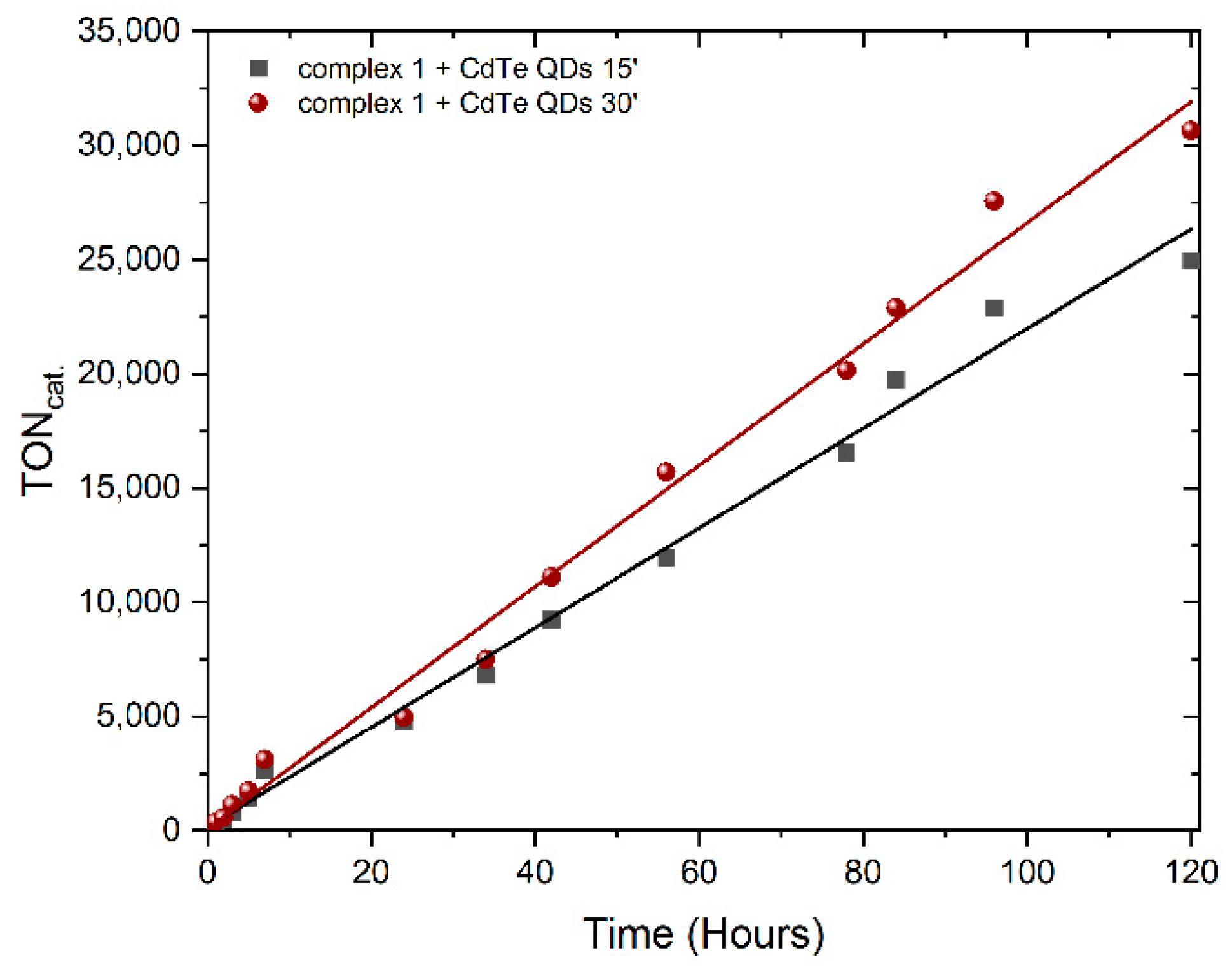
Six distinct TGA-CdTe quantum dots (QDs) were synthesized utilizing established literature protocols [22], and they are denoted according to their reaction duration as 15 min, 30 min, 1 h, 3 h, 7 h, and 13 h, respectively. The resultant QDs exhibited sizes ranging from 35 nm to 70 nm. Their characterization was conducted by comparing their UV–vis absorption and emission spectra with those reported in the literature [22]. Notably, the emission peaks of the synthesized QDs were observed to red-shift from 497 nm to 631 nm (Figure S7). In the photocatalytic systems, containing TGA-QDs as the PS and 1, 2, or 3 as catalysts with a solvent system of DMF: H2O (1:2), two distinct electron donors, triethanolamine (TEOA, pH 10.55) and ascorbic acid (AscOH, pH 4.5), were incorporated. The experimental data are summarized in Table S2 and are the mean of three independent experiments. When AscOH served as the electron donor, the highest TONCat was achieved with 30 min CdTe-QDs for complex 1, yielding a TONCat of 30,665. This was followed by complexes 2 and 3, with TONCat values of 4778 and 114, respectively. Conversely, when TEOA was employed as the electron donor, the highest TONCat obtained was 1340. Notably, the photocatalytic systems demonstrated sustained activity for at least 120 h under the conditions evaluated, as illustrated in Figure 4. The data obtained from all the photocatalytic investigations reveal that the chemical structure of the PS influences the photocatalytic activity of the catalysts, as has been observed before [19].

3.3. Electrocatalytic Hydrogen Production

Electrocatalysis and photocatalysis, though distinct processes, offer valuable insights into mechanistic elucidation. In this regard, we conducted an electrocatalytic investigation aimed at evaluating the hydrogen evolution activity of all three complexes. Trifluoroacetic acid (TFA with pKa = 6 ± 0.03 in DMF) [37] was employed as the proton source under argon and incremental equivalents of it were introduced into solutions of the complexes in DMF. Identical experimental conditions were applied to all complexes as described in the experimental section. Upon the addition of increasing concentrations of TFA, complexes 1, 2, and 3 exhibited catalytic waves at −2.23 V, −2.27 V, and −2.31 V, respectively (Figure 5), with their current response proportional to TFA concentration and attributed to catalytic H2 evolution. The catalytic wave, subsequent to the reduction of Ni(II)/Ni(I), exhibits a negative potential shift with increasing acid concentration. Such behavior is consistent across all three complexes and implies an electron transfer-catalyzed chemical exchange (EC) mechanism [14,38,39,40]. To elucidate the contribution of nickel complexes to the observed increase in current, a cyclic voltammogram of TFA devoid of nickel complexes was also obtained and is illustrated in Figure 5 (dashed line). Moreover, in the CV of all three complexes, a new wave at around –1.00 V appeared, which intensity was increased upon the gradual addition of TFA. This new wave is attributed to the protonation of the o—diamine ligand (which is common for all three complexes) with the simultaneous disappearing of the non-protonated diamine reduction at around −0.90 V. The new wave is reversible as indicated by the oxidative wave at +0.6 V. The investigation of whether this wave is a catalytic process involved recording the CV of the complexes under the same conditions as before but scanning from −0.5 to −1.5 V (Figure S8). The intensity of the wave is proportional to the added concentration of TFA, whereas its potentials shifts towards more positive values, indicating a PCET process [38]. It is worth mentioning that the solution changed from green to brown due to the protonation of the diimine ligand and the disruption of the LL’MCT band around 800 nm with increasing aliquots of TFA, as evidenced by monitoring the UV–vis spectra of all three complexes (Figure S9). These spectra were obtained by monitoring the solutions of complexes by adding 1 eq TFA in 1 eq of each complex in DMF. These control experiments provide compelling evidence suggesting that the initial step of the hydrogen evolution reaction (HER) involves a protonation event. Furthermore, the molecular structure of the catalyst was elucidated through rinse test experiments, which demonstrated that the catalytic activity primarily originates from the complex rather than the nickel nanoparticles [14,38,39,40,41,42,43,44]. Following each measurement, the working electrode was extracted from the solution and immersed in a fresh DMF solution containing 0.1 M NBu4PF6. A negligible background signal was observed across all series when scanning towards the negative potential region (Figure S10). Upon the addition of 10 mM TFA, subsequent potential sweeps exclusively revealed the response attributable to the acid (red lines, Figure S10), confirming the homogeneity of our systems.
Overpotentials (η) [45] were determined for all complexes using the methodology we published before [19], resulting in values of −1.27 V, −1.08 V, and −1.05 V for 1, 2, and 3, respectively. Remarkably, the distinctive S-shaped voltammogram commonly associated with such electrochemical systems was notably absent upon the introduction of adequate acid concentrations. This observation suggests potential causes such as geometric alterations from square planar to tetrahedral configurations or substrate depletion during the catalytic process [38]. Furthermore, it is noteworthy to mention that the observed linearity between the catalytic current (ic) and the anodic peak current (ip) with the square root of the scan rate (v½) served as an indication of the molecular nature of the catalysts (Figure S11) [46,47,48]. This relationship was discerned through experimental investigations involving the alteration of scan rates in cyclic voltammograms of complexes’ solutions, each containing a predetermined quantity of TFA (5 mM).
In addition, we conducted chronocoulometric experiments over a duration of 3 h, employing a potential of −1.7 V with a graphite electrode (sized 3 × 8 cm) serving as the working electrode. The complexes 1, 2, and 3 exhibited turnover frequencies (TOF/h) of 15.30, 12.37, and 7.13, respectively. Calculation of the faradaic yield resulted in values of 74%, 72%, and 65% for complexes 1, 2, and 3, respectively. Notably, the linearity observed in the coulometric experiment suggests the stability of the complexes throughout electrocatalysis (Figure S12). Further analysis revealed distinct performance characteristics among the complexes, particularly concerning the influence of different diimine ligands. Specifically, complex 1, featuring 1,10-phenanthroline as a diimine ligand, demonstrated a twofold increase in yield compared to 4,4′-dimethyl-2,2′-bi-pyridine, underscoring the pivotal role of 1,10-phenanthroline in stabilizing the electron cloud within the heteroleptic complexes. Moreover, the superior electron-donating ability of the m-CH3 group resulted in a higher TOF for complex 2 compared to complex 3, consistent with the observations made during photocatalysis experiments. Contrary to the existing literature where nickel diimine complexes demonstrate limited efficacy as both electrocatalysts and photocatalysts for hydrogen production, our findings suggest enhanced hydrogen production efficiency facilitated by the inclusion of o-phenylene diamine.
To elucidate the electrocatalytic mechanisms involved in our experimental investigations, cyclic voltammetry was performed under consistent conditions. The TFA concentration was maintained at 40 mM in a solution of DMF containing 0.1 M n-Bu4NPF6, with a scan rate of 100 mV/s (Figure S13a–c). The addition of catalysts exhibited similar catalytic behavior, with the catalytic wave shifted to a more positive potential. The ratio of catalytic to peak current (ic/ip) displayed a linear dependence on the catalyst concentration, suggesting a first-order relationship (Figure S10a–c—inset) [49].
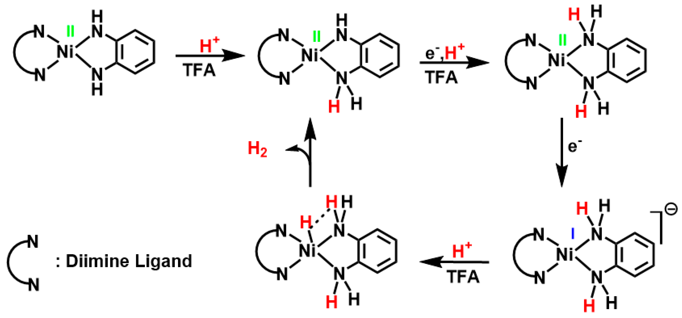
Taking into account all the aforementioned results, particularly the absence of a significant loss of reversibility in the cyclic voltammogram at approximately −1.00 V in the presence of acid, indicative of a rapid chemical reaction [50], we propose a PCET process. In this proposed mechanism, the reduction of the ligand occurs concomitantly with the protonation of the second nitrogen atom of the diamine, and this is followed by an Electron Coupling Electron Transfer (ECET) process, leading to the formation of the metal hydride and an intermediate species involved in the evolution of H2 [14,23,51,52]. Thus, a combined PCET/ECET mechanism is suggested for the H2 evolution (Figure 6) which aligns well with the molecular structure of the catalyst.

4. Conclusions

In this study, we synthesized and characterized three heteroleptic, square planar Ni(II) complexes: [Ni(1,10-phenanthroline)(o-phenylene diamine)] (1), [Ni(2,2-dimethyl-2,2-bipyridine)(o-phenylene diamine)] (2), and [Ni(5,5-dimethyl-2,2-bipyridine)(o-phenylene diamine)] (3). These complexes feature a common o-phenylene diamine ligand and were investigated for their catalytic activity in light-driven hydrogen (H2) evolution, aiming to develop systems that align with the principles of a cyclic economy and environmental sustainability.
Complex 2 exhibited the highest efficiency in photocatalysis, highlighting the significance of the ligand substituent’s position and the molecular nature of all three catalysts. Although all three complexes demonstrated suboptimal electrocatalytic performance, their electrochemical studies yielded valuable insights into their mechanistic details, suggesting a combined proton-coupled electron transfer/electron transfer (PCET/ECET) mechanism.
Complex 1 emerged as the most efficient electrocatalyst, while complex 3 exhibited the lowest activity. Interestingly, the overpotentials across the complexes were relatively consistent due to the shared diamine ligand, with complex 3 demonstrating the lowest overpotential. In contrast, complex 1 displayed the highest faradaic yield. These findings underscore the importance of both the ligand substituent’s position and the chemical nature of the ligands, as the performance of this new series surpassed that of our previous work [19].
The electrocatalytic data reveal a combined PCET/ECET mechanism, proceeding through the formation of a transient intermediate, namely Ni(I)-hydride. This suggests the superiority of nickel for this type of catalyst, as Ni-hydrides are known to exhibit greater stability as transient intermediates in catalytic processes compared to, for instance, copper hydrides. Furthermore, the poor stability of the resultant intermediate Cu(I)-hydride with these ligands may compromise the stability of the complexes. The transformation from a distorted octahedral Cu(II) complex to a tetrahedral Cu(I) species could occur, potentially leading to the formation of nanoparticles, as previously reported [36]. Additional experimental and theoretical investigations are underway to further elucidate these phenomena, exploring analogous complexes with various first-row transition metal ions.
For future research endeavors aiming to develop benchmarked molecular catalysts, we intend to explore novel diamine ligands with substituents at different positions. Additionally, theoretical calculations could provide valuable insights to support and elucidate the observed experimental results.

Supplementary Materials

The following supporting information can be downloaded at https://www.mdpi.com/article/10.3390/en17112777/s1, Figure S1: HRMS of the complexes 1 (a), 2 (b), 3 (c); Figure S2: IR spectra of complex 1 (black), 2 (red), and 3 (blue); Figure S3: 1H-NMR spectrum of complex 1 in DMSO-d6; Figure S4: 1H-NMR spectrum of complex 2 in DMSO-d6; Figure S5: 1H-NMR spectrum of complex 3 in DMSO-d6; Figure S6: UV–vis spectra of a. complex 1 (black), complex 2 (red), complex 3 (blue), in DMF in C = 5·10−5 M and (a), (b), (c) zoomed in 400–1100 nm region for complexes 1, 2, and 3, respectively, in C = 2.9·10−4 M; Figure S7: Stability study of complex 1 (a), complex 2 (b), and complex 3 (c) in DMF for 24 h in dark; Figure S8: Fluorescent spectroscopy of CdTe QDs: 15′ (black), 30′ (red),1 h (blue), 3 h (pink), 7 h (green), and 13 h (dark blue); Figure S9: Electrocatalytic hydrogen production of the complex 1 (3·10−3 M) in DMF with TFA as proton source, 0.1 M n-Bu4NPF6, a glassy carbon as a working electrode, Ag/AgCl as a reference electrode, and a Pt wire as a counter electrode. The scan rate was 100 mV/s under Ar (scan window: (−0.5)–(−1.5)V vs Fc/Fc+); Figure S10: UV–vis spectra of complexes (a) 1 (3·10−4 M), (b) 2 (5·10−5 M), and (c) 3 (3·10−5 M) in DMF adding equivalents of TFA; Figure S11 Cyclic voltammograms of complexes 1 (a), 2 (b), and 3 (c) in the presence of 10 mM of TFA (black), and a subsequent CV (red) using the same electrode after performing electrolysis under constant potential (−1.7 V) for 5 min, then rinsing with DMF and transfer to fresh DMF/0.1 M Bu4NPF6 solution with the absence of complexes with 10 mM TFA; Figure S12: Scan rate-dependent cyclic voltammograms of complexes (a) 1 (10−3 M), (b) 2 (10−3 M), and (c) 3 (10−3 M) recorded from 0.1 V/s to 2 V/s in DMF with TFA and Cottrell plots of peak current versus the square root of scan rate (icat./ip versus ν1/2). CVs were collected in DMF with 0.1 M n-Bu4NPF6 as supp. electrolyte using a glassy carbon (work elec.), platinum wire (count elec.), and Ag/Ag+ (ref. elec.). All the potentials were referenced vs Fc/Fc+; Figure S13: Chronocoulometry for bulk electrolysis in DMF solution with 10 mM TFA, with complexes (a) 1 (10−4 M), (b) 2 (10−4 M), and (c) 3 (10−4 M) at −1.7 V with 0.1 M n-Bu4NPF6, a graphite rod as a working electrode, Ag/AgCl as a reference electrode, and a Pt wire as a counter electrode being used; Figure S14: Cyclic voltammograms in DMF with 0.1 M n-Bu4NPF6 containing 40 mM TFA with addition of aliquots (0.6–3 mM) of complexes (a) 1, (b) 2, and (c) 3 at scan rate 100 mV/s; Figure S15: Cyclic voltammograms in DMF with 0.1 M n-Bu4NPF6 containing increasing equivalents of TFA, with a glassy carbon working electrode, a Ag/AgCl reference electrode, and a Pt wire counter electrode. At scan rate 100 mV/s, under Ar atmosphere; Table S1: 1H-NMR shifts in ppm for complexes 13 and the free ligands; Table S2: Photocatalytic hydrogen evolution of complexes 1, 2, and 3 using CdTe QDs as photosensitizers in acidic and alkaline environment. References [53,54,55,56] are cited in the supplementary materials.

Author Contributions

Conceptualization, C.A.M.; methodology, F.K. and C.A.M.; synthesis, F.K. and M.K.; photocatalytic experiments, M.K. and F.K.; electrocatalytic experiments, F.K. and M.K.; data curation, M.K.; writing—review and editing, M.K., F.K. and C.A.M.; supervision, C.A.M.; project administration, C.A.M. All authors have read and agreed to the published version of the manuscript.

Funding

We gratefully acknowledge financial support from the Special Research Account of NKUA (No: 19712).

Data Availability Statement

The original contributions presented in the study are included in the article/Supplementary Materials, further inquiries can be directed to the corresponding author.

Conflicts of Interest

The authors declare no conflicts of interest.

References

  1. Ibn Shamsah, S.M. Earth-Abundant Electrocatalysts for Water Splitting: Current and Future Directions. Catalysts 2021, 11, 429. [Google Scholar] [CrossRef]
  2. Lewis, N.S.; Nocera, D.G. Powering the planet: Chemical challenges in solar energy utilization. Proc. Natl. Acad. Sci. USA 2006, 103, 15729. [Google Scholar] [CrossRef] [PubMed]
  3. Zhu, J.; Hu, L.; Zhao, P.; Lee, L.Y.S.; Wong, K.-Y. Recent Advances in Electrocatalytic Hydrogen Evolution Using Nanoparticles. Chem. Rev. 2020, 120, 851–918. [Google Scholar] [CrossRef] [PubMed]
  4. Deng, Y.; Lai, W.; Xu, B. A Mini Review on Doped Nickel-Based Electrocatalysts for Hydrogen Evolution Reaction. Energies 2020, 13, 4651. [Google Scholar] [CrossRef]
  5. Ferriday, T.B.; Middleton, P.H.; Kolhe, M.L. Review of the Hydrogen Evolution Reaction—A Basic Approach. Energies 2021, 14, 8535. [Google Scholar] [CrossRef]
  6. Benghanem, M.; Mellit, A.; Almohamadi, H.; Haddad, S.; Chettibi, N.; Alanazi, A.M.; Dasalla, D.; Alzahrani, A. Hydrogen Production Methods Based on Solar and Wind Energy: A Review. Energies 2023, 16, 757. [Google Scholar] [CrossRef]
  7. Gray, H.B. Powering the planet with solar fuel. Nat. Chem. 2009, 1, 7. [Google Scholar] [CrossRef] [PubMed]
  8. Bockris, J.O.; Conway, B.E. Studies in hydrogen overpotential. The effect of catalytic poisons at platinized platinum and nickel. Trans. Faraday Soc. 1949, 45, 989–999. [Google Scholar] [CrossRef]
  9. Thoi, V.S.; Sun, Y.; Long, J.R.; Chang, C.J. Complexes of earth-abundant metals for catalytic electrochemical hydrogen generation under aqueous conditions. Chem. Soc. Rev. 2013, 42, 2388–2400. [Google Scholar] [CrossRef]
  10. McKone, J.R.; Marinescu, S.C.; Brunschwig, B.S.; Winkler, J.R.; Gray, H.B. Earth-abundant hydrogen evolution electrocatalysts. Chem. Sci. 2014, 5, 865–878. [Google Scholar] [CrossRef]
  11. Zhang, T.; Wang, C.; Liu, S.; Wang, J.-L.; Lin, W. A Biomimetic Copper Water Oxidation Catalyst with Low Overpotential. J. Am. Chem. Soc. 2014, 136, 273–281. [Google Scholar] [CrossRef] [PubMed]
  12. Sutra, P.; Igau, A. Emerging Earth-abundant (Fe, Co, Ni, Cu) molecular complexes for solar fuel catalysis. Curr. Opin. Green Sustain. Chem. 2018, 10, 60–67. [Google Scholar] [CrossRef]
  13. Hu, C.; Lv, C.; Liu, S.; Shi, Y.; Song, J.; Zhang, Z.; Cai, J.; Watanabe, A. Nickel Phosphide Electrocatalysts for Hydrogen Evolution Reaction. Catalysts 2020, 10, 188. [Google Scholar] [CrossRef]
  14. Drosou, M.; Kamatsos, F.; Mitsopoulou, C.A. Recent advances in the mechanisms of the hydrogen evolution reaction by non-innocent sulfur-coordinating metal complexes. Inorg. Chem. Front. 2020, 7, 37–71. [Google Scholar] [CrossRef]
  15. Mitsopoulou, C.A. Identifying of charge-transfer transitions and reactive centers in M (diimine)(dithiolate) complexes by DFT techniques. Coord. Chem. Rev. 2010, 254, 1448–1456. [Google Scholar] [CrossRef]
  16. Zarkadoulas, A.; Koutsouri, E.; Mitsopoulou, C.A. A perspective on solar energy conversion and water photosplitting by dithiolene complexes. Coord. Chem. Rev. 2012, 256, 2424–2434. [Google Scholar] [CrossRef]
  17. Zarkadoulas, A.; Field, M.J.; Papatriantafyllopoulou, C.; Fize, J.; Artero, V.; Mitsopoulou, C.A. Experimental and Theoretical Insight into Electrocatalytic Hydrogen Evolution with Nickel Bis(aryldithiolene) Complexes as Catalysts. Inorg. Chem. 2016, 55, 432–444. [Google Scholar] [CrossRef] [PubMed]
  18. Koutsouri, E.; Mitsopoulou, C.A. Photocatalytic Hydrogen Evolution by tris-dithiolene tungsten complexes. Open Chem. 2016, 14, 393–403. [Google Scholar] [CrossRef]
  19. Kamatsos, F.; Bethanis, K.; Mitsopoulou, C.A. Synthesis of Novel Heteroleptic Oxothiolate Ni(II) Complexes and Evaluation of Their Catalytic Activity for Hydrogen Evolution. Catalysts 2021, 11, 401. [Google Scholar] [CrossRef]
  20. Lelj, F.; Rosa, A.; Ricciardi, G.; Casarin, M.; Cristinziano, P.; Morelli, G. On the spectroscopic behaviour of o-bis(o-phenylendiimido) nickel. Chem. Phys. Lett. 1989, 160, 39–42. [Google Scholar] [CrossRef]
  21. Bachler, V.; Olbrich, G.; Neese, F.; Wieghardt, K. Theoretical Evidence for the Singlet Diradical Character of Square Planar Nickel Complexes Containing Two o-Semiquinonato Type Ligands. Inorg. Chem. 2002, 41, 4179–4193. [Google Scholar] [CrossRef] [PubMed]
  22. Wu, S.; Dou, J.; Zhang, J.; Zhang, S. A simple and economical one-pot method to synthesize high-quality water soluble CdTe QDs. J. Mater. Chem. 2012, 22, 14573. [Google Scholar] [CrossRef]
  23. Kamatsos, F.; Drosou, M.; Mitsopoulou, C.A. Heteroleptic thiolate diamine nickel complexes: Noble-free-metal catalysts in electrocatalytic and light-driven hydrogen evolution reaction. Int. J. Hydrogen Energy 2021, 46, 19705–19716. [Google Scholar] [CrossRef]
  24. Himmel, H.-J.; Manceron, L. Ni(N2)4 revisited: An analysis of the Ni–N2 bonding properties of this benchmark system on the basis of UV/Vis, IR and Raman spectroscopy. Dalton Trans. 2005, 15, 2615–2623. [Google Scholar] [CrossRef] [PubMed]
  25. Ramos, J.M.; Versiane, O.; Felcman, J.; Soto, C.A.T. FT-IR vibrational spectrum and DFT:B3LYP/6-31G structure and vibrational analysis of guanidinoaceticserinenickel(II) complex: [Ni(GAA)(Ser)]. Spectrochim. Acta Part A Mol. Biomol. Spectrosc. 2007, 67, 1037–1045. [Google Scholar] [CrossRef] [PubMed]
  26. Halli, M.B.; Patil, V.B. Synthesis, spectroscopic, thermal, and biological activities of mixed ligand complexes containing E-N′-(3,4,5-trimethoxybenzylidene)benzofuran-2-carbohydrazide and 2-aminothiophenol. J. Coord. Chem. 2011, 64, 3740–3750. [Google Scholar] [CrossRef]
  27. Ghiasi, Z.; Amani, V.; Mirzaei, P.; Safari, N.; Abedi, A. Trichloridothallium(III) Complexes with Bipyridine Derivatives: From Structure to Luminescence Properties. Aust. J. Chem. 2013, 66, 676. [Google Scholar] [CrossRef]
  28. Lever, A.B.P. Inorganic Electronic Spectroscopy; Elsevier: Amsterdam, The Netherlands, 1968. [Google Scholar]
  29. Patra, A.K.; Mukherjee, R. Bivalent, trivalent, and tetravalent nickel complexes with a common tridentate deprotonated pyridine bis-amide ligand. Molecular structures of nickel (II) and nickel (IV) and redox activity. Inorg. Chem. 1999, 38, 1388–1393. [Google Scholar] [CrossRef]
  30. Kaasjager, V.E.; Puglisi, L.; Bouwman, E.; Driessen, W.L.; Reedijk, J. Synthesis, characterization and crystal structures of nickel complexes with dissymmetric tetradentate ligands containing a mixed-donor sphere. Inorg. Chim. Acta 2000, 310, 183–190. [Google Scholar] [CrossRef]
  31. Herebian, D.; Bothe, E.; Bill, E.; Weyhermüller, T.; Wieghardt, K. Experimental Evidence for the Noninnocence of o-Aminothiophenolates:  Coordination Chemistry of o-Iminothionebenzosemiquinonate(1-) π-Radicals with Ni(II), Pd(II), Pt(II). J. Am. Chem. Soc. 2001, 123, 10012–10023. [Google Scholar] [CrossRef]
  32. Das, A.; Han, Z.; Brennessel, W.W.; Holland, P.L.; Eisenberg, R. Nickel Complexes for Robust Light-Driven and Electrocatalytic Hydrogen Production from Water. ACS Catal. 2015, 5, 1397–1406. [Google Scholar] [CrossRef]
  33. Goss, C.A.; Abruna, H.D. Spectral, electrochemical and electrocatalytic properties of 1,10-phenanthroline-5,6-dione complexes of transition metals. Inorg. Chem. 1985, 24, 4263–4267. [Google Scholar] [CrossRef]
  34. Saravani, H.; Rezvani, A.R.; Mansouri, G.; Rad, A.R.S.; Khavasi, H.R.; Hadadzadeh, H. Crystal structure, magnetic and electrochemical properties of five-coordinate copper (II) complexes with 1,10-phenanthroline-5,6-dione. Inorg. Chim. Acta 2007, 360, 2829–2834. [Google Scholar] [CrossRef]
  35. Hadadzadeh, H.; Mansouri, G.; Rezvani, A.R.; Khavasi, H.R. A Novel 1:1 Co-Crystal of Bis(1,10-Phenanthroline)(1,10-Phenanthroline-5,6-Dione)Nickel(II) Hexafluorophosphate and Tris(1,10-Phenanthroline)Nickel(II) Hexafluorophosphate Complexes, [Ni(phen)2(phen-dione)] [Ni(phen)3] (PF6)4. J. Chem. Crystallogr. 2012, 42, 486–493. [Google Scholar] [CrossRef]
  36. Drosou, M.; Kamatsos, F.; Ioannidis, G.; Zarkadoulas, A.; Mitsopoulou, C.A.; Papatriantafyllopoulou, C.; Tzeli, D. Reactivity and Mechanism of Photo- and Electrocatalytic Hydrogen Evolution by a Diimine Copper(I) Complex. Catalysts 2020, 10, 1302. [Google Scholar] [CrossRef]
  37. Fourmond, V.; Canaguier, S.; Golly, B.; Field, M.J.; Fontecave, M.; Artero, V. A nickel–manganese catalyst as a biomimic of the active site of NiFe hydrogenases: A combined electrocatalytical and DFT mechanistic study. Energy Environ. Sci. 2011, 4, 2417–2427. [Google Scholar] [CrossRef]
  38. Elgrishi, N.; McCarthy, B.D.; Rountree, E.S.; Dempsey, J.L. Reaction Pathways of Hydrogen-Evolving Electrocatalysts: Electrochemical and Spectroscopic Studies of Proton-Coupled Electron Transfer Processes. ACS Catal. 2016, 6, 3644–3659. [Google Scholar] [CrossRef]
  39. Fang, M.; Engelhard, M.H.; Zhu, Z.; Helm, M.L.; Roberts, J.A.S. Electrodeposition from Acidic Solutions of Nickel Bis(benzenedithiolate) Produces a Hydrogen-Evolving Ni–S Film on Glassy Carbon. ACS Catal. 2014, 4, 90–98. [Google Scholar] [CrossRef]
  40. Rountree, E.S.; McCarthy, B.D.; Eisenhart, T.T.; Dempsey, J.L. Evaluation of Homogeneous Electrocatalysts by Cyclic Voltammetry. Inorg. Chem. 2014, 53, 9983–10002. [Google Scholar] [CrossRef]
  41. Costentin, C.; Savéant, J.-M. Multielectron, Multistep Molecular Catalysis of Electrochemical Reactions: Benchmarking of Homogeneous Catalysts. ChemElectroChem 2014, 1, 1226–1236. [Google Scholar] [CrossRef]
  42. Cavell, A.C.; Hartley, C.L.; Liu, D.; Tribble, C.S.; McNamara, W.R. Sulfinato Iron(III) Complex for Electrocatalytic Proton Reduction. Inorg. Chem. 2015, 54, 3325–3330. [Google Scholar] [CrossRef] [PubMed]
  43. Eckenhoff, W.T. Molecular catalysts of Co, Ni, Fe, and Mo for hydrogen generation in artificial photosynthetic systems. Coord. Chem. Rev. 2018, 373, 295–316. [Google Scholar] [CrossRef]
  44. Zhao, P.-H.; Li, J.-R.; Gu, X.-L.; Jing, X.-B.; Liu, X.-F. Diiron and trinuclear NiFe2 dithiolate complexes chelating by PCNCP ligands: Synthetic models of [FeFe]- and [NiFe]-hydrogenases. J. Inorg. Biochem. 2020, 210, 111126. [Google Scholar] [CrossRef] [PubMed]
  45. Appel, A.M.; Helm, M.L. Determining the Overpotential for a Molecular Electrocatalyst. ACS Catal. 2014, 4, 630–633. [Google Scholar] [CrossRef]
  46. Fourmond, V.; Jacques, P.-A.; Fontecave, M.; Artero, V. H2 Evolution and Molecular Electrocatalysts: Determination of Overpotentials and Effect of Homoconjugation. Inorg. Chem. 2010, 49, 10338–10347. [Google Scholar] [CrossRef] [PubMed]
  47. Gomez-Mingot, M.; Porcher, J.-P.; Todorova, T.K.; Fogeron, T.; Mellot-Draznieks, C.; Li, Y.; Fontecave, M. Bioinspired Tungsten Dithiolene Catalysts for Hydrogen Evolution: A Combined Electrochemical, Photochemical, and Computational Study. J. Phys. Chem. B 2015, 119, 13524–13533. [Google Scholar] [CrossRef] [PubMed]
  48. Haddad, A.Z.; Cronin, S.P.; Mashuta, M.S.; Buchanan, R.M.; Grapperhaus, C.A. Metal-Assisted Ligand-Centered Electrocatalytic Hydrogen Evolution upon Reduction of a Bis(thiosemicarbazonato)Cu(II) Complex. Inorg. Chem. 2017, 56, 11254–11265. [Google Scholar] [CrossRef] [PubMed]
  49. McCarthy, B.D.; Martin, D.J.; Rountree, E.S.; Ullman, A.C.; Dempsey, J.L. Electrochemical Reduction of Brønsted Acids by Glassy Carbon in Acetonitrile—Implications for Electrocatalytic Hydrogen Evolution. Inorg. Chem. 2014, 53, 8350–8361. [Google Scholar] [CrossRef] [PubMed]
  50. Zarkadoulas, A.; Koutsouri, E.; Kefalidi, C.; Mitsopoulou, C.A. Rhenium complexes in homogeneous hydrogen evolution. Coord. Chem. Rev. 2015, 304, 55–72. [Google Scholar] [CrossRef]
  51. Solis, B.H.; Hammes-Schiffer, S. Proton-Coupled Electron Transfer in Molecular Electrocatalysis: Theoretical Methods and Design Principles. Inorg. Chem. 2014, 53, 6427–6443. [Google Scholar] [CrossRef]
  52. Greene, B.L.; Wu, C.-H.; McTernan, P.M.; Adams MW, W.; Dyer, R.B. Proton-Coupled Electron Transfer Dynamics in the Catalytic Mechanism of a [NiFe]-Hydrogenase. J. Am. Chem. Soc. 2015, 137, 4558–4566. [Google Scholar] [CrossRef] [PubMed]
  53. Zhang, C.; Li, G.; Cai, X. Study on deactivation and reaction mechanism of Co thiolate complexes in photocatalytic hydrogen production system. Int. J. Energy Res. 2018, 42, 977–984. [Google Scholar] [CrossRef]
  54. Costentin, C.; Drouet, S.; Robert, M.; Savéant, J.-M. Turnover numbers, turnover frequencies, and overpotential in molecular catalysis of electrochemical reactions. Cyclic voltammetry and preparative-scale electrolysis. J. Am. Chem. Soc. 2012, 134, 11235–11242. [Google Scholar] [PubMed]
  55. Schröder, D.; Shaik, S.; Schwarz, H. Two-State Reactivity as a New Concept in Organometallic Chemistry. Acc. Chem. Res. 2000, 33, 139–145. [Google Scholar] [CrossRef]
  56. Artero, V.; Saveant, J.-M. Toward the rational benchmarking of homogeneous H2-evolving catalysts. Energy Environ. Sci. 2014, 7, 3808–3814. [Google Scholar] [CrossRef]
Figure 1. Structures of the complexes 13.
Energies 17 02777 g001
Figure 2. Cyclic voltammograms of 1 (10−3 M) (black), 2 (10−3 M) (red), 3 (10−3 M) (blue) in DMF, 0.1 M n-Bu4NPF6, a glassy carbon as a working electrode, Ag/AgCl as a reference electrode, and a Pt wire as a counter electrode. The scan rate was 100 mV/s.
Energies 17 02777 g002
Figure 3. Photocatalytic hydrogen evolution of the solutions containing the complexes at C = 10–5 M/fluorescein (1 mM)/TEOA (0.5 M)/DMF:H2O (1:45) with and without Hg (a) 1, (b) 2, and (c) 3.
Energies 17 02777 g003
Figure 4. Photocatalytic hydrogen evolution of complex 1 in presence of CdTe QDs 15′ (black) and CdTe QDs 30′ (red) with AscOH as sacrificial electron donor.
Energies 17 02777 g004
Figure 5. Electrocatalytic hydrogen production at a glassy carbon as a working electrode in a 10−3 M solution of complexes (a) 1, (b) 2, and (c) 3, in DMF (0.1 M n-Bu4NPF6) in the presence of increasing aliquots of TFA as a proton source. The black dotted CV corresponds to 100 mM TFA in the absence of catalyst. Reference electrode: Ag/AgCl, counter electrode: Pt wire, scan rate: 100 mV/s under Ar.
Energies 17 02777 g005
Figure 6. Proposed mechanism for complexes 13.
Energies 17 02777 g006
Table 1. UV–vis data for the complexes 1, 2, and 3.
[Complex].Solventλmax (ε *)λmax (ε *)λmax (ε *)λmax (ε *)
1
(5 × 10⁻⁵ M)
DMF274
(80,000)
295
(43,400)
634
(1080)
785
(8200)
2
(5 × 10⁻⁵ M)
DMF274
(5600)
298
(800)
635
(12,400)
787
(23,000)
3
(5 × 10⁻⁵ M)
DMF275
(3800)
295
(480)
643
(25,800)
784
(40,600)
* ε, Μ−1cm−1.
Table 2. Cyclic voltametric data.
ComplexEdiimine0/−1
V,(ip,a/ip,c)
Ediamine0/−1
V,(ip,a/ip,c)
E0NiII/I
V,(ip,a/ip,c)
1Ep,a = −0.45 *
Ep,c = −0.30
Ep,a = −0.95
Ep,c = −0.89
Ep,a = −1.38 ***
Ep,c = −1.32
Ep,a = −2.12 *
Ep,c = −1.96
2--Ep,a = −1.74 **Ep,a = −2.12 **
3-Ep,a = −0.94 ***
Ep,c = −0.92
Ep,a = −1.38 ***
Ep,c = −1.32
Ep,a = −2.04
Ep,c = −1.93
E(V) vs. Fc/Fc+, * semi. = semi-reversible, ** ir. = Irreversible, *** rev. = reversible.
Disclaimer/Publisher’s Note: The statements, opinions and data contained in all publications are solely those of the individual author(s) and contributor(s) and not of MDPI and/or the editor(s). MDPI and/or the editor(s) disclaim responsibility for any injury to people or property resulting from any ideas, methods, instructions or products referred to in the content.