Article

Microalgae as an Effective Recovery Agent for Vanadium in Aquatic Environment

1
Daegu Center, Korea Basic Science Institute (KBSI), 80 Daehak-ro, Daegu 41566, Korea
2
Cell Factory Research Center, Korea Research Institute of Bioscience and Biotechnology (KRIBB), 125 Gwahak-ro, Daejeon 34141, Korea
3
Department of Chemistry and Energy Engineering, Sangmyung University, 20 Hongimun 2-gil, Jongno-gu, Seoul 03016, Korea
4
Biorefinery and Bioenergy Research Laboratory, Amity Institute of Biotechnology, Amity University Uttar Pradesh, Noida 201313, India
5
Library of Marine Samples, Korea Institute of Ocean Science & Technology, Geoje 53201, Korea
6
Department of Applied Marine Bioresource Science, National Marine Biodiversity Institute of Korea (MABIK), Seocheon-gun 33662, Korea
7
School of Fisheries Science & Environmental Studies, Nagasaki University, 1-14 Bunkyo-machi, Nagasaki 852-8521, Japan
*
Authors to whom correspondence should be addressed.
Energies 2022, 15(12), 4467; https://doi.org/10.3390/en15124467
Submission received: 22 May 2022 / Revised: 14 June 2022 / Accepted: 17 June 2022 / Published: 19 June 2022

Abstract

:
Given that vanadium is a valuable material, the implementation of vanadium recycling processes is thus necessary to enhance the element’s value chain as well as minimize its undesirable environmental consequences. Among various remediation methods available, a biological method based on microalgal adsorption is known to be eco-friendly and calls for further investigations. Herein, we evaluated V2O5 adsorption efficiencies of four different microalgal strains: Nannochloropsis oculata, Heterocapsa circularisquama, Chattonella marina, and Chattonella antiqua. Inductively coupled plasma mass spectrometry (ICP-MS) data indicated that vanadium concentration in the culture medium of Nannochloropsis oculata was reduced from 4.61 ± 0.11 mg L−1 to 1.85 ± 0.21 mg L−1 after being exposed to V2O5 solution for 24 h, whereas the supernatants of the other three strains displayed no change in vanadium ion concentration. Therefore, our results indicated a strong potential of Nannochloropsis oculata for recycling vanadium with approximately 59.9% of vanadium ion removal efficiency. Furthermore, morphological observation of Nannochloropsis oculata using scanning electron microscopy (SEM) indicated that the cells were able to maintain their intact morphology even under the presence of high concentrations of heavy metals. Due to the high adsorption efficiency and robustness of Nannochloropsis oculata, the results collectively support it as a potential strain for V2O5 recovery.

1. Introduction

Scientists across the world are studying various issues associated with heavy metal recycling because of: (1) environmental pollution and (2) high commercial values of some of the rare elements [1,2,3,4,5]. Among various heavy metals, vanadium (V) is directly used in real-life applications as catalysts in the steel and semiconductor industries [6], and vanadium-based batteries are considered a promising energy storage device [7,8,9,10]. Vanadium ore is mined only in a few countries, including China, the United States, and South Africa, therefore it is an industrially valuable commodity [11]. However, vanadium is often discarded in various industrial wastewaters [12,13], even though its different forms have been acknowledged to be toxic [6]. Thus, recycling vanadium ions present in aquatic environments will serve as an effective approach to addressing both environmental and industrial-associated issues [14,15].
Currently, different types of vanadium sources and various methods for its recovery are being actively studied [16,17]. In the case of solid sources such as vanadium-bearing steel slags, chemical methods are mainly implemented to recover vanadium [17] as such methods are simple and economically feasible. However, vanadium pentoxide (V2O5), which is the most common commercial form of vanadium, is soluble in water [16,17]. Although chemical treatments can easily recover dissolved vanadium, deploying chemical processes for the treatment of a large volume of water resources can often lead to unintended environmental pollution, which could seriously impact our daily lives [18]. Therefore, a non-chemical method for recycling vanadium pentoxide should be developed, and eco-friendly biological methods are considered one of the best options. Among various types of biological methods used for recovering heavy metals, microalgal treatment is considered to be promising, owing to the high tolerance of microalgae to toxic environments [19,20]. Several active studies on heavy metal removal using microalgae indicated that microalgae can purify the aquatic ecosystem by removing heavy metals [19,21]. Unfortunately, the mechanism associated with the recovery of heavy metals using microalgae is not well understood, therefore the industrial-scale implementation of microalgae-based heavy metal remediation technologies is currently limited [19,22].
Heavy metals such as vanadium carry a cationic group, and they are attracted to the anionic (i.e., negative charges) functional groups on microbial cell walls [22]. The mechanism of heavy metal recycling is expected to be similar to that of heavy metal remediation using microalgae, which is related to the adhesion of heavy metals on the cell surface [19,23]. However, microalgae must maintain their bioactivity even under the presence of toxic heavy metals, to be deployed as an active remediation agent. In general, microalgae with large cellular sizes were considered to be advantageous for withstanding the toxicity of heavy metals [24]. However, small-sized microalgae are also capable of withstanding the toxicity of heavy metals if the surface area to volume ratio is high enough [25]. On the other hand, heavy metal removal is also affected by pH [26], as heavy metals are positively charged in dissolved forms, they likely bond with OH [27]. As pH is lowered, metal ions are more likely to be present in dissolved ionic forms [22]; those unstable metal ions are toxic to the cell by interfering with negatively charged functional groups on the cellular surface.
There are various methods for removing heavy metals by microalgae, including passive diffusion, ion exchange, complexation, and mediated transport [27]; however, all adsorption-based removal of heavy metals could compromise cellular integrity and bioactivity. Nonetheless, when metal ions are indirectly removed by microalgae, the pH is increased [28], which could substantially influence the ionic strength of heavy metals and associated cellular toxicity [29]. Therefore, the close interplay between bioabsorption and heavy metal forms modulated by an expected shift in pH levels should be carefully considered when screening a novel bioreagent for heavy metal recovery in an aquatic environment.
In this study, we compared V2O5 adsorption efficiencies of four different marine microalgal strains: Nannochloropsis oculata, Heterocapsa circularisquama, Chattonella marina, and Chattonella antiqua [30,31,32,33]; these strains are commonly found in the oceans of Korea and Japan, and well-studied in earlier scientific publications [33,34,35,36,37]. For the experiment, V2O5 was first dissolved in water, and the same concentration of V was then added to different T-flasks containing distinct microalgal strains.

2. Materials and Methods

2.1. Strain and Maintenance Condition

A non-toxic microalgal strain, Nannochloropsis oculata (N. oculata) [32,36]; two conventional harmful algae, Chattonella marina (C. marina) and Chattonella antiqua (C. antiqua) [33,35]; and a harmful alga, Heterocapsa circularisquama (H. circularisquama) were isolated from Yeong-deok seawater. These four seawater microalgal strains were cultivated in 40-mL cell culture flasks (T-flask, SPL, Pocheon-si, Korea) containing 30 mL of L1 medium. The L1 medium was composed of 35 g L−1 sea salt, 75 mg L−1 NaNO3, 5 mg L−1 NaH2PO4·2H2O, 0.5 g L−1 Tris-(hydroxymethyl) aminomethane, 1 mL trace element solution (4.36 g L−1 Na2EDTA.2H2O, 3.15 g L−1 FeCl3, 178.1 g L−1 MnCl2·4H2O, 23 g L−1 ZnSO4, 11.9 g L−1 CoCl2. 4H2O, 2.5 g L−1 CuSO4, 19.9 g L−1 Na2MoO4, 1.29 g L−1 H2SeO3), and 0.5 mL f/2 vitamin solution (1 mg L−1 vitamin B12, 1 mg L−1 biotin, and 200 mg L−1 thiamine hydrochloride). The flask was continuously illuminated with light at an intensity of 4200 µmol m−2 s−1 under a light/dark cycle of 12:12, and its temperature and pH were set at 25 °C and 7.6, respectively.

2.2. Cellular Growth Measurement

Subculturing each microalgal strain was performed every 2 weeks. To analyze the V2O5 absorption efficiencies of blooming microalgae, their cellular growth was measured every 24 h, and culture broth from day 7 was used for the absorption experiment. Cellular growth was analyzed by measuring the optical density (OD) at 680 nm wavelength using a UV spectrophotometer (Jenway, Stone, UK) [37]. The strains utilized in this study were adherent cells on the floor, and, therefore, their cultivation was conducted without agitation. Consequently, the culture broths were shaken only during sampling periods for the analyses of OD, which is the most efficient method to measure the concentration of adherent cells in flask-scale cultivation. For N. oculata, the dry cell weight was calculated using the following correlative equation:
DCW (g L−1) = 4.2872 × OD680nm − 0.1066
where DCW represents the dry cell weight of N. oculata, and OD680nm refers to the optical density of N. oculata.

2.3. Vanadium Ion Measurement Using ICP-MS

To estimate the vanadium adsorption efficiencies of the four microalgae, V2O5 solution was added to the culture broths of day 7 and gently mixed. Adsorption of vanadium by the microalgae was observed for 24 h without any mixing process, and the pH of the culture broths was measured at the end of both cultivation and vanadium adsorption processes, respectively. In the case of N. oculata, cell lysis was performed after 24 h-long cultivation. Three cell lysis methods were tested: sonication, acid treatment, and base treatment. Prior to the sonication method, cells were washed twice using distilled water for ICP-MS analysis of adsorption or absorption of vanadium ions. Thereafter, washed cells were treated with a sonicator at 20,000 Hz for 1 min to lyse them completely, which was then followed by ICP-MS analysis. In addition, acid and base reactions were performed with washed cells using either nitric acid or potassium hydroxide at a concentration of 2 N; ICP-MS analysis was performed after treating cells with either acidic or alkaline reagent for 30 min.
Vanadium pentoxide solution was purchased from Merck & Co. (Sigma Aldrich, St. Louis, MO, USA), and the concentration of vanadium ions was analyzed using 7700 ICP-MS (Agilent, Santa Clara, CA, USA) [38]. To prepare samples for ICP-MS analyses, the supernatants of culture broths were collected after centrifugation (Thermo Fisher, Waltham, MA, USA). The standard solutions were prepared, and the concentrations of the supernatants were adjusted to be within a linear range for the analysis. Since Agilent 7700 ICP-MS has an analytical range of up to 500 ppb, the samples must be diluted to ≤0.5 mg L−1 for the analysis of metal ion concentration. Given this, we made a 100-fold dilute solution for all of our samples. In addition, ICP-MS analysis also requires that all insoluble components in the samples must be completely dissolved to form an aqueous solution, and hence all our samples were diluted with 0.5% nitric acid as a pretreatment process. During the analysis, the temperature and humidity were maintained at 20 °C and 18%, respectively. All experiments were conducted thrice.

2.4. Scanning Electron Microscope (SEM)

The cell pellets were collected through centrifugation, and their morphology was analyzed by scanning electron microscope (SEM). The details of various sample preparation steps for the scanning electron microscopy such as fixation, conductive staining, dehydration, and coating [39,40] are as follows: (1) For fixation, cells were immobilized by 0.5% glutaraldehyde solution, followed by washing with phosphate buffer saline (PBS) solution, (2) during the conductive staining step, the cells were exposed to 1% OsO4 for approximately 15 min, this step was also followed by washing with PBS, (3) for dehydration, both freeze-drying and critical point drying were used, and (4) for Pt-Pd coating, an ion sputter [41] was used. To quantify the relative concentration of ions on the cell surface, the lyophilized cells were studied using SEM (SUPRA 55VP, Carl Zeiss AG, Oberkochen, Germany) [42]. Additionally, the morphology of cells was also observed using an optical microscope.

2.5. Statistical Analysis

The cellular growth rates and vanadium concentrations are reported as mean values of the data obtained from experiments ± 2 standard deviation (2 SD). Differences in V concentration between various groups were analyzed by one-way analysis of variance (ANOVA) with Tukey’s post hoc comparison at significance levels of p < 0.05 and p < 0.01 for cellular growth rate and vanadium concentration, respectively.

3. Results and Discussion

3.1. Cellular Growth and Physiology of Microalgae

Four species of marine microalgae, Nannochloropsis oculata, Heterocapsa circularisquama, Chattonella marina, and Chattonella antiqua, were cultivated. These four species are commonly found in seawater and have been used in various studies. All four species grew very slowly, with a life cycle of two weeks. The microalgal cells used in the experiment were cultured for a week (Table 1), and each strain was exposed to the same light and humidity conditions during the cultivation period. Their growth profiles, shown in Figure S1, reflect that the four species exhibit similar trends in their growth curve, with the final OD680nm of 0.9–0.95.
It was observed that N. oculata grew relatively faster, but differences in its OD value relative to that of the other three microalgae were insignificant (less than ± 2 SD). In addition, cells at the exponential phase from day 7 were sampled and studied under an optical microscope to verify cellular morphology, the results of which are presented in Figure S2. The cultivation process was aimed at determining the effectiveness of V2O5 adsorption by environmentally prevalent species. The algal growth of four different species with similar OD, and the absence of any bacterial contamination confirmed that the culture broth resulting from microalgal growth was suitable for analyzing the adsorption efficiency for V2O5 recovery.

3.2. Vanadium Ion Concentration Measurement by ICP-MS

Vanadium pentoxide has a solubility of 8 g L−1 at room temperature [42], and the color of the solution changes depending on the ambient pH [43]. In this study, distilled water at room temperature was used to dissolve vanadium (Table 1). The entire experimental procedure used to study the vanadium recycling process is illustrated in Figure 1. To enhance the possibility of industrial applications, the initial concentration of vanadium ions considered in this study was 5 mg L−1 (Table 1), which is the maximum amount of heavy metal concentration that can be released into industrial wastewaters according to the environmental regulation law of South Korea [44]. Therefore, 5 mg L−1 vanadium solution was prepared for each flask culture, and the supernatant was sampled every 3 h between 0 h and 12 h to verify the robustness of the proposed method in vanadium recycling (Figure 1). After 12 h of reaction, samples were collected every 6 h for 24 h.
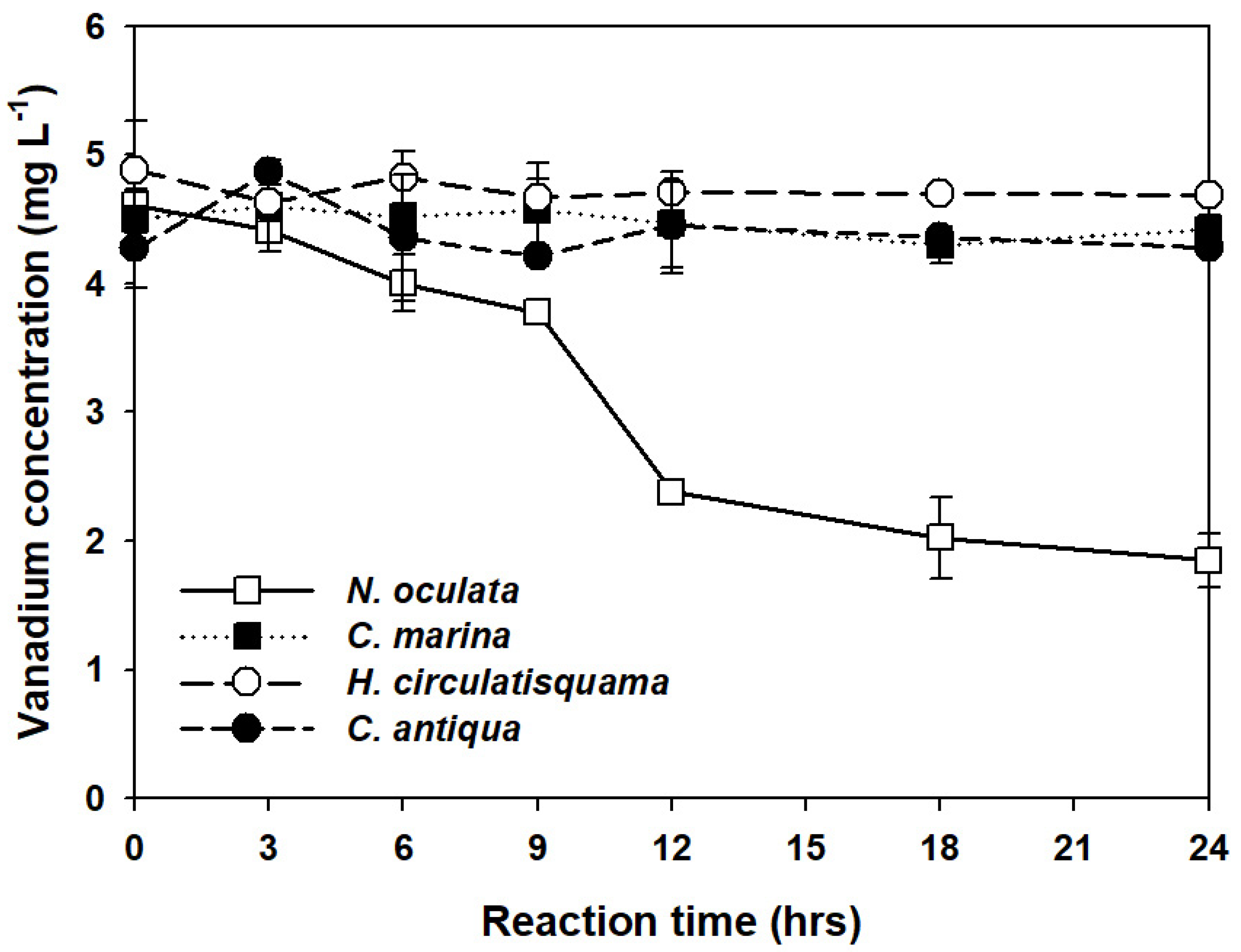
As shown in Figure 2, it is evident that the vanadium solution was added to each sample at a uniform concentration. In the case of C. marina, C. antiqua, and H. circularisquama, the respective changes in V concentration from 4.5 ± 0.07 mg L−1 to 4.47 ± 0.28 mg L−1, from 4.28 ± 0.22 mg L−1 to 4.45 ± 0.23 mg L−1, and from 4.89 ± 0.27 L−1 to 4.71 ± 0.1 mg L−1, were insignificant. However, N. oculata exhibited a gradual decrease in vanadium concentration from 4.61 ± 0.11 mg L−1 to 2.38 ± 0.07 mg L−1 in a period of 12 h. This observation demonstrated the possibility of removing a significant amount of vanadium ions down to approximately half of the initial concentration. For most species, microalgae soaked in vanadium solution for more than 12 h displayed cellular degradation. On the contrary, N. oculata treatment reduced the vanadium ion concentration to 1.85 mg L−1 in the supernatant after 24 h, indicating that the removal rate of vanadium ion between 0 h and 12 h was about 4.2 times greater than that observed between 12 h and 24 h.
While the maximum vanadium removal of 59.9% was observed after 24 h, the maximum removal rate was calculated as 0.00157 g−1 h−1 following Equation (1) and considering the concentration of N. oculata inoculum as 3.94 g L−1. This implies that 0.1858 mg of vanadium per 118.3 mg of N. oculata could be removed after 12 h. In future studies to establish the vanadium recovery process with the same amount of microalgae, 12 h of reaction period would be the most efficient, considering that the hydraulic retention time of the wastewater treatment process is one of the most cost-dominant factors.
In addition to checking the vanadium concentration in the pellet, various cell lysis methods were attempted to confirm the vanadium recovery from the cell. In this respect, sonication, acid treatment, and base treatment were performed (Table 2), and the results indicated vanadium concentration in the lysates obtained with the corresponding methods as 2.57 ± 0.27 mg L−1, 0.89 ± 0.11 mg L−1, and 2.07 ± 0.31 mg L−1. In particular, the method that showed the highest vanadium recovery rate was sonication, which showed a recovery rate of about 93%. In addition, a recovery rate of about 85% was measured by the base treatment (2N KOH), whereas a low recovery rate of about 37% was measured with the acid treatment (2N HNO3). While this seems to be associated with the effectiveness of each method in cell wall disruption, the results suggested cell lysis through sonication as the most effective method for recovering vanadium. Given that most of the vanadium was recovered following a relatively simple treatment, the results supported that the recycling of vanadium would be possible cost-effectively.
To estimate the significance of vanadium recovery using N. oculata, a series of one-way ANOVAs with Tukey’s post hoc tests were conducted for 6, 9, and 12 h of reaction time, and the results are listed in Tables S1 and S2, and Figure S3. All four experimental groups considered in this study had no difference at 6 h, except that the pair-wise differences between HC and NO (H. circularisquama-N. oculata) seemed to be substantial. Furthermore, after 12 h of reaction time, the significance of ANOVA increased further with p < 0.01, and a comparison with the other three microalgal species indicated that the V2O5 adsorption efficiency of N. oculata was significantly different compared to the other groups.
Our comparative analysis of four candidate strains indicated N. oculata as a promising strain that can effectively remove vanadium ions present in an aquatic environment. Moreover, the recovery rate results of this study were quite noteworthy compared to other studies demonstrating the removal of heavy metals using microalgae. For instance, removal rates for Cr, Cd, and Cu varied from 14% to 90% during a week-long treatment period [27]. Although direct comparison is difficult because the initial concentrations of metal ions and microalgae are different, the vanadium removal of N. oculata used in this study was more than 59.9% following just a day-long treatment. Surprisingly, the results also indicated a high recovery rate (i.e., ~93% recovery of vanadium from microalgae) using simple pretreatment, reiterating the great industrial potential of N. oculata.

3.3. SEM Observation of Cellular Morphology

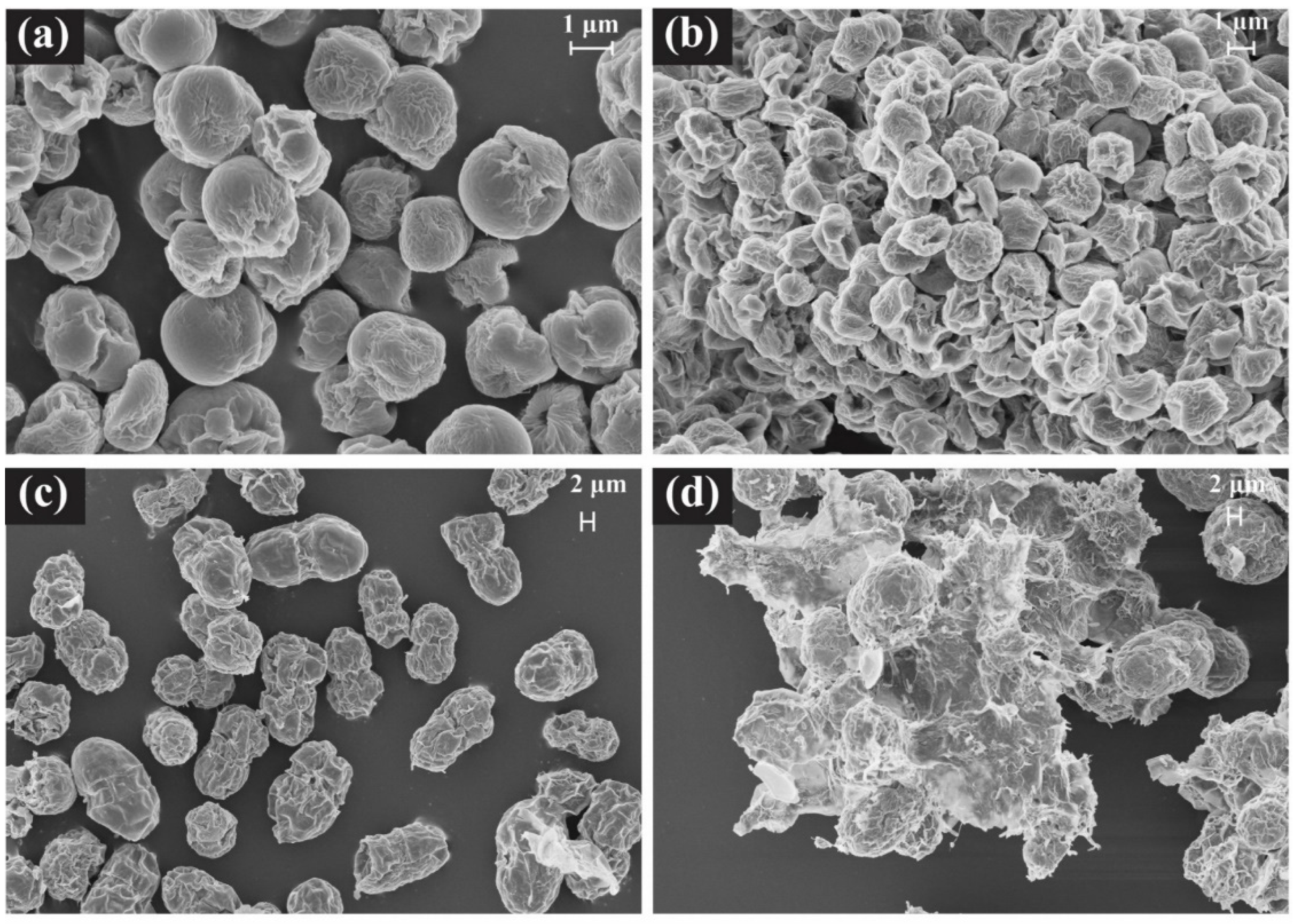
The morphology of two species, N. oculata (NO) and H. circularisquama (HC) were studied using SEM. Additionally, both (1) fresh cells and (2) cells obtained following vanadium-addition treatment were analyzed for strains N. oculata and H. circularisquama. N. oculata was found to possess a rounded coccus-like shape with a size of approximately 2 μm (Figure 3a). On the other hand, H. circularisquama was rod-shaped with an approximate diameter of 10 μm (Figure 3c). During this experiment, the same amount (OD-based) of microalgal biomass was inoculated, which was proportional to the biovolume. Notably, the cellular size of each species differed in diameter, i.e., 2–3 μm for N. oculata and 10–15 μm for H. circularisquama, respectively.
The fresh cells had a smooth surface with some wrinkles, although some cells had mild disruption and rough surfaces (Figure 3a,c). One possible reason for this observation could be associated with the pretreatment process required for SEM analysis, which involves a moisture removal process after fixation of the cell wall. During the moisture removal process, it is possible that the surface might have been damaged. Microalgae with weak cell walls are more susceptible to such surface damages.
The difference in cell morphology was highly evident when the state of the cells without vanadium solution and with vanadium solution were compared. In the case of N. oculata, it was difficult to find any cells that had undergone serious damage in the vanadium solution mixed condition (Figure 3b). This ability of N. oculata to maintain intact morphology even under the addition of vanadium could be associated with its vanadium tolerance, which seemingly led to its high removability of soluble vanadium. In contrast, serious damage to the cells of H. circularisquama were observed in a vanadium addition treatment (Figure 3d). The ICP-MS results of H. circularisquama also indicated that it has no possibility of recovering V at any sampling stage. Therefore, it can be assumed that the activity of H. circularisquama was negatively impacted by the addition of the vanadium solution due to toxicity or change in pH conditions (pH 4.3) [18]. Consequently, it was difficult to find normal cells in H. circularisquama as most H. circularisquama cells were disrupted.
Contrary to the results of H. circularisquama, N. oculata maintained its bioactivity even when mixed with vanadium solution for more than 12 h (Figure 3b). Given that the live cell showed higher heavy metal removal efficiency than dead cells because of the maintenance of a negatively-charged cell wall [45,46], N. oculata was reconfirmed for its potential as a vanadium recovery/removal agent. Previously, there have been studies on the use of substances with high molecular weight such as azo dyes in wastewater in the process of adsorption as well by N. oculata [47]. Notably, although the pH of experimental groups was significantly decreased (Table 1), it was observed that N. oculata can survive in such low pH conditions. In summary, it was reaffirmed through SEM analysis that the cellular viability of N. oculata can be maintained without any evidence of cellular disruption even in the presence of toxic vanadium.

4. Conclusions

Seawater microalgae have a cell wall system that can withstand high salinity, and are resistant to bacterial contamination that may occur in a cultivation system. This is a great advantage for the development of industrial processes because a high salinity environment can prevent contamination. This study is the first to analyze the adsorption abilities of four different marine microalgal species of V2O5. We found that one of the tested strains, Nannochloropsis oculata, successfully recovered 59.9% of dissolved V2O5 in 24 h.
In this study, we not only demonstrated the potential of N. oculata for heavy metal recovery processes but also analyzed the changes in the morphological state of N. oculate before and after the exposure of vanadium mixed acidic solution. Further studies on the effect of different pH levels should be conducted to improve the efficiency of the vanadium recycling process. Given that a number of marine microalgae are known to possess vanadium-dependent enzymes, future work will also be necessary to confirm the presence of these enzymes in Nanochloropsis sp., the understanding of which could provide additional opportunities to enhance the removal/recycling efficiencies of the valuable heavy metal. In addition, studying different groups of Nannochloropsis would be needed to determine suitable candidates for industrial-scale heavy metal recovery under optimized operation conditions. As an expandable part of another thesis, research using actual wastewater can be considered. In particular, in the case of wastewater containing heavy metals, not only wastewater that is well dissolved in water but also wastewater in the form of sludge. By directly applying these various wastewaters, it is expected that the basis of a process that can be used in actual industry will be established.

Supplementary Materials

The following supporting information can be downloaded at: https://www.mdpi.com/article/10.3390/en15124467/s1, Table S1. Differences between groups after 6, 9, and 12 h of reaction analyzed by one-way ANOVA with significance level of *: p < 0.05 and **: p < 0.01. Table S2. Pairwise tests using Tukey’s HSD for vanadium concentration after 6, 9, and 12 h of reaction with significance level of *: p < 0.05 and **: p < 0.01. NO, CM, HC, and CA represents N. oculata, C. marina, H. circularisquama, and C. antiqua, respectively. Figure S1. Growth curves of Nannochloropsis oculata, Heterocapsa circularisquama, Chattonella marina, and Chattonella antiqua. Figure S2. Size of N. oculata (up) and H. circularisquama (down) analyzed with optical microscope. Figure S3. Differences in vanadium ion concentration obtained between the four experimental groups using ANOVA with Tukey posthoc test at 95% family-wise confidence level at (a) 6 h, (b) 9 h, and (c) 12 h of reaction, respectively.

Author Contributions

H.S.K. designed and performed most of the experiments. H.S.K., W.-K.P., W.-G.Y., M.N., H.H.S. and T.O. analyzed the data. D.K. and H.H.S. initiated and coordinated the study and contributed to the experimental design and data interpretation. M.K. and K.C. assisted in writing the manuscript. H.S.K. and D.K. wrote the manuscript. All authors have read and agreed to the published version of the manuscript.

Funding

This work was supported by the Daegu Center of Korea Basic Science Institute (KBSI) funded by the Ministry of Science and ICT of Korea (C030330) and by grants from MarineBiotics (20210469) and KIOST (PEA0021) projects.

Institutional Review Board Statement

Not applicable.

Informed Consent Statement

Not applicable.

Data Availability Statement

Not applicable.

Acknowledgments

The authors would like to thank Jin-Ho Yun at Korea Research Institute of Bioscience and Biotechnology (KRIBB) for his kind invitation to submit a manuscript to the special issue on “Innovative Technologies for Biomass Valorization in Sustainable Energy System” as well as his thoughtful comments on different versions of the manuscript.

Conflicts of Interest

The authors declare no conflict of interest.

References

  1. Shi, J.; Xiang, L.; Wang, X.; Ren, H.; Wei, L.; Chen, P. Residual effects of organochlorine pesticides (OCPs) in an e-waste recycling area compared with heavy metal pollution. Ecotoxicol. Environ. Saf. 2020, 198, 110651. [Google Scholar] [CrossRef] [PubMed]
  2. Xu, Q.; Xiang, J.; Ko, J.H. Municipal plastic recycling at two areas in China and heavy metal leachability of plastic in municipal solid waste. Environ. Pollut. 2020, 260, 114074. [Google Scholar] [CrossRef] [PubMed]
  3. Lou, X.-Y.; Xu, Z.; Bai, A.-P.; Resina-Gallego, M.; Ji, Z.-G. Separation and Recycling of Concentrated Heavy Metal Wastewater by Tube Membrane Distillation Integrated with Crystallization. Membranes 2020, 10, 19. [Google Scholar] [CrossRef] [Green Version]
  4. Platzer, S.; Kar, M.; Leyma, R.; Chib, S.; Roller, A.; Jirsa, F.; Krachler, R.; MacFarlane, D.R.; Kandioller, W.; Keppler, B.K. Task-specific thioglycolate ionic liquids for heavy metal extraction: Synthesis, extraction efficacies and recycling properties. J. Hazard. Mater. 2017, 324, 241–249. [Google Scholar] [CrossRef] [Green Version]
  5. Paulose, B.; Chhikara, S.; Coomey, J.; Jung, H.I.; Vatamaniuk, O.; Dhankher, O.P. A gamma-glutamyl cyclotransferase protects Arabidopsis plants from heavy metal toxicity by recycling glutamate to maintain glutathione homeostasis. Plant Cell 2013, 25, 4580–4595. [Google Scholar] [CrossRef] [PubMed] [Green Version]
  6. Moskalyk, R.R.; Alfantazi, A.M. Processing of vanadium: A review. Miner. Eng. 2003, 16, 793–805. [Google Scholar] [CrossRef]
  7. Santos Andrade, T.; Keramidas, A.; Lianos, P. Use of Chalcogenide-Semiconductor-Sensitized Titania to Directly Charge a Vanadium Redox Battery. Nanomaterials 2020, 10, 1137. [Google Scholar] [CrossRef] [PubMed]
  8. Yun, S.J.; Duong, D.L.; Ha, D.M.; Singh, K.; Phan, T.L.; Choi, W.; Kim, Y.-M.; Lee, Y.H. Ferromagnetic Order at Room Temperature in Monolayer WSe2 Semiconductor via Vanadium Dopant. Adv. Sci. 2020, 7, 1903076. [Google Scholar] [CrossRef] [Green Version]
  9. Zhang, F.; Zheng, B.; Sebastian, A.; Olson, D.H.; Liu, M.; Fujisawa, K.; Pham, Y.T.H.; Jimenez, V.O.; Kalappattil, V.; Miao, L.; et al. Monolayer Vanadium-Doped Tungsten Disulfide: A Room-Temperature Dilute Magnetic Semiconductor. Adv. Sci. 2020, 7, 2001174. [Google Scholar] [CrossRef]
  10. Cho, H.; Atanasov, V.; Krieg, H.M.; Kerres, J.A. Novel Anion Exchange Membrane Based on Poly(Pentafluorostyrene) Substituted with Mercaptotetrazole Pendant Groups and Its Blend with Polybenzimidazole for Vanadium Redox Flow Battery Applications. Polymers 2020, 12, 915. [Google Scholar] [CrossRef] [Green Version]
  11. Jeon, H.; Baek, S.H.; Han, Y.; Go, B.; Jeong, D.; Kim, S.M. Developing technology necessary to produce domestic vanadium resources. J. Korean Soc. Miner. Energy Resour. 2021, 58, 66–74. [Google Scholar] [CrossRef]
  12. Wold, A.; Ward, R. Perowskite-type oxides of cobalt, chromium and vanadium with some rare earth elements. J. Am. Chem. Soc. 1954, 76, 1029–1030. [Google Scholar] [CrossRef]
  13. Baroch, E.F. Updated by Staff. Vanadium and vanadium alloys. In Kirk-Othmer Encyclopedia of Chemical Technology; Wiley: New York, NY, USA, 2000; pp. 1–18. [Google Scholar]
  14. Peng, H. A literature review on leaching and recovery of vanadium. J. Environ. Chem. Eng. 2020, 7, 103313. [Google Scholar] [CrossRef]
  15. Touarssi, I.; Mourtah, I.; Chaouqi, Y.; Kamal, O.; Sefiani, N.; Lebrun, L.; Hlaïbi, M. Conceptualization and quantification of oriented membrane processes for recovering vanadium ions from acidic industrial discharges. J. Environ. Chem. Eng. 2020, 7, 103182. [Google Scholar] [CrossRef]
  16. Petranikova, M.; Tkaczyk, A.H.; Bartl, A.; Amato, A.; Lapkovskis, V.; Tunsu, C. Vanadium sustainability in the context of innovative recycling and sourcing development. Waste Manag. 2020, 113, 521–544. [Google Scholar] [CrossRef] [PubMed]
  17. Li, K.; Jiang, Q.; Gao, L.; Chen, J.; Peng, J.; Koppala, S.; Omran, M.; Chen, G. Investigations on the microwave absorption properties and thermal behavior of vanadium slag: Improvement in microwave oxidation roasting for recycling vanadium and chromium. J. Hazard. Mater. 2020, 395, 122698. [Google Scholar] [CrossRef] [PubMed]
  18. Awasthi, A.K.; Li, J. An overview of the potential of eco-friendly hybrid strategy for metal recycling from WEEE. Resour. Conserv. Recycl. 2017, 126, 228–239. [Google Scholar] [CrossRef]
  19. Kumar, K.S.; Dahms, H.-U.; Won, E.-J.; Lee, J.-S.; Shin, K.-H. Microalgae–A promising tool for heavy metal remediation. Ecotoxicol. Environ. Saf. 2015, 113, 329–352. [Google Scholar] [CrossRef]
  20. Van Hille, R.P.; Boshoff, G.A.; Rose, P.D.; Duncan, J.R. A continuous process for the biological treatment of heavy metal contaminated acid mine water. Resour. Conserv. Recycl. 1999, 27, 157–167. [Google Scholar] [CrossRef] [Green Version]
  21. Chang, D.; Fukushi, K.; Ghosh, S. Stimulation of activated sludge cultures for enhanced heavy metal removal. Water Environ. Res. 1995, 67, 822–827. [Google Scholar] [CrossRef]
  22. Monteiro, C.M.; Castro, P.M.; Malcata, F.X. Microalga-mediated bioremediation of heavy metal–contaminated surface waters. In Biomanagement of Metal-Contaminated Soils; Springer: Dordrecht, The Netherlands, 2011; pp. 365–385. [Google Scholar]
  23. Dirbaz, M.; Roosta, A. Adsorption, kinetic and thermodynamic studies for the biosorption of cadmium onto microalgae Parachlorella sp. J. Environ. Chem. Eng. 2018, 6, 2302–2309. [Google Scholar] [CrossRef]
  24. Hein, M.; Pedersen, M.F.; Sand-Jensen, K. Size-dependent nitrogen uptake in micro-and macroalgae. Mar. Ecol. Prog. Ser. 1995, 118, 247–253. [Google Scholar] [CrossRef] [Green Version]
  25. Khoshmanesh, A.; Lawson, F.; Prince, I.G. Cell surface area as a major parameter in the uptake of cadmium by unicellular green microalgae. Chem. Eng. J. 1997, 65, 13–19. [Google Scholar] [CrossRef]
  26. Jia, W.; Wang, B.; Wang, C.; Sun, H. Tourmaline and biochar for the remediation of acid soil polluted with heavy metals. J. Environ. Chem. Eng. 2017, 5, 2107–2114. [Google Scholar] [CrossRef]
  27. Monteiro, C.M.; Castro, P.M.; Malcata, F.X. Metal uptake by microalgae: Underlying mechanisms and practical applications. Biotechnol. Prog. 2012, 28, 299–311. [Google Scholar] [CrossRef] [PubMed]
  28. Monteiro, C.M.; Marques, A.P.; Castro, P.M.; Xavier Malcata, F. Characterization of Desmodesmus pleiomorphus isolated from a heavy metal-contaminated site: Biosorption of zinc. Biodegradation 2009, 20, 629–641. [Google Scholar] [CrossRef]
  29. Schiewer, S.; Wong, M.H. Ionic strength effects in biosorption of metals by marine algae. Chemosphere 2000, 41, 271–282. [Google Scholar] [CrossRef]
  30. Kim, D.; Nakamura, A.; Okamoto, T.; Komatsu, N.; Oda, T.; Iida, T.; Ishimatsu, A.; Muramatsu, T. Mechanism of superoxide anion generation in the toxic red tide phytoplankton Chattonella marina: Possible involvement of NAD (P) H oxidase. Biochim. Biophys. Acta 2000, 1524, 220–227. [Google Scholar] [CrossRef]
  31. Kim, D.; Sato, Y.; Oda, T.; Muramatsu, T.; Matsuyama, Y.; Honjo, T. Specific toxic effect of dinoflagellate Heterocapsa circularisquama on the rotifer Brachionus plicatilis. Biosci. Biotechnol. Biochem. 2000, 64, 2719–2722. [Google Scholar] [CrossRef]
  32. Samarakoon, K.W.; Kwon, O.-N.; Ko, J.-Y.; Lee, J.-H.; Kang, M.-C.; Kim, D.; Lee, J.B.; Lee, J.-S.; Jeon, Y.-J. Purification and identification of novel angiotensin-I converting enzyme (ACE) inhibitory peptides from cultured marine microalgae (Nannochloropsis oculata) protein hydrolysate. J. Appl. Phycol. 2013, 25, 1595–1606. [Google Scholar] [CrossRef]
  33. Kim, D.; Watanabe, M.; Nakayasu, Y.; Kohata, K. Production of superoxide anion and hydrogen peroxide associated with cell growth of Chattonella antiqua. Aquat. Microb. Ecol. 2004, 35, 57–64. [Google Scholar] [CrossRef]
  34. Kim, D.; Choi, K.-S.; Hong, H.-K.; Jiang, Z.; Zou, Y.; Choi, K.-S.; Yamasaki, Y.; Matsuyama, Y.; Yamaguchi, K.; Oda, T. Comparative study on the toxic effects of red tide flagellates Heterocapsa circularisquama and Chattonella marina on the short-necked clam (Ruditapes philippinarum). Biosci. Biotechnol. Biochem. 2011, 75, 2052–2055. [Google Scholar] [CrossRef] [PubMed] [Green Version]
  35. Kim, D.; Oda, T. Production of Nitric Oxide by Marine Unicellular Red Tide Phytoplankton, Chattonella marina. In Nitric Oxide in Plants: Metabolism and Role in Stress Physiology; Springer: Cham, Switzerland, 2014; pp. 75–84. [Google Scholar]
  36. Cha, S.-H.; Kim, M.-J.; Yang, H.-Y.; Jin, C.-B.; Jeon, Y.-J.; Oda, T.; Kim, D.-K. ACE, α-glucosidase and cancer cell growth inhibitory activities of extracts and fractions from marine microalgae, Nannochloropsis oculata. Korean J. Fish. Aquat. Sci. 2010, 43, 437–444. [Google Scholar]
  37. Kim, H.S.; Park, W.; Lee, B.; Seon, G.; Suh, W.I.; Moon, M.; Chang, Y.K. Optimization of heterotrophic cultivation of Chlorella sp. HS2 using screening, statistical assessment, and validation. Sci. Rep. 2019, 9, 19383. [Google Scholar] [CrossRef] [PubMed] [Green Version]
  38. Šebestová, A.; Baron, D.; Pechancová, R.; Pluháček, T.; Petr, J. Determination of oxaliplatin enantiomers at attomolar levels by capillary electrophoresis connected with inductively coupled plasma mass spectrometry. Talanta 2019, 205, 120151. [Google Scholar] [CrossRef] [PubMed]
  39. Salim, S.; Kosterink, N.; Wacka, N.T.; Vermuë, M.; Wijffels, R. Mechanism behind autoflocculation of unicellular green microalgae Ettlia texensis. J. Biotechnol. 2014, 174, 34–38. [Google Scholar] [CrossRef]
  40. Lee, H.; Nam, K.; Yang, J.-W.; Han, J.-I.; Chang, Y.K. Synergistic interaction between metal ions in the sea salts and the extracellular polymeric substances for efficient microalgal harvesting. Algal Res. 2016, 14, 79–82. [Google Scholar] [CrossRef]
  41. Huang, H.; Winchester, K.J.; Suvorova, A.; Lawn, B.R.; Liu, Y.; Hu, X.Z.; Dell, J.M.; Faraone, L. Effect of deposition conditions on mechanical properties of low-temperature PECVD silicon nitride films. Mater. Sci. 2006, 435, 453–459. [Google Scholar] [CrossRef] [Green Version]
  42. Lide, D.R. CRC Handbook of Chemistry and Physics; CRC Press: Boca Raton, FL, USA, 2004; Volume 85. [Google Scholar]
  43. Brauer, G. Vanadium, Niobium, Tantalum. In Handbook of Preparative Inorganic Chemistry V2, 2nd ed.; Academic Press: Cambridge, MA, USA, 2012; p. 1252. [Google Scholar]
  44. Emission Permitted Standards, Article 34, Section 1, Chapter 3, Ministry of Environment Decree No. 553, Republic of KOREA. Available online: http://www.law.go.kr/lsInfoP.do?lsiSeq=153570&efYd=20140430#AJAX (accessed on 16 June 2022).
  45. Ellendersen, L.S.N.; Milinsk, M.C.; Feroldi, M.; Zadinelo, I.V.; Santos, L.D.; Muniz, G.I.B.; Gasparrini, L.J.; Alves, H.J. Biopolymer foam for remediation of aquatic environments contaminated with particulates and heavy metals. J. Environ. Chem. Eng. 2018, 6, 6131–6138. [Google Scholar] [CrossRef]
  46. Cui, Z.; Zhang, X.; Yang, H.; Sun, L. Bioremediation of heavy metal pollution utilizing composite microbial agent of Mucor circinelloides, Actinomucor sp. and Mortierella sp. J. Environ. Chem. Eng. 2017, 5, 3616–3621. [Google Scholar] [CrossRef]
  47. Zuorro, A.; Maffei, G.; Lavecchia, R. Kinetic modeling of azo dye adsorption on non-living cells of Nannochloropsis oceanica. J. Environ. Chem. Eng. 2017, 5, 4121–4127. [Google Scholar] [CrossRef]
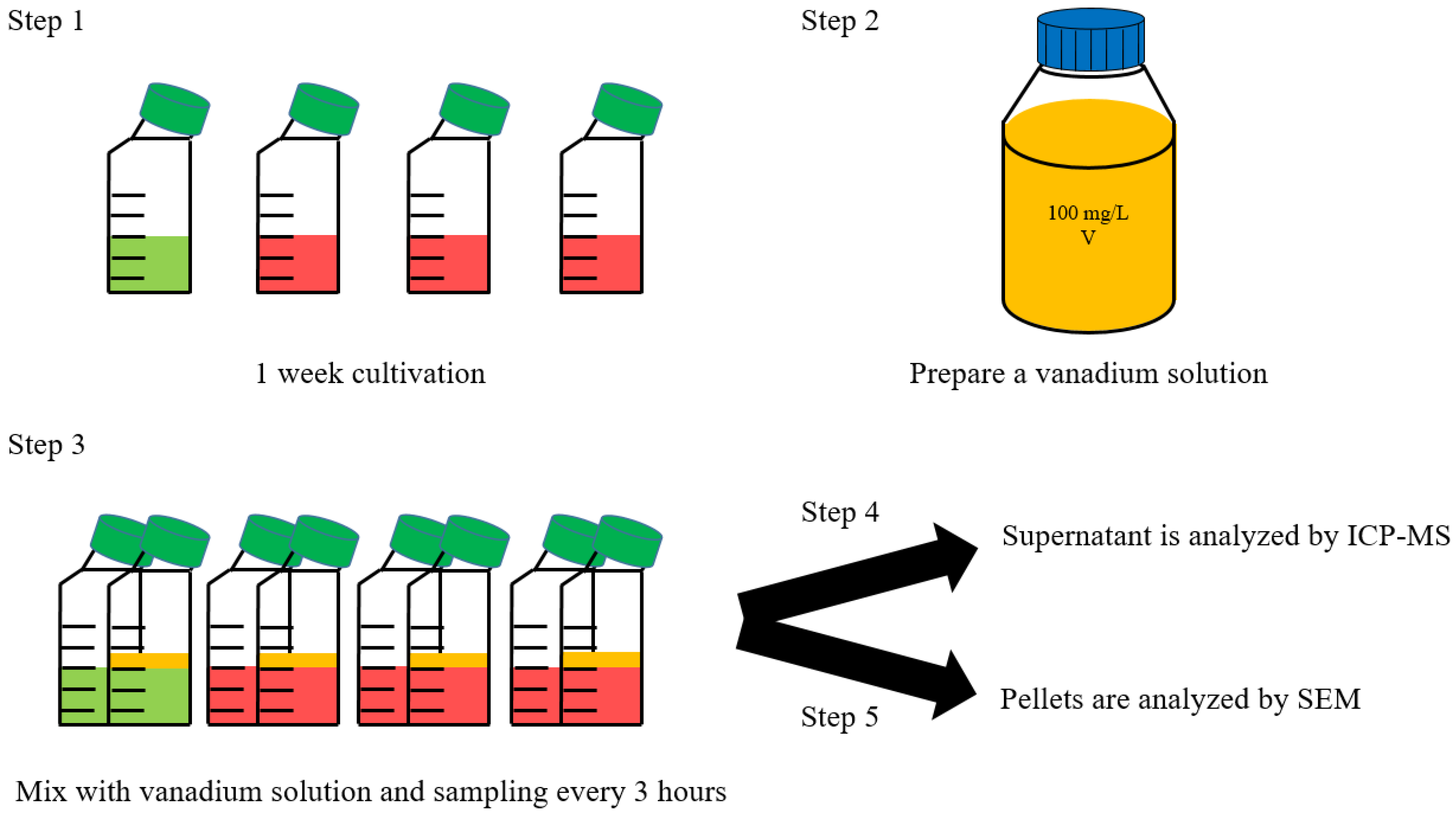
Figure 1. Schematic diagram of scientific vanadium pentoxide ion recycling process.
Energies 15 04467 g001
Figure 2. Vanadium ion concentration in the supernatant by ICP−MS after mixing with vanadium solution.
Energies 15 04467 g002
Figure 3. Scanning electron micrograph of N. oculata (a,b) and H. circularisquama (c,d) under normal and vanadium solution mixed conditions, respectively.
Energies 15 04467 g003
Table 1. List of various physical and chemical parameters used for cell culture. Basic information applies equally to all species in this study.
Cell ConditionV2O5 Solution Condition
(Vanadium Ion Concentration)
ODCultivation Period
(day)
Temperature
(℃)
pHStock
(mg L−1)
Inoculation
(mg L−1)
pH
0.9–0.957207.510054.3
Table 2. Vanadium concentrations of supernatant and pellet under various methods.
N. oculataVanadium Concentration (mg L−1)
Supernatant0 h4.61 ± 0.11
24 h1.85 ± 0.21
PelletSonication2.57 ± 0.27
Acid * treatment1.01 ± 0.07
Base ** treatment2.34 ± 0.08
* 2N HNO3, ** 2N KOH.
Publisher’s Note: MDPI stays neutral with regard to jurisdictional claims in published maps and institutional affiliations.