Article

Anti-Aging, Anti-Acne, and Cytotoxic Activities of Houttuynia cordata Extracts and Phytochemicals Analysis by LC-MS/MS

by
Santi Phosri
1,
Kanokwan Kiattisin
2,3,
Aekkhaluck Intharuksa
3,
Raveeporn Janon
4,
Tanat Na Nongkhai
4 and
Tinnakorn Theansungnoen
4,5,*
1
Department of Chemical Engineering, Faculty of Engineering, Burapha University, Chonburi 20131, Thailand
2
Innovation Center for Holistic Health, Nutraceuticals, and Cosmeceuticals, Faculty of Pharmacy, Chiang Mai University, Chiang Mai 50200, Thailand
3
Department of Pharmaceutical Sciences, Faculty of Pharmacy, Chiang Mai University, Chiang Mai 50200, Thailand
4
School of Cosmetic Science, Mae Fah Luang University, Chiang Rai 57100, Thailand
5
Green Cosmetic Technology Research Group, School of Cosmetic Science, Mae Fah Luang University, Chiang Rai 57100, Thailand
*
Author to whom correspondence should be addressed.
Cosmetics 2022, 9(6), 136; https://doi.org/10.3390/cosmetics9060136
Submission received: 14 November 2022 / Revised: 3 December 2022 / Accepted: 5 December 2022 / Published: 7 December 2022

Abstract

:
Although many biological properties of Houttuynia cordata have been found, its anti-aging and anti-acne effects have not yet been investigated. This study was aimed to evaluate the in vitro anti-aging and anti-acne activities of H. cordata extracts and their cytotoxic activities and phytochemicals analyzed with liquid chromatography with tandem mass spectrometry (LC-MS/MS). Dried aerial parts of H. cordata were given different extractions. The aqueous and ethanolic extracts obtained were named HCA and HCE, respectively, and used to screen total phenolic and flavonoid contents. In vitro anti-aging, skin-related antimicrobial, scanning electron microscopy (SEM), in vitro cytotoxic, and LC-MS/MS analyses were performed. The total phenolic contents of the HCA and HCE were 5.11 ± 0.25 and 27.02 ± 1.07 mg gallic acid equivalent (GAE)/g dry extract while their total flavonoid contents were 104.94 ± 5.16 and 571.86 ± 2.86 mg quercetin equivalent (QE)/g dry extract, respectively. The HCA and HCE inhibited the activities of collagenase (28.33–46.00%), elastase (30.00–34.33%), and hyaluronidase (93.87–98.72%). The minimum inhibitory concentration (MIC) and minimum bactericidal concentration (MBC) of the HCA against Cutibacterium acnes DMST14916 were 5.77 and 5.77 mg/mL while those of the HCE were 2.47 and 2.47 mg/mL, respectively. Cell collapses of C. acnes after treatment with the extracts were observed with SEM. The HCE was not toxic to macrophages, keratinocytes, and fibroblasts up to 400 mg/mL. The HCA showed toxicity against macrophages at 62.5 mg/mL and both skin cells at 250 mg/mL. The main phytochemicals in the extracts were identified with LC-MS/MS. Phenolic compounds, flavonoids, and flavonoid derivatives in H. cordata extracts could be major phytochemicals to possess a broad spectrum of biological activities including antioxidant, antimicrobial, and anti-aging activities. The findings from this study showed that the HCE has potential anti-aging and anti-acne properties while having non-cytotoxic activities on the immune and skin cells. These results indicate that the extract is probably advantageous in the development of skincare cosmeceutics and beauty treatments.

1. Introduction

Skin health and beauty are important fundamental factors representing overall “well-being” as well as the perception of “health” in humans [1]. Skin aging is a part of the natural age progression for humans and is influenced by endogenous or intrinsic factors, including genetics, cellular metabolism, hormones, and metabolic processes. It is also promoted by exogenous or extrinsic factors, such as light exposure, pollution, ionizing radiation, chemicals, and toxins [2]. The endogenous and exogenous factors cause cumulative structural and physiological alterations as well as progressive changes in the skin layers and even the skin appearance, especially photoaging-damaged skin [3]. With normal body development, acne vulgaris is a common skin disorder that affects approximately 85% of adolescents and may continue to young adulthood [4]. Acne vulgaris can reduce skin appearance, such as permanent physical scarring, leading to lifelong problems regarding self-esteem, depression, and even unemployment [5]. Because of the negative effects of skin aging and acne vulgaris on skin health and beauty, naturally active agents with anti-aging and anti-acne have been discovered and developed during the past few years. Much scientific research has revealed that many plants have great potential to be anti-aging [6,7] and anti-acne [8,9] agents.
Houttuynia cordata Thunb. is a perennial plant spread widely across many countries in Asia. H. cordata has been used as medicine and also as food. The plant is grown and harvested for daily vegetable consumption, particularly in the North and Northeast of Thailand [10]. As a medicinal plant, it has also been studied for a long time. Scientific evidence has revealed the efficacy of H. cordata in traditional Asian medicines. It has long been used to treat many disorders including constipation, hypertension, pneumonia, and hyperglycemia via detoxification, the reduction of heat, and diuretic action [11]. Moreover, a large number of research results have shown that H. cordata extracts have the potential to exhibit a broad spectrum of biological activities. The ethanol extracts of H. cordata induced apoptotic programmed cell death in human primary colorectal cancer cells [12]. The methanolic extract of H. cordata could possess a free radical scavenging activity using 2, 2-diphenyl1-picrylhydrazyl (DPPH), ferric reducing antioxidant power (FRAP), and Trolox equivalent anti-oxidant capacity assays [13]. Water extracts of H. cordata have shown anti-inflammatory activity on lipoteichoic acid (LTA)-induced inflammation in dermal fibroblasts by suppressing the metabolic pathway of tumor necrosis factor-α (TNF-α) [14]. An aqueous extract of H. cordata exhibits immunomodulatory and anti-severe acute respiratory syndrome (SARS) activities [15]. An aqueous extract of H. cordata could inhibit Murine salmonellosis and Salmonella typhimurium that infected the RAW 264.7 macrophage [16]. The ethanolic extract of H. cordata poultice exhibited antibacterial and antibiofilm activities against methicillin-resistant Staphylococcus aureus (MRSA), suggesting that the extract is effective for skin abscesses [17]. Although many biological properties of H. cordata extracts have been found, their anti-aging and anti-acne effects have not yet been investigated. Therefore, the aim of this research was to evaluate the in vitro anti-aging and anti-acne activities of H. cordata extracts, and their cytotoxic activities and phytochemicals are analyzed with LC-MS/MS.

2. Materials and Methods

2.1. Preparation of Plant Extracts

The aerial part of H. cordata was obtained from a garden in Chiang Rai Province, Thailand, and air-dried. The dried part was ground into a powder with an electric blender (EM-11, Sharp, Bangkok, Thailand). The condition of plant extraction was modified slightly from the previous study [18]. H. cordata powder (10 g) was added to DI water or absolute ethanol (100 mL) and stirred continuously in an incubator shaker (Thermo Fisher Scientific, Waltham, MA, USA) 24 h at room temperature. Subsequently, the mixtures were centrifuged at 5000 rpm for 10 min and filtered through Whatman filter paper No.1. The extracts were subjected to evaporation with a rotary evaporator and dried further at 45 °C overnight. Finally, the aqueous H. cordata (HCA) and ethanolic H. cordata (HCE) extracts were weighed and stored at −20 °C until use.

2.2. Determination of Total Phenolic Content

The total phenolic content in H. cordata extracts was evaluated using the Folin-Ciocalteu method [19]. Gallic acid was used as a standard and prepared in distilled water with various concentrations from 0.01–0.12 mg/mL. H. cordata extracts were prepared in absolute ethanol. All standard and extract samples (0.5 mL) were added individually to 2 mL of diluted Folin-Ciocalteu reagent. Then, 7.5% sodium carbonate (4 mL) was mixed gently with all reactions and incubated for 30 min at room temperature. The absorbance of all reactions was measured with a UV-Vis spectrophotometer (UV-2600i, Shimadzu, Kyoto, Japan) at a wavelength of 765 nm. The total phenolic content of H. cordata extracts was compared with the standard equilibration curve of gallic acid and then expressed as mg GAE/g dry weight extract sample. All experiments were performed in triplicate.

2.3. Determination of Total Flavonoid Content

The total flavonoid contents in H. cordata extracts were assayed by the aluminum chloride colorimetric method [19]. Quercetin was used as a standard and prepared in absolute ethanol with various concentrations from 0.1–2.6 mg/mL. The H. cordata extracts were also prepared in absolute ethanol, and 1 mL of the extracts was added to distilled water and adjusted to a final volume of 5 mL. After that, 0.3 mL of 5% NaNO2 solution was added and incubated for 5 min. The mixture was mixed with 10% AlCl3 solution and incubated for 1 min, then added to 2 mL of 1 M NaOH and adjusted immediately with distilled water to a final volume of 10 mL. The absorbance of all reactions was measured with a UV-Vis spectrophotometer (UV-2600i, Shimadzu, Kyoto, Japan) at a wavelength of 510 nm. The total flavonoid content of H. cordata extracts was compared with the standard equilibration curve of quercetin and then expressed as mg QE/g dry weight extract sample. All experiments were performed in triplicate.

2.4. DPPH Radical Scavenging Assay

The antioxidant activity of H. cordata extracts (HCA and HCE) was measured by DPPH radical scavenging assay with slight modification from a previous study [20]. DPPH solution (0.2 mM) was prepared in absolute ethanol. Various concentrations of Trolox standard were used at 6.25, 12.5, 50, and 100 µg/mL, and the concentration of the HCA and HCE was used at 2 mg/mL. A test solution of each Trolox standard or extracts (10 µL) was added to 100 µL of DPPH solution. The mixture of each reaction was stirred gently and kept completely in the dark at room temperature for 30 min. The absorbance of the reactions was measured at 517 nm using a microplate reader (SpectraMax M3, Molecular Devices, San Jose, CA, USA). All reactions were performed in triplicate. The scavenging effect of the extracts on DPPH radicals was calculated using the following equation: % DPPH scavenged = ((A − B)/A) × 100, where A is the absorbance of the DPPH solution and B is the absorbance of Trolox standard or extracts. A calibration curve was plotted with % DPPH scavenged versus the various concentrations of the Trolox standard.

2.5. ABTS Radical Scavenging Assay

The ABTS radical scavenging assay for H. cordata extracts (HCA and HCE) was modified slightly from a previous study [21]. The ABTS cationic radical (ABTS•+) solution was produced by mixing 7 mM ABTS and 2.45 Potassium persulphate at a ratio 1:1 and then incubating at room temperature for 12–16 h. The ABTS+ solution was diluted with acetate buffer and reached 0.074 ± 0.02 at 734 nm. The various concentrations of Trolox standard used were 6.25, 12.5, 50, 100, and 200 µg/mL, and the concentration of the HCA and HCE used was 2 mg/mL. A test solution of each Trolox standard or extracts (10 µL) was added to 200 µL of the diluted ABTS•+ solution. The mixture for each reaction was stirred gently and incubated at room temperature for 7 min. The absorbance of the reactions was measured at 734 nm using a microplate reader (SpectraMax M3, Molecular Devices, San Jose, CA, USA). All reactions were performed in triplicate. The scavenging effect of the extracts on ABTS radicals was calculated using the following equation: % ABTS•+ scavenged = ((A − B)/A) × 100, where A is the absorbance of ABTS•+ solution and B is the absorbance of Trolox standard or extracts. A calibration curve was plotted with % ABTS•+ scavenged versus the various concentrations of Trolox standard.

2.6. Ferric Reducing Antioxidant Power Assay

The FRAP assay for H. cordata extracts (HCA and HCE) was modified slightly from a previous study [22]. The fresh working solution of the FRAP was prepared by mixing 20 mL of acetate buffer (300 mmol/L), 2 mL of TPTZ solution (10 mmol/L), and 2 mL of ferric chloride hexahydrate solution (20 mmol/L). The various concentrations of Trolox standard used were 6.25, 12.5, 50, 100, 200, and 400 µg/mL, and the concentration of the HCA and HCE used was 2 mg/mL. A test solution of each Trolox standard or extracts (10 µL) was added to 140 µL of the fresh FRAP solution. The mixture for each reaction was stirred gently and incubated in the dark at room temperature for 30 min. The absorbance of the reactions was measured at 593 nm using a microplate reader (Molecular Devices SpectraMax ABS). All reactions were performed in triplicate. The FRAP efficacy of reactions was calculated using the following equation: % FRAP = ((A − B)/A) × 100, where A is the absorbance of the FRAP solution and B is the absorbance of Trolox standard or extracts. A calibration curve was plotted with % FRAP versus the various concentrations of the Trolox standard.

2.7. Collagenase Inhibitory Activity

The collagenase inhibitory activity of H. cordata extracts was assayed according to the method with a slight modification [23]. The inhibition of collagenase activity was evaluated via the reduction of the FALGPA substrate, and Epigallocatechin gallate (EGCG) was used as a positive control. The enzymatic activity of collagenase was determined before testing each experiment, with only more than 90% of the activity taken for evaluation of the collagenase inhibitory activity. In the experiment, 10 µL of 1 mg/mL H. cordata extracts was added to 40 µL of 0.25 units/mL collagenase from Clostridium histolyticum (ChC—E.C. 3.3.23.3) in tricine buffer, pH 7.5 for 15 min at room temperature. The reaction was mixed with 50 µL of 2 mM FALGPA in tricine buffer, immediately measured at a wavelength of 335 nm, and continuously measured for 20 min with kinetic mode using a microplate reader (SPECTROstar Nano, BMG Labtech, Aylesbury, Buckinghamshire, UK). All experiments were performed in triplicate. The collagenase inhibition of H. cordata extracts was determined by the following equation: %Collagenase inhibition = ((A − B)/A) × 100, where A is the rate of the reaction containing collagenase and substrate and B is the rate of the reaction containing a sample, collagenase, and substrate.

2.8. Elastase Inhibitory Activity

The elastase inhibitory activity of H. cordata extracts was assayed according to the method with a slight modification [23]. The inhibition of elastase activity was evaluated via the increase of the product generated from the reaction of the enzyme and AAAVPN as a substrate. Epigallocatechin gallate (EGCG) was used as a positive control. The enzymatic activity of elastase was determined before testing each experiment, and only more than 90% enzyme activity were taken for the experiment. In the experiment, 50 µL of 1 mg/mL H. cordata extracts was added to 25 µL of 4.4 mM AAAVPN in Tris-HCl buffer, pH 8.0, and then incubated for 20 min at room temperature. After that, the reaction was mixed with 25 µL of 0.05 units/mL elastase from a porcine pancreas (E.C. 3.4.21.11) in Tris-HCl buffer, immediately measured at a wavelength of 410 nm, and continuously measured for 20 min with kinetic mode using a microplate reader (SPECTROstar Nano, BMG Labtech, Aylesbury, UK). All experiments were performed in triplicate. The elastase inhibition of H. cordata extracts was determined by the following equation: %Elastase inhibition = ((A − B)/A) × 100, where A is the rate of the reaction containing elastase and substrate and B is the rate of the reaction containing sample, elastase, and substrate.

2.9. Hyaluronidase Inhibitory Assay

The hyaluronidase inhibitory activity of H. cordata extracts was evaluated by the turbidimetric assay according to the sigma protocol with a slight modification [24]. Ascorbic acid was used as a positive control. The enzymatic activity of hyaluronidase was determined before testing each experiment, and only more than 90% enzyme activity was taken for the experiment. In the experiment, 50 µL of 1 mg/mL H. cordata extracts was added to 100 µL of 800 units/mL hyaluronidase from bovine testes (E.C. 3.2.1.3.5) in enzyme diluent. The mixture was incubated for 10 min at 37.5 °C in a water bath (Memmert GmbH, Büchenbach, Roth, Germany). After that, 100 µL of 0.03% (w/v) hyaluronic acid in phosphate buffer, pH 5.35 was added and then incubated for 45 min. After incubation, the mixture was mixed with 1 mL of bovine serum albumin acid solution containing 24 mM sodium acetate, 79 mM acetic acid and 0.1% (w/v) bovine serum albumin (pH 3.75). The reaction was incubated for 10 min at room temperature, and then measured with a microplate reader (SPECTROstar Nano, BMG Labtech, Aylesbury, UK). All experiments were performed in triplicate. The hyaluronidase inhibition of H. cordata extracts was determined by the following equation: %Hyaluronidase inhibition = ((−(B − C))/A) × 100, where A is the absorbance of the mixture containing hyaluronidase, hyaluronic acid, and bovine serum albumin acid solution; B is the absorbance of the mixture containing a sample, hyaluronidase, hyaluronic acid, and bovine serum albumin acid solution; and C is the absorbance of the mixture without hyaluronidase.

2.10. Antimicrobial Activity Assay

The minimal inhibitory concentrations (MICs) of H. cordata extracts were determined with a broth microdilution technique with modifications [25,26]. The HCA and HCE were diluted to 10% DMSO with two-fold dilution and used in the final concentrations between 0.33 and 11.54 mg/mL. C. acnes DMST 14916 was cultured in brain heart infusion broth as well as Staphylococcus aureus TISTR 746 and Staphyloccus epidermidis TISTR 2141 were cultured in nutrient broth to log phase (OD600 nm = 0.5–0.8). The bacterial culture was diluted to 0.001 of OD600 nm (approximate cell density of 106 CFU/mL). The diluted bacterial solution (100 µL) was mixed with 30 µL of the HCA or HCE samples in a 96-well plate. The sample tests were incubated under anaerobic conditions at 37 °C for 72 h (C. acnes) and under aerobic condition at 37 °C for 16–20 h (S. aureus and S. epidermidis). After that, 10 µL of 0.06% resazurin dye was added to each sample in a 96-well plate and incubated further for 4 h. The MICs were defined as the lowest concentration of the HCA or HCE at which C. acnes showed no color change after incubation. The MBCs were investigated by the colony-count technique [27]. The samples of at least the MICs in the 96-well plate were taken to streak on brain heart infusion agar and incubated at the same conditions for 72 h (C. acnes) and for 16–20 h (S. aureus and S. epidermidis). The MBCs were defined as the lowest concentration of the HCA or HCE at which any colony of C. acnes could not grow.

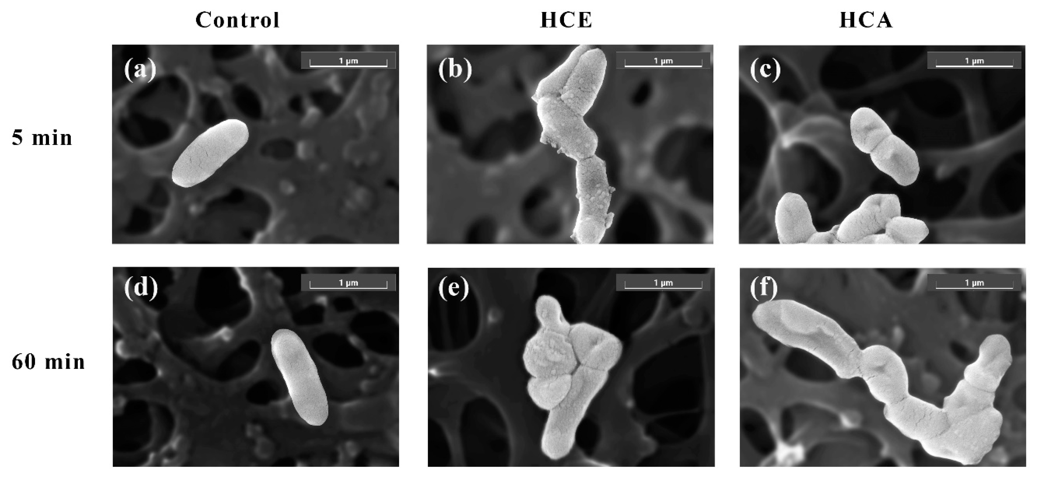
2.11. Scanning Electron Microscopy

The antimicrobial effects of the HCA and HCE on C. acnes were investigated by scanning electron microscopy (SEM) with a modified method of Lau et al., 2004 [28]. C. acnes were cultured in brain heart infusion broth under an anaerobic condition at 37 °C and reached the log phase. The cell culture was centrifuged at 8000 rpm for 3 min, washed twice with PBS (pH 7.4), and had its cells harvested. The bacterial cells were resuspended and diluted in new brain–heart infusion broth to a cell density of ~108 cells/mL (OD600 nm = 0.1). The C. acnes suspensions were incubated in the presence of the HCA or HCE at 10× MICs for 5 min and 60 min under the same conditions. Cells without any extract treatment were controls. Each bacterial solution (100 µL) was applied to a cellulose acetate membrane for 60 min and fixed on the membrane with 300 µL of 2.5% glutaraldehyde overnight. The bacterial cells on the membrane were dehydrated with a series of ethanol solutions starting at 30%, 50%, 70%, 90%, 90%, 100%, and 100%, respectively, for 30 min in each solution. The bacterial cells were further dried, coated with gold-palladium, and observed with a Field Emission Scanning Electron microscope (TESCAN MIRA4, Brno, Czech Republic).

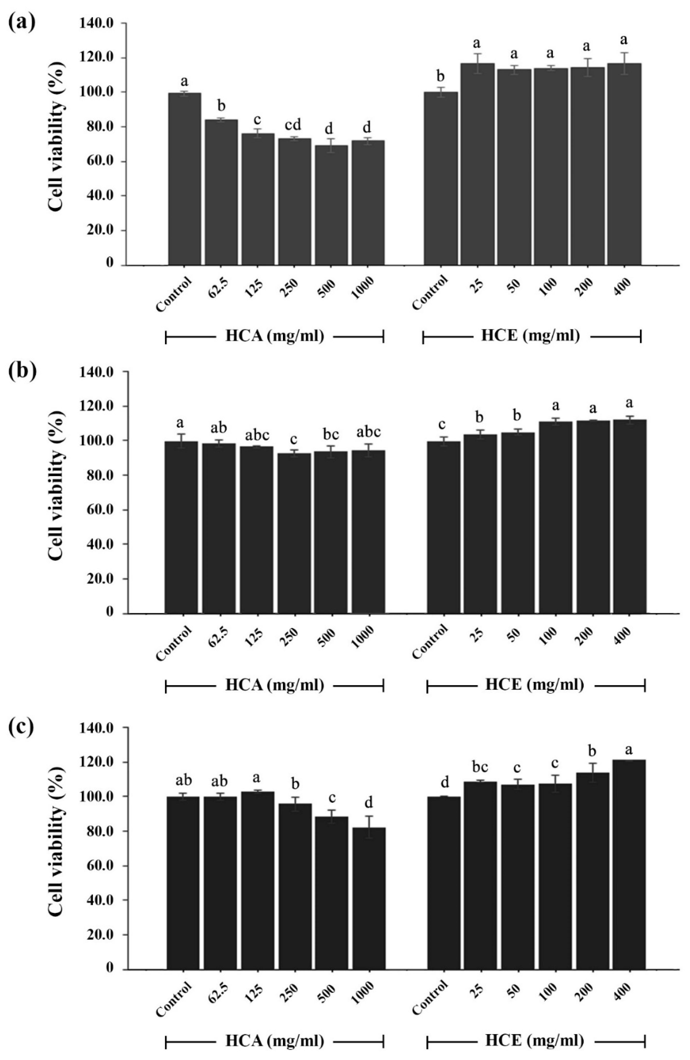
2.12. Cytotoxic Assays on Mouse Immune and Human Skin Cells

The cytotoxic activities of the HCA and HEA were evaluated on human cell lines with an MTT assay with a slight modification of the previous study [29]. The sample concentrations of the HCA and HCE were prepared in the range of 62.5–1000 mg/mL and 25–400 mg/mL, respectively. Mouse macrophage (RAW 264.7 cell line) and human skin cells (HaCaT and NHDF cell lines) were seeded onto 96-well plates at 5 × 10⁴ cells/well in new DMEM medium and 1 × 10⁴ cells/well in new RPMI−1640 medium for 24-h incubation, respectively. Cells were treated with the HCA or HCE and incubated under a humidified atmosphere of 5% CO2 at 37 °C for 24 h. Then, a 150-µL MTT solution (0.5 mg/mL) was added to each sample well and incubated for 1 h. DMSO (100 µL) was added to each well to solubilize the formazan salt product. The solubilized solutions were measured at 550 nm, and the cell viability of each sample was evaluated by comparing the absorbance of extracts-treated and untreated cells.

2.13. LC-MS/MS

Phytochemical compounds in H. cordata extracts were determined with LC-MS/MS as well as performed by an expert of the Scientific and Technological Instruments Center, Mae Fah Luang University. For sample preparation, the HCA and HCE were diluted in absolute methanol and filtered through a WHATMAN 0.2 µm NYL filter. The final concentration of samples was 100 ppm. For LC acquisition, the LC system was operated using a combination of Agilent 1290 UHPLC (Agilent Technologies, Santa Clara, CA, USA) and Agilent Poroshell EC-C18 (2.1 mm × 150 mm, 2.7 µm) column with Agilent Poroshell EC-C18 (4.6 mm × 5 mm, 2.7 µm) guard column. The injection volume was 1 mL. The column and autosampler temperatures were set at 30 °C and 5 °C, respectively. The LC separation was achieved at a flow rate of 0.2 mL/min under a time and gradient program in which mobile phase A was composed of 0.1% (v/v) formic acid in water and mobile phase B was composed of 0.1% (v/v) formic acid in acetonitrile. The gradient program was applied as follows: t = 0 min, 5% B; t = 1 min, 5% B; t = 10 min, 17% B; t = 13 min, 17% B; t = 20 min, 100% B; t = 25 min, 100% B. The gradient was taken to re-equilibrium as follows: t = 27 min, 5% B, and hold 5% B for 6 min. The stop time was 33 min. For MS acquisition, the data was obtained with an Agilent G6454B Q-TOF Mass Spectrometry (Agilent Technologies, California, USA). The instrument parameters of the mass spectrometry were set initially by controlling the gas temperature at 300 °C, gas flow at the rate of 11 l/min, nebulizer pressure at 45 psig, sheath gas temperature at 300 °C, and sheath gas flow at 12 l/min. The operation system of the mass spectrometer used a Dual AJS ESI ion source. The capillary voltage (VCap) and nozzle voltage of both ion modes was set at 4000 V and 500 V, respectively. The voltages of the fragmentor, skimmer1, and OctopoleRFPeak were kept at 150 V, 65 V, and 750 V, respectively. The scan range was adjusted to 100–1100 m/z at the scan rate of 1.00 spectra/sec. Agilent reference mass solution containing internal reference compounds with m/z 112.98558700 and m/z 1033.98810900 for the negative mode, and m/z 121.05087300 and m/z 922.00979800 for the positive mode was infused into the MS by Agilent 1260 isocratic pump. For MS/MS acquisition, the data was achieved by setting at the same scan source parameters of the MS acquisition, as well as at 10, 20, or 40 eV of collision energy.

3. Results and Discussion

3.1. Yield Percentage, Total Phenolic Content, and Total Flavonoid Content of H. cordata Extracts

The aerial part of H. cordata was extracted with DI water and ethanol. After solvent removal and sample drying, the crude aqueous extract (HCA) and ethanolic extract (HCE) were obtained for the evaluation of yield, total phenolic content, and total flavonoid content. The yields of the HCA and HCE were 9.68 ± 3.10% and 3.42 ± 1.97%, respectively. The total phenolic contents in the HCA and HCE were 5.11 ± 0.25 and 27.02 ± 1.07 mg GAE/g dry extract while their total flavonoid contents were 104.94 ± 5.16 and 571.86 ± 2.86 mg QE/g dry extract, respectively (Table 1). The results indicate that the aqueous extraction obtained a higher yield from H. cordata than the ethanol extraction. The phenolic and flavonoid contents of the aqueous extract (HCA) were lower than those of the ethanol extract (HCE). These results are similar to a previous study. The extraction yield increased with increasing polarity of the solvents. The aqueous extraction was less effective to extract phenolic and flavonoid compounds than the ethanol extraction [30].

3.2. Antioxidant Activities of H. cordata Extracts

DPPH, ABTS•+, and FRAP assays are members of the single electron transfer (SET) assays. They are simple, cost-effective, easily interpreted, and display either reduction capacity (FRAP) or direct free radical inhibition (DPPH and ABTS•+) [31]. The methods were used to assay the antioxidant activities of H. cordata extracts (HCA and HCE). As shown in Table 1, the antioxidant efficacies of the HCA and HCE to scavenge DPPH radicals were 11.44 ± 0.13 and 13.55 ± 0.42 mg Trolox/g dry extract while their efficacies against ABTS radicals were 41.98 ± 3.90 and 103.46 ± 5.15 mg Trolox/g dry extract, respectively. The FRAP value of the HCE (136.88 ± 4.71 mg Trolox/g dry extract) was higher than that of the HCA (46.11 ± 1.20 mg Trolox/g dry extract). The results indicate that both the HCA and HCE possessed a broad spectrum of antioxidant abilities on DPPH, ABTS, and FRAP. The HCE had greater antioxidant activities than the HCA. It is believed that the phenolic and flavonoid compounds in the HCE are higher than in the HCA. As in previous studies, plants are a wonderful source of diverse bioactives and antioxidants. Much research has shown many antioxidants in plants including phenolics, flavonoids, tannins, vitamins, quinines, coumarins, lignans, and ligins [32,33]. Furthermore, to reveal a broad spectrum of antioxidant activities, H. cordata extracts could be taken for additional antioxidant assays, such as lipid peroxidation, superoxide radical scavenging activity, hydroxyl radical scavenging activity, and hydrogen peroxide scavenging activity assays [34].

3.3. Anti-Aging Activity of H. cordata Extracts

Aging of the skin is primarily related to the reduction in the levels of the extracellular matrix (ECM), in which the main structural components are collagen, elastin, and hyaluronic acid. The reductions in the structural components are caused by aging-related enzymes including collagenase, elastase, and hyaluronidase [23]. Collagenase is responsible for collagen degradation, which leads to reduced skin elasticity and tensile strength of the skin [35]. Elastase, a member of the chymotrypsin family of protease, is responsible primarily for the breakdown of elastin [23]. Hyaluronidase is the key enzyme that controls the turnover of hyaluronic acid in human skin as well as the retention of skin moisture [36].
To prevent the skin from aging, natural phytochemical sources are desirable. In this study, the anti-aging properties of H. cordata extracts were evaluated via the inhibition against collagenase, elastase, and hyaluronidase. The results of extracts against collagenase activity are shown in Table 2. At the 1 mg/mL of samples, the inhibitory activities of the HCA and HCE against collagenase were 46.00 ± 3.61% and 28.33 ± 5.13%, respectively, while the activity of EGCG was 23.67 ± 5.86%. The HCA and HCE showed an elastase inhibitory activity of 34.33 ± 1.49% and 30.00 ± 3.38%, respectively, compared to the activity of EGCG (33.02 ± 1.65%) at 1 mg/mL. The HCA and HCE exhibited strong hyaluronidase inhibitory activity of 98.72 ± 0.38% and 93.87 ± 1.85%, respectively, while ascorbic acid had an activity of 91.61 ± 0.35%. The findings show that the HCA and HCE have the potential to inhibit skin aging-related enzymes, collagenase, elastase, and hyaluronidase. The results presumably suggest that H. cordata extracts may contribute a good anti-wrinkle activity by preventing the degradation of collagen, elastin, and hyaluronic acid in the skin dermis [37,38].

3.4. Antimicrobial Activity of H. cordata Extracts

The antimicrobial activities of H. cordata extracts against acne-related pathogens including C. acnes DMST 14916, S. aureus TISTR 746, and S. epidermidis TISTR 2141 were determined using the broth-microdilution method. The MICs and MBCs of the extracts were evaluated for the determination of their antimicrobial efficacies (Table 3). The HCA and HCE did not inhibit S. aureus and S. epidermidis but could inhibit C. acnes. The MICs and MBCs of the HCA against C. acnes were found at the same concentration at 5.77 mg/mL while those of the HCE were both at 2.47 mg/mL. The results indicate that the HCE obtained from the ethanolic extraction revealed higher antimicrobial activity against C. acnes than the HCA. From a previous study, the flower of Mesua ferrea (M. ferrea) was extracted by ethanolic extraction. The M. ferrea flower extract showed bacteriostatic efficacy against C. acnes with MIC and MBC of 3.12 and 25.00 mg/mL, respectively [39]. The ethanolic extract of cinnamon bark was reported to inhibit C. acnes. The MIC of cinnamon bark extract was 256 µg/mL [40].
The effects of the HCA and HCE on cell surfaces of C. acnes were further investigated with scanning electron microscopy (SEM). The results are shown in Figure 1. Control cells were C. acnes treated without any extract and had smooth cell surfaces without any obvious damage for 5 min and 60 min incubation (Figure 1a,d). As shown in Figure 1b,c, the HCE and HCA could not cause bleb formation but induced slight cell shrinkages and collapses on bacterial cells for 5 min incubation. After 60 min incubation with the HCE and HCA, it was clear that the morphologies of C. acnes possessed visible, extreme damage with cell shrinkage and collapse (Figure 1e,f). These results indicate that the HCA and HCE can inhibit C. acnes by interrupting the cell surface of the bacteria. From a previous study, Surian (Toona sinensis) leaf extract and their fractions showed anti-acne activity. Among the fractions, fraction 3 could cause cellular destruction of C. acne observed with scanning electron microscopy [41].

3.5. Cytotoxic Activity of H. cordata Extracts

The cytotoxicity of H. cordata extracts was determined on RAW 264.7 macrophages, HaCaT keratinocytes, and NHDF fibroblasts. Cells treated without any extract were taken as 100% (Controls). The effects of H. cordata extracts on RAW 264.7 cells are shown in Figure 2a. The HCE could promote cell viability to be higher than the control, and the percentage of cell viability was significantly increased at 25 mg/mL (117.49 ± 5.84%). The HCA caused toxicity on RAW 264.7 cells and significantly decreased at 62.5 mg/mL (84.64 ± 1.03%). The results indicate that there was no cytotoxicity of the HCE on macrophages, while that of the HCA was only slight. In a previous study, Antirrhinum majus extraction was performed with ethanol. The cytotoxic effect of A. majus extract was tested on RAW 264.7 cells. The cell viability was not significantly altered by the extract at concentrations of 0–300 µg/mL [42]. Moreover, the aqueous, acetone, and methanol extracts of Celosia argentea flowering stage were used for cytotoxic evaluation on RAW 264.7 macrophages. The flowering stage extracts of C. argentea were not toxic to the cells [43].
The effects of H. cordata extracts on HaCaT cells are shown in Figure 2b. The HCE had the potential to promote the cell viability of HaCaT cells at the initial concentration of 25 mg/mL (103.82 ± 2.65%) and greatly increased at 100 mg/mL (111.60 ± 2.17%). The cell viability of the HaCaT cells treated with the HCA decreased slightly but significantly at 250 mg/mL (93.04 ± 2.29%). The effects of H. cordata extracts on NHDF cells are shown in Figure 2c. The HCE could increase the cell viability of NHDF cells at the initial concentration of 25 mg/mL (108.90 ± 0.60%) and increased constantly until 400 mg/mL (121.04 ± 0.13%), while the HCA could decrease the cell viability at the initial concentration of 500 mg/mLµ (88.55 ± 3.63%). The results indicate that the HCE obtained from ethanolic extraction showed no toxicity to keratinocytes and fibroblasts, but the HCA obtained from aqueous extraction caused slight toxicities. From previous studies, the vitro-cytotoxicity of tuber and leaf extracts from Helianthus tuberosus L. was evaluated on HaCaT and BJ fibroblast. The extracts were made with the ultrasound-assisted extraction method. The tuber extract at all tested concentrations did not show a toxic effect on HaCaT cells but exhibited a positive effect on fibroblast growth. The smallest concentration of the leaf extract exhibited proliferative properties on both HaCat and BJ cells, but when the dose increased, the cell viabilities decreased [44].

3.6. Phytochemical Analysis of H. cordata Extracts

The HCE was taken for phytochemical analysis with LC-QTOF-MS/MS. Twenty compounds were found in the HCE and are shown in Table 4. These compounds included sugars (sucrose and mannobiose), amino acid derivatives (N-octanoyl-L-valine), benzenesulfonic acid (N-undecylbenzenesulfonic acid and 4-dodecylbenzenesulfonic acid), alkyl-phenylketones (fluanisone), sodium tetradecyl sulfate, fatty acids and fatty acid derivatives (floionolic acid and lauroyl diethanolamide), ubiquinones (myrsinone), phenolic compounds (quinic acid, chlorogenic acid, and 4-O-caffeoylquinic acid), flavonoids and flavonoid derivatives (rutin, kaempferol 7-rhamnoside, quercetin 3-O-glucoside and quercitrin), uvaribonone, safghanoside D, and acetic acid—5-(2-methoxypropan-2-yl)-2-methylphenol (1/1). From a literature review, among these compounds, quinic acid [45,46], chlorogenic acid [47,48], 4-O-caffeoylquinic acid [49,50], and rutin [49,51] exhibited both antioxidant and antimicrobial activities, while quercetin 3-O-glucoside [52], quercitrin [52], and safghanoside D [53] possessed only antioxidant activity. Chlorogenic acid showed the potential to reduce ear swelling, redness, and erythema skin in the ears of ICR mice induced by C. acnes [54]. Chlorogenic acid was reported to be a strong matrix metalloproteinase-9 inhibitor [55]. Rutin could reduce the elevated matrix metalloproteinase-9 level in rat models [56]. Rutin also inhibited the hyaluronidase reaction [57]. A molecular docking study was made of flavonoid compounds for possible matrix metalloproteinase-13 inhibition. Nine flavonoids including rutin, nicotiflorin, orientin, vitexin, apigenin-7-glucoside, quercitrin, isoquercitrin, quercitrin-3-rhamnoside, and vicenin-2 had considerable estimated free energy of a binding and inhibition constant [58].

4. Conclusions

In conclusion, this study revealed that H. cordata extracts (HCA and HCE) obtained from aqueous and ethanol extraction, respectively, provide anti-C. acnes, antioxidant, and anti-aging activity against collagenase, elastase, and hyaluronidase. The in vitro cytotoxicity of the HCE against human skin cells and macrophages was not found while the HCA showed the cytotoxicity in a dose-dependent manner. In LC-MS/MS analysis, the phenolic compounds, flavonoids, and flavonoid derivatives in the HCE could be major phytochemicals to possess a broad spectrum of biological activities including antioxidant, antimicrobial, and anti-aging activities. Chlorogenic acid was expected to show the antimicrobial activity against C. acnes. Rutin, chlorogenic acid, and quercetin derivatives contributed to exhibit the anti-aging activity against collagenase, elastase, and hyaluronidase. In conclusion, the ethanolic extract of H. cordata is a promising plant-based ingredient that could be used to further develop skin care, beauty, and cosmeceutic products, especially topical anti-acne and antiaging products. For further studies, the project may perform other antioxidant assays based on different types of free radicals, such as lipid peroxidation, superoxide radical scavenging activity, hydroxyl radical scavenging activity, and hydrogen peroxide scavenging activity assays, for showing a broad spectrum of antioxidant efficacy of H. cordata extracts. Moreover, we attempt to develop topical formulations containing H. cordata extracts, evaluate the efficacy and stability of the formulations, determine the skin irritation, and measure the satisfaction with human subjects in clinical trials.

Author Contributions

Conceptualization, T.T.; methodology, S.P., K.K., R.J., T.N.N. and T.T.; software, S.P. and T.T.; validation, S.P. and T.T.; formal analysis, S.P., A.I. and T.T.; investigation, S.P. and T.T.; data curation, S.P. and T.T.; writing—original draft preparation, S.P., A.I., K.K. and T.T.; writing—review and editing, S.P. and T.T.; supervision, T.T.; project administration, T.T.; funding acquisition, T.T. All authors have read and agreed to the published version of the manuscript.

Funding

This work was financially supported by the Thailand Science Research and Innovation and Mae Fah Luang University [Grant no. 652A02033].

Institutional Review Board Statement

Not applicable.

Informed Consent Statement

Not applicable.

Data Availability Statement

Not applicable.

Acknowledgments

This work conducted and supported by the School of Cosmetic Science, Mae Fah Luang University. We would like to thank the Protein and Proteomics Research Center for Commercial and Industrial Purposes (ProCCI), Department of Biochemistry, Faculty of Science, Khon Kaen University, Thailand. We appreciate Tinnakorn Bang-ngoen and Poramet Nachalaem of the Scientific and Technological Instruments Center, Mae Fah Luang University, for providing access to the scanning electron microscopic and mass spectrometric equipment.

Conflicts of Interest

The authors declare no conflict of interest.

References

  1. Ganceviciene, R.; Liakou, A.I.; Theodoridis, A.; Makrantonaki, E.; Zouboulis, C.C. Skin anti-aging strategies. Derm. Endocrinol. 2012, 4, 308–319. [Google Scholar] [CrossRef] [PubMed] [Green Version]
  2. Cevenini, E.; Invidia, L.; Lescai, F.; Salvioli, S.; Tieri, P.; Castellani, G.; Franceschi, C. Human models of aging and longevity. Expert Opin. Biol. Ther. 2008, 8, 1393–1405. [Google Scholar] [CrossRef] [PubMed]
  3. Gromkowska-Kępka, K.J.; Puścion-Jakubik, A.; Markiewicz-Żukowska, R.; Socha, K. The impact of ultraviolet radiation on skin photoaging—Review of in vitro studies. J. Cosmet. Dermatol. 2021, 20, 3427–3431. [Google Scholar] [CrossRef] [PubMed]
  4. Williams, H.C.; Dellavalle, R.P.; Garner, S. Acne vulgaris. Lancet. 2012, 379, 361–372. [Google Scholar] [CrossRef]
  5. Mallon, E.; Newton, J.N.; Klassen, A.; Stewart-Brown, S.L.; Ryan, T.J.; Finlay, A.Y. The quality of life in acne: A comparison with general medical conditions using generic questionnaires. Br. J. Dermatol. 1999, 140, 672–676. [Google Scholar] [CrossRef]
  6. Binic, I.; Lazarevic, V.; Ljubenovic, M.; Mojsa, J.; Sokolovic, D. Skin ageing: Natural weapons and strategies. Evid. Based Complement. Alternat. Med. 2013, 2013, 827248. [Google Scholar] [CrossRef] [Green Version]
  7. Rungruang, R.; Ratanathavorn, W.; Boohuad, N.; Selamassakul, O.; Kaisangsri, N. Antioxidant and anti-aging enzyme activities of bioactive compounds isolated from selected Zingiberaceae plants. Agric. Nat. Resour. 2021, 55, 153–160. [Google Scholar]
  8. Sinha, P.; Srivastava, S.; Mishra, N.; Yadav, N.P. New perspectives on antiacne plant drugs: Contribution to modern therapeutics. Biomed. Res. Int. 2014, 2014, 301304. [Google Scholar] [CrossRef] [Green Version]
  9. Nasri, H.; Bahmani, M.; Shahinfard, N.; Moradi Nafchi, A.; Saberianpour, S.; Rafieian Kopaei, M. Medicinal plants for the treatment of acne vulgaris: A review of recent evidences. Jundishapur J. Microbiol. 2015, 8, e25580. [Google Scholar] [CrossRef] [Green Version]
  10. Woranam, K.; Senawong, G.; Utaiwat, S.; Yunchalard, S.; Sattayasai, J.; Senawong, T. Anti-inflammatory activity of the dietary supplement Houttuynia cordata fermentation product in RAW264.7 cells and wistar rats. PLoS ONE 2020, 15, e0230645. [Google Scholar] [CrossRef] [Green Version]
  11. Wang, J.H.; Bose, S.; Shin, N.R.; Chin, Y.W.; Choi, Y.H.; Kim, H. Pharmaceutical impact of Houttuynia cordata and metformin combination on high-fat-diet-induced metabolic disorders: Link to intestinal microbiota and metabolic endotoxemia. Front. Endocrinol. 2018, 9, 620. [Google Scholar] [CrossRef] [PubMed]
  12. Lai, K.-C.; Chiu, Y.-J.; Tang, Y.-J.; Lin, K.-L.; Chiang, J.-H.; Jiang, Y.-L.; Jen, H.-F.; Kuo, Y.-H.; Agamaya, S.; Chung, J.-G.; et al. Houttuynia cordata Thunb extract inhibits cell growth and induces apoptosis in human primary colorectal cancer. Anticancer Res. 2010, 30, 3549–3556. [Google Scholar]
  13. Li, H.B.; Wonga, C.C.; Chenga, K.W.; Chena, F. Antioxidant properties in vitro and total phenolic contents in methanol extracts from medicinal plants. LWT 2008, 41, 385–390. [Google Scholar] [CrossRef]
  14. Jee, Y.C.; Jung, A.L.; Jee, B.L.; Sook, J.Y.; Seung, C.L. Anti-inflammatory activity of Houttuynia cordata against lipoteichoic acid-induced inflammation in human dermal fibroblasts. Chonnam Med. J. 2010, 46, 140–147. [Google Scholar]
  15. Lau, K.-M.; Lee, K.-M.; Koon, C.-M.; Cheung, C.S.-F.; Lau, C.-P.; Ho, H.-M.; Lee, M.Y.-H.; Au, S.W.-N.; Cheng, C.H.-K.; Lau, C.B.-S.; et al. Immunomodulatory and anti-SARS activities of Houttuynia cordata. J. Ethnopharmacol. 2008, 118, 79–85. [Google Scholar] [CrossRef]
  16. Kim, G.S.; Kim, D.H.; Lim, J.J.; Lee, J.J.; Han, D.Y.; Lee, W.M.; Jung, W.C.; Min, W.G.; Gil Won, C.; Rhee, M.H.; et al. Biological and antibacterial activities of the natural herb Houttuynia cordata water extract against the intracellular bacterial pathogen Salmonella within the RAW 264.7 macrophage. Biol. Pharm. Bull. 2008, 31, 2012–2017. [Google Scholar] [CrossRef] [Green Version]
  17. Sekita, Y.; Murakami, K.; Yumoto, H.; Mizuguchi, H.; Amoh, T.; Ogino, S.; Matsuo, T.; Miyake, Y.; Fukui, H.; Kashiwada, Y. Anti-bacterial and anti-inflammatory effects of ethanol extract from Houttuynia cordata poultice. Biosci. Biotechnol. Biochem. 2016, 80, 1205–1213. [Google Scholar] [CrossRef] [Green Version]
  18. Kumnerdkhonkaen, P.; Saenglee, S.; Asgar, M.A.; Senawong, G.; Khongsukwiwat, K.; Senawong, T. Antiproliferative activities and phenolic acid content of water and ethanolic extracts of the powdered formula of Houttuynia cordata Thunb. fermented broth and Phyllanthus emblica Linn. fruit. BMC Complement Altern. Med. 2018, 18, 130. [Google Scholar] [CrossRef] [Green Version]
  19. Poomanee, W.; Wattananapakasem, I.; Panjan, W.; Kiattisin, K. Optimizing anthocyanins extraction and the effect of cold plasma treatment on the anti-aging potential of purple glutinous rice (Oryza sativa L.) extract. Cereal Chem. 2021, 98, 571–582. [Google Scholar] [CrossRef]
  20. Rajurkar, N.S.; Hande, S.M. Estimation of phytochemical content and antioxidant activity of some selected traditional Indian medicinal plants. Indian J. Pharm. Sci. 2011, 73, 146–151. [Google Scholar] [CrossRef] [Green Version]
  21. Phosri, S.; Mahakunakorn, P.; Lueangsakulthai, J.; Jangpromma, N.; Swatsitang, P.; Daduang, S.; Dhiravisit, A.; Thammasirirak, S. An investigation of antioxidant and anti-inflammatory activities from blood components of crocodile (Crocodylus siamensis). Protein J. 2014, 33, 484–492. [Google Scholar] [CrossRef]
  22. Ishtiaq, S.; Ahmad, M.; Hanif, U.; Akbar, S.; Mehjabeen; Kamran, S.H. Phytochemical and in vitro antioxidant evaluation of different fractions of Amaranthus graecizans subsp. silvestris (Vill.). Brenan. Asian Pac. J. Trop. Med. 2014, 7S1, S342–S347. [Google Scholar] [CrossRef] [Green Version]
  23. Thring, T.S.; Hili, P.; Naughton, D.P. Anti-collagenase, anti-elastase and anti-oxidant activities of extracts from 21 plants. BMC Complement Altern. Med. 2009, 9, 27. [Google Scholar] [CrossRef] [PubMed]
  24. Widowati, W.; Rani, A.P.; Hamzah, R.A.; Arumwardana, S.; Afifah, E.; Kusuma, H.S.W.; Rihibiha, D.D.; Nufus, H.; Amalia, A. Antioxidant and antiaging assays of Hibiscus sabdariffa extract and its compounds. Nat. Prod. Sci. 2017, 23, 192–200. [Google Scholar] [CrossRef] [Green Version]
  25. Wu, Y.; Qiang, Y.; Cao, K.; Zhang, W.; Zhang, G. Inhibitory effect of the antimicrobial peptide BLP-7 against Propionibacterium acnes and its anti-inflammatory effect on acne vulgaris. Toxicon 2020, 184, 109–115. [Google Scholar] [CrossRef]
  26. Pata, S.; Yaraksa, N.; Daduang, S.; Temsiripong, Y.; Svasti, J.; Araki, T.; Thammasirirak, S. Characterization of the novel antibacterial peptide Leucrocin from crocodile (Crocodylus siamensis) white blood cell extracts. Dev. Comp. Immunol. 2011, 35, 545–553. [Google Scholar] [CrossRef]
  27. Elshikh, M.; Ahmed, S.; Funston, S.; Dunlop, P.; McGaw, M.; Marchant, R.; Banat, I.M. Resazurin-based 96-well plate microdilution method for the determination of minimum inhibitory concentration of biosurfactants. Biotechnol. Lett. 2016, 38, 1015–1019. [Google Scholar] [CrossRef] [Green Version]
  28. Lau, S.K.; Woo, P.C.; Woo, G.K.; Fung, A.M.; Wong, M.K.; Chan, K.-M.; Tam, D.M.; Yuen, K.-Y. Eggerthella hongkongensis sp. nov. and Eggerthella sinensis sp. nov., two novel Eggerthella species, account for half of the cases of Eggerthella bacteremia. Diagn. Microbiol. Infect. Dis. 2004, 49, 255–263. [Google Scholar] [CrossRef] [PubMed]
  29. Jangpromma, N.; Suttee, K.; Phosri, S.; Theansungnoen, T.; Lueangsakulthai, J.; Payoungkiattikun, W.; Daduang, S.; Klaynongsruang, S. Antioxidant properties of Crocodylus siamensis blood components on H2O2-induced human skin fibroblast cells. Chiang Mai J. Sci. 2018, 45, 1359–1371. [Google Scholar]
  30. Do, Q.D.; Angkawijaya, A.E.; Tran-Nguyen, P.L.; Huynh, L.H.; Soetaredjo, F.E.; Ismadji, S.; Ju, Y.-H. Effect of extraction solvent on total phenol content, total flavonoid content, and antioxidant activity of Limnophila aromatica. JFDA 2014, 22, 296–302. [Google Scholar] [CrossRef] [Green Version]
  31. Wootton-Beard, P.C.; Moran, A.; Ryan, L. Stability of the total antioxidant capacity and total polyphenol content of 23 commercially available vegetable juices before and after in vitro digestion measured by FRAP, DPPH, ABTS and Folin–Ciocalteu methods. Int. Food Res. J. 2011, 44, 217–224. [Google Scholar] [CrossRef]
  32. Cai, Y.; Luo, Q.; Sun, M.; Corke, H. Antioxidant activity and phenolic compounds of 112 traditional Chinese medicinal plants associated with anticancer. Life Sci. 2004, 74, 2157–2184. [Google Scholar] [CrossRef] [PubMed]
  33. Amarowicz, R.; Pegg, R.B.; Rahimi-Moghaddam, P.; Barl, B.; Weil, J.A. Free-radical scavenging capacity and antioxidant activity of selected plant species from the Canadian prairies. Food Chem. 2004, 84, 551–562. [Google Scholar] [CrossRef]
  34. Mazumder, K.; Nabila, A.; Aktar, A.; Farahnaky, A. Bioactive variability and in vitro and in vivo antioxidant activity of unprocessed and processed flour of nine cultivars of Australian lupin species: A comprehensive substantiation. Antioxidants 2020, 9, 282. [Google Scholar] [CrossRef] [PubMed]
  35. Roy, A.; Sahu, R.K.; Matlam, M.; Deshmukh, V.K.; Dwivedi, J.; Jha, A.K. In vitro techniques to assess the proficiency of skin care cosmetic formulations. Pharmacogn. Rev. 2013, 7, 97–106. [Google Scholar]
  36. ChChaiyana, W.; Anuchapreeda, S.; Punyoyai, C.; Neimkhum, W.; Lee, K.-H.; Lin, W.-C.; Lue, S.-C.; Viernstein, H.; Mueller, M. Ocimum sanctum Linn. as a natural source of skin anti-ageing compounds. Ind. Crops Prod. 2019, 127, 217–224. [Google Scholar] [CrossRef]
  37. Li, T.S.C.; Mazza, G.; Cottrell, A.A.C.; Gao, L. Ginsenosides in roots and leaves of American ginseng. Agric. Food Chem. 1996, 44, 717–720. [Google Scholar] [CrossRef]
  38. Jiratchayamaethasakul, C.; Ding, Y.; Hwang, O.; Im, S.; Jang, Y.; Myung, S.; Lee, J.M.; Kim, H.; Ko, S.; Lee, S. In vitro screening of elastase, collagenase, hyaluronidase, and tyrosinase inhibitory and antioxidant activities of 22 halophyte plant extracts for novel cosmeceuticals. Fish Aquat. Sci. 2020, 23, 6. [Google Scholar] [CrossRef] [Green Version]
  39. Nakyai, W.; Pabuprapap, W.; Sroimee, W.; Ajavakom, V.; Yingyongnarongkul, B.; Suksamrarn, A. Anti-acne vulgaris potential of the ethanolic extract of Mesua ferrea L. flowers. Cosmetics 2021, 8, 107. [Google Scholar] [CrossRef]
  40. Julianti, E.; Rajah, K.K.; Fidrianny, I. Antibacterial activity of ethanolic extract of cinnamon bark, honey, and their combination effects against acne-causing bacteria. Sci. Pharm. 2017, 85, 19. [Google Scholar] [CrossRef]
  41. Febriyani, E.; Falah, S.; Andrianto, D.; Lastini, T. Identification of active compounds and anti-acne activity from extracts and fractions of surian (Toona sinensis) leaves planted in Sumedang, West Java, Indonesia. Biodiversitas 2018, 19, 1406–1412. [Google Scholar] [CrossRef]
  42. Jang, M.; Hwang, I.; Hwang, B.; Kim, G. Anti-inflammatory effect of Antirrhinum majus extract in lipopolysaccharide-stimulated RAW 264.7 macrophages. Food Sci. Nutr. 2020, 8, 5063–5070. [Google Scholar] [CrossRef]
  43. Adegbaju, O.D.; Otunola, G.A.; Afolayan, A.J. Anti-inflammatory and cytotoxic evaluation of extracts from the flowering stage of Celosia argentea. BMC Complement Med. Ther. 2020, 20, 152. [Google Scholar] [CrossRef]
  44. Nizioł-Łukaszewska, Z.; Furman-Toczek, D.; Zagórska-Dziok, M. Antioxidant activity and cytotoxicity of Jerusalem artichoke tubers and leaves extract on HaCaT and BJ fibroblast cells. Lipids Health Dis. 2018, 17, 280. [Google Scholar] [CrossRef] [Green Version]
  45. Pero, R.W.; Lund, H.; Leanderson, T. Antioxidant metabolism induced by quinic acid. Increased urinary excretion of tryptophan and nicotinamide. Phytother. Res. 2009, 23, 335–346. [Google Scholar] [CrossRef]
  46. Bai, J.; Wu, Y.; Bu, Q.; Zhong, K.; Gao, H. Comparative study on antibacterial mechanism of shikimic acid and quinic acid against Staphylococcus aureus through transcriptomic and metabolomic approaches. LWT 2022, 153, 112441. [Google Scholar] [CrossRef]
  47. Tomac, I.; Šeruga, M.; Labuda, J. Evaluation of antioxidant activity of chlorogenic acids and coffee extracts by an electrochemical DNA-based biosensor. Food Chem. 2020, 325, 126787. [Google Scholar] [CrossRef] [PubMed]
  48. Lou, Z.; Wang, H.; Zhu, S.; Ma, C.; Wang, Z. Antibacterial activity and mechanism of action of chlorogenic acid. J. Food Sci. 2011, 76, M398–M403. [Google Scholar] [CrossRef] [PubMed]
  49. Ganzon, J.G.; Chen, L.G.; Wang, C.C. 4-O-Caffeoylquinic acid as an antioxidant marker for mulberry leaves rich in phenolic compounds. J. Food Drug Anal. 2018, 26, 985–993. [Google Scholar] [CrossRef] [PubMed]
  50. Guzman, J.D. Natural cinnamic acids, synthetic derivatives and hybrids with antimicrobial activity. Molecules 2014, 19, 19292–19349. [Google Scholar] [CrossRef]
  51. Ganeshpurkar, A.; Saluja, A.K. The pharmacological potential of rutin. Saudi Pharm. J. 2017, 25, 149–164. [Google Scholar] [CrossRef] [PubMed] [Green Version]
  52. Li, X.; Jiang, Q.; Wang, T.; Liu, J.; Chen, D. Comparison of the antioxidant effects of quercitrin and isoquercitrin: Understanding the role of the 6″-OH group. Molecules 2016, 21, 1246. [Google Scholar] [CrossRef] [PubMed]
  53. Macahig, R.A.; Harinantenaina, L.; Matsunami, K.; Otsuka, H.; Takeda, Y.; Shinzato, T. Secoiridoid and iridoid glucosides from the leaves of Fraxinus griffithii. J. Nat. Med. 2010, 64, 1–8. [Google Scholar] [CrossRef] [PubMed]
  54. Luo, J.; He, W.; Li, X.; Ji, X.; Liu, J. Anti-acne vulgaris effects of chlorogenic acid by anti-inflammatory activity and lipogenesis inhibition. Exp. Dermatol. 2021, 30, 865–871. [Google Scholar] [CrossRef] [PubMed]
  55. Jin, U.-H.; Lee, J.-Y.; Kang, S.-K.; Kim, J.-K.; Park, W.-H.; Kim, J.-G.; Moon, S.-K.; Kim, C.-H. A phenolic compound, 5-caffeoylquinic acid (chlorogenic acid), is a new type and strong matrix metalloproteinase-9 inhibitor: Isolation and identification from methanol extract of Euonymus alatus. Life Sci. 2005, 77, 2760–2769. [Google Scholar] [CrossRef] [PubMed]
  56. Jang, J.W.; Lee, J.K.; Hur, H.; Kim, T.W.; Joo, S.P.; Piao, M.S. Rutin improves functional outcome via reducing the elevated matrix metalloproteinase-9 level in a photothrombotic focal ischemic model of rats. J. Neurol. Sci. 2014, 339, 75–80. [Google Scholar] [CrossRef]
  57. Lee, J.H.; Kim, G.H. Evaluation of antioxidant and inhibitory activities for different subclasses flavonoids on enzymes for rheumatoid arthritis. J. Food Sci. 2010, 75, H212–H217. [Google Scholar] [CrossRef]
  58. Taherkhani, A.; Moradkhani, S.; Orangi, A.; Jalalvand, A.; Khamverdi, Z. Molecular docking study of flavonoid compounds for possible matrix metalloproteinase-13 inhibition. J. Basic Clin. Physiol. Pharmacol. 2020, 32, 1105–1119. [Google Scholar] [CrossRef]
Figure 1. SEM images of C. acnes after incubation with H. cordata extracts at 10× MICs for 5 min and 60 min. Control cells were treated without any sample for 5 min (a) and 60 min (d). Bacterial cells were treated with HCE or HCA for 5 min ((b) or (c)) and 60 min ((e) or (f)), respectively.
Cosmetics 09 00136 g001
Figure 2. Cytotoxicity activities of H. cordata extracts with different concentrations determined by MTT assay. Mouse macrophages (RAW 264.7) (a), human keratinocytes (HaCat) (b), human fibroblasts (NHDF) (c) were used for the assay. The different letters among the bars indicate that the values of the bars are significantly different (p <0.05). On the other hand, if some bars have same letters, it indicates that the values of the bars are not different (p > 0.05).
Cosmetics 09 00136 g002
Table 1. Total phenolic content, total flavonoid content, and antioxidant activities of H. cordata extracts.
ExtractTotal Phenolic Content
(mg GAE/g Dry Extract wt)
Total Flavonoid Content
(mg QE/g Dry Extract wt)
DPPH
(mg Trolox/g Dry Extract wt)
ABTS
(mg Trolox/g Dry Extract wt)
FRAP
(mg Trolox/g Dry Extract wt)
HCA5.11 ± 0.25104.94 ± 5.1611.44 ± 0.1341.98 ± 3.9046.11 ± 1.20
HCE27.02 ± 1.07571.86 ± 2.8613.55 ± 0.42103.46 ± 5.15136.88 ± 4.71
HCA: aqueous extract of H. cordata; HCE: ethanolic extract of H. cordata; GAE: gallic acid equivalents; QE: quercetin equivalents; wt: weight; DPPH: 2,2-diphenyl-1-picrylhydrazyl; ABTS: 2,2′-azino-bis (3-ethylbenzthiazoline-6-sulphonic acid); FRAP: ferric reducing antioxidant power. The values are from triplicate experiments.
Table 2. Skin aging-related enzyme inhibitory effects of H. cordata extracts at 1 mg/mL.
SampleCollagenase Inhibition (%)Elastase Inhibition (%)Hyaluronidase Inhibition (%)
HCA46.00 ± 3.6134.33 ± 1.4998.72 ± 0.38
HCE28.33 ± 5.1330.00 ± 3.3893.87 ± 1.85
EGCG23.67 ± 5.8633.02 ± 1.65NA
Ascorbic acidNANA91.61 ± 0.35
HCA: aqueous extract of H. cordata; HCE: ethanolic extract of H. cordata; EGCG: epigallocatechin gallate; NA: Not assayed. The values are from triplicate experiments.
Table 3. Antimicrobial activities of H. cordata extracts against acne-related bacteria.
ExtractC. acnes
DMST 14916
S. aureus
TISTR 746
S. epidermidis
TISTR 2141
MIC (mg/mL)MBC (mg/mL)MIC (mg/mL)MBC (mg/mL)MIC (mg/mL)MBC (mg/mL)
HCA5.775.77NDNDNDND
HCE2.472.47NDNDNDND
HCA: aqueous extract of H. cordata; HCE: ethanolic extract of H. cordata; C. acnes: Cutibacterium acnes; S. aureus: Staphylococcus aureus; S. epidermidis: Staphylococcus epidermidis; MIC: minimum inhibitory concentration; MBC: minimum bactericidal concentration; ND: Not detected. The values are from triplicate experiments.
Table 4. Phytochemicals identified in HCE by LC-QTOF-MS-MS data in the negative ion mode.
No.RT
(Min)
m/zTentative IdentificationFormulaMassIon SpeciesMS/MS FragmentsMatch Score
12.009341.1094SucroseC12 H22 O11342.1166[M-H]119.0344, 179.0561, 341.109198.34
22.017387.1147MannobioseC12 H22 O11342.1164[M+HCOO]179.0563, 321.0142, 341.1088, 387.108499.21
32.039191.0571Quinic acidC7 H12 O6192.0643[M-H]127.0410, 191.056895.55
48.064353.0882Chlorogenic acidC16 H18 O9354.0954[M-H]135.0468, 191.0561, 353.089793.31
511.000353.08744-O-Caffeoylquinic acidC16 H18 O9354.0948[M-H]173.0455, 353.089097.36
616.571609.146RutinC27 H30 O16610.1536[M-H]151.0044, 301.0357, 609.146098.68
716.643431.0988Kaempferol 7-rhamnosideC21 H20 O10432.1059[M-H]311.0567, 431.098798.63
816.826463.0891Quercetin 3-O-glucosideC21 H20 O12464.0961[M-H]151.0042, 300.0278, 463.087797.74
917.076723.5029UvaribononeC39 H68 O8664.489[M+CH3COO]677.4972, 723.502890.52
1017.178447.0943QuercitrinC21 H20 O11448.1013[M-H]178.9991, 301.0355, 447.093996.76
1118.718242.1767N-Octanoyl-L-valineC13 H25 N O3243.1839[M-H]181.1602, 242.177198.82
1219.554311.1696N-Undecylbenzenesulfonic acidC17 H28 O3 S312.1768[M-H]183.0130, 311.169495.66
1319.592401.1871FluanisoneC21 H25 F N2 O2356.189[M+HCOO]146.0612, 280.1339, 401.186492.89
1419.633293.1797Sodium tetradecyl sulfateC14 H30 O4 S294.1869[M-H]114.9871, 293.179498.39
1519.724645.2179Safghanoside DC32 H38 O14646.2249[M-H]397.1642, 465.1552, 645.217396.31
1619.789325.18474-Dodecylbenzenesulfonic acidC18 H30 O3 S326.1921[M-H]183.0122, 325.184596.54
1719.796331.2496Floionolic acidC18 H36 O5332.2568[M-H]185.1199, 265.1410, 331.249197.07
1819.996332.2446Lauroyl diethanolamideC16 H33 N O3287.2465[M+HCOO]158.1190, 286.239896.68
1920.165293.1769MyrsinoneC17 H26 O4294.1841[M-H]236.1065, 293.177494.58
2020.624239.1288Acetic acid—5-(2-methoxypropan-2-yl)-2-methylphenol (1/1)C13 H20 O4240.136[M-H]195.1395, 239.126698.42
Publisher’s Note: MDPI stays neutral with regard to jurisdictional claims in published maps and institutional affiliations.

