Next Article in Journal
Clinical Experience with a Less Invasive Surgical Transparotid Approach and Trapezoidal Plate for Neck and Base Condylar Fractures: A Retrospective Study
Previous Article in Journal
Endoscopic Dacryocystorhinostomy Following Nasolacrimal Drainage System Trauma and Medial Orbital Wall Reconstruction
 
 
Craniomaxillofacial Trauma & Reconstruction is published by MDPI from Volume 18 Issue 1 (2025). Previous articles were published by another publisher in Open Access under a CC-BY (or CC-BY-NC-ND) licence, and they are hosted by MDPI on mdpi.com as a courtesy and upon agreement with Sage.
Article

Reconstruction of Composite Mandible Defects Using a Cellular Bone Allograft and Soft Tissue Free Flap Coverage

by
Kevin J. Carlson
1,2,
Robert M. Liebman
2,3,
Matthew J. Bak
2,
William M. Dougherty
2 and
Jonathan R. Mark
2,*
1
Division of Otolaryngology–Head and Neck Surgery, Department of Surgery, University of Wisconsin, Madison, WI, USA
2
Department of Otolaryngology–Head and Neck Surgery, Eastern Virginia Medical School, 600 Gresham Drive, Suite 1100, Norfolk, VA 23507, USA
3
Memorial Health University Medical Center, Savannah, GA, USA
*
Author to whom correspondence should be addressed.
Craniomaxillofac. Trauma Reconstr. 2024, 17(4), 63; https://doi.org/10.1177/19433875241237920
Submission received: 1 November 2023 / Revised: 1 December 2023 / Accepted: 1 January 2024 / Published: 6 March 2024

Abstract

Study Design: Retrospective case series. Objective: Cellular bone allografts (CBAs) contain the components of a successful bone graft with no autologous component and have been used extensively outside the head and neck. Descriptions of their utilization for mandible reconstruction are limited. We review our experience utilizing a CBA, with no autologous component, for the reconstruction of mandible defects. Methods: Patients undergoing reconstruction of a composite mandible defect with a CBA, no added autologous component, within a patient-specific graft cage and soft tissue free flap coverage were retrospectively identified. Graft survival and defect management are assessed and results of post-operative imaging reported. Results: Five subjects, aged 23–56 years, underwent reconstruction of mandible defects with the described technique. Defects resulted from gunshot wounds in 4 patients and the composite resection of a low-grade malignancy in one. The defect was definitively managed in 4 subjects, 3 of which had post-operative imaging demonstrating bone formation. The fifth experienced graft failure after developing an orocutaneous fistula and was successful salvaged with an osteocutaneous fibula free flap. Conclusions: Our early experience is promising that a CBA, with no autologous component, and soft tissue free flap coverage can be used for the reconstruction of composite mandible defects in select patients.

Introduction

Osteocutaneous free tissue transfer provides a durable option for the reconstruction of almost all composite defects of the mandible [1]. However, this technique is associated with significant donor site morbidity, prolonged operative times, and soft tissue is tethered to the bone graft, limiting reconstructive options. Non-vascularized autologous and allogenic bone grafts offer an alternative for select defects when post-operative radiation is not anticipated. Use for larger composite mandible defects has been limited, and most surgeons have relied on an autologous component [2].
Cellular bone allografts (CBAs) have been utilized extensively outside the head and neck and can satisfy the characteristics of a successful bone graft with no autologous component [3,4,5,6]. There have been minimal reports of using these grafts for mandible reconstruction [7,8]. We initially utilized CBAs as an additive in cases of mandibular non-union after traumatic injuries, similar to that described by Ryu et al. [7]. The benefits of PSIs for mandible reconstruction have been well described [9,10], and others have demonstrated how implants can be designed to house nonvascularized bone grafts as cages [7,11]. We subsequently expanded our use of CBAs to treat select larger, segmental, mandible defects by housing them within a patient-specific graft cage, covered with a soft tissue free flap. In this case series, we retrospectively assess our experience with this technique in 5 patients.

Methods

A retrospective series of patients undergoing reconstruction of composite mandible defects at a single institution between January 1, 2020 and January 1, 2023 is presented. Subjects over the age of 18 years were identified via review of the authors operative schedules and were included if they had undergone repair of a composite mandible defect using a CBA, with no autologous component, within a patient-specific graft cage and soft tissue free flap coverage. The primary outcome measure was definitive management of the composite defect and bone formation assessed using postoperative computed tomography (CT) imaging. Institutional review board approval was obtained from Eastern Virginia Medical School (Norfolk, VA, USA).

Patient Selection and Management
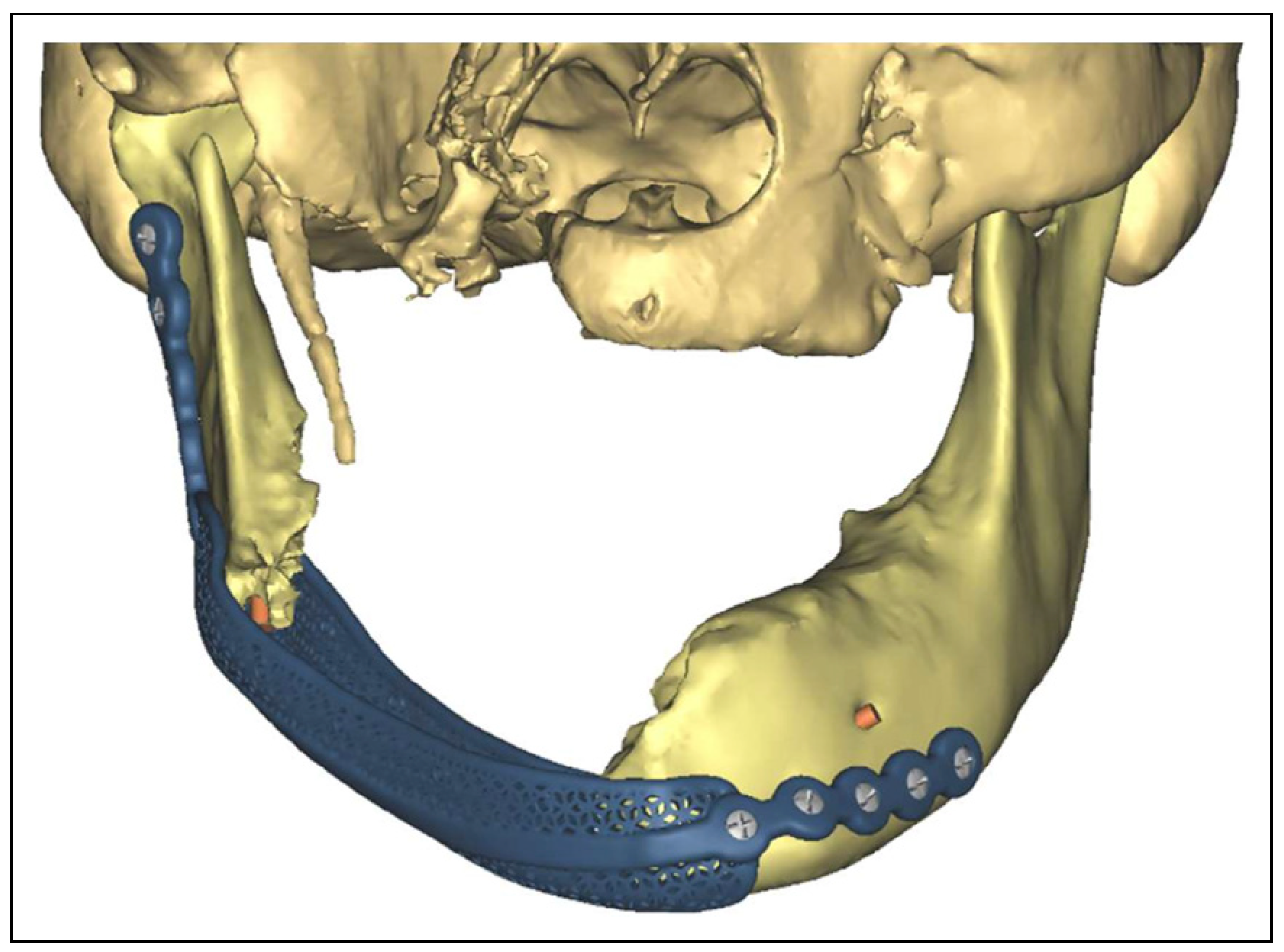
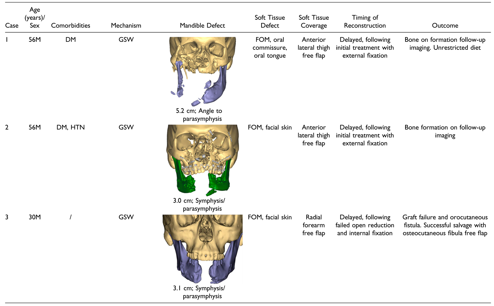
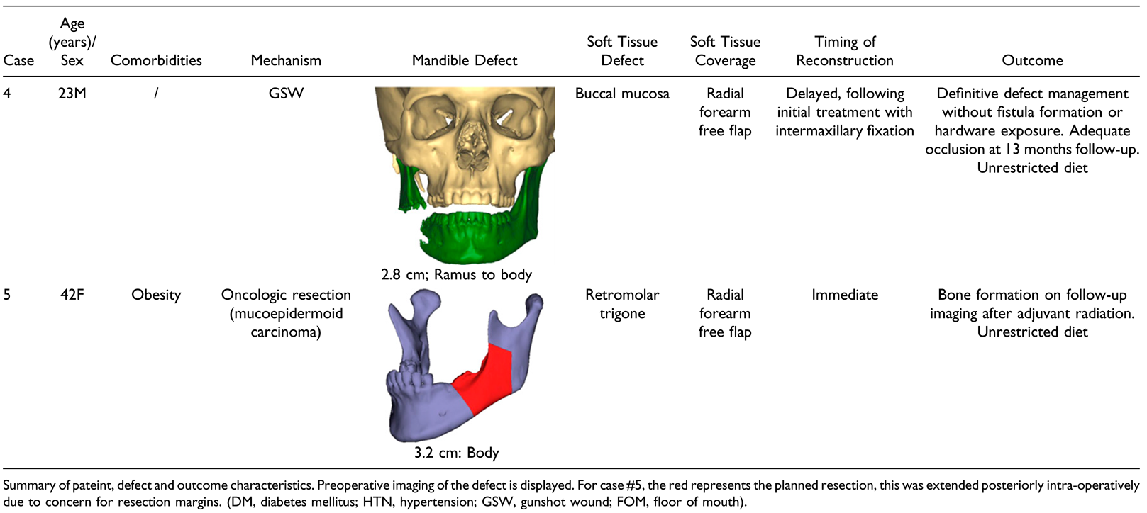
Initial patient selection focused on traumatic defects as these patients would not be expected to receive radiation to the surgical field and often have minimal medical comorbidities. Our group often manages traumatic composite mandible defects in a staged fashion to allow for some healing of the wound bed secondarily as well as stabilization of other injuries. This practice allowed time for creation of the PSI. Simple continuity defects of the mandible body are often managed with a single segment osteocutaneous free tissue reconstruction, and we focused on defects including the symphyseal region or angle where a patient-specific graft cage might offer improved ease of recreating a natural contour. After some initial success in managing traumatic defects, we briefly expanded application to reconstruction following resection of a low-grade malignancy, in which post-operative radiation was not anticipated (Case 5).
Virtual surgical planning was used to create a patient-specific titanium graft cage (TruMatch ®, DePuy Synthes, Raynham, MA, USA) spanning the anticipated defect (Figure 1). The planned defect included removal of most mono-cortical bone fragments. In defects with large bicortical fragments, a reconstruction bar was planned in continuity with the graft cage (case 2). Each graft cage was designed with placement of at least 4, 2.4 mm bi-cortical screws on either side of the defect. Patient-specific drill guides were designed for appropriate screw and cage placement.
The mandible was exposed intraoperatively via a cervical incision and small bone fragments removed from the defect. Patient-specific drill guides were affixed to proper position of the lateral surface of the mandible with temporary mono-cortical screws and the implant placed with bi-cortical locking screws (Figure 2). The graft cage was tightly filled with ViviGen Formable (LifeNet Health, Virginia Beach, VA, USA) CBA following placement. The amount of graft utilized varied on the size of the defect. Collagen membranes or other implantable products were not used to cover the graft, and recombinant bone morphogenic protein 2 was not used. Rather, the soft tissue reconstruction was placed directly over the implant and graft. This was performed with musculocutaneous (anterior lateral thigh) or fasciocutaneous (radial forearm) free tissue transfer depending on the extent of the defect using standard techniques. A portion of vastus lateralis was harvested with each anterior lateral thigh flap as is the preference of the authors. Postoperative maxillomandibular fixation (MMF) was not performed. If MMF had been placed in a previous, initial surgery, it was removed following implant placement. A minimum 7-day course of post-operative antibiotics with oral flora coverage (Ampicillin/Sulbactam or Clindamycin) and topical chlorhexidine mouth rinse were administered. Patients were kept on a nil per os diet for a minimum of 1 week.

Results

Five patients, aged 23–56 years, underwent reconstruction of a composite mandible defect with the described technique. Patient comorbidities, defect, and surgical characteristics are summarized in Table 1. Each traumatic injury was managed in a staged fashion. Cases 1 and 2 were initially managed with external fixation for 50 and 83 days, respectively, prior to definitive repair. The repair of case 2 was delayed due to a prolonged intensive care unit stay complicated by ventilator-associated pneumonia and cardiac arrest. Case 3 underwent attempted open reduction and internal fixation, augmented with a CBA, closure of the soft tissue defect with adjacent tissue transfer, and postoperative MMF 2 days after the initial injury. After development of an orocutaneous fistula and mandibular nonunion, repair was attempted using a CBA within a PSI and radial forearm free flap (index procedure), 114 days following injury. Case 4 was initially managed with MMF utilizing intermaxillary fixation screws and underwent definitive repair 48 days following injury. Case 5 underwent immediate reconstruction following the composite resection of an intermediate grade mucoepidermoid carcinoma.
Defects were definitively managed in 4 patients. There were no failures of the soft tissue free flap or microvascular revisions. One subject (case 3) developed an orocutaneous fistula and allograft failure despite a viable soft tissue flap and was successfully salvaged with osteocutaneous fibular free flap. During the salvage procedure, the graft cage was empty with no residual CBA or bone formation. There were no other major complications following the index procedure though case 1 underwent 2 soft tissue flap debulking procedures and case 4 underwent excision of a forearm neuroma at the radial forearm donor site. Cases 1 and 2 had post-operative CT imaging at 6- and 5-months post-procedure, respectively, demonstrating bone formation (Figure 3 and Figure 4). Case 4 had no post-operative imaging, through on clinical examination at 13 months post-procedure had no abnormal mandibular mobility or implant exposure. The patient reported adequate occlusion while tolerating an unrestricted diet. Final pathology of the resection specimen of case 5 demonstrated a positive bone margin, and adjuvant radiation was given to 60 Gy over 30 fractions beginning at 50 days postoperatively. Subsequent CT imaging demonstrated stable bone formation at 2 years following reconstruction (Figure 5).

Discussion

Our initial experience is promising that select composite mandible defects maybe definitively managed using a CBA, with no autologous component, when paired with soft tissue free flap coverage. In our series of 5 patients, no autologous component was utilized for the bone graft with only a CBA providing definitive management of the osseous defect in 4 subjects. Three patients had follow-up imaging showing graft take and boney union (Figure 3, Figure 4 and Figure 5).
Owing to its robust ability to restore oromandibular function, osteocutaneous free tissue transfer must be considered the standard of care for large defects and following resection of high-grade malignancies in which post-operative radiation is anticipated. For other defects, including post-traumatic composite defects and following resection of benign neoplasms and low-grade malignancies, surgeons have utilized a variety of non-vascularized bone grafts [12,13]. These techniques may avoid the prolonged operative time and donor site morbidity associated with osteocutaneous free issue transfer. Traditionally, autologous bone harvested from the iliac crest or rib is used [12,14]. The success of this technique is variable and usually limited to smaller defects without a significant soft tissue component.
Allogenic bone is an alternative to autologous grafts, though usually requires the addition of an autologous component. Melville et al. demonstrated the efficacy of allogenic bone grafts, describing a 90% success rate in 31 patients treated for mandibular continuity defects, 5.6 cm in length on average. Their protocol utilized milled allogenic bone chips with the addition of autologous bone marrow and bone morphogenic protein 2 [15]. While this protocol has demonstrated considerable success, it relies on the harvest of autologous bone marrow, a technique many craniofacial surgeons are likely to be unfamiliar with. Many similar reconstructive protocols exist for mandible reconstruction, though often similarly utilize autologous bone marrow aspirate [13]. A completely allogenic graft could allow for more widespread application.
The addition of an autologous component to an allogenic bone graft creates an osteo-inductive, conductive, and integrative environment. CBAs aim to satisfy these characteristics of a successful graft with the inclusion of autologous osteogenic cells within a mixture of demineralized bone and corticocancellous scaffold. There are numerous commercially available CBAs, including Osteocel Plus (NuVasive, San Diego, CA, USA), Trinity Evolution (Orthofix, Lewisville, TX, USA), Map3 Cellular Allogenic Bone Graft (RTI Surgical, Alachua, FL, USA), VIA Graft (Vivex Biologics, Atlanta, GA, USA), PrimaGen Advanced Allograft (Zimmer Biomet, Warsaw, IN, USA), Cellentra Viable Cell Bone Matrix (Zimmer Biomet, Warsaw, IN, USA), and ViviGen and ViviGen Formable (DePuy Synthes, Warsaw, IN, USA/LifeNet Health, Virginia Beach, VA, USA) [6], with the majority of published reports focusing on their use for spine or foot and ankle surgery [3,4,5,6]. The CBA used in this series (Vivigen Formable; LifeNet Health, Virginia Beach, VA, USA) utilizes lineage-committed osteogenic cells within a demineralized bone and cortico-cancellous scaffold. Preclinical research of this product demonstrated osteoblast-related gene and protein expression without immunogenicity [16].
Use of CBAs for mandible reconstruction is limited to case reports. Ryu et al. [7]. described the use of ViviGen (DePuy Synthes, Warsaw, IN, USA) CBA within a patient-specific graft cage for the treatment of mandibular non-union after initial treatment with mandibulomaxillary fixation (MMF). Alfi et al. [8]. described the use of ViviGen Formable (LifeNet Health, Virginia Beach, VA, USA) CBA for immediate reconstruction of a 6.5 cm mandible defect in a pediatric patient after resection of an ossifying fibroma. Osseous consolidation was demonstrated on CT imaging 7 months post-operation. It is difficult to draw conclusions regarding the more widespread applications of CBAs for mandible defects from these limited case reports. While Alfi demonstrates impressive bone formation within a large defect this is within a single pediatric patient, undergoing immediate reconstruction following transoral excision. Conversely, Ryu utilized a CBA for bilateral comminuted mandible fractures resulting from a gun-shot wound. This patient had initially failed treatment with MMF and underwent reconstruction with the CBA 7 weeks after the injury. While the patient reported pain-free function at 7 months post-procedure, available postoperative imaging shows less than complete osseous consolidation within the graft cage.
Soft-tissue microvascular reconstruction has been useful in expanding the application of autologous and allogenic bone grafts for mandible reconstruction. Stoor et al. [11]. covered a patient-specific graft cage filled with a autologous bone graft, BMP-2, and betatricalciumphosphate granules with either an anterior lateral thigh or radial forearm free flap in 12 patients. Schlund and co-authors described the use of a radial forearm free flap to cover a fresh frozen humeral allograft seeded with autologous bone marrow aspirate for treatment of a post-traumatic continuity defect of the mandible body [17]. In their previous referenced series of 31 patients, Melville et al. [15]. utilized a radial forearm free flap for soft tissue reconstruction in 1 patient treated for ameloblastoma and staged boney reconstruction, placing the allograft 4 months later. The vascular environment provided by free tissue to the CBA may allow improved graft take, expanding their use to larger defects.
We found initial utility in the use of CBAs for the treatment of non-union of mandibular fractures, similar to Ryu et al. In this series, we expand on the use of CBAs, reporting our initial experience for the use of mandibular continuity defects, used with soft tissue free flap coverage of the allograft and implant, with promising results. Three dimensional reconstructions of CT images may better demonstrate the outcome of bone formation in the present study, though available imaging which does demonstrate long-term bone formation in cases 1, 2, and 5. While case 4 did not undergo any post-operative imaging, this patient was followed clinically to 13 months post-procedure. Bone formation in this patient cannot be definitively assessed though the technique did allow for definitive management of the composite defect with adequate occlusive and cosmetic outcome.
One subject experienced total loss of the CBA and was successfully salvaged with a fibular free flap (case 3). The initial management of this patient varied from that of others treated for post-traumatic defects (cases 1, 2, and 4), each of which were treated initially with external or mandibular maxillary fixation. Case 3 was initially treated with ORIF and underwent attempted salvage with the described technique after developing mandibular non-union and orocutaneous fistula formation. Findings during the index procedure included purulence within the wound bed, and ongoing infection may have resulted in graft failure and fistula formation.
This study is limited by its size, with only 5 patients undergoing reconstruction using the described technique, one without post-operative imaging. Given this, we are unable to draw larger conclusions regarding the ideal usage of CBAs for mandible reconstruction and comparisons to osteocutaneous free tissue transfer or bone allografts with the addition of an autologous component cannot be made. It is moreover difficult to assess the quality of the regenerated bone in this small case series. The feasibility placement of dental implants after this technique is not known. If such rehabilitation is planned, traditional osteocutaneous free tissue transfer would be recommended. In patients with traumatic defects, the time from injury to definitive reconstruction with this technique may limit its broader application. It is unclear if this could be shortened, though case 3 suggests benefit to a wound that has partially healed secondarily. Moreover, it is unknown how important the cellular component of the CBA is, with some literature suggesting no benefit of a CBA over demineralized bone matrix for spine fusion [6].
Our initial experience suggests that some composite mandible defects can be managed with a CBA and soft-tissue free flap coverage. While we do not believe this to be an appropriate reconstructive option when post-operative radiation is anticipated, or in a patient with a history of radiation treatment, case 5 suggests the durability to the technique. For traumatic defects, we have found staged reconstruction to be most effective, with patients initially managed with external or intermaxillary fixation. Watertight closure of the soft tissue defect and maintenance of a clean wound bed are of the upmost importance, with one patient experiencing graft failure after fistula formation.

Conclusion

Our initial experience is promising that select composite mandible defects from traumatic injuries and resection of low-grade neoplasms can be managed using a CBA, with no autologous component, and soft tissue free flap coverage. Further work with larger patient cohorts is needed to better make comparisons to previously described techniques and understand ideal patient selection and long-term function outcomes.

Funding

The author(s) received no financial support for the research, authorship, and/or publication of this article.

Acknowledgments

The authors would like to acknowledge Thomas Markley of Depuy Synthes Orthopedics for his work obtaining in virtual surgical plans and product information for this publication. This study ws presented at the Triological Society Combined Sections Meeting, Coronado, CA, USA; January 26–28, 2023.

Conflicts of Interest

The author(s) declared no potential conflicts of interest with respect to the research, authorship, and/or publication of this article.

References

  1. Bak, M.; Jacobson, A.S.; Buchbinder, D.; Urken, M.L. Contemporary reconstruction of the mandible. Oral Oncol. 2010, 46, 71–76. [Google Scholar] [CrossRef] [PubMed]
  2. Pu, J.J.; Hakim, S.G.; Melville, J.C.; Su, Y.X. Current trends in the reconstruction and rehabilitation of jaw following ablative surgery. Cancers. 2022, 14, 3308. [Google Scholar] [CrossRef] [PubMed]
  3. Elgafy, H.; Wetzell, B.; Gillette, M.; et al. Lumbar spine fusion outcomes using a cellular bone allograft with lineage-committed bone-forming cells in 96 patients. BMC Musculoskelet Disord. 2021, 22, 699. [Google Scholar] [CrossRef] [PubMed]
  4. Moran, T.E.; Sequeira, S.; Cooper, M.T.; Park, J. A retrospective analysis of outcomes from foot and ankle arthrodesis and open reduction and internal fixation using cellular bone allograft augmentation. Foot Ankle Spec. 2022, 15, 312–320. [Google Scholar] [CrossRef] [PubMed]
  5. Jones, C.P.; Loveland, J.; Atkinson, B.L.; Ryaby, J.T.; Linovitz, R.J.; Nunley, J.A. Prospective, multicenter evaluation of allogeneic bone matrix containing viable osteogenic cells in foot and/or ankle arthrodesis. Foot Ankle Int. 2015, 36, 1129–1137. [Google Scholar] [CrossRef] [PubMed]
  6. Pinter, Z.W.; Elder, B.D.; Kaye, I.D.; et al. A review of commercially available cellular-based allografts. Clin Spine Surg. 2022, 35, E77–E86. [Google Scholar] [CrossRef] [PubMed]
  7. Ryu, B.; Abraham, C.; Polido, W.D. Treatment of mandibular non-union using patient specific crib cage plates and cellular bone allograft: A case report. Craniomaxillofac Trauma Reconstr Open 2021, 6, 247275122110059. [Google Scholar] [CrossRef]
  8. Alfi, D.M.; Hassan, A.; East, S.M.; Gianulis, E.C. Immediate mandibular reconstruction using a cellular bone allograft following tumor resection in a pediatric patient. FACE. 2021, 2, 490–495. [Google Scholar] [CrossRef]
  9. Yang, W.F.; Choi, W.S.; Wong, M.C.; et al. Three-dimensionally printed patient-specific surgical plates increase accuracy of oncologic head and neck reconstruction versus conventional surgical plates: A comparative study. Ann Surg Oncol. 2021, 28, 363–375. [Google Scholar] [CrossRef] [PubMed]
  10. Rana, M.; Chin, S.J.; Muecke, T.; et al. Increasing the accuracy of mandibular reconstruction with free fibula flaps using functionalized selective laser-melted patient-specific implants: A retrospective multicenter analysis. J CranioMaxillo-Fac Surg. 2017, 45, 1212–1219. [Google Scholar] [CrossRef] [PubMed]
  11. Stoor, P.; Suomalainen, A.; Mesima¨ki, K.; Kontio, R. Rapid prototyped patient specific guiding implants in critical mandibular reconstruction. J Cranio-Maxillo-Fac Surg. 2017, 45, 63–70. [Google Scholar] [CrossRef] [PubMed]
  12. Moura, L.B.; Carvalho, P.H.A.; Xavier, C.B.; et al. Autogenous non-vascularized bone graft in segmental mandibular reconstruction: A systematic review. Int J Oral Maxillofac Surg. 2016, 45, 1388–1394. [Google Scholar] [CrossRef] [PubMed]
  13. Ramezanzade, S.; Aeinehvand, M.; Ziaei, H.; et al. Reconstruction of critical sized maxillofacial defects using composite allogeneic tissue engineering: Systematic review of current literature. Biomimetics 2023, 8, 142. [Google Scholar] [CrossRef] [PubMed]
  14. Habib, A.M.A.; Hassan, S.A. The feasibility of rib grafts in long span mandibular defects reconstruction: A long term follow up. J Cranio-Maxillo-Fac Surg. 2019, 47, 15–22. [Google Scholar] [CrossRef] [PubMed]
  15. Melville, J.C.; Tran, H.Q.; Bhatti, A.K.; Manon, V.; Young, S.; Wong, M.E. Is reconstruction of large mandibular defects using bioengineering materials effective? J Oral Maxillofac Surg. 2020, 78, 661. [Google Scholar] [CrossRef] [PubMed]
  16. Gianulis, E.; Wetzell, B.; Scheunemann, D.; et al. Characterization of an advanced viable bone allograft with preserved native bone-forming cells. Cell Tissue Bank. 2023, 24, 417–434. [Google Scholar] [CrossRef] [PubMed]
  17. Schlund, M.; Nicot, R.; Depeyre, A.; Alkasbi, J.; Ferri, J. Reconstruction of a large posttraumatic mandibular defect using bone tissue engineering with fresh-frozen humeral allograft seeded with autologous bone marrow aspirate and vascularized with a radial forearm flap. J Craniofac Surg. 2019, 30, 2085–2087. [Google Scholar] [CrossRef] [PubMed]
Figure 1. Virtual surgical planning was used to create patient-specific graft cages spanning each mandible defect (case 1).
Cmtr 17 00063 g001
Figure 2. Intra-operative view of case 1 following placement of the graft cage after transcervical exposure. The harvested anterior lateral thigh free flap is shown adjacent to the incision and later used to provide coverage of the implant and graft.
Cmtr 17 00063 g002
Figure 3. CT post-operative imaging of case 1 demonstrating bone formation within the implant cage at 6 months.
Cmtr 17 00063 g003
Figure 4. Case 2 (A) 3D reconstruction of initial injury demonstrating 3 cm symphyseal defect and bilateral body fracture. (B) PSI designed to plate bilateral body fractures with intervening symphyseal graft cage. (C) CT imaging 5 months postoperation demonstrating bone formation in continuity to the native mandible.
Cmtr 17 00063 g004
Figure 5. Post-operative imaging of case 5 demonstrating bone formation with the graft cage, in continuity to the native mandible, stable at 2 years post-operative. The patient received adjuvant radiation (60 Gy, 30 Fx), beginning on post-operative day 50, due to a positive margin on final pathology following resection of an intermediate grade mucoepidermoid carcinoma.
Cmtr 17 00063 g005
Table 1. Patient, Defect, and Surgical Characteristics of Included Subjects.
Cmtr 17 00063 i001Cmtr 17 00063 i002

