Next Article in Journal
Incidence and Factors Associated with the Development of Calvarial Osteoradionecrosis in Patients Treated for Cutaneous Malignancies
Previous Article in Journal
Reconstruction of Composite Mandible Defects Using a Cellular Bone Allograft and Soft Tissue Free Flap Coverage
 
 
Craniomaxillofacial Trauma & Reconstruction is published by MDPI from Volume 18 Issue 1 (2025). Previous articles were published by another publisher in Open Access under a CC-BY (or CC-BY-NC-ND) licence, and they are hosted by MDPI on mdpi.com as a courtesy and upon agreement with Sage.
Article

Clinical Experience with a Less Invasive Surgical Transparotid Approach and Trapezoidal Plate for Neck and Base Condylar Fractures: A Retrospective Study

by
Carlos Cortez Fuentes
1,2,
Felipe Astorga Mori
1,
Salvador Valladares Pérez
1,2,
Osvaldo Gahona Gutiérrez
1,3,
Gerson Sepúlveda Troncoso
1,3,
Matias Dallaserra Albertini
3 and
Juan Pablo Vargas Buratovic
2,*
1
Oral and Maxillofacial Surgery Unit, Hospital Clínico Metropolitano El Carmen, Maipú, Chile
2
Oral and Maxillofacial Surgery, Escuela de Odontología, Faculty of Medicine, Pontificia Universidad Católica de Chile, Vicuña Mackenna 4860, Santiago, Chile
3
Facultad de Odontología, Universidad de Chile, Santiago, Chile
*
Author to whom correspondence should be addressed.
Craniomaxillofac. Trauma Reconstr. 2024, 17(4), 64; https://doi.org/10.1177/19433875241242938
Submission received: 1 November 2023 / Revised: 1 December 2023 / Accepted: 1 January 2024 / Published: 29 March 2024

Abstract

Study Design: This is a retrospective observational study. Objective: To evaluate the clinical outcomes after an open reduction and internal fixation (ORIF) of condylar neck and base fractures (CNBFs) with a trapezoidal shape plate in patients using a less invasive transparotid approach. Methods: Fifteen patients with condylar fractures were classified according to the Arbeitsgemeinschaft für Osteosynthesefragen Craniomaxillofacial (AOCMF) classification system, treated by ORIF with a trapezoidal plate. All patients were evaluated by clinical examination (pain, occlusion, and mandibular dynamics) and imaging through computed axial tomography. Postoperative evaluations considered a favourable clinical outcome of fragment stability, stable occlusion, absence of pain, and regular mouth opening ranges. In addition, any signs of local infection, malocclusion, facial nerve damage, or failure of the fixation system were recorded. Results: According to the AOCMF classification for condylar fractures, eight fractures affected the condylar base and seven involved the condylar neck. No patients presented pain or joint sounds in the TMJ. All patients obtained a buccal opening greater than or equal to 35 mm. Two patients showed postoperative malocclusion which was corrected by intermaxillary elastics therapy. In addition, two patients had transient paresis during the postoperative period. One of them had paresis of the frontal and buccal branches of the facial nerve while the second patient had paresis of the zygomatic and buccal branches. Conclusions: Using trapezoidal miniplates provides functionally stable fixation for neck and base condylar fractures and allows for a less invasive surgical transparotid approach (2 cm extension). They also reduce the amount of osteosynthesis material and are easier to adapt and fix in the author’s opinion.

Introduction

The condylar process fractures represent the most common pattern of mandibular fracture [1]. To describe the fracture patterns in this region, several classifications have been proposed. The AO Foundation introduced a classification of condylar fractures [2], identifying three subregions: head, neck, and base of the condylar process. The last two subregions share specific diagnostic parameters according to this classification. Both, the neck and base fractures, run below the condyle head and above the angle/ramus region.
Even though there is not a gold standard treatment for the condylar fractures, the open reduction and internal fixation (ORIF) is generally indicated when a neck or base condyle is fractured in an adult patient, especially in displaced and dislocated fractures [3]. The surgical approach to the fracture depends on the localization of the fracture and the surgeon experience, but by far the most common are the retromandibular (36.4%) and preauricular (25%) approaches [4]. Some authors have proposed transoral endoscopic-assisted osteosynthesis to treat these fractures [5].
To fix the condylar neck and base fractures (CNBFs), several number and shape of plates had been proposed when the ORIF is selected by the surgeon. Delta, rhombic, strut, and lambda are some shapes of tridimensional plates used to achieve the ORIF in CNBF [5,6,7,8,9].
The aim of this study was to evaluate the clinical outcomes after an ORIF of a mandible CNBF with a trapezoidal shape plate in a group of patients.

Patients and Methods

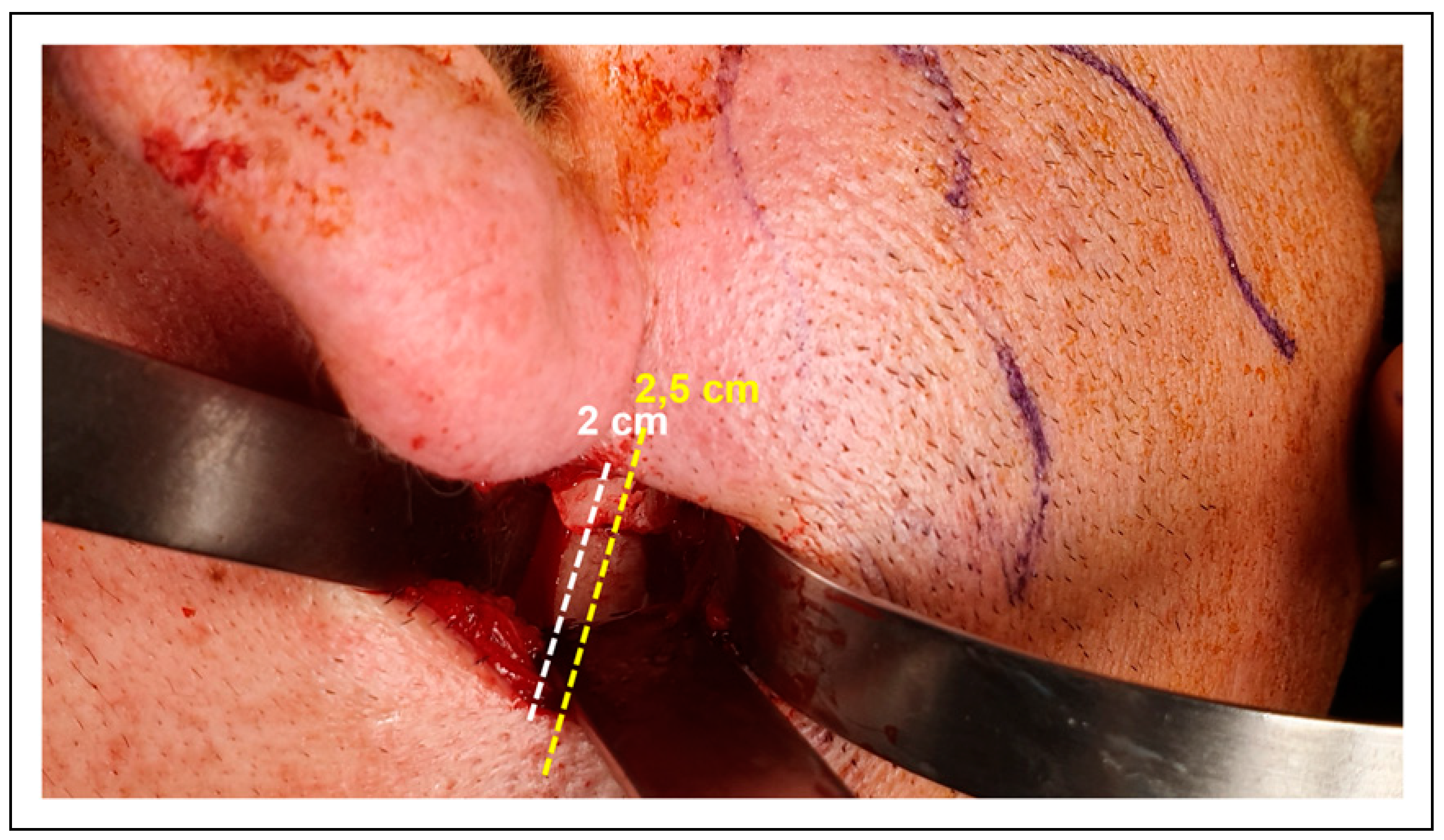
This retrospective observational study included 15 patients with condylar fractures in isolation or combination with other mandible fractures or mid/upper third facial, presenting to the Unit of Maxillofacial Surgery, Hospital El Carmen, Santiago, Chile, from 2019 to 2022. Condylar fractures were classified according to the AOCMF classification system. Patients who accepted and consented to surgical treatment for resolution of their fracture and who were monitored for a minimum of three months were included. All patients were evaluated by clinical examination (pain, occlusion, and mandibular dynamics) and imaging through computed axial tomography (1 mm slices) pre- and post-surgery. The fractures were treated by open reduction and rigid internal fixation with a trapezoidal plate (MatrixMANDIBLE, DePuy Synthes) (Figure 1). Intermaxillary locking was performed with IMF 2.0 screws (MatrixMANDIBLE, DePuy Synthes). The surgeries were performed under general anaesthesia with nasotracheal intubation, and a preauricular or a less invasive transparotid approach was used to access the affected site. The conservative transparotid approach had a 2 cm skin extension that is described in Figure 2. Postoperative clinical and imaging information was evaluated retrospectively through the individuals’ electronic medical records. Postoperative evaluations considered a favourable clinical outcome of fragment stability, stable occlusion, absence of pain, and regular mouth opening ranges. In addition, any signs of local infection, malocclusion, facial nerve damage, or failure of the fixation system were recorded.

Results

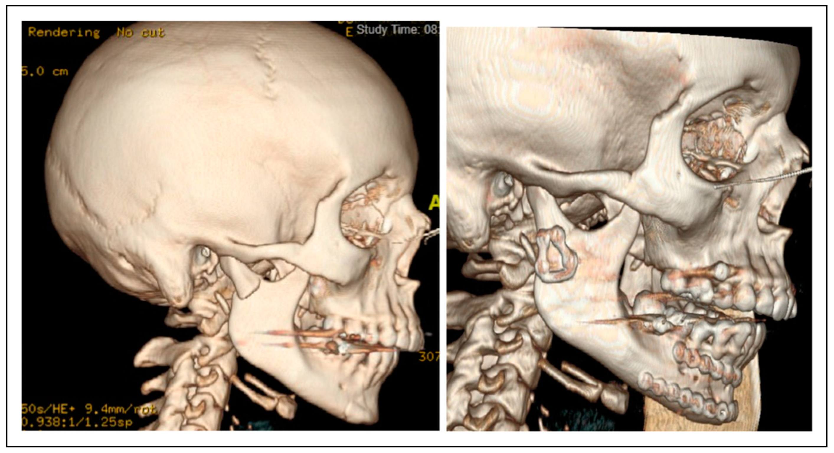
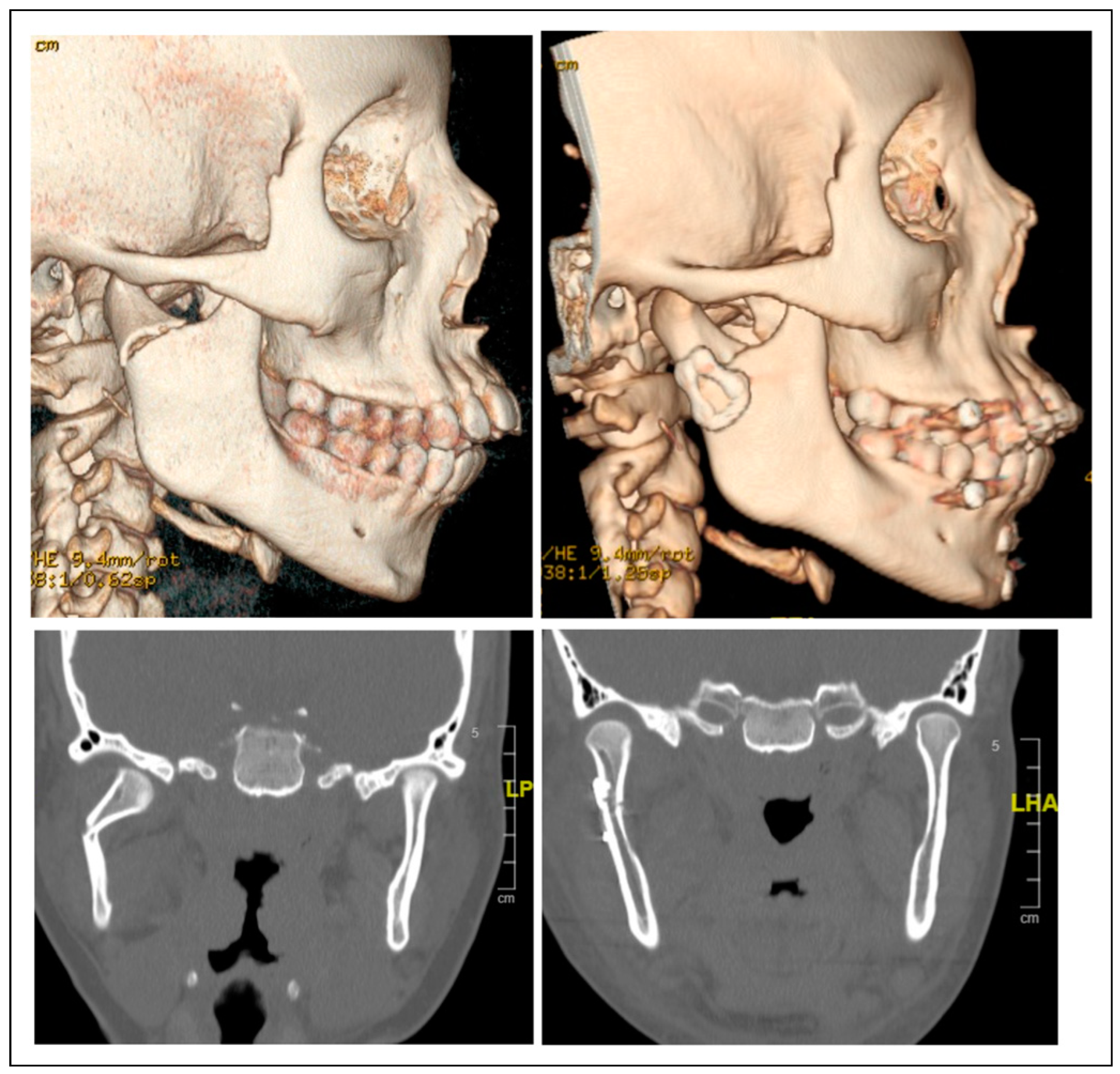
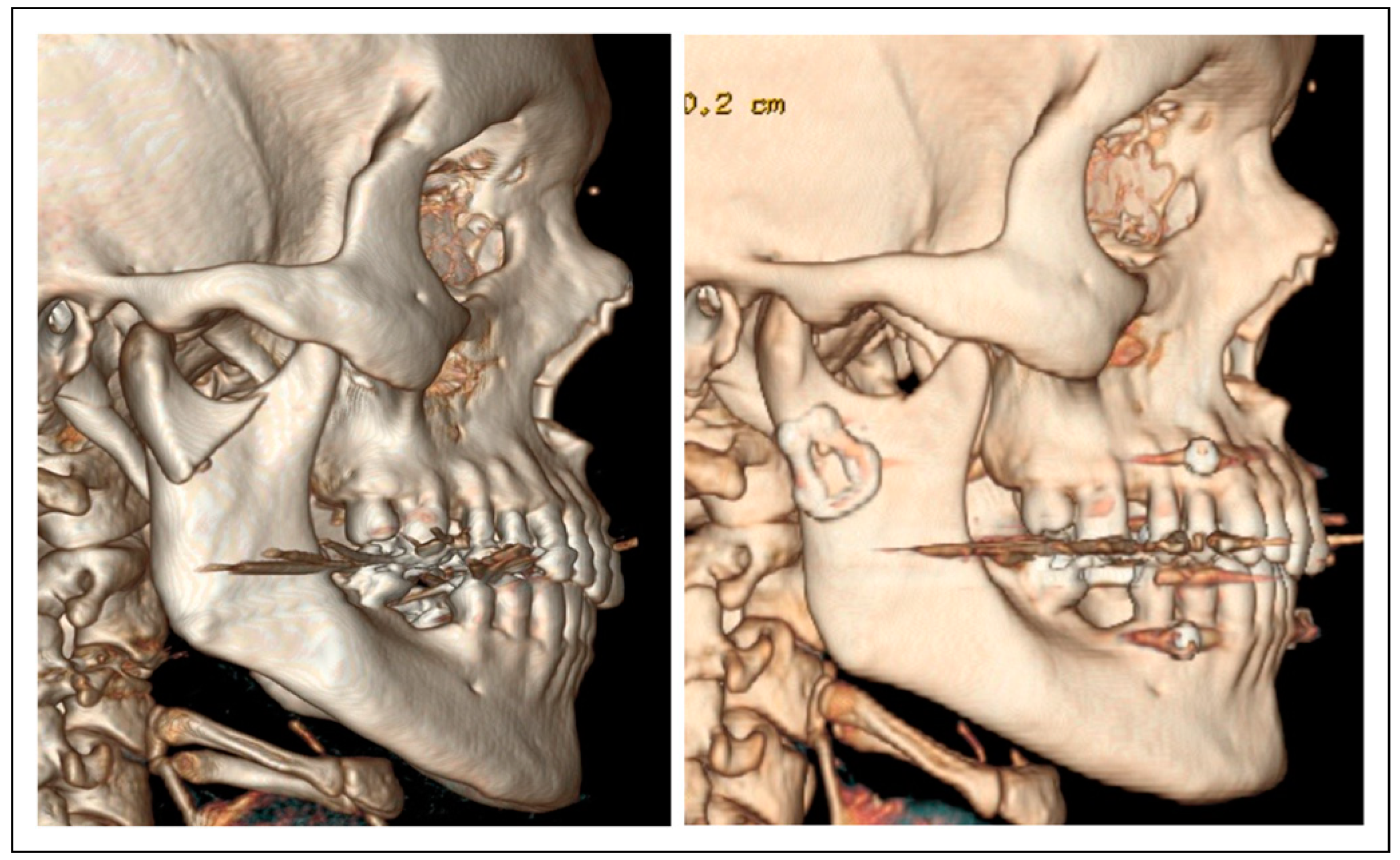
Between January 2019 and December 2022, a total of 30 patients were diagnosed with mandible condyle fracture and 15 patients underwent open reduction and rigid internal fixation surgery with trapezoidal shape miniplates. Regarding the gender of the surgically treated patients, a 73.3% (11 patients) were male, a 26.7% (4 patients) were female, and the average age of the patients was 32.14 years with a standard deviation of 13.55 years. The follow-up time of the patients was a minimum of 3 months. According to the AOCMF classification for condylar fractures, eight fractures affected the condylar base and seven involved the condylar neck. Regarding the affected side, nine fractures of the condylar process were located on the right side, five on the left side, and one affected both condyles. Additional fractures were found in 10 of 15 patients, which were located in the right para-symphyseal region (4), left parasymphyseal (2), left body (2), right body (1), and the middle and upper third face skeleton(1). Figure 4, Figure 5 and Figure 6 show computed tomography data of pre-surgery and post-surgery comparisons of condylar fractures treated with ORIF.
Regarding the surgical approach used in the patients, 15 underwent a retromandibular transparotid (TP) approach, while one underwent an endaural approach plus TP. Regarding the outcomes evaluated in the postoperative follow-up, no patients presented pain or joint sounds in the TMJ. All patients obtained a buccal opening greater than or equal to 35 mm, which is defined as normal. Two patients showed postoperative malocclusion, one due to an anterior open bite and the other due to a posterior premature contact. In both cases, the postoperative malocclusion was corrected by intermaxillary elastics therapy. In addition, two patients had transient paresis during the postoperative period, one of them had paresis of the frontal and buccal branches of the facial nerve and a transparotid plus endaural approach was performed, while the second patient had paresis of the zygomatic and buccal branches, and the fracture was accessed through a transparotid approach. Both patients with facial transient paresis were managed with physiotherapy and totally resolved.

Discussion

The treatment of mandibular condylar fractures, open or closed, is still widely discussed. If ORIF is desired, sufficiently rigid fixation is necessary to withstand physiological strains during function [10]. During functional activity, there are tensile strains at the anterolateral border and compressive strains along the posterior-medial border, causing lateromedial bending of the condyle [4]. These principles mandate that the plates be placed along the ideal lines of osteosynthesis to provide the best possible functionality.
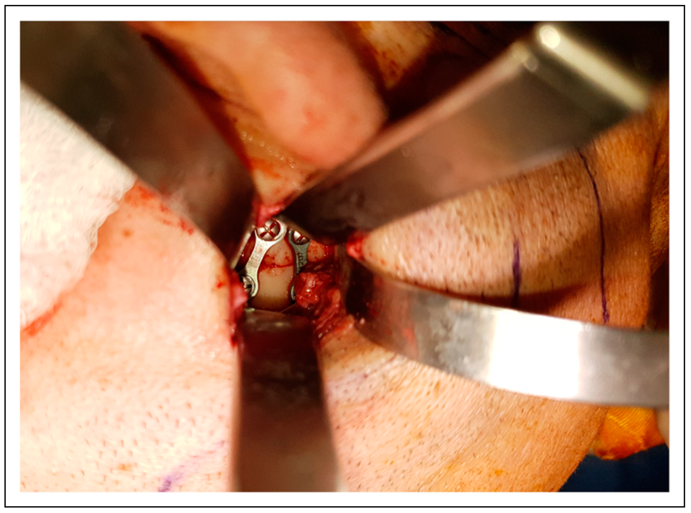
The osteosynthesis method for subcondylar fracture of the mandible traditionally involves two non-parallel mini plates; one plate placed parallel to the condylar axis along the posterior border of the ramus and the other parallel to the sigmoid notch [11]. In this study, all patients were treated with ORIF and trapezoidal plates. The plate is trapezoidal so that the anterior arm of the plate can be superimposed over the tension lines under the sigmoid notch (Figure 3). The 3D plates were developed to offer enhanced stability, less periosteal destruction, osteosynthesis of small fragments, minimum osteosynthesis material, and ease of placement. The authors believe that trapezoidal plates are more accessible to adapt than two non-parallel mini plates, as has been reported by previous literature [8,12], and it allows for less invasive surgical access. It has been published that sometimes it is difficult to place two mini plates due to the requirement for screws in a small proximal segment [12]. To overcome this disadvantage, trapezoidal plates can be used to fix the condylar segment. The trapezoidal plate helps by neutralizing the changing strains at different sites of the condyle (anterior, posterior, and lateral) [12]. Also, trapezoidal plates have a small profile; these plates can be placed in confined spaces, as in the case of subcondylar fracture [5].
Another controversial issue for the open treatment of condylar fractures concerns the design and arrangement of the titanium miniplates used to achieve the maximum stability of fixed bony fragments. A feasible way to simulate and evaluate the performance of different titanium miniplates is using finite element analysis (FEA), a mathematical method used to predict bone behaviour under a range of loading conditions [13]. FEA has widely been used to explore the behaviour of plating systems in trauma and other maxillofacial fields [14]. Previous studies have compared different miniplate designs focussing on stresses in miniplates under loads but not presenting information related to the displacement of fracture fragments, which is considered a limitation to obtaining any conclusion of what miniplate provides the most rigid fixation [15]. Hassan et al. compared five designs of miniplates for condylar fractures in terms of displacement of fracture segments, strains in bone, and stresses in miniplates. They found significant differences in mechanical performance among the evaluated miniplates and that the trapezoidal miniplate showed the best behaviour. In this FEA model, the trapezoidal plate generated the lowest strains in bone, and it has been published that excessive strains would lead to the loosening of screws and failure of fixation [16]. Also, they found that minor peak stresses in hardware were expected in the trapezoidal miniplate [16]. The available clinical evidence is consistent with these findings. Previous clinical studies reported a low risk of osteosynthesis failure with trapezoid miniplates [17,18].
A previous randomized controlled trial that compared one trapezoidal plate against two conventional miniplates for subcondylar fracture ORIF reported no difference concerning system failure [12]. They also reported that the trapezoidal plates were more accessible to adapt than two non-parallel miniplates and that sometimes it was difficult to fix two miniplates due to the screw requirement in small proximal segments. Also, the intraoperative time taken for the fixation of the trapezoidal plate was statistically significantly less than for two miniplates [12]. The median time for fixation in the two-miniplate group was 15 minutes and 6 minutes in the trapezoidal plate group. A reduction in time could benefit patients because it is hypothesized that a decreased intraoperative time in the fixation of plates decreases the duration in which the soft tissues are retracted, so the incidence of facial nerve paresis could be decreased [12].
A limitation of this study could be the observational and retrospective design, the small sample size, the limited follow-up, and the absence of a control group. Future clinical studies should compare the performance of trapezoidal plates against other plate designs, such as the lambda, strut, delta, or rhombic miniplates, to evaluate which plate has the ideal clinical results. Also, future investigation should compare the performance of the presented less invasive transparotid approach and trapezoidal plates with other percutaneous approaches, as the preauricular approach or even with an intraoral endoscopic management.

Conclusions

Using trapezoidal miniplates provides functionally stable fixation for neck and base condylar fractures and allows for a less invasive surgical transparotid approach (2 cm extension). They also reduce the amount of osteosynthesis material and are easier to adapt and fix in the author’s opinion.

Funding

The author(s) received no financial support for the research, authorship, and/or publication of this article.

Conflicts of Interest

The author(s) declared no potential conflicts of interest with respect to the research, authorship, and/or publication of this article.

Ethical Statement

Ethical Approval. This research was conducted in full accordance with the ethical standards of the institutional research committee and with the 1964 Helsinki Declaration and its later amendments or comparable ethical standards. For this non-interventional observational retrospective study, patient data were referenced with the understanding and written consent of the patient, and all data were also anonymized and de-identified prior to analysis.

References

  1. Afrooz, P.; Bykowski, M.R.; James, I.; Daniali, L.N.; Clavijo-Alvarez, J.A. The epidemiology of mandibular fractures in the United States, Part 1: a review of 13,142 cases from the US National trauma data bank. J Oral Maxillofac Surg. 2015, 73, 2361–2366. [Google Scholar] [CrossRef] [PubMed]
  2. Neff, A.; Cornelius, C.; Rasse, M.; Torre, D.D.; Audige´, L. The comprehensive AOCMF classification system: condylar process fractures—Level 3 Tutorial. Craniomaxillofac Trauma Reconstr. 2014, 7 (Suppl. 1), S44–S58. [Google Scholar] [CrossRef] [PubMed]
  3. Neff, A.; Chossegros, C.; Blanc, J.; et al. Position paper from the IBRA Symposium on surgery of the head—The 2nd International Symposium for condylar fracture osteosynthesis, Marseille, France 2012. J Cranio-Maxillo-Fac Surg. 2012, 42, 1234–1249. [Google Scholar] [CrossRef] [PubMed]
  4. Rozeboom, A.; Dubois, L.; Bos, R.; Spijker, R.; de Lange, J. Open treatment of condylar fractures via extraoral approaches: a review of complications. J Cranio-Maxillo-Fac Surg. 2018, 46, 1232–1240. [Google Scholar] [CrossRef] [PubMed]
  5. Lauer, G.; Pradel, W.; Schneider, M.; Eckelt, U. A new 3-dimensional plate for transoral endoscopic-assisted osteosynthesis of condylar neck fractures. J Oral Maxillofac Surg. 2007, 65, 964–971. [Google Scholar] [CrossRef] [PubMed]
  6. Leonhardt, H.; Franke, A.; Nowak, A.; McLeod, N.; Lauer, G. Clinical experience and results with a rhombic plate for transoral endoscopically-assisted osteosynthesis of fractures of the condylar neck. Br J Oral Maxillofac Surg. 2019, 57, 1063–1067. [Google Scholar] [CrossRef] [PubMed]
  7. Lechler, C.; Probst, F.; Cornelius, C.P.; Smolka, W. Open reduction and internal fixation of mandibular condylar base and neck fractures using strut plates. J Oral Maxillofac Surg. 2018, 76, 1494–1503. [Google Scholar] [CrossRef] [PubMed]
  8. Chaudhary, M.; Pant, H.; Singh, M.; Vashistha, A.; Kaur, G. Evaluation of trapezoidal-shaped 3-D plates for internal fixation of mandibular subcondylar fractures in adults. J Oral Biol Craniofac Res. 2015, 5, 134–139. [Google Scholar] [CrossRef] [PubMed]
  9. Smolka, W.; Liokatis, P.; Cornelius, C.P. Open reduction and internal fixation of unilateral mandibular condylar base and neck fractures using a lambda plate: selection criteria for application. J Oral Maxillofac Surg. 2020, 78, 979–985. [Google Scholar] [CrossRef] [PubMed]
  10. Meyer, C.; Serhir, L.; Boutemi, P. Experimental evaluation of three osteosynthesis devices used for stabilizing condylar fractures of the mandible. J Cranio-Maxillo-Fac Surg. 2006, 34, 173–181. [Google Scholar] [CrossRef] [PubMed]
  11. Cortelazzi, R.; Altacera, M.; Turco, M.; Antonicelli, V.; De Benedittis, M. Development and clinical evaluation of MatrixMANDIBLE subcondylar plates system (Synthes). Craniomaxillofac Trauma Reconstr. 2015, 8, 94–99. [Google Scholar] [CrossRef] [PubMed]
  12. Adhikari, M.; Bhatt, K.; Yadav, R.; Mandal, J.; Bhutia, O.; Roychoudhury, A. Fixation of subcondylar fractures of the mandible: a randomized clinical trial comparing one trapezoidal plate with two miniplates. Int J Oral Maxillofac Surg. 2021, 50, 756–762. [Google Scholar] [CrossRef] [PubMed]
  13. Darwich, M.A.; Albogha, M.H.; Abdelmajeed, A.; Darwich, K. Assessment of the biomechanical performance of 5 plating techniques in fixation of mandibular subcondylar fracture using finite element analysis. J Oral Maxillofac Surg. 2016, 74, 794.e1–794.e8. [Google Scholar] [CrossRef] [PubMed]
  14. Lisiak-Myszke, M.; Marciniak, D.; Bielin´ski, M.; Sobczak, H.; Garbacewicz, Ł.; Drogoszewska, B. Application of finite element analysis in oral and maxillofacial surgery-A literature review. Materials 2020, 13, 3063. [Google Scholar] [CrossRef] [PubMed]
  15. Murakami, K.; Yamamoto, K.; Sugiura, T.; Horita, S.; Matsusue, Y.; Kirita, T. Computed tomography–based 3-dimensional finite element analyses of various types of plates placed for a virtually reduced unilateral condylar fracture of the mandible of a patient. J Oral Maxillofac Surg. 2017, 75, e1–e1239. [Google Scholar] [CrossRef] [PubMed]
  16. Shantavasinkul, P.; Akkus, O.; Palomo, J.M.; Baumgaertel, S. Surface strain distribution of orthodontic miniscrews under load. Am J Orthod Dentofac Orthop. 2016, 150, 444–450. [Google Scholar] [CrossRef] [PubMed]
  17. Meyer, C.; Zink, S.; Chatelain, B.; Wilk, A. Clinical experience with osteosynthesis of subcondylar fractures of the mandible using TCP plates. J Cranio-Maxillo-Fac Surg. 2008, 36, 260–268. [Google Scholar] [CrossRef] [PubMed]
  18. Zrounba, H.; Lutz, J.C.; Zink, S.; Wilk, A. Epidemiology and treatment outcome of surgically treated mandibular condyle fractures. A five years retrospective study. J Cranio-Maxillo-Fac Surg. 2014, 42, 879–884. [Google Scholar] [CrossRef] [PubMed]
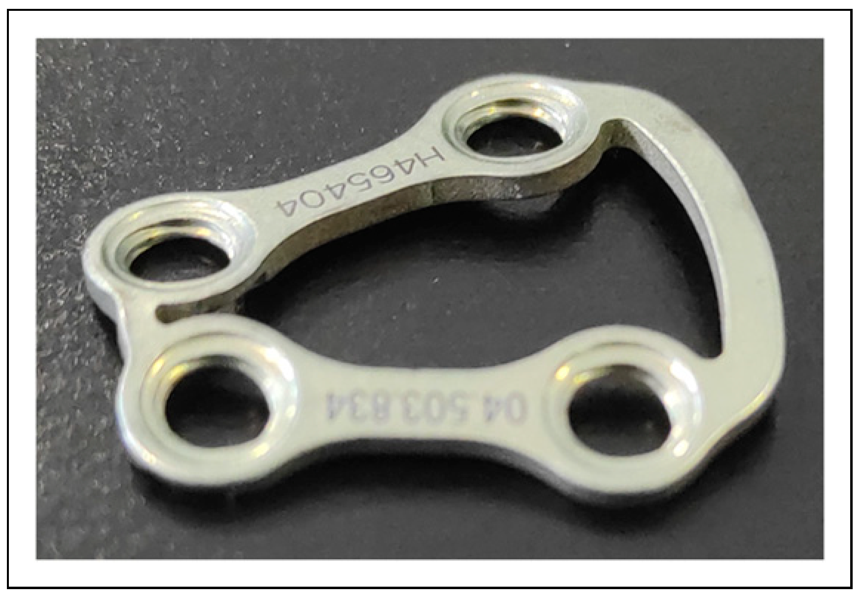
Figure 1. MatrixMANDIBLE Subcondylar Trapezoidal Plate (Synthes). The trapezoidal plate consists of a 4-hole 1.0-mm plate precontoured to fit the convex anatomy of the subcondylar region.
Cmtr 17 00064 g001
Figure 2. Conservative transparotid surgical approach. 1: Vertical incision of 2 cm in length through the skin and subcutaneous tissue below the ear lobe and parallel to the posterior border of the mandible ramus. 2: Subcutaneous blunt dissection in order to improve visualization. 3: Exposure of the superficial musculoaponeurotic system (SMAS) with a vertical incision of 2.5 cm into the parotid capsule. 4: Blunt dissection of the parotid gland parallel to the direction of the facial nerve branches until the identification of the pterygomasseteric sling. 5: Subperiostal dissection with periostal elevator and superior dissection to expose the condylar process.
Cmtr 17 00064 g002
Figure 3. Trapezoidal plate fixed in fracture condylar process. The anterior arm acts as a tension-banding plate and the posterior arm, placed along the axis of the condylar neck, lies in the compression zone.
Cmtr 17 00064 g003
Figure 4. Computed tomography data pre-surgery and post-surgery comparisons. Patient with a right base condylar fracture and right mandibular body fracture, before and after surgery.
Cmtr 17 00064 g004
Figure 5. Computed tomography data pre-surgery and post-surgery comparisons. Patient with a right base condylar fracture with 45° medial inclination, before and after surgery.
Cmtr 17 00064 g005
Figure 6. Computed tomography data pre-surgery and post-surgery comparison. Patient with a right base condylar fracture with lateral displacement.
Cmtr 17 00064 g006