Next Article in Journal
Anatomical Landmarks and Branching Patterns of the Greater Auricular Nerve
Previous Article in Journal
Precision and Progress: Key Developments in Craniomaxillofacial Surgery
 
 
Craniomaxillofacial Trauma & Reconstruction is published by MDPI from Volume 18 Issue 1 (2025). Previous articles were published by another publisher in Open Access under a CC-BY (or CC-BY-NC-ND) licence, and they are hosted by MDPI on mdpi.com as a courtesy and upon agreement with Sage.
Article

Tomographic Evaluation of Bone Height Between the Mandibular Canal and the Inferior Cortex of the Mandible Related to Bicortical Screws Fixation

by
Raissa Dias Fares
1,2,*,
João Victor Borges Leal
3,
Manuella Zanela da Silva Areas
4,
Hernando Valentim da Rocha
4,
Sylvio Luiz Costa de Moraes
1,
Nicolas Homsi
5 and
Jonathan Ribeiro da Silva
1
1
Department of Oral and Maxillofacial Surgery, Centro Universitário Serra dos Órgãos (UNIFESO), Alameda Euclydes Cotrim, n°53, casa 2. Vale do Paraíso, Teresópolis, Rio de Janeiro 25976187, Brazil
2
Master’s Student of Oral and Maxillofacial Surgery, Universidade Federal Fluminense (UFF), Rio de Janeiro, Brazil
3
Department of Oral and Maxillofacial Surgery, Neo Face Clinic, São Paulo, Brazil
4
Department of Oral and Maxillofacial Surgery, Hospital Geral de Nova Iguaçu (HGNI), Rio de Janeiro, Brazil
5
Department of Oral and Maxillofacial Surgery, Universidade Federal Fluminense (UFF), Rio de Janeiro, Brazil
*
Author to whom correspondence should be addressed.
Craniomaxillofac. Trauma Reconstr. 2024, 17(3), 181-185; https://doi.org/10.1177/19433875231213892
Submission received: 1 November 2022 / Revised: 1 December 2022 / Accepted: 1 January 2023 / Published: 3 November 2023

Abstract

:
Study Design: Original Article. Objective: The surgical treatment of mandibular fractures has undergone several changes over the past 100 years, since the use of wires and intermaxillary fixation, until today where most of the fractures are treated with internal fixation using plates and screws. For the correct use of this hardware, the surgeon must have adequate knowledge of jaw anatomy to prevent complications during the insertion of screws like in fractures of the mandibular body and angle, when the screws of the plate installed in the compression zone can damage the inferior alveolar nerve within the mandibular canal. Methods: To minimize this complication, 66 hemimandibles of adult patients were analyzed in CT scans and manipulated with Dental Slice® software, (BioParts®, Brazil) and measured the shortest distance from the inferior cortex of the mandibular canal to the inferior cortex of the mandibular body during the path of the inferior alveolar nerve, since the mandibular foramen until the mental foramen. Results: The result obtained showed a wide variation of the shortest distance measured (3.52 mm–11.5 mm) with an average of 7.99 mm. Conclusion: The conclusion demonstrated a minimum distance of 3.52 mm below the cortical mandibular canal to the lower border of the mandible and should be considered as a safety margin during the application of fixing the material in the region of the mandibular body.

Introduction

The methods of treatment in maxillofacial and trauma surgery suffered several changes in the past 100 years. Several approaches have been described for the management of facial fractures since the non-use of open reduction to avoid the risk of osteomyelitis, which was warned by Waldron in 1942. Those approaches fade out from open reduction and wiring, reasoned by Shira in 1954, to nowadays with the use of rigid internal fixation for most fractures face [1].
The structural and functional restoration without violating important anatomical structures is one of the basic principles for the treatment of any fracture [2,3]. To perform osteotomies and fracture reduction before applying the material fixation at body mandibular region, one must take great care to prevent inferior alveolar nerve injury [3]. A precise knowledge of mandibular canal anatomy is essential for dental surgeons who will perform simple local anesthesia as much as surgeons who treat a fractured jaw.
Studies have been conducted on the corpse to assist in understanding the alveolar canal anatomy and its inferior route [3]. Tieu Li et al. published a study assessing three hundred eighty-six panoramic radiographs, in which they pointed out several ways to route the inferior alveolar canal [4]. Lun-Jou Lo et al. conducted a study analyzing 75 CT scans, in which they identified some relationships between the inferior alveolar canal and specific points at the mandibular angle region [5]. These relationships led him to evaluate the possible variations in distances, a fact that, in turn, resulted in a security increase by reducing mandibular angle fractures.
The goal of this study is to analyze the relationship between the inferior alveolar canal and the lower border of the mandible, analyzing CT scan images to obtain the structure distances, which will lead in a safer application of hardware at the mandibular body region, consequently, avoiding the risk of inferior alveolar nerve injury.

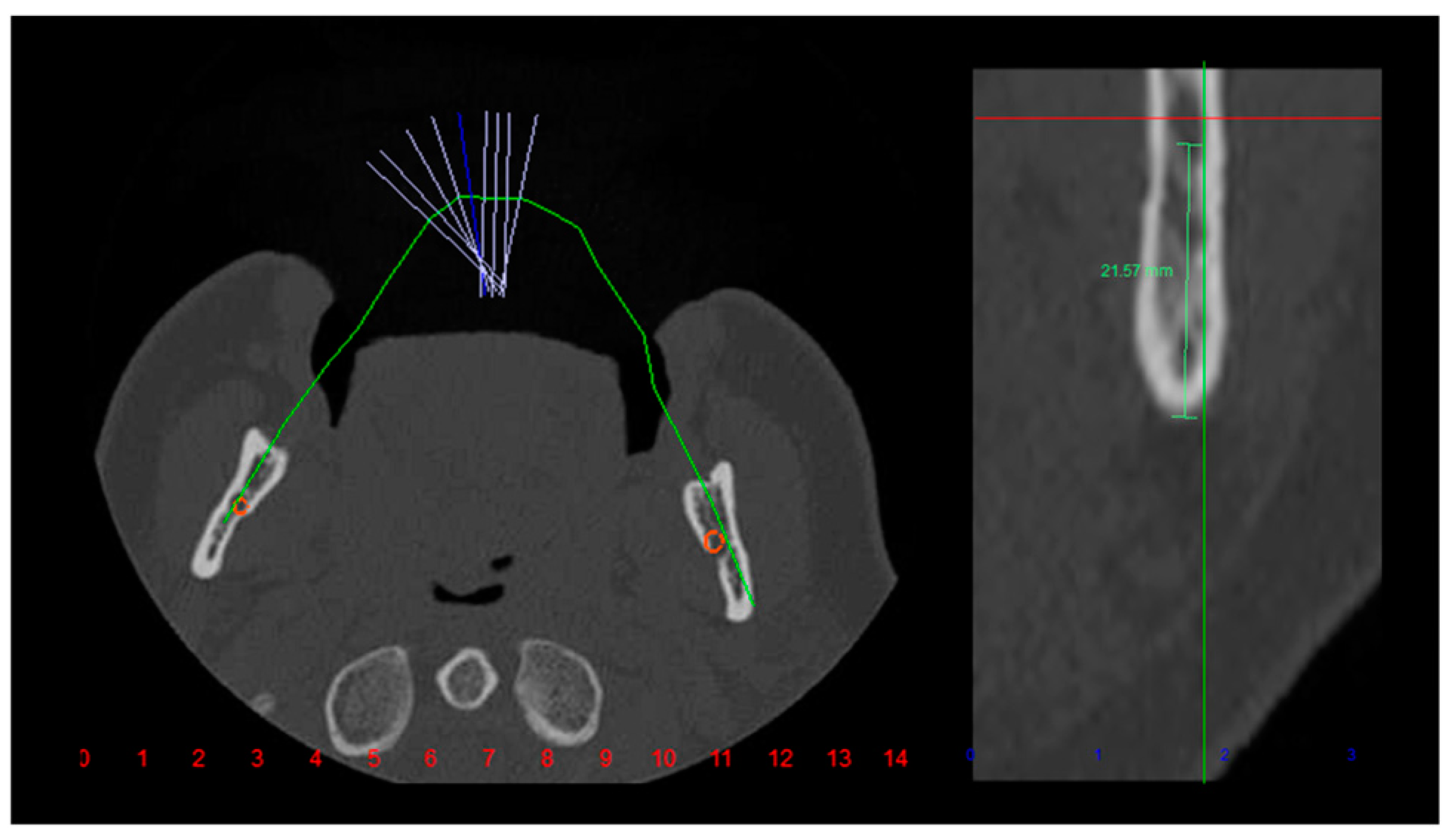
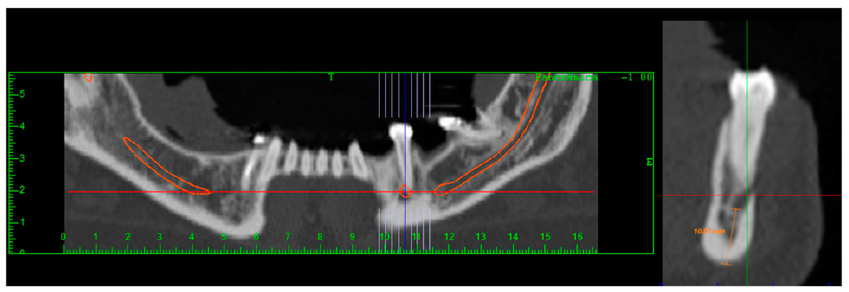
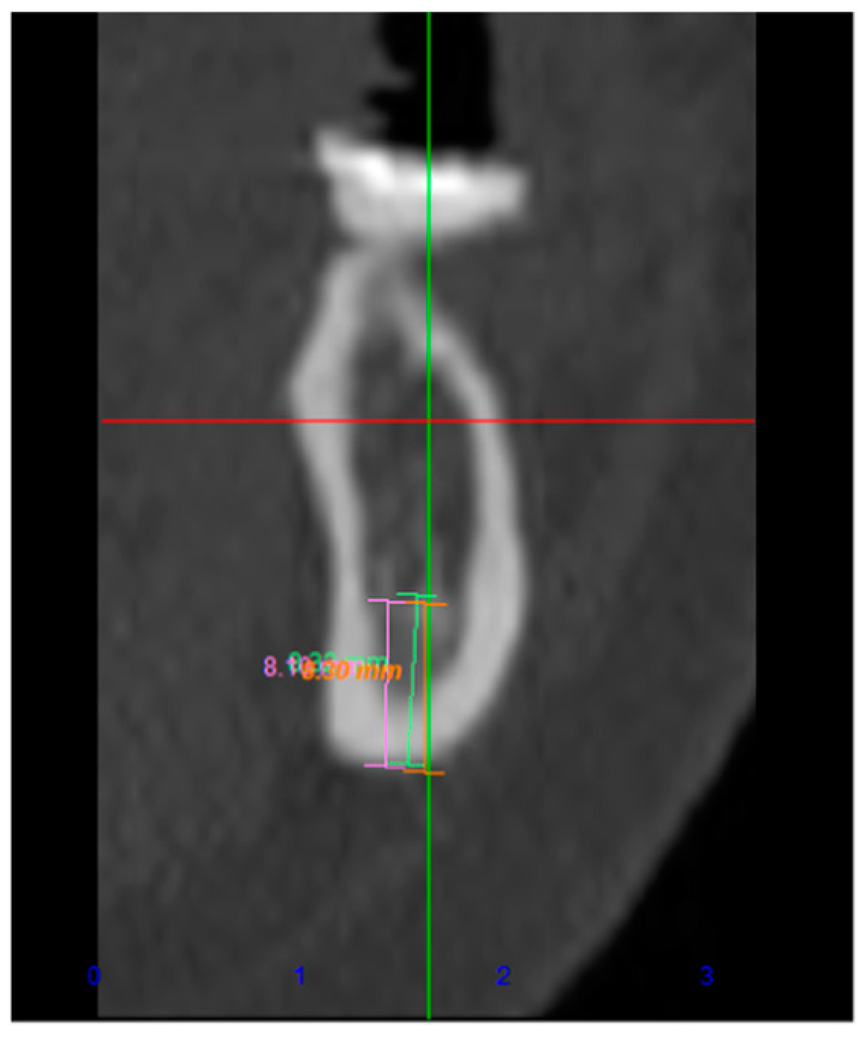
Materials and Methods
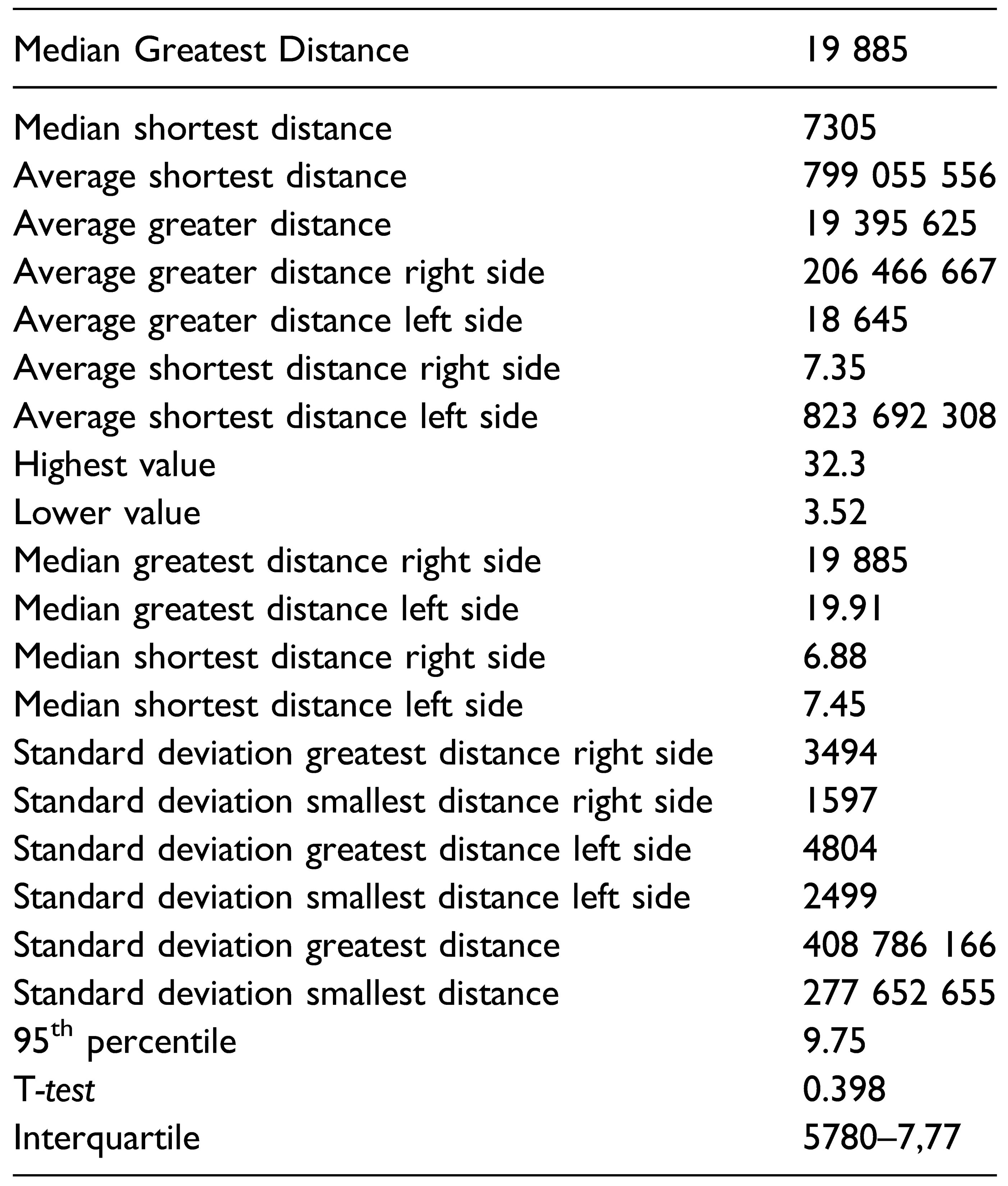
This study was approved by the ethics committee of Nova Iguaçu General Hospital with number 2.015.670 and performed at the maxillofacial surgery department from Nova Iguaçu General Hospital in partnership with ScanFace radiologic clinic (Rio de Janeiro, Brazil). The data in this study were obtained from CT scans performed in the multislice tomography Aquilion 64 TSX-101 A/E (Toshiba 64 channels), and, subsequently, analyzed with Dental Slice program (BioParts, Brazil). A sample of 70 hemimandibles from 35 patients was measured. All those individuals were young adults, 21 females and 14 males. It was excluded 4 hemimandibles because they presented variation in the inferior alveolar canal, fact that altered the shape or number of channels. A final sample of 66 hemimandibles resulted from those eliminations. None of the patients had ever had a historical trauma to the jaw or any surgery that involved the region of interest in this study. The entire assessment using the Dental Slice program was made by the same professional, who analyzed the major and minor distances between the lower cortical mandibular canal and the lower border of the mandible, starting in the region of the mental foramen until the mandibular foramen using the sagittal section. (Figure 1, Figure 2 and Figure 3). Measurement was performed every 1 mm of the path between the mental and mandibular foramen in each hemimandible.

Results

From the evaluation of the sample of 66 hemimandibles, it was found that the major distance between the mandibular canal and the inferior border of the mandible was 32.3 mm, and the minor was 3.52 mm. Specifically, in female patients, the major distance was 25.93 mm and the minor was 3.52 mm, while in male patients the major distance was 32.3 mm, and the minor was 3.88 mm. The average of major distances was 19.39 mm with a standard deviation of 4.08 mm, while the average of minor distance was 7.99 mm with a standard deviation of 2.77 mm (Table 1).
Comparing the right and left side of the patients, it was found that the average of the major distance was 20.64 mm and 18.64 mm, consecutively, while the average of the minor distance was 7.35 mm and 8.23 mm, following the same sequence. Using the Student T-test, both comparisons mentioned above were not statistically significant (P > 0.05).

Discussion

Internal fixation with plates and screws has become the gold standard for the treatment of mandibular fractures. It presents significant advantages over intermaxillary fixation such as faster recovery, and better hygiene, speech, and stability [1].
Despite its advantages, this treatment method is not free from complications. Kocaelli et al. reported in their study a case of drilling tooth roots during application of mini plates, consequently, they warned the possibility of mandibular canal perforation during mandibular fracture treatment [5].
When the mandibular canal is perforated and the inferior alveolar nerve injured, the patient may experience sensorial changes such as paresthesia, allodynia, hypesthesia, dysesthesia, anesthesia, and all these changes may cause considerable harm to patient [6].
In a retrospective study of 10 years, Seeman et al. found 5 patients with severe nerve damage from a total sample of 355 mandible fractures. However, there was no specification whether the injury was due to operative handling or drilling to apply hardware [7].
In the epidemiologic study of mandibular fractures by Bede [8]; It was observed that in a sample of 112 patients, 2 patients presented sensory disturbances of the inferior alveolar nerve after treatment, without recovery after 6 months post-treatment. Evaluating the incidence of sensorial changes in patients suffering from mandibular fractures presents some difficulties because many of these patients have some degree of sensorial variations before the treatment [9].
Intending to minimize injuries to the mandibular canal structure, studies analyzing the course of the mandibular canal increased the knowledge of this anatomical landmark.
Tiu Liu et al. evaluated 386 panoramic radiographs, and as a result, they identified four shapes of mandibular canal path, in which the elliptical form was the most common [4]. Hernandez et al. evaluated the safe distances for application of orthodontic mini-implants through the use of CT scans, but this device was not installed low enough; therefore, this study did not measure the distance between the mandibular canal and the basilar [10].
Simonton et al. assessed the distance between the tooth roots and the mandibular canal and found variations of 0–13.9 mm in males and 0–11.3 mm in females [11]. Although the distinct anatomical landmarks compared with the one in our study, Simonton et al also discovered significant variations in the mandibular canal path.
Ribeiro et al. performed a study with the proposal to measure the bone thickness between the cortical external to the mandibular canal in the premolars and molars. After evaluating 62 hemimandibles by computed tomography, the result was 11.78 mm for the largest thickness and .63 for the smallest, and also confirmed the information that the thickness increases from the premolar region to the molars, giving more safety in the application of monocortical screws [12].
In a similar study compared to ours, Lun-Jou Lo et al. evaluated the distance between the mandibular canal and the lower mandible border at the mandibular angle region of 75 patients through CT scans. They obtained results ranging from 16.48 mm (+- 1.60 mm) to 36.28 mm (+- 2.28 mm) in males, and 14.47 mm (+- 1.98 mm) to 34.88 mm (+- 2.25 mm) in females [5]. The distance found by them was similar to our result on the region of antigonial notch and mandibular foramen. However, they measured 32.03 mm as the major result because of the upward curve of the mandibular canal at this region.
According to Manson et al. [12], fractures of the mandibular body region should be treated by using two plates of 2.0 system, one at tension zone and one at compression zone, or a reconstruction plate of the 2.4 system at compression zone. The screws installed on the plates placed at compression zone must necessarily be bicortical, which may cause sensorial complications in the proximity of the mandibular canal region.
In order to avoid such problems, our study examined the minor distances found between the inferior cortical of the mandibular canal and the lower border of the mandible, in this way, a safety zone can be established. Measuring the distance mentioned above, we obtained a high variation ranging from 3.52 mm to 11.5 mm with an average of 7.99 mm. Based on these results, we advise using a distance of 3.51 mm starting from the basilar during the application of hardware on the mandibular body area, it can be considered as a safe distance because it reduce considerably the risk of damage to the inferior alveolar neurovascular bundle. The study focuses on establishing a safe distance of 3.51 mm for screw placement, but it is also imperative considering each patient’s unique anatomical characteristics. It is essential for clinicians to utilize CT imaging to assess these measurements and tailor their approach accordingly. This approach not only allows for the recognition and accommodation of anatomic variants, which were excluded from this study, but also ensures a higher level of patient-specific care and precision in surgical interventions.

Conclusions

The results in this study showed that all major distances measured were found in the path of the mandibular canal, in the region of the mandibular foramen. These measurements reached a maximum of 32.03 mm, with an average of 19.03 mm. Regarding the minor distance measured, it was found a dimension of 3.52 mm between the lower cortical mandibular canal and the lower border of the mandible. We advise using the 3.51 mm as a safe distance.

Funding

The author(s) received no financial support for the research, authorship, and/or publication of this article.

Declaration of Conflicting Interests

The author(s) declared no potential conflicts of interest with respect to the research, authorship, and/or publication of this article.

References

  1. Ellis, E., 3rd; Dean, J. Rigid fixation of mandibular condyle fractures. Oral Surg Oral Med Oral Pathol. 1993, 76(1), 6–15. [Google Scholar] [CrossRef]
  2. Cutright, B.; Quillopa, N.; Schubert, W. An anthropometric analysis of the key foramina for maxillofacial surgery. J Oral Maxillofac Surg. 2003, 61(3), 354–357. [Google Scholar] [CrossRef] [PubMed]
  3. Levine, M.H.; Goddard, A.L.; Dodson, T.B. Inferior alveolar nerve canal position: a clinical and radiographic study. J Oral Maxillofac Surg. 2007, 65(3), 470–474. [Google Scholar] [CrossRef]
  4. Liu, T.; Xia, B.; Gu, Z. Inferior alveolar canal course: a radiographic study. Clin Oral Implants Res. 2009, 20(11), 1212–1218. [Google Scholar] [CrossRef]
  5. Kocaelli, H.A.; Arikan, H.; Kaptan, F.; Kayahan, B.; Haznedaroğlu, F. Management of the perforations due to miniplate application. J Endod. 2006, 32(5), 482–485. [Google Scholar] [CrossRef] [PubMed]
  6. Hillerup, S. Iatrogenic injury to the inferior alveolar nerve: etiology, signs and symptoms, and observations on recovery. Int J Oral Maxillofac Surg. 2008, 37(8), 704–709. [Google Scholar] [CrossRef] [PubMed]
  7. Seemann, R.; Schicho, K.; Wutzl, A.; et al. Complication rates in the operative treatment of mandibular angle fractures: a 10-year retrospective. J Oral Maxillofac Surg 2010, 68(3), 647–650. [Google Scholar] [CrossRef]
  8. Bede, S. Mandibular fractures in Iraq: an epidemiological study. Craniomaxillofac Trauma Reconstr. 2015, 8(01), 59–63. [Google Scholar] [CrossRef]
  9. Lo, L.J.; Wong, F.H.; Chen, Y.R. The position of the inferior alveolar nerve at the mandibular angle:: an anatomic consideration for aesthetic mandibular angle reduction. Ann Plast Surg. 2004, 53(1), 50–55. [Google Scholar] [CrossRef]
  10. Hernandez, L.C.; Montoto, G.; Puente Rodriguez, M.; Galban, L.; Martinez, V. Bone map’ for a safe placement of miniscrews generated by computed tomography. Clin Oral Implants Res. 2008, 19(6), 576–581. [Google Scholar] [CrossRef]
  11. Simonton, J.D.; Azevedo, B.; Schindler, W.G.; Hargreaves, K.M. Ageand gender-related differences in the position of the inferior alveolar nerve by using cone beam computed tomography. J Endod. 2009, 35(7), 944–949. [Google Scholar] [CrossRef] [PubMed]
  12. Ribeiro, J.; Marin, C.; Homsi, N.; et al. Tomographic evaluation of mandibular thickness on premolar and molar regions related to monocortical screws. Craniomaxillofac Trauma Reconstr. 2016, 9(02), 105–108. [Google Scholar] [CrossRef] [PubMed]
Figure 1. Mandibular and mental foramen identification with Dental Slice (Bioparts, Brazil) software on axial window.
Cmtr 17 00029 g001
Figure 2. Mandibular and mental foramen identification with Dental Slice (Bioparts, Brazil) software on panoramic window.
Cmtr 17 00029 g002
Figure 3. Measurement in cross section.
Cmtr 17 00029 g003
Table 1. Results of the Measurements of the Major and Minor Distances of the Mandibular Canal Path.
Cmtr 17 00029 i001

