Next Article in Journal
CAD/CAM Engineered Patient-Specific Impants as a Reposition Device in Le Fort I and Modified Subcondylar Osteotomies: Case Report of Facial Deformity Correction in Acromegaly
Previous Article in Journal
The Anomalous Radial Artery: A Rare Vascular Variant and Its Implications in Radial Forearm Free Tissue Transfer
 
 
Craniomaxillofacial Trauma & Reconstruction is published by MDPI from Volume 18 Issue 1 (2025). Previous articles were published by another publisher in Open Access under a CC-BY (or CC-BY-NC-ND) licence, and they are hosted by MDPI on mdpi.com as a courtesy and upon agreement with Sage.
Review

Etiopathogenesis of Trismus in Patients With Head and Neck Cancer: An Exploratory Literature Review

by
Radhu Raj
1,
Krishnakumar Thankappan
2,*,
Chandrasekhar Janakiram
3,
Subramania Iyer
2 and
Anil Mathew
1
1
Department of Prosthodontics, Amrita School of Dentistry, Amrita Vishwa Vidyapeetham, Kochi, Kerala, India
2
Department of Head and Neck Surgery and Oncology, Amrita Institute of Medical Sciences, Amrita Vishwa Vidyapeetham, Kochi, Kerala 682041, India
3
Department of Public Health Dentistry, Amrita School of Dentistry, Amrita Vishwa Vidyapeetham, Kochi, Kerala, India
*
Author to whom correspondence should be addressed.
Craniomaxillofac. Trauma Reconstr. 2020, 13(3), 219-225; https://doi.org/10.1177/1943387520917518
Submission received: 1 December 2019 / Revised: 31 December 2019 / Accepted: 1 February 2020 / Published: 27 April 2020

Abstract

:
Trismus refers to a person’s inability to normally open his or her mouth. Trismus can occur as a symptom due to tumor ingrowth or it can occur postsurgical following the treatment for head and neck cancer. Radiation-induced trismus is also a relatively common oral complication. This review aimed at reviewing the etiopathogenesis of trismus in patients with head and neck cancer. Of the 16 publications included after final screening, of which one was a nonrandomized control trial, one a randomized control trial, 6 prospective cohort studies, and 8 retrospective cohort studies. Among them, 6 articles addressed the possible mechanism for trismus related to tumor ingrowth, 8 articles suggested the likely reason for trismus in patients who had undergone radiation therapy and 2 articles addressed the postsurgical cause for trismus. This review highlights the possible involvement of infratemporal fossa as a predetermining factor for developing trismus related to tumor extension. The molecular mechanism of radiation-induced fibrosis is well studied in the literature.

Introduction

Trismus indicates severely restricted mouth opening related to any etiology. The word trismus is derived from the Greek word “trismos” meaning grating, grinding, or squeaking.[1] Trismus is defined as a tonic contraction of the muscles of mastication and as mandibular hypomobility, which results in a limited ability to open the mouth.[2] Although, by definition, it refers to bilateral process that results from increased tone mediated by the efferent portion of the reflex arch of trigeminal nerve, unilateral restriction may also occur clinically.[3] Dijkstra et al. defined it as mouth opening of ≤35 mm (either the interincisal distance or the distance between the upper and lower alveolus) and this measurement has been accepted universally.[4,5,6,7]
Trismus in patients with head and neck cancer may be caused by ingrowths of tumor in the masticatory muscles or by fibrosis after surgery or radiotherapy (RT). The prevalence of trismus after head and neck oncology treatment ranges from 5% to 38%.[8] This limitation in the ability to open the mouth can have serious health implications. Management of trismus can be conservative (exercise therapy) or surgical. If the clinical examination reveals the presence of limited mouth opening and diagnosis determines the condition due to be trismus, treatment should begin as soon as it is practical. Interventions such as spatulas or a passive jaw mobilizer should be targeted at patients at high risk for trismus early in the postoperative phase.[2,9,10,11,12,13] The muscles that elevate the mandible to close the mouth are the temporalis, masseter, and internal pterygoid. The external pterygoid (or lateral pterygoid) inserts on the articular disc of the temporomandibular joint (TMJ) and into a small depression in front of the neck of the condyle. Its action is to protrude the mandible and pull the neck of the mandible forward. This primary opening muscle is assisted by the mylohyoid, anterior belly of the digastric, geniohyoid, and infrahyoid muscles while opening the mouth. The muscles of mouth closure can exert a combined force of 100 to 300 pounds; those that open, about 25 pounds.[14] This relative weakness of the opening musculature is a prime factor in the patient’s inability to open the mouth when pathology limiting the mandibular motion exists.
So far there have been very few articles addressing the prevalence and various exercise therapies to manage this complication. But there has been no gold standard of treatment to prevent or treat the condition, the possible reason could be a lack of understanding of the causative mechanism of trismus in patients with cancer. This exploratory review aims to address the etiopathogenesis of trismus in such patients.

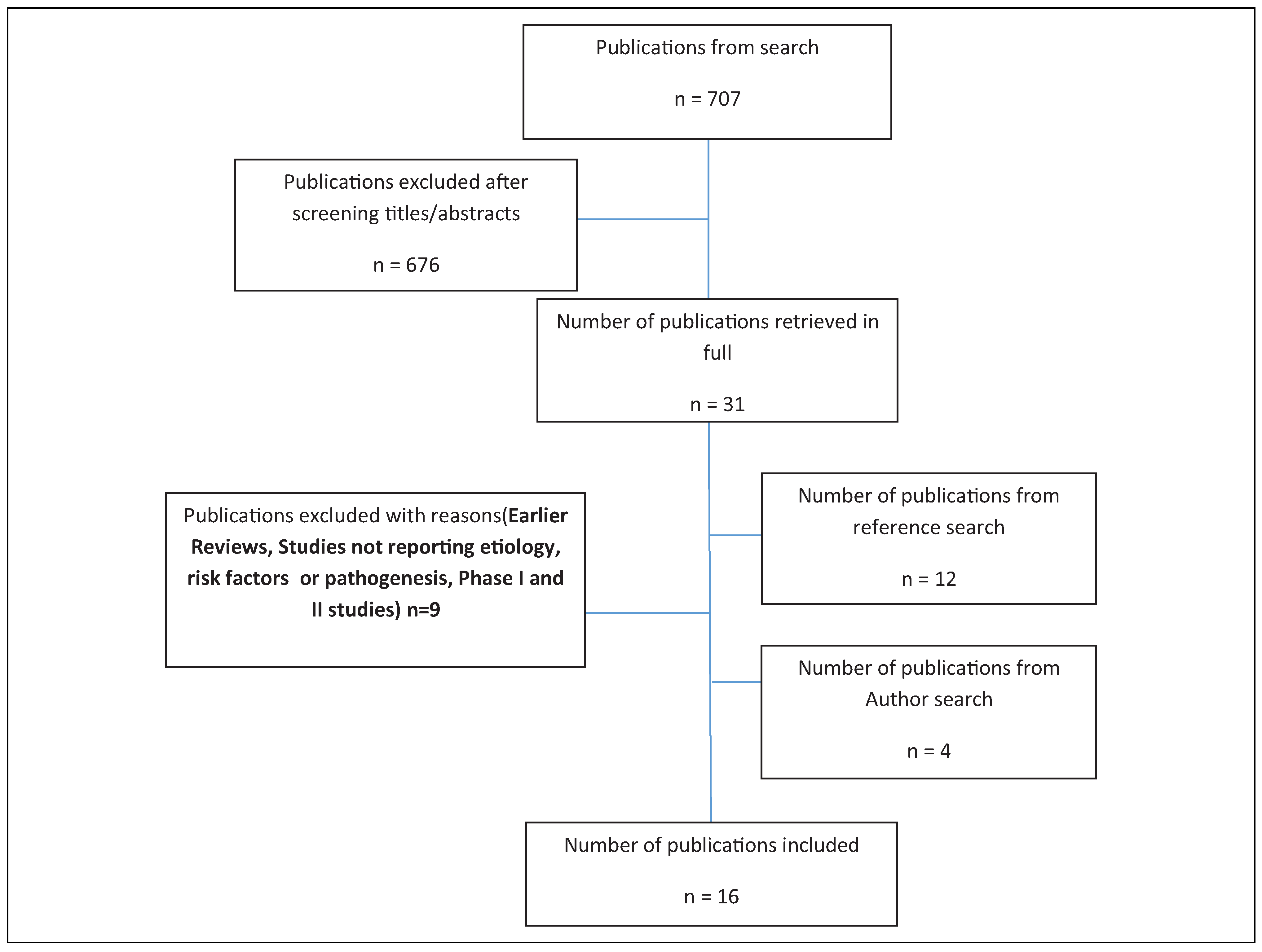
Materials and Methods

Search Methods

This review was conducted for all published articles containing data regarding the etiology and pathogenic mechanism of trismus in patients with cancer. The relevant literature was identified by searching 3 electronic databases through the PubMed, Clinical key, and EBSCO Host platforms. Subject headings were mapped using search terms related to etiology and pathogenesis of trismus in patients with cancer. The Boolean operator “AND” was used to combine the search string with subject headings related to trismus. Three authors (C.J., R.R., and K.K.) independently eliminated any duplicate from the gathered results and examined the remaining articles by title and abstract. Subsequently, the full texts were obtained and analyzed for further inclusion/exclusion. The article full text of those identified after the title and abstract were screened.

Inclusion and Exclusion Criteria

This review included only articles that studied the outcomes in terms of the risk factor/anatomical site that are more prone to develop trismus in patients with cancer and the subsequent sequelae of trismus in such patients. The search was limited to English language. Gender and age restrictions were not applied. The following publication types were eliminated by the review panel from the present review: systematic and nonsystematic reviews; studies not reporting actual data on risk factors and pathogenesis of trismus; studies reporting redundant data from previous publications; and phase I and II studies, opinion articles. There was no lower limit for the analyzed time frame.
Data collection process and data item. For every included study, using Microsoft Excel sheet, the study design, interventions, outcomes, results, and other items were collected for each study by 2 reviewers.

Results

Of the 707 publications identified from the initial search, 31 publications were retrieved following screening of the title and abstract (Figure 1). Of these publications, 676 were excluded following examination of the full text. Thirty one further publications were identified from the initial reference search and 6 additional publication identified from the expert author search. Of the 16 publications included after final screening, 1 was nonrandomized control trial, 8 were retrospective cohort studies, and 6 prospective cohort study (Table 1). Among them, 6 articles addressed the possible causative mechanism for trismus related to tumor, 8 articles suggested the likely reason for trismus in radiation therapy, and 2 articles addressed the surgery-related cause for trismus.

Discussion

This review identified and summarized the etiology and pathophysiological mechanism of trismus in patients with cancer. In the head and neck cancer setting, trismus can result from local invasion of the primary or metastatic tumor into critical structures of mastication including the masseter and pterygoid muscles, their neural innervation, the TMJ, and/or other supportive tissues. Also, surgery and RT are well known to cause trismus in those patients undergoing treatment for the head and neck cancer. Most cases of cancer-related trismus are extra-articular in nature.
  • Trismus due to tumor extension: The primary sites of malignant tumors that induce trismus are commonly the tongue, maxillary sinus, nasopharynx, retromolar trigone, infratemporal fossa, nasal cavity, hypopharynx and cervical esophagus, parotid gland, floor of the mouth, buccal mucosa, larynx, oropharynx, thyroid, palate, ear, and submandibular gland.[15] Among them, the patients with cancer of the tonsils are the one most prone to develop trismus.[16] The proximity of this site to the masticator space and infratemporal fossa could be the reason.
  • Metastatic: It is a complex multistep process in which the primary tumor spreads to distant organs. Metastatic malignancies of the mandible are rare. When they do occur, they usually come from the lung, breast, and thyroid gland.[17,18]
  • Primary, benign: Benign neoplasms such as angioma, neurofibroma, neurolemmoma, osteoma, meningioma, and adamantinoma may arise in the infratemporal fossa or invade it from surrounding areas and give rise to trismus.[15] Osteochondromas, myxomas, osteomas, and hemangiomas have also been described as lesions which can cause limitation of mandibular movement.
  • Primary, malignant: These are more likely to cause trismus. Carcinomas are tumors of epithelial origin. Epithelial tumors of the buccal mucosa, retromolar area, palate, pharynx, parotid, or maxillary sinus may infiltrate infratemporal fossa. Since carcinoma cells are genetically unstable, they start from an activated phenotype.
Ichimura and Tanaka conducted a study in 1993 to expound the causal mechanism of trismus in head and neck cancer. In their study, of 21 patients manifesting trismus, 9 patients developed trismus either by infiltration of the muscles of mastication or the ascending ramus of the mandible or induced by reflex spasm. In some cases of oropharyngeal cancer, computed tomography (CT) revealed no evidence of tumor invasion into the infratemporal fossa when trismus occurred, suggesting that trismus was caused either by the reflex spasm of muscles or by microinvasion too small to be seen in CT films. Maxillary sinus tumors were often without trismus even when they extended posteriorly to the infratemporal fossa. The findings of their study points to the observation that benign neoplasms in the parapharyngeal space and simple compression of the muscles do not induce trismus.[15]
Malignancy enters the mouth cavity by direct extension from the tonsillar fossa or retromolar trigone or via perineural tumor spread along the inferior alveolar nerve (V3 sensory branch). According to Kveton and Pillsbury, stimulation of these fibers may transmit sensation to the principal sensory trigeminal nucleus in the pons. The result is an increased tonus which develops trismus. The muscles of mouth closure exert a power 10 times greater than the opening muscles.[19,20] Tumors invading the mandible have a prosperity for perineural tumor spread both distally, along the inferior alveolar nerve in the inferior alveolar canal, and proximally, along the main trunk of V3. Such irritation can cause reflectory trismus.[19]
The study conducted by Ozyar et al. is the first to report the response of trismus and nasopharyngeal carcinoma (NPC). It is their observation that trismus is a common symptom in NPC patients with younger age and an indicator of advanced T stage. Most interesting finding of their study was the relatively high rate of trismus observed in the pediatric patients. They also found that 80% of the patients with trismus were less than 30 years.[20]

Postsurgical Trismus

Fibrosis as a result of surgical intervention is known to occur after excision of lesions involving the buccal mucosa, retromolar area, and tonsillar fossa.[14] Postsurgical fibrosis occurs after cold-knife excision or electrocoagulation of lesions involving the buccal mucosa, retromolar area, and/or tonsillar fossa. In such conditions, the internal pterygoid muscle and pterygomandibular ligament are caught up in the healing process because of their proximity to these structures, resulting in their progressive shortening. Trismus following maxillectomy is observed in case of posterior extension of the tumor into the infratemporal fossa, necessitating removal of its contents, and the pterygoid plates. The formation of a hematoma in the area around the coronoid process or ascending ramus sometimes allows fibrosis to develop, which leads to progressive decrease in its motion. Due to the relative weakness of the depressor muscles as mentioned previously, the patient will not be able to open his mouth voluntarily. This is often reported as a complication of masseter muscle surgery.
The findings of longitudinal study by Scott and colleagues concluded that surgery involving the buccal mucosa, tonsillar fossa, and retromolar trigone area has been shown to cause trismus, which has been attributed to fibrosis and shortening of the pterygoid muscle and pterygomandibular ligament. They also showed that patients having laser excisions, primary closure, or split skin grafting been proved to be able to open their mouths better than patients having soft-tissue flaps or composite flaps. Similar significant adverse results were observed with the use of local or regional flaps for reconstruction. Lesser reduction in mouth opening was seen with the use of skin or dermal grafts or when only primary closure was performed.[5,21]

Postirradiation Trismus

Radiotherapy is one of the major treatment modalities used for the management of head and neck malignancies. It can be used as a primary therapy for many early stage malignancies, as an adjuvant therapy following surgical resection, and in conjunction with concurrent CT for late-stage and unresectable head and neck malignancies. The most common cause of cancer-related trismus is radiationinduced fibrosis.
Ionizing radiation is a form of high-speed particles that is enough to displace bound electrons. Ionizing radiation refers to 3 types of emissions, namely α, β, and γ with therapeutic radiation being predominantly γ.[22] Radiation injury may result from direct DNA damage or may be due to the generation of reactive oxygen species.[23] Free radicals damage all components of cells, including proteins, nucleic acids, and lipids.[24] Superoxide dismutase, catalase, and glutathione peroxidase are responsible for controlling free radical damage. A deficiency in these enzymes leads to oxidative stress in tissues.[25] Furthermore, thrombosis and ischemia exacerbate local injury leading to further release of inflammatory chemokines and cytokines.[26] Neutrophils are the first inflammatory cells to arrive at the site of injury. Increased expression of intercellular adhesion molecule 1 and platelet endothelial cell adhesion molecule 1 on disrupted endothelial surfaces contributes to neutrophil extravasation and transmigration into tissues.[27] When these cells come into contact with collagen fragments and fibronectin, they release proinflammatory cytokines such as tumor necrosis factor α, interleukin (IL) 1, and IL-6 that lead to even greater local inflammation.[28] The next cells to arrive are the monocytes and lymphocytes, which interact with each other to lead to the differentiation of monocytes into 2 subsets of macrophages namely classically activated proinflammatory M1 or alternatively activated anti-inflammatory M2.[29] Platelet-derived growth factor secreted from the M2 subset promotes neoangiogenesis and stimulates the migration of fibroblasts into the injured tissue from either the surrounding stroma or from circulating mesenchymal stem cells.[30] They also secrete transforming growth factor-beta (TGF-β), which is heavily implicated in radiation-induced fibrosis (RIF). Indeed, TGF-β is responsible for a number of functions that contribute to the pathogenesis of this condition, including the production of fibroblasts from bone marrow progenitors and the differentiation of fibroblasts into myofbroblasts,[31] whereby a phenotypic change in the fibroblasts results in increased expression of α-smooth muscle actin, followed by subsequent transformation into protomyofibroblasts and eventual maturation into myofibroblasts. These myofibroblasts may also derive from circulating bone marrow-derived progenitor cells known as fibrocytes or from epithelial cells undergoing epithelial–mesenchymal transition.[32] In response to TGF-β, myofibroblasts secrete excess collagen, fibronectin, and proteoglycans which are responsible for the increased stiffness and thickening of the tissue. Furthermore, TGF-β promotes decreased matrix metalloproteinase (MMP) activity (especially MMP-2 and MMP-9) and increased activity of tissue inhibitors of metalloproteinases, compounding the already excessive extracellular matrix deposition.[33] Although myofibroblasts promote endothelial cell proliferation and angiogenesis through the secretion of basic fibroblast growth factor, excess collagen reduces vascularity over time. This makes fibrotic areas susceptible to physical trauma and gradual ischemia, which may lead to loss of function, tissue atrophy, reduction in the number fibroblasts, or necrosis.[34]
Radiological studies of the long-term effects of high-dose irradiation showed chronic signal changes in skeletal muscles including high T2W signal and T1W enhancement. Interestingly, a higher frequency of signal abnormalities was identified in the pterygoid muscles compared with the temporalis and masseter muscles in patients with trismus.[35]
In a study by Sophie A. Kraaijenga et al., they studied the relationship between trismus (maximum interincisor opening [MIO] ≤35 mm) and the dose to the ipsilateral masseter muscle (iMM) and ipsilateral medial pterygoid muscle (iMPM). The results of their study suggested that dose levels of iMM and iMPM were highly correlated due to proximity. Both iMPM and iMM dose parameters were predictive for trismus, especially mean dose and intermediate dose–volume parameters. Optimal cutoffs were 58 Gy (mean dose iMPM), 22 Gy (mean dose iMM), and 46 mm (baseline MIO).[36]
The nature and timing of stimuli responsible for many fibrotic diseases are uncertain, on the other hand, both are known for radiation fibrosis. Immediate oxidative damage of DNA, protein, and lipid is the initiating chemical event. Microscopically, fibrosis is often focal in distribution and associated with an interstitial fibrin network for many years after exposure, with the evidence of continuing microvascular injury.[37] Observations on the timing of fibrotic changes following radiation exposure in humans are limited by tissue availability (autopsy or surgery).
The limitation of the study is that it is not a complete systematic review. The quality of some publications was poor and the level of evidence was weak. No statistical analysis was attempted in this article. This review could serve as a basis for an analytic systematic review with meta-analysis, seeking answers to the questions related to the etiopathogenesis of trismus.
Table 1. Summary of Studies Reviewed.
AuthorYearStudy TypeFindings
Chon-Jong Wang et al.[37]2005Prospective, single-armed measurementPatients with nasopharyngeal cancer had a mean decrease in initial interincisal distance of 32% at 4 years after radiotherapy. It was rapid at 1 to 9 months after radiation therapy, then became slower and protracted over later years.
Bhatia et al.[35]2009RetrospectiveThe prevalence of trismus increases with increasing doses of RT, and levels in excess of 60 Gy are more likely to cause trismus. An abnormal proliferation of fibroblasts is an important initial event
Paul Okunieff et al.[38]2004Non-RCTFGF2 is chemotactic and mitotic for fibroblasts, whereas TGFβ1 stimulates fibroblast proliferation and premature end differentiation, leading to excessive extracellular matrix glycoprotein production
Maria GebreMedhin et al.[39]2016Cohort retrospectiveMean radiation dose to the ipsilateral masseter muscle is an important risk factor for trismus development.
Hsuing et al.[40]2008Cohort prospectiveRadiation-induced trismus results from postradiotherapy muscle fibrosis and scarring, fibrosis of the ligaments around the TMJ, and scarring of the pterygomandibular raphes.
Louise Kent et al.[41]2008Retrospective cohortDirect cell damage combined with regional loss of vascular perfusion results in bone and soft tissue necrosis and finally leads to fibrosis to the muscles of mastication.
Marc Goldstein et al.[42]1999Prospective cohortAs dose to the temporomandibular joint and pterygoid muscles increases, maximal jaw opening decreases linearly. Doses as low as 1493 cGy to the TMJ resulted in functional impairment.
Lisette van der Molen et al.[45]2013Prospective longitudinal studyDose parameters of masseter and pterygoid muscles were significant predictors of trismus at 10 weeks posttreatment. And at 1-year posttreatment therapy, dose parameters of all mastication structures are strong predictors for subjective mouth-opening problems.
Teguh et al.[43]2008Retrospective cohortIn radiation-induced fibrosis, there is presence of infiltrating inflammatory cells, atypical fibroblasts, and large amounts of various extracellular matrix components.
Ichimura and Tanaka[15]1993Retrospective Benign neoplasms in the parapharyngeal space and simple compression of the muscles do not induce trismus.
George P. Katsantonis et al.[44]1989Retrospective cohort studyLesions arising in the deep lobe of the parotid will naturally extend into the parapharyngeal space and the infratemporal fossa. Infratemporal fossa invasion is usually associated with trismus and pain along the distribution of V3 and occasionally V2
Lisette van der Molen et al.[45]2013ProspectiveDose parameters of masseter and pterygoid muscles were significant predictors of trismus
Frank R. Miller et al.[46]1996Retrospective Compared with benign parapharyngeal space neoplasms, the malignant tumors were much more likely to be associated with pain, trismus.
R. Lee et al.[47]2012ProspectiveCurrent or previous heavy drinkers had a substantially smaller chance of developing trismus.
The alcohol also acts as a muscle relaxant and counteracts the laying down of collagen.
Joakim Johnson et al.[10]2010Retrospective Trismus incidence (≤35 mm) was highest in the patients treated for parotid gland tumors, followed by patients treated for nasopharyngeal cancers and submandibular gland tumors
Christina Hague et al.[48]2018Prospective randomized trialRestricted mouth opening developed when radiation mean doses was >40 Gy to the ipsilateral block.

Conclusion

While acknowledging the methodological limitations, most of the included studies addressed the possible etiology and pathological mechanism of trismus in particularly patients with head and neck cancer. In particular, the highlighted possible involvement of infratemporal fossa as a predetermining factor for developing trismus is related to tumor extension. The molecular mechanism of RIF and subsequent onset of trismus is well studied in literature.

Funding

The author(s) received no financial support for the research, authorship, and/or publication of this article.

Conflicts of Interest

The author(s) declared no potential conflicts of interest with respect to the research, authorship, and/or publication of this article.

References

  1. Krogman, W.M. Illustrated Dictionary of Dentistry. By S. Jablonski; W.B. Saunders: Philadelphia, 1982; p. xvii + 919. [Google Scholar]
  2. Bensadoun, R.J.; Riesenbeck, D.; Lockhart, P.B.; et al. A systematic review of trismus induced by cancer therapies in head and neck cancer patients. Support Care Cancer. 2010, 18, 1033–1038. [Google Scholar] [PubMed]
  3. Santiago-Rosado, L.M.; Lewison, C.S. Trismus. In StatPearls; StatPearls Publishing: Treasure Island (FL). Available online: http://www.ncbi.nlm.nih.gov/books/NBK493203/ (accessed on 11 May 2019).
  4. Dijkstra, P.U.; Huisman, P.M.; Roodenburg, J.L. Criteria for trismus in head and neck oncology. Int J Oral Maxillofac Surg. 2006, 35, 337–342. [Google Scholar]
  5. Scott, B.; Butterworth, C.; Lowe, D.; Rogers, S.N. Factors associated with restricted mouth opening and its relationship to health-related quality of life in patients attending a Maxillofacial Oncology Clinic. Oral Oncol. 2008, 44, 430–438. [Google Scholar]
  6. Jen, Y.M.; Lin, Y.S.; Su, W.F.; et al. Dose escalation using twicedaily radiotherapy for nasopharyngeal carcinoma: does heavier dosing result in a happier ending? Int J Radiat Oncol Biol Phys. 2002, 54, 14–22. [Google Scholar]
  7. Nguyen, T.D.; Panis, X.; Froissart, D.; Legros, M.; Coninx, P.; Loirette, M. Analysis of late complications after rapid hyperfractionated radiotherapy in advanced head and neck cancers. Int J Radiat Oncol Biol Phys. 1988, 14, 23–25. [Google Scholar] [PubMed]
  8. Dijkstra, P.U.; Kalk, W.W.I.; Roodenburg, J.L.N. Trismus in head and neck oncology: a systematic review. Oral Oncol. 2004, 40, 879–889. [Google Scholar] [PubMed]
  9. Weber, C.; Dommerich, S.; Pau, H.W.; Kramp, B. Limited mouth opening after primary therapy of head and neck cancer. Oral Maxillofac Surg. 2010, 14, 169–173. [Google Scholar]
  10. Johnson, J.; van As-Brooks, C.J.; Fagerberg-Mohlin, B.; Finizia, C. Trismus in head and neck cancer patients in Sweden: incidence and risk factors. Med Sci Monit Int Med J Exp Clin Res. 2010, 16, CR278–CR282. [Google Scholar]
  11. Van Cann, E.M.; Dom, M.; Koole, R.; Merkx, M.A.; Stoelinga, P.J. Health related quality of life after mandibular resection for oral and oropharyngeal squamous cell carcinoma. Oral Oncol. 2005, 41, 687–693. [Google Scholar]
  12. Melchers, L.J.; Van Weert, E.; Beurskens, C.H.; et al. Exercise adherence in patients with trismus due to head and neck oncology: a qualitative study into the use of the Therabite. Int J Oral Maxillofac Surg. 2009, 38, 947–954. [Google Scholar]
  13. Wranicz, P.; Herlofson, B.B.; Evensen, J.F.; Kongsgaard, U.E. Prevention and treatment of trismus in head and neck cancer: a case report and a systematic review of the literature. Scand J Pain. 2010, 1, 84–88. [Google Scholar] [PubMed]
  14. Beekhuis, G.J.; Harrington, E.B. Trismus. Etiology and management of inability to open the mouth. Laryngoscope. 1965, 75, 1234–1258. [Google Scholar] [PubMed]
  15. Rapidis, A.D.; Dijkstra, P.U.; Roodenburg, J.L.; et al. Trismus in patients with head and neck cancer:etiopathogenesis, diagnosis and management. Clin Otolaryngol. 2015, 40, 516–526. [Google Scholar] [PubMed]
  16. Pauli, N.; Johnson, J.; Finizia, C.; Andréll, P. The incidence of trismus and long-term impact on health-related quality of life in patients with head and neck cancer. Acta Oncol. 2013, 52, 1137–1145. [Google Scholar]
  17. Irani, S. Metastasis to the jawbones: a review of 453 cases. J Int Soc Prev Community Dent. 2017, 7, 71–81. [Google Scholar]
  18. Varadarajan, V.V.; Pace, E.K.; Patel, V.; Sawhney, R.; Amdur, R.J.; Dziegielewski, P.T. Follicular thyroid carcinoma metastasis to the facial skeleton: a systematic review. BMC Cancer. 2017, 17, 225. [Google Scholar]
  19. Kveton, J.F.; Pillsbury, H.C. Breaking trismus to facilitate drainage of peritonsillar abscess. Laryngoscope. 1980, 90(Pt 1), 1892–1893. [Google Scholar]
  20. Ozyar, E.; Cengiz, M.; Gurkaynak, M.; Atahan, I.L. Trismus as a presenting symptom in nasopharyngeal carcinoma. Radiother Oncol. 2005, 77, 73–76. [Google Scholar]
  21. Scott, B.; D’Souza, J.; Perinparajah, N.; Lowe, D.; Rogers, S.N. Longitudinal evaluation of restricted mouth opening (trismus) in patients following primary surgery for oral and oropharyngeal squamous cell carcinoma. Br J Oral Maxillofac Surg. 2011, 49, 106–111. [Google Scholar]
  22. Harrison, J.D.; Stather, J.W. The assessment of doses and effects from intakes of radioactive particles. J Anat. 1996, 189(Pt 3), 521–530. [Google Scholar]
  23. Travis, E.L. Organizational response of normal tissues to irradiation. Semin Radiat Oncol. 2001, 11, 184–196. [Google Scholar] [PubMed]
  24. Terasaki, Y.; Ohsawa, I.; Terasaki, M.; et al. Hydrogen therapy attenuates irradiation-induced lung damage by reducing oxidative stress. Am J Physiol Lung Cell Mol Physiol. 2011, 301, L415–L426. [Google Scholar] [PubMed]
  25. Chaudière, J.; Ferrari-Iliou, R. Intracellular antioxidants: From chemical to biochemical mechanisms. Food Chem Toxicol Int J Publ Br Ind Biol Res Assoc. 1999, 37, 949–962. [Google Scholar]
  26. Boerma, M.; Hauer-Jensen, M. Potential targets for intervention in radiation-induced heart disease. Curr Drug Targets. 2010, 11, 1405–1412. [Google Scholar]
  27. Lefaix, J.L.; Daburon, F. Diagnosis of acute localized irradiation lesions: review of the French experimental experience. Health Phys. 1998, 75, 375–384. [Google Scholar] [CrossRef]
  28. Calveley, V.L.; Khan, M.A.; Yeung, I.W.T.; Vandyk, J.; Hill, R.P. Partial volume rat lung irradiation: Temporal fluctuations of in-field and out-of-field DNA damage and inflammatory cytokines following irradiation. Int J Radiat Biol. 2005, 81, 887–899. [Google Scholar]
  29. Sica, A.; Mantovani, A. Macrophage plasticity and polarization: In vivo veritas. J Clin Invest. 2012, 122, 787–795. [Google Scholar]
  30. Mathew, M.; Thomas, S.M. The Cellular Microenvironment of Head and Neck Squamous Cell Carcinoma. 2012. Available online: https://www.intechopen.com/books/squamous-cell-carcinoma/the-cellular-microenvironment-of-head-and-neck-squamous-cell-carcinoma (accessed on 23 July 2019).
  31. Yarnold, J.; Brotons, M.C. Pathogenetic mechanisms in radiation fibrosis. Radiother Oncol. 2010, 97, 149–161. [Google Scholar]
  32. Darby, I.A.; Hewitson, T.D. Fibroblast differentiation in wound healing and fibrosis. Int Rev Cytol. 2007, 257, 143–179. [Google Scholar]
  33. Pardo, A. Matrix metalloproteases in aberrant fibrotic tissue remodeling. Proc Am Thorac Soc. 2006, 3, 383–388. [Google Scholar]
  34. Toussaint, O.; Remacle, J.; Dierick, J.F.; et al. Approach of evolutionary theories of ageing, stress, senescence-like phenotypes, calorie restriction and hormesis from the view point of far-from-equilibrium thermodynamics. Mech Ageing Dev. 2002, 123, 937–946. [Google Scholar] [CrossRef] [PubMed]
  35. Bhatia, K.S.S.; King, A.D.; Paunipagar, B.K.; et al. MRI findings in patients with severe trismus following radiotherapy for nasopharyngeal carcinoma. Eur Radiol. 2009, 19, 2586–2593. [Google Scholar] [CrossRef]
  36. Kraaijenga, S.A.; Hamming Vrieze, O.; Verheijen, S.; et al. Radiation dose to the masseter and medial pterygoid muscle in relation to trismus after chemoradiotherapy for advanced head and neck cancer. Head Neck. 2019, 41, 1387–1394. [Google Scholar] [PubMed]
  37. Wang, C.-J.; Huang, E.-Y.; Hsu, H.-C.; Chen, H.-C.; Fang, F.-M.; Hsiung, C.-Y. The degree and timecourse assessment of radiation-induced trismus occurring after radiotherapy for nasopharyngeal cancer. Laryngoscope. 2005, 115, 1458–1460. [Google Scholar]
  38. Okunieff, P.; Augustine, E.; Hicks, J.E.; et al. Pentoxifylline in the Treatment of Radiation-Induced Fibrosis. J Clin Oncol. 2004, 22, 2207–2213. [Google Scholar] [PubMed]
  39. Gebre-Medhin, M.; Haghanegi, M.; Robért, L.; Kjellén, E.; Nilsson, P. Dose-volume analysis of radiation-induced trismus in head and neck cancer patients. Acta Oncol. 2016, 55, 1313–1317. [Google Scholar]
  40. Hsiung, C.-Y.; Huang, E.-Y.; Ting, H.-M.; Huang, H.-Y. Intensitymodulated radiotherapy for nasopharyngeal carcinoma: The reduction of radiation-induced trismus. Br J Radiol. 2008, 81, 809–814. [Google Scholar]
  41. Louise Kent, M.; Brennan, M.T.; Noll, J.L.; et al. Radiationinduced trismus in head and neck cancer patients. Support Care Cancer. 2008, 16, 305–309. [Google Scholar]
  42. Goldstein, M.; Maxymiw, W.G.; Cummings, B.J.; Wood, R.E. The effects of antitumor irradiation on mandibular opening and mobility: A prospective study of 58 patients. Oral Surg Oral Med Oral Pathol Oral Radiol Endodontology. 1999, 88, 365–373. [Google Scholar] [CrossRef]
  43. Teguh, D.N.; Levendag, P.C.; Voet, P.; et al. Trismus in patients with oropharyngeal cancer: Relationship with dose in structures of mastication apparatus. Head Neck. 2008, 30, 622–630. [Google Scholar]
  44. Katsantonis, G.P.; Friedman, W.H.; Rosenblum, B.N. The surgical management of advanced malignancies of the parotid gland. Otolaryngol Head Neck Surg. 1989, 101, 633–640. [Google Scholar] [CrossRef] [PubMed]
  45. van der Molen, L.; Heemsbergen, W.D.; de Jong, R.; et al. Dysphagia and trismus after concomitant chemo-IntensityModulated Radiation Therapy (chemo-IMRT) in advanced head and neck cancer; dose-effect relationships for swallowing and mastication structures. Radiother Oncol J Eur Soc Ther Radiol Oncol. 2013, 106, 364–369. [Google Scholar] [CrossRef] [PubMed]
  46. Miller, F.R.; Wanamaker, J.R.; Lavertu, P.; Wood, B.G. Magnetic resonance imaging and the management of parapharyngeal space tumors. Head Neck. 1996, 18, 67–77. [Google Scholar] [CrossRef]
  47. Lee, R.; Slevin, N.; Musgrove, B.; Swindell, R.; Molassiotis, A. Prediction of post-treatment trismus in head and neck cancer patients. Br J Oral Maxillofac Surg. 2012, 50, 328–332. [Google Scholar] [CrossRef]
  48. Hague, C.; Beasley, W.; Garcez, K.; et al. Prospective evaluation of relationships between radiotherapy dose to masticatory apparatus and trismus. Acta Oncol. 2018, 57, 1038–1042. [Google Scholar] [CrossRef]
Figure 1. Flowchart of included studies in the review.
Cmtr 13 00033 g001

