Next Article in Journal
Psychological Consequences of Maxillofacial Trauma in the Indian Population: A Preliminary Study
Previous Article in Journal
Endoscopic-Assisted Intraoral Open Reduction Internal Fixation of Mandibular Subcondylar Fractures: Initial Experiences from a Tertiary-Care Maxillofacial Center in India
 
 
Craniomaxillofacial Trauma & Reconstruction is published by MDPI from Volume 18 Issue 1 (2025). Previous articles were published by another publisher in Open Access under a CC-BY (or CC-BY-NC-ND) licence, and they are hosted by MDPI on mdpi.com as a courtesy and upon agreement with Sage.
Article

The Effect of Bone Marrow Mesenchymal Stem Cells Application on Distracted Bone Quality during Rapid Rate of Distraction Osteogenesis

by
Marwa El Kassaby
1,
Khaled Abd El Kader
1,
Nahed Khamis
2,
Alaa Al Hammoud
1,
Alaa Ben Talb
1 and
Yasser Nabil el Hadidi
1,*
1
Department of Oral and Maxillofacial Surgery, Faculty of Dentistry, Ain Shams University, Cairo 11566, Egypt
2
Department of General Pathology, Faculty of Medicine, Ain Shams University, Cairo, Egypt
*
Author to whom correspondence should be addressed.
Craniomaxillofac. Trauma Reconstr. 2018, 11(3), 192-198; https://doi.org/10.1055/s-0037-1604070
Submission received: 27 January 2017 / Revised: 1 February 2017 / Accepted: 2 April 2017 / Published: 19 July 2017

Abstract

:
Distraction osteogenesis (DO) bone regenerate usually suffers from an inferior quality especially with rapid rate. This study was conducted to investigate the effect of mesenchymal stem cells (MSCs) application on different rates of distraction bone quality. Twenty-four goats were divided into group A with standard DO and group B with rapid distraction osteogenesis (RDO) both aided by MSCs. Group C with standard DO and group (D) with RDO were controls. Kruskal–Wallis test and Conover’s post hoc analysis was used to evaluate significance (p = 0.05). Histomorphometry showed a strongly significant (SS) increase (p = 0.00036) in trabecular bone (TB) in group A (TB = 174.7 µm, SD = 33.5) and group B (TB = 166.8 µm, SD = 14) compared with group C (TB = 115.4 µm, SD = 19.6) and group D (TB = 86.1 µm, SD = 9.3). There was SS decrease (p = 0.00093) in osteoid percentage (OP) in group A (OP = 13.4%, SD = 2) and group B (OP = 11.5%, SD = 6.5) compared with group C (OP = 27.3, SD = 3.5) and group D (OP = 26.2%, SD = 2.6). Energy dispersive X-ray showed a nonsignificant increase (p = 0.11) in calcification (Ca2+%) in group A (Ca2+% = 17.6%, SD = 4.9) and group B (Ca2+% = 17.6%, SD = 4.3) compared with group C (Ca2+% = 14.2%, SD = 6.7) and group D (Ca2+% = 11.5%, SD = 2.4). MSCs application improved microscopic bone quality during standard DO and RDO. However, macroscopic bone quality improvement still needs further investigation.

Distraction osteogenesis (DO) is a surgical technique used to regenerate bone. McCarthy et al were the first to perform clinical trial for mandibular distraction in 1992.[1]
Rapid rate distraction osteogenesis (RDO) was proposed to decrease the distraction time to avoid consequences of the long distraction procedures. Djasim et al[2] reviewed variable DO rates ranging from 1 to 4 mm/day. According to Djasim et al,[2] the use of rapid distraction is debatable and not always recommended and they recommended a rate of 1 mm/day as the ideal rate and even recommended splitting it to half mm every 12 hours.
Tee and Sun in their review found that the addition of mesenchymal stem cells (MSCs) improved bone quality and quantity.[3] Kitoh et al were the first to use bone marrow concentrate (BMAC) clinically as a source for MSCs in distraction gap to decrease time needed for healing of the distracted bone.[4]
The hypothesis of this study is the assumption that RDO decreases bone quality.[5] This study aimed to improve RDO bone quality by the addition of MSCs to the distracted bone.

Materials and Methods

This article is a modification of a study published in 2016 to evaluate the effect of the application of MSCs on standard rate of DO bone regeneration[6]; the previous article was published by the same authors of this article. The current study is an independent extension and expansion of the previous study.[6] The standard DO results of the previous study were included and used as a control for this study. This study focuses on the application of MSCs during RDO.
Animal Grouping and Housing
This study was conducted in respect to the guidelines of the research ethical committee of the Faculty of Dentistry of Ain Shams University. This research got the ethical approval before the beginning of the experiment.
Twenty-four goats, Capra aegagrus hircus, of average weight of 10 to 15 kg were included in the study. The goats were between 2 and 3 months old.[7] and were of same sex (female goats). The goats were divided into four groups: groups A, B, C, and D and each group consisted of six goats. These goats were assigned in each group using shuffled deck of cards (simple randomization technique).[6]
Group A had a standard distraction rate of 1 mm/day and adjunctive MSCs (injected in inert physiological saline carrier) were injected in the distraction gap; group A is considered as a positive control. Group B had a rapid distraction rate of 2 mm/day and adjunctive MSCs (injected in inert physiological saline carrier) were injected in the distraction gap. Saline was injected in groups C and D (control groups) because they were considered as negative control.
The study design necessitates the use of three control groups, as two factors affected the quality of the distracted bone: the increase in rate usually decreases bone quality and the addition of MSCs is added to improve bone quality. Group C presented the classic unassisted normal rate distraction, group D presented the rapid rate unassisted distraction, and group A presented the classic normal rate distraction assisted by MSCs.
Surgical Protocol
The submandibular area was prepared and shaved prior to the procedure. Intravenous ketamine and propofol were administered to anesthetize the goats. A submandibular incision was followed by an osteotomy performed by fissure bur of no. 1 size using a low speed hand piece associated with copious saline irrigation. The distractor was fixed at both osteotomy ends by 2.0 mm self-tapping mini screws. A skin puncture in the lip region allowed the exit of the distractor activation screw (Figure 1).[6]
Distraction Protocol
Distractors were kept in the state of latency for 5 days in all groups. Active distraction of the goats followed 5 days of latency in a rate of 1 mm/day for 10 days to create a distraction gap of 10 mm in groups A and C. Active distraction of the goats followed latency at a rate of 2 mm/day for 5 days to create a distraction gap of 10 mm in groups B and D. Three million MSCs were injected in groups A and B using 18-gauge needles in the distracted gap on two doses (each dose had 1.5 million MSCs) at days 10 and 20 of consolidation to improve bone quality of the distracted bone. All groups had 30 days of consolidation following the active distraction.[6]
Stem Cell Preparation (Ficoll Protocol)
MSCs were prepared to be injected in groups A and B. Bone marrow aspirated from iliac crest during the surgical procedure was diluted by Dulbecco’s Phosphate Buffered Saline (PBS; Biochrom, AG, Germany) in a ratio of 4:1. The dilute was titrated and suspended over BIOCOLL (Biochrom AG), separating solution and then centrifuged at 2,000 revolutions per minute for 30 minutes.[5] Extraction of the buffy layer which is rich with undifferentiated MSCs was done by pipette. Centrifugation was performed twice to ensure high concentrate of undifferentiated MSCs. The MSCs were allowed to multiply in an incubator for 2 weeks suspended in bovine serum and high streptomycin–penicillin concentrate at 37 °C. One and a half million MSCs counted using an inverted microscope were injected twice (at day 10 and day 20 of consolidation) in every goat in the study groups A and B. The cells were extracted from three goats and multiplied and injected in all other goats, as theoretically the MSCs do not induce immune reaction in same species.[8] The MSCs were injected in the soft bony callus slowly under pressure to avoid inducing pain; no local anesthesia was added to avoid any disturbance or effect on the cells and the procedure did not require sedation or general anesthesia of the goats.
Killing (Euthanasia Procedure)
All the goats of all the groups (regardless of rate or addition of MSCs) were killed after 30 days of consolidation after the end of active distraction using overdose thiopental sodium, and their dead bodies were incinerated.
Sampling
Samples were extracted carefully after soft-tissue dissection after the killing of all animals after 30 days of consolidation. After radiographic assessment was done, smaller sections were made and preserved in formalin 10% solution prior to energy dispersive X-ray (EDX) assessment and histological assessment.
Assessment
Assessment of the bone quality was done histologically, as histological assessment is considered the gold standard in bone quality assessment.[[9[] Sections were stained by hematoxylin and eosin (H&E) and Masson’s trichrome (MT). The stained samples were evaluated histomorphometrically.[6] After decalcification, bony samples were sectioned longitudinally. Each histological section was divided into three zones: proximal, middle, and distal zones. The average of histomorphometric results for each section was calculated and tabulated.
Histological assessment was assisted by two radiological methods (cone beam computed tomography [CBCT] and EDX) to confirm results. Percentage of calcium ion was assessed by EDX, and an X-ray technique was used to identify element composition in a sample to evaluate mineralization and calcium content. CBCT results were excluded and removed from the current study because the concordance correlation coefficient between examiners was 0.5, indicating poor agreement between examiners.
Statistical Evaluation
Results were tabulated and evaluated statistically by the Kruskal–Wallis test (one-way ANOVA on ranks because of inclusion of different variables in the study and Conover’s post hoc analysis) and p-value was set at 0.05. Eta-squared test recommended by Cohen was used to measure the effective size of sample. According to Cohen, if the Eta-square value is 0.02, the sample is small; if the value is 0.13, the sample size is medium, and if the value is bigger than 0.26, the sample size is large.[10]

Results

The results of the study are presented in the following subsections.
Histomorphometric Evaluation
Histomorphometric assessment of H&E-stained sections showed a strongly significant (SS) increase (p = 0.00036) in trabecular bone (TB) in group A (TB = 174.7 µm, standard deviation [SD] = 33.5) and group B (TB = 166.8 µm, SD = 14) compared with group C (TB = 115.4 µm, SD = 19.6) and group D (TB = 86.1 µm, SD = 9.3). The Eta-square value regarding this test was 0.8 indicating that the sample size is large enough to give statistical data (Figure 2a–d and Figure 3; Table 1).
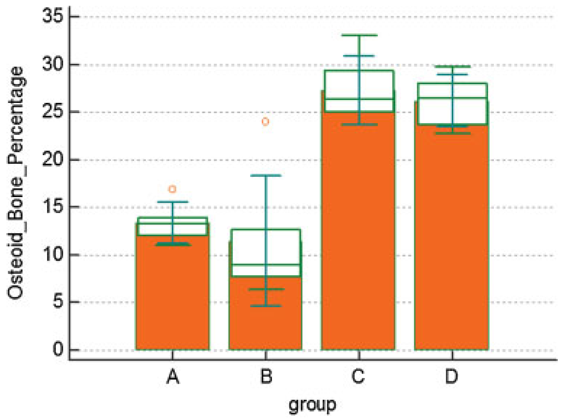
Histomorphometric assessment of MT-stained sections showed a SS decrease (p = 0.0009) in osteoid percentage (OP) in group A (OP = 13.4%, SD = 2) and group B (OP = 11.5%, SD = 6.5) compared with group C (OP = 27.3, SD = 3.5) and group D (OP = 26.2%, SD = 2.6). The Eta-square value regarding this test was 0.8 indicating that the sample size is large enough to give statistical data (Figure 4a–d and Figure 5; Table 1).
Evaluation of Radiographic Calcium Ion Concentration by EDX
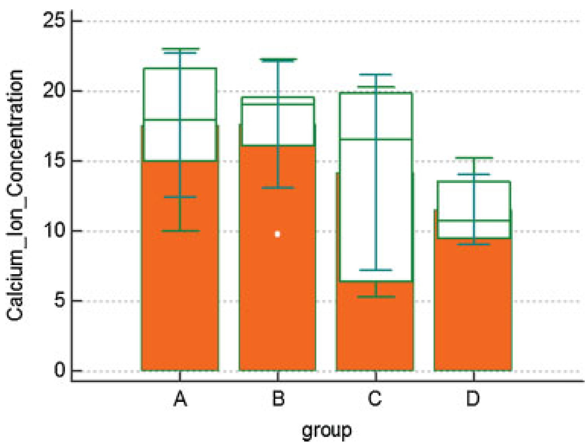
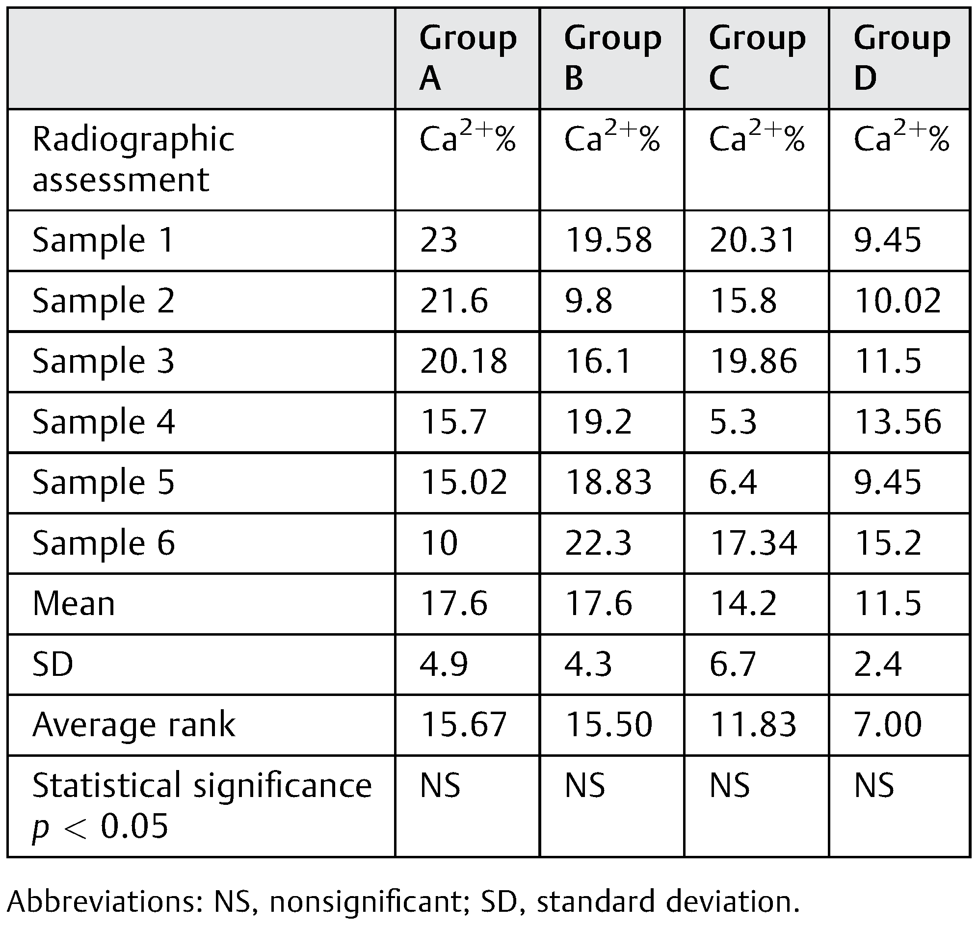
EDX showed a nonsignificant (NS) increase (p = 0.11) in calcification in group A (Ca2+% = 17.6%, SD = 4.9) and group B (Ca2+% = 17.6%, SD = 4.3) compared with group C (Ca2+% = 14.2%, SD = 6.7) and group D (Ca2+% = 11.5%, SD = 2.4). The Eta-square value regarding this test was 0.25, indicating that the sample size is of acceptable medium strength to give statistical data (Figure 6; Table 2).
Discussion
A wide range of DO rates was applied and reported previously in the literature.[11] Rates applied on experimental small animals such as rats and rabbits reached 4 mm, while large animals’ rates varied between 1, 1.5, and 2 mm/day.[11,12,13] Those studies conducted on the rapid distraction rate of 2 mm/day were highly debatable; RDO was associated usually with inferior bone quality compared with the normal DO.[14]
One of the main drawbacks of DO is the long duration taken through the procedure. Patients are at risk of pain, fracture, infection, and discomfort. In contrast to long bones, the craniofacial bones have unique characteristics such as abundant blood supply and rapid rate of bone healing. Hence, a rapid distraction rate might be a possible alternative to the normal distraction rate. Rapid distraction rate will shorten the distraction period reducing the associated complications of the DO procedure.[15]
The current research idea is based on the previously mentioned facts: the diversity of rates of distraction in literature and on the need to decrease the time of distraction to decrease complications. However, the literature showed that rapid distraction usually decreases the bone quality. Therefore, the aim of this study was to assess the effect of MSCs on bone quality of the rapidly distracted bone.
The distraction protocol of this study was based on two systematic reviews published by and Djasim et al[2] and Swennen et al.[16] The standard distraction rate of groups A and C was set at 1 mm/day and the rapid distraction rate was set at 2 mm/day in agreement with several studies that advocated this rate.[17,18,19,20,21,22,23]
In a meta-analysis, Tee and Sun[3] found that only 19 articles were published in literature concerned with the evaluation of the effect of MSCs application and tissue culture on the distracted bone. Most of the studies were conducted on small animals such as rodents; few studies were conducted only on large mammals. Siegel and Mooney,[24] O’Loughlin et al,[25] and Mills and Simpson[26] claimed that bones of higher-order mammals and nonhuman primates mimic bones of humans more effectively than the rodents.
The systematic review performed by Tee and Sun mentioned only four studies that were conducted on MSCs applications during DO performed on large animals. Kroczek et al[27] conducted a study on minipigs, Castro-Govea et al[28] on dogs, Sun et al[29] on dogs, and Aykan et al[30] on goats. All the previously mentioned studies conducted on large animals were concerned with normal DO,[27,28,29,30] not the RDO. Hence, to the best of our knowledge, there is a deficiency in literature concerned with the assessment of the effect of MSCs application during RDO in large animals.
The assessment of H&E samples showed that there was a significant increase (p = 0.00036) in the TB thickness in study groups enhanced by MSCs (groups A and B) compared with control groups (groups C and D), which is in agreement with Qi et al.[5] The TB-thickness improvement is attributed to the addition of the MSCs.
Histomorphometrical assessment of the MT samples showed a statistical significant decrease (p = 0.00093) in the percentage of the osteoid bone in study groups enhanced by MSCs (groups A and B) compared with the control groups (groups C and D). This decrease in the osteoid bone percentage indicates more rapid bone maturation associated with MSCs application, which is in agreement with Song et al.[31]
CBCT is considered to be a reliable method in the assessment of bone quality and comparable even to micro-CT,[32] multislice CT,[33] and dual-energy X-ray.[34] However, in this study, there was poor reliability between examiners. The authors attribute this finding to the human error in reproducibility of bone evaluation points on CBCT. The poor agreement between examiners was documented by Pauwels et al,[35] which showed that the inter-examiner reliability of CBCT results is poor and not reliable.
Busse et al[36] considered EDX to be a reliable method for the assessment of bone quality. Rachmiel et al[37] used EDX to assess the newly formed distracted bone. Evaluation of Ca2+ ion concentration in specimens was done using EDX to validate and support the CBCT results. In this study, there was an increase in the bone Ca2+ ion concentration in groups A and B assisted by the addition of MSCs, indicating better ossification and more rapid healing in MSCs-assisted groups. EDX and CBCT findings were correlated confirming the increase in calcification. However, the increase was not statistically significant (p = 0.11), which is in agreement with Tai et al.[38] Tai et al evaluated bone healing enhanced by MSCs in an experimental study. The nonsignificant increase may be attributed to early killing of the animals (the bone is newly formed; not fully calcified yet).
Histological assessment is considered the gold standard for assessing bone because it analyzes the bone microstructure.[9] In conclusion, this study showed that the RDO rate (2 mm/day) had a negative effect on bone quality, which is in agreement with Djasim et al,[2] since the trabecular bone thickness of RDO bone regenerate was significantly less compared with standard distraction rate.
However, the addition of MSCs application was capable of improving the bone quality of the distracted bone generated in RDO at microscopic level. There was a strong correlation between the results, since the increase in the size of the calcified trabeculae was associated with a decrease in the osteoid bone percentage and an increase in the calcium level content in the sample.
Recommendations
This study recommends an immune histochemical assessment of the MSCs before their injection to ensure their nature. The aim of immune histochemical addition is to ensure that the MSCs are the effective factor in the improvement of bone microstructure.
This study recommends further evaluation of the associated soft tissue such as muscle and nerves generated by RDO which may not be fixed by MSCs.

Conflicts of Interest

All authors disclose no financial and personal relationships with other people or organizations that could inappropriately influence (bias) their work.

References

  1. McCarthy, J.G.; Schreiber, J.; Karp, N.; Thorne, C.H.; Grayson, B.H. Lengthening the human mandible by gradual distraction. Plast Reconstr Surg 1992, 89, 1–8, discussion 9. [Google Scholar] [CrossRef] [PubMed]
  2. Djasim, U.M.; Wolvius, E.B.; van Neck, J.W.; Weinans, H.; van der Wal, K.G. Recommendations for optimal distraction protocols for various animal models on the basis of a systematic review of the literature. Int J Oral Maxillofac Surg 2007, 36, 877–883. [Google Scholar] [CrossRef] [PubMed]
  3. Tee, B.C.; Sun, Z. Mandibular distraction osteogenesis assisted by cell-based tissue engineering: a systematic review. Orthod Craniofac Res 2015, 18 (Suppl. S1), 39–49. [Google Scholar] [CrossRef]
  4. Kitoh, H.; Kitakoji, T.; Tsuchiya, H.; et al. Transplantation of marrowderived mesenchymal stem cells and platelet-rich plasma during distraction osteogenesis–a preliminary result of three cases. Bone 2004, 35, 892–898. [Google Scholar] [CrossRef]
  5. Qi, M.; Hu, J.; Zou, S.; Zhou, H.; Han, L. Mandibular distraction osteogenesis enhanced by bone marrow mesenchymal stem cells in rats. J Craniomaxillofac Surg 2006, 34, 283–289. [Google Scholar] [CrossRef]
  6. El Hadidi, Y.N.; El Kassaby, M.; El Fatah Ahmed, S.A.; Khamis, N.S. The effect of mesenchymal stem cells application on the distracted bone microstructure: an experimental study. J Oral Maxillofac Surg 2016, 74, 1463.e1–1463e11. [Google Scholar] [CrossRef]
  7. Spector, W.S. Handbook of Biological Data; W.B. Saunders: Philadelphia, PA, USA, 1956. [Google Scholar]
  8. Uccelli, A.; Moretta, L.; Pistoia, V. Immunoregulatory function of mesenchymal stem cells. Eur J Immunol 2006, 36, 2566–2573. [Google Scholar] [CrossRef]
  9. Brandi, M.L. Microarchitecture, the key to bone quality. Rheumatology (Oxford) 2009, 48 (Suppl. S4), iv3–iv8. [Google Scholar] [CrossRef]
  10. Field, A.P. Eta and Eta squared. Wiley StatsRef: Statistics Reference Online. Wiley Online Library; 2014.
  11. al Ruhaimi, K.A. Comparison of different distraction rates in the mandible: an experimental investigation. Int J Oral Maxillofac Surg 2001, 30, 220–227. [Google Scholar] [CrossRef]
  12. Stewart, K.J.; Lvoff, G.O.; White, S.A.; et al. Mandibular distraction osteogenesis: a comparison of distraction rates in the rabbit model. J Craniomaxillofac Surg 1998, 26, 43–49. [Google Scholar] [CrossRef]
  13. Schiller, J.R.; Moore, D.C.; Ehrlich, M.G. Increased lengthening rate decreases expression of fibroblast growth factor 2, platelet-derived growth factor, vascular endothelial growth factor, and CD31 in a rat model of distraction osteogenesis. J Pediatr Orthop 2007, 27, 961–968. [Google Scholar] [CrossRef] [PubMed]
  14. Troulis, M.J.; Glowacki, J.; Perrott, D.H.; Kaban, L.B. Effects of latency and rate on bone formation in a porcine mandibular distraction model. J Oral Maxillofac Surg 2000, 58, 507–513, discussion 514. [Google Scholar] [CrossRef] [PubMed]
  15. Mofid, M.M.; Manson, P.N.; Robertson, B.C.; Tufaro, A.P.; Elias, J.J.; Vander Kolk, C.A. Craniofacial distraction osteogenesis: a review of 3278 cases. Plast Reconstr Surg 2001, 108, 1103–1114, discussion 1115. [Google Scholar] [CrossRef] [PubMed]
  16. Swennen, G.; Dempf, R.; Schliephake, H. Cranio-facial distraction osteogenesis: a review of the literature. Part II: Experimental studies. Int J Oral Maxillofac Surg 2002, 31, 123–135. [Google Scholar]
  17. Glowacki, J.; Shusterman, E.M.; Troulis, M.; Holmes, R.; Perrott, D.; Kaban, L.B. Distraction osteogenesis of the porcine mandible: histomorphometric evaluation of bone. Plast Reconstr Surg 2004, 113, 566–573. [Google Scholar] [CrossRef]
  18. Kaban, L.B.; Thurmüller, P.; Troulis, M.J.; et al. Correlation of biomechanical stiffness with plain radiographic and ultrasound data in an experimental mandibular distraction wound. Int J Oral Maxillofac Surg 2003, 32, 296–304. [Google Scholar] [CrossRef]
  19. Farhadieh, R.D.; Gianoutsos, M.P.; Dickinson, R.; Walsh, W.R. Effect of distraction rate on biomechanical, mineralization, and histologic properties of an ovine mandible model. Plast Reconstr Surg 2000, 105, 889–895. [Google Scholar] [CrossRef]
  20. Hasse, A.R.; Pörksen, M.; Zimmermann, C.E. Bilateral mandibular distraction in adult dogs with an epiperiosteal distractor. Br J Oral Maxillofac Surg 2005, 43, 105–112. [Google Scholar] [CrossRef]
  21. Zimmermann, C.E.; Harris, G.; Thurmüller, P.; et al. Assessment of bone formation in a porcine mandibular distraction wound by computed tomography. Int J Oral Maxillofac Surg 2004, 33, 569–574. [Google Scholar] [CrossRef]
  22. Zimmermann, C.E.; Thurmüller, P.; Troulis, M.J.; Perrott, D.H.; Rahn, B.; Kaban, L.B. Histology of the porcine mandibular distraction wound. Int J Oral Maxillofac Surg 2005, 34, 411–419. [Google Scholar] [CrossRef]
  23. Perrott, D.H.; Rahn, B.; Wahl, D.; et al. Development of a mechanical testing system for a mandibular distraction wound. Int J Oral Maxillofac Surg 2003, 32, 523–527. [Google Scholar] [CrossRef] [PubMed]
  24. Siegel, M.I.; Mooney, M.P. Appropriate animal models for craniofacial biology. Cleft Palate J 1990, 27, 18–25. [Google Scholar] [PubMed]
  25. O’Loughlin, P.F.; Morr, S.; Bogunovic, L.; Kim, A.D.; Park, B.; Lane, J.M. Selection and development of preclinical models in fracture-healing research. J Bone Joint Surg Am 2008, Suppl. S1, 79–84. [Google Scholar] [CrossRef] [PubMed]
  26. Mills, L.A.; Simpson, A.H. In vivo models of bone repair. J Bone Joint Surg Br 2012, 94, 865–874. [Google Scholar] [CrossRef]
  27. Kroczek, A.; Park, J.; Birkholz, T.; Neukam, F.W.; Wiltfang, J.; Kessler, P. Effects of osteoinduction on bone regeneration in distraction: results of a pilot study. J Craniomaxillofac Surg 2010, 38, 334–344. [Google Scholar] [CrossRef]
  28. Castro-Govea, Y.; Cervantes-Kardasch, V.H.; Borrego-Soto, G.; et al. Human bone morphogenetic protein 2-transduced mesenchymal stem cells improve bone regeneration in a model of mandible distraction surgery. J Craniofac Surg 2012, 23, 392–396. [Google Scholar] [CrossRef]
  29. Sun, Z.; Tee, B.C.; Kennedy, K.S.; et al. Scaffold-based delivery of autologous mesenchymal stem cells for mandibular distraction osteogenesis: preliminary studies in a porcine model. PLoS One 2013, 8, e74672. [Google Scholar] [CrossRef]
  30. Aykan, A.; Ozturk, S.; Sahin, I.; et al. Biomechanical analysis of the effect of mesenchymal stem cells on mandibular distraction osteogenesis. J Craniofac Surg 2013, 24, e169–e175. [Google Scholar] [CrossRef]
  31. Song, S.H.; Yun, Y.P.; Kim, H.J.; Park, K.; Kim, S.E.; Song, H.R. Bone formation in a rat tibial defect model using carboxymethyl cellulose/BioC/ bone morphogenic protein-2 hybrid materials. BioMed Res Int 2014, 2014, 230152. [Google Scholar] [CrossRef]
  32. Hsu, J.T.; Wang, S.P.; Huang, H.L.; Chen, Y.J.; Wu, J.; Tsai, M.T. The assessment of trabecular bone parameters and cortical bone strength: a comparison of micro-CT and dental cone-beam CT. J Biomech 2013, 46, 2611–2618. [Google Scholar] [CrossRef]
  33. Parsa, A.; Ibrahim, N.; Hassan, B.; van der Stelt, P.; Wismeijer, D. Bone quality evaluation at dental implant site using multislice CT, micro-CT, and cone beam CT. Clin Oral Implants Res 2015, 26, e1–e7. [Google Scholar] [CrossRef] [PubMed]
  34. Hsu, J.T.; Chen, Y.J.; Tsai, M.T.; et al. Predicting cortical bone strength from DXA and dental cone-beam CT. PLoS One 2012, 7, e50008. [Google Scholar] [CrossRef]
  35. Pauwels, R.; Nackaerts, O.; Bellaiche, N.; et al. SEDENTEXCT Project Consortium. Variability of dental cone beam CT grey values for density estimations. Br J Radiol 2013, 86, 20120135. [Google Scholar] [PubMed]
  36. Busse, B.; Hahn, M.; Soltau, M.; et al. Increased calcium content and inhomogeneity of mineralization render bone toughness in osteoporosis: mineralization, morphology and biomechanics of human single trabeculae. Bone 2009, 45, 1034–1043. [Google Scholar] [CrossRef]
  37. Rachmiel, A.; Laufer, D.; Jackson, I.T.; Lewinson, D. Midface membranous bone lengthening: A one-year histological and morphological follow-up of distraction osteogenesis. Calcif Tissue Int 1998, 62, 370–376. [Google Scholar] [CrossRef]
  38. Tai, K.; Pelled, G.; Sheyn, D.; et al. Nanobiomechanics of repair bone regenerated by genetically modified mesenchymal stem cells. Tissue Eng Part A 2008, 14, 1709–1720. [Google Scholar] [CrossRef]
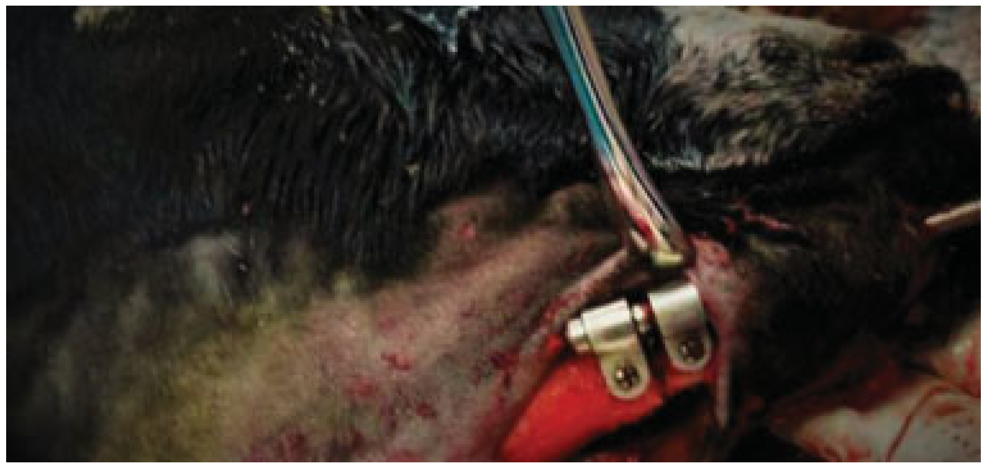
Figure 1. Distractor fixed in place.
Cmtr 11 00033 g001
Figure 2. (ad) Histological section at ×10 of H&E-stained samples showing bone trabeculae in group a, b, c and d, respectively.
Cmtr 11 00033 g002
Figure 3. Bar chart showing means of trabecular bone thickness.
Cmtr 11 00033 g003
Figure 4. (ad) Histological section at ×10 magnification of Masson’s trichrome (MT)-stained samples showing osteoid immature bone in green and mature bone in red in group a, b, c and d, respectively.
Cmtr 11 00033 g004
Figure 5. Bar chart showing difference in osteoid bone percentage between groups.
Cmtr 11 00033 g005
Figure 6. Bar chart showing mean of calcium ion concentration percentage of groups.
Cmtr 11 00033 g006
Table 1. Difference in trabecular bone thickness and osteoid bone percentage statistical data (Kruskal–Wallis test [one-way ANOVA on ranks] and post hoc; p-value is set at 0.05).
Cmtr 11 00033 i001
Table 2. Difference in calcium ion percentage statistical data (Kruskal–Wallis test [one-way ANOVA on ranks] and post hoc; p-value is set at 0.05).
Cmtr 11 00033 i002