Chronic wounds significantly lower patient quality of life and impose a large financial burden on today’s health-care system. The average cost of therapy for a lower-extremity ulcer has been estimated to be
$28,000 in the 2 years after diagnosis. To date, amputation costs total up to
$6 billion in the United States alone.[
1] Most lower-extremity ulcers are caused by venous disease, arterial insufficiency, neuropathy, or a combination of these factors.[
2]
Patients with diabetes mellitus are 17 times more likely than people without diabetes to develop gangrene and account for five of every six major limb amputations. In the United States, foot problems account for approximately 20% of all diabetic hospital admissions and 50% of all nontraumatic amputations.[
3] On average, 20% of diabetic patients with foot ulcers will require amputation. In 2001 there were approximately 85,000 lower-extremity amputations related to diabetes performed in the United States.[
4]
Venous ulcers affect approximately 1% of the world’s population, increasing health-care expenditures and decreasing quality of life, and account for almost 70% of all chronic leg ulcers.[
5,
6] Incompetent veins, valves, and impaired muscle function may lead to abnormal calf muscle pump function that can elevate ambulatory venous pressure.[
5] Venous hypertension causes disturbed microcirculation and pathologic changes in the capillaries, which eventually transform the condition into a self-amplifying, detrimental cascade, with persistent elevated levels and activities of pro-inflammatory cytokines and proteases preventing progress into a healing phase.[
7] A study by Raffetto et al[
8] showed that venous ulcer wound fluid directly inhibits the signal-transduction mechanism that regulates growth, differentiation, and apoptosis in eukaryotic cells and has negative trophic factors that affect fibroblast proliferation and ulcer healing.
Peripheral arterial disease is a slow and insidious disease process that affects an estimated 8 to 12 million Americans and accounts for approximately 20% of all leg ulcers.[
9] Depending on its severity, lower-extremity arterial disease can present in different ways, including 1) asymptomatic arterial insufficiency, 2) symptomatic disease presenting as intermittent claudication with positive noninvasive test results, and 3) critical leg ischemia, which defines the subgroup of patients with symptomatic lower-extremity arterial disease in which the ischemic process endangers part or all of the lower extremity.[
10] Atherosclerosis is the most common cause of this disease state, but acute arterial ischemia, commonly associated with an embolic event, is also prevalent.[
9]
General management of lower-extremity ulcers includes control of disease processes and associated risk factors and local treatment of the wound, whereby an attempt is made to foster the process of physiologic healing and closure through the use of various procedures and, especially, dressings.[
11] Three basic principles must be followed in all wound care: 1) modulation of the hostile wound environment, 2) adequate debridement of the wound, and 3) application of the appropriate dressing or wound-care products with off-loading of the affected area. In addition, new and innovative research is being directed at stimulating the complex orchestration of cell-mediated growth factors and chemotactic signals, ultimately resulting in increased mitotic figures and reepithelialization.[
12]
During the past several decades, the discovery of growth factors has led to excitement and renewed hope regarding the treatment of chronic wounds. Several studies[
7,
13] have been performed that demonstrate growth factor deficiencies in chronic ulcers. The concentration of growth factors was shown to be markedly decreased compared with acute wounds, and the quantity and quality of cytokines and proteases and their inhibitors were different in acute and chronic ulcers.[
13] Thus it has been hypothesized that supplementing these missing factors may stimulate wound healing. The source of such growth factors is the thrombocyte or platelet. Platelets are described as nonnucleated cells arising from megakaryocytes that produce complex protein mitogens.[
14] These mitogens are stored in the platelet α-granules and are released on exposure to thrombin. As mitogenic proteins, growth factors participate in the regulation of normal cell proliferation (mitosis), differentiation, cell migration (chemotaxis), angiogenesis, production and degradation of the extracellular matrix, the production of growth factors by other cells, and organ growth.[
13,
15] These factors are essential for wound repair. We propose that adequate wound debridement with the application of concentrated autologous platelet-derived growth factors can stimulate healing and eliminate a portal for infection.
Materials and Methods
Patient Selection
Patients were enrolled at the Mount Sinai Hospital of Queens (Astoria, New York) and at the private office of one of the authors (G.P.) (Astoria). Eligible for this study were individuals with chronic nonhealing lower-extremity wounds treated for at least 6 months with traditional wound-healing methods and exhibiting no reduction in total surface area. Previous treatment methods included a combination of enzymatic debridement and off-loading with custom-molded extra-depth orthopedic shoes and surgical debridement with subsequent use of daily wet-to-dry dressing applications.
On initial presentation, patients underwent a complete history, a physical examination, and a detailed lower-extremity examination. Vascular assessment was performed by means of manual palpation and Doppler examination of all lower-extremity pulses. Patients were excluded from the study if they had ankle-arm indices less than 0.60, if they exhibited signs of systemic or gross lower-extremity soft-tissue infection, if there was radiographic evidence of osteomyelitis, or if gangrenous changes were present at the time of enrollment. All of the patients provided informed consent and were advised of the risks and benefits of the procedure as well as alternatives.
Wound Assessment and Preparation
Wounds were evaluated on the basis of surface area and underlying pathologic processes. Surface area (SA) was calculated using the following formula: SA = π(r1)(r2). Digital photographs were taken at the time of initial evaluation and at each subsequent visit to further monitor and assess the progression of wound healing.
Before application of the concentrated autologous platelet-derived growth factors, all of the wounds were thoroughly debrided under local anesthesia. Care was taken to completely remove all hyperkeratotic, fibrotic, and necrotic tissue from the wound environment. Chronic nonhealing wounds were converted to acute bleeding wounds, thus increasing graft incorporation potential and localized chemotaxis.
Preparation of Platelet Concentrate
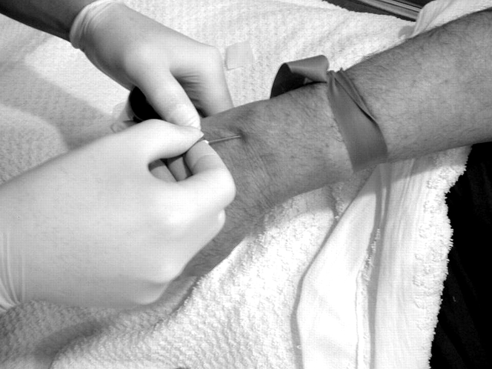
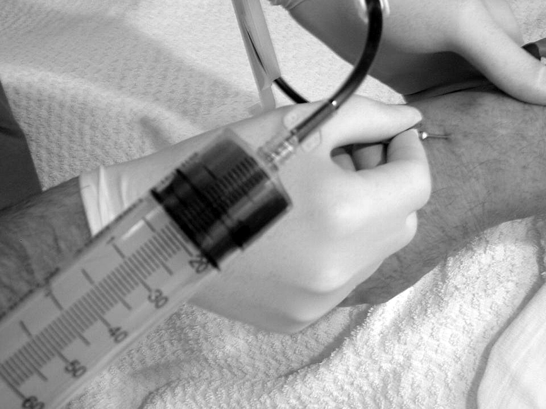
The platelet concentrate was prepared using the Gravitational Platelet Separation System (Biomet Biologics, Warsaw, Indiana) according to manufacturer guidelines. We collected 55 mL of venous blood, to which 5 mL of anticoagulant citrate dextrose solution A (Citra Anticoagulants, Braintree, Massachusetts) was added (Figs. 1
and 2
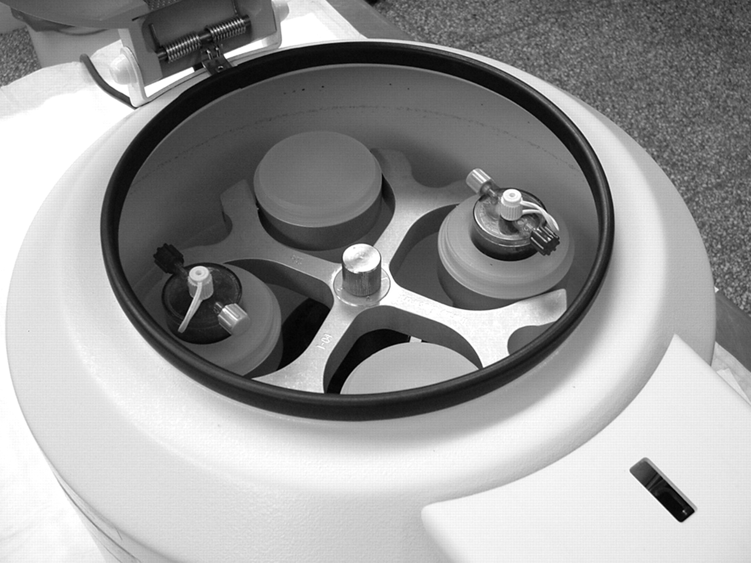
). The 60-mL solution was then centrifuged in the Gravitational Platelet Separation System (Fig. 3
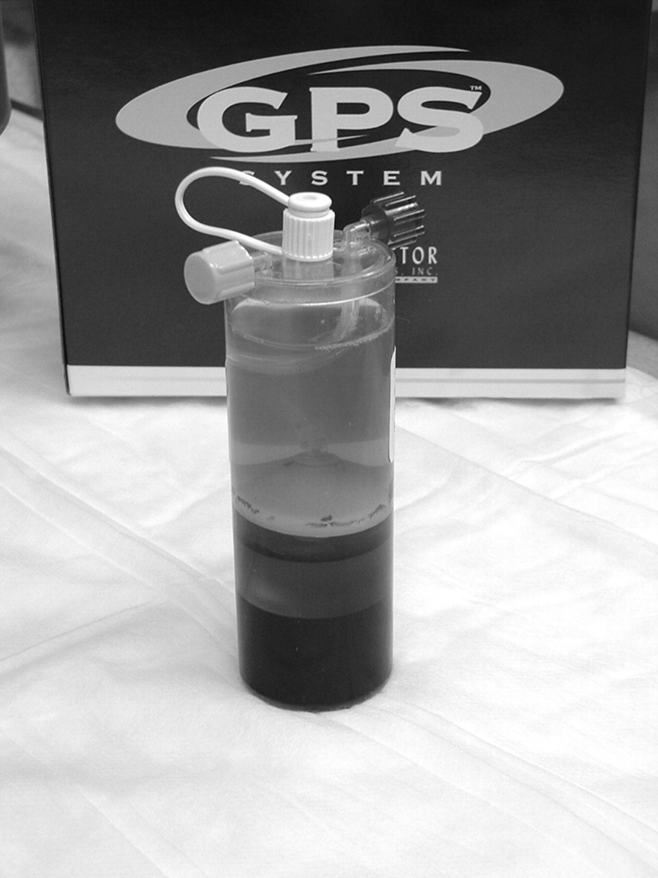
) for 15 min at 3,200 rpm (Fig. 4
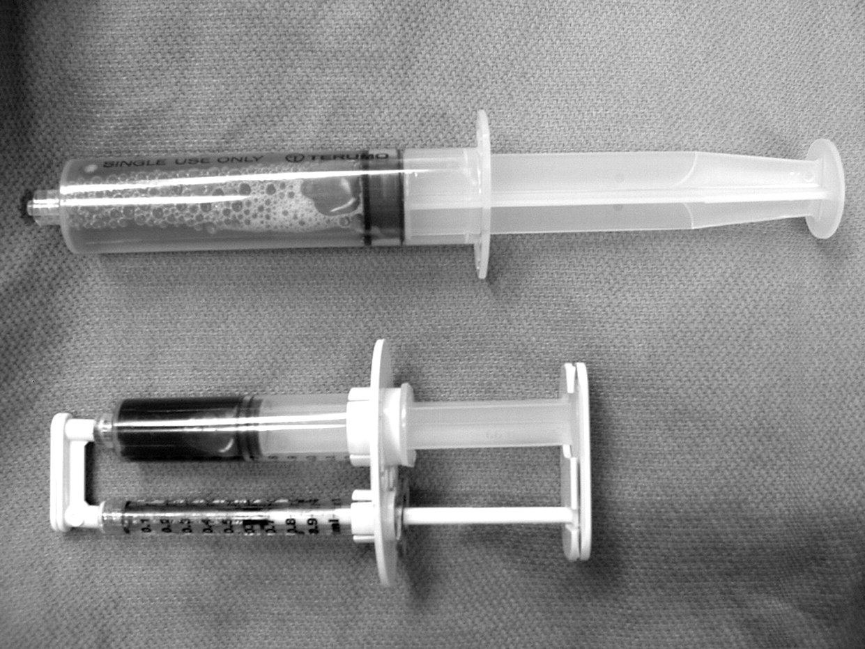
). The platelets were suspended by means of manual agitation, and the isolated buffy coat containing platelet-rich plasma was drawn into a 10-mL syringe from the Gravitational Platelet Separation System disposable centrifugation unit (Fig. 5
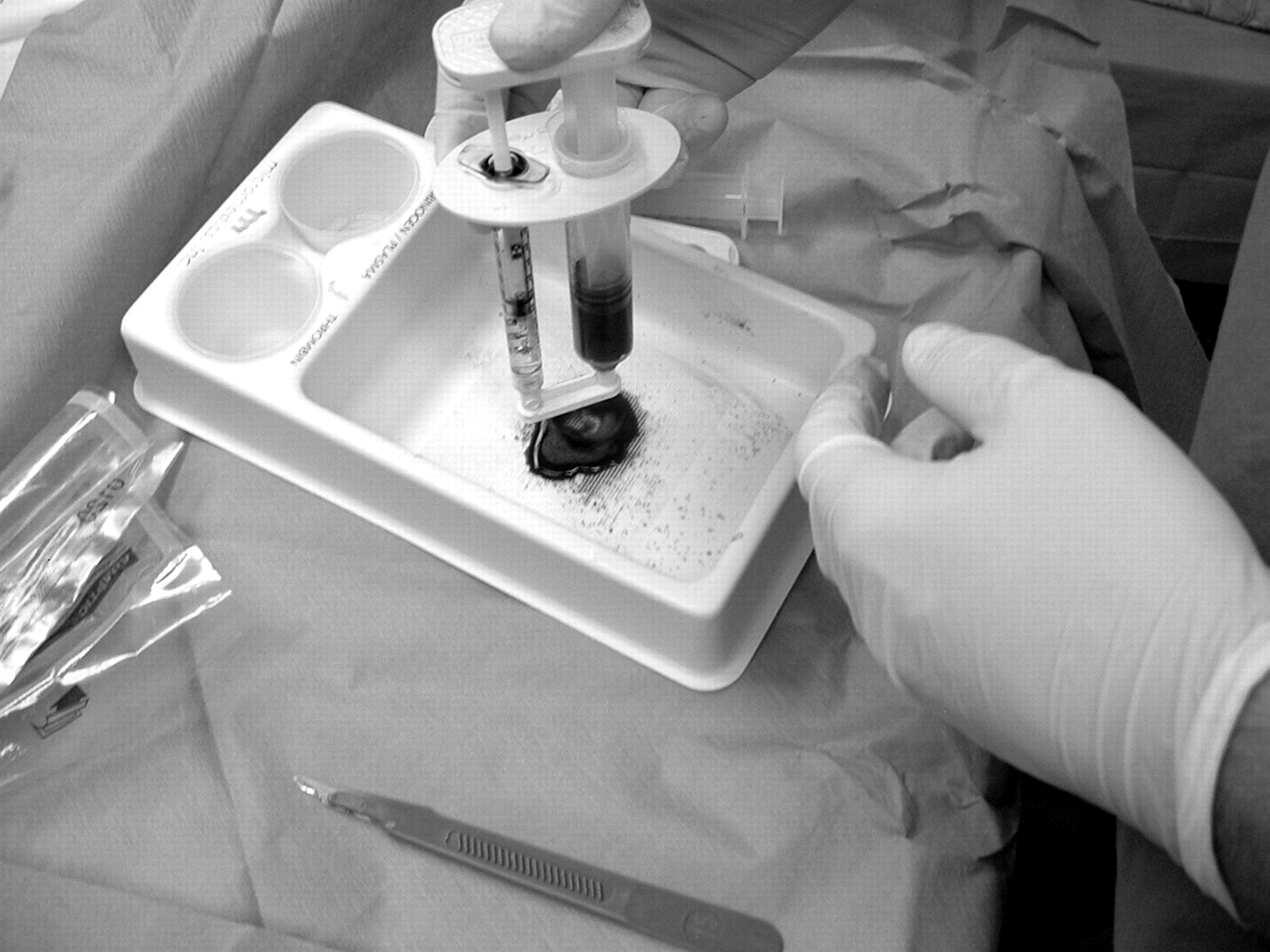
). The platelet-rich plasma was combined with thrombin and 10% calcium chloride (both from Sigma-Aldrich Corp, St Louis, Missouri), initiating platelet α-granule degranulation and growth factor release. The soft-tissue graft material was formed by combining the calcium chloride–thrombin mixture (5 mL:5,000 U) with the concentrated growth factors in a 1:10 ratio (Figs. 6
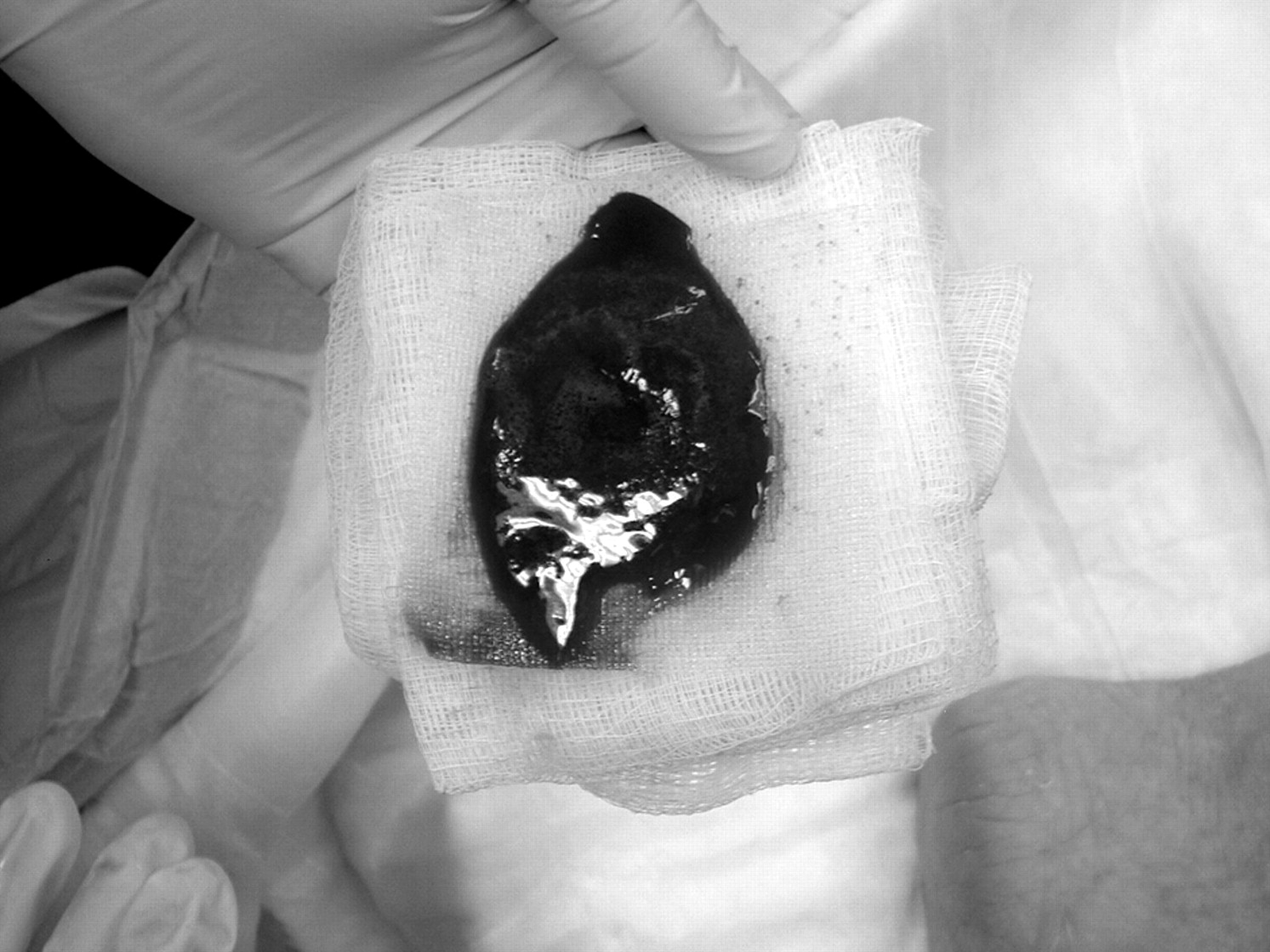
and 7
). The average yield of this process is a concentration of 1.6 × 10
6 platelets per microliter.[
16]
Graft Application Protocol
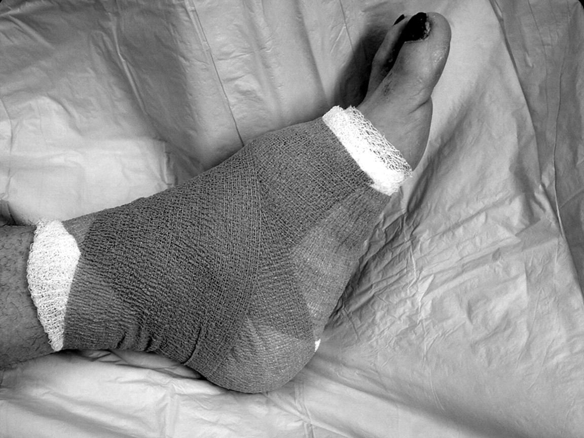
The concentrated platelet-derived autologous growth factor graft material was applied to the debrided wound bed and dressed with nonadhering sterile gauze (Adaptic; Johnson & Johnson, New Brunswick, New Jersey), a sterile antimicrobial dressing (Kerlix; Kendall, Mansfield, Massachusetts), and an elastic compressive dressing (Fig. 8
). The dressing remained clean, dry, and intact for 7 days. Patients were dispensed a surgical shoe and cane postoperatively and were instructed to limit weightbearing to essential activities of daily living. Wounds were evaluated weekly, and subsequent debridement and reapplication was performed every 2 weeks until wound closure was achieved.
Results
The study consisted of 24 patients with a total of 33 chronic nonhealing lower-extremity wounds previously treated for at least 6 months with traditional wound-healing methods and exhibiting no reduction in total surface area. Patients ranged in age from 25 to 91 years (mean age, 61.9 years). The group was composed of 13 women and 11 men and consisted of three patients with venous stasis ulcerations, two with decubitus ulcerations, five with arterial insufficiency ulcerations, eight with ulcerations due to diabetes with traumatic events prior to enrollment, and six with ulcerations due to diabetes with neuropathic pathology.
Traumatic events occurred in eight patients prior to enrollment in this study. Six patients underwent partial forefoot amputation to manage infection and had no residual bacterial colonization at the time of enrollment, one underwent several surgeries to manage an ankle fracture and later to remove the hardware, and one experienced ulceration from a short-leg cast placed after an Achilles tendon rupture.
Two patients were discontinued from the study after they developed severe lower-extremity infections that resulted in below-the-knee amputations. Two patients were lost to follow-up. One patient achieved wound closure by means of a split-thickness skin graft after undergoing several successful applications of the platelet graft. Surgical correction of forefoot neuroarthropathy was performed on another patient to fully address aberrant foot architecture and to provide primary wound closure. The reduction in wound diameter in the latter patients was recorded and included in the final data set.
The population was treated during a 10-month period (March 1, 2004 to January 1, 2005). Wound closure and complete epithelialization was achieved in 20 wounds. Seventy-five percent or greater wound closure was obtained in three wounds, 50% to 74% closure in three wounds, and 25% to 49% closure in two wounds. Five wounds displayed no improvement. Mean time to complete closure was 11.15 weeks. These findings are significant considering the failure of past treatment methods in this patient group.
Discussion
Wound Healing and Growth Factors
Wound healing occurs in three distinct phases. The first, the inflammatory or substrate phase, takes place during the first week of acute wound healing. Platelets and leukocytes migrate via chemotactic gradients to the site of injury, where cytokines and mediators begin to function. Activated platelets produce basic fibroblast growth factor and platelet-derived growth factor.
The second phase of wound healing, the proliferative phase, begins approximately 2 to 3 days after injury. Fibroblasts become the dominant cell type and produce transforming growth factor β, keratinocyte growth factor, insulin-like growth factor I, basic fibroblast growth factor, and platelet-derived growth factor. These assist in the production of collagen fibers, wound epithelialization, and angiogenesis.
The final phase is the remodeling or maturation phase, during which matrix is deposited and collagen is refashioned. Each phase of wound healing is mediated and modulated by interacting molecular signals, primarily cytokines and growth factors that stimulate and modulate the main cellular activities that underlie the healing process.[
17]
Platelet-derived growth factor was discovered in 1974, at which time research was initiated to elucidate its function. It has been determined that platelet-derived growth factor augments wound healing through stimulation of protein and collagen synthesis and remodeling, components of which are lacking in the chronic wound. Chronic wounds are characterized by defective remodeling of the extracellular matrix, failure to reepithelialize, and prolonged inflammation.[
13] Platelet-derived growth factor protein mitogens induce chemotactic responses in connective tissue as a means of attracting monocytes and neutrophils, which are necessary cellular components in chronic and acute wound repair.
In addition to platelet-derived growth factor, the functions of several other important growth factors have been elucidated. Epidermal growth factor and transforming growth factor β have been found to promote cell growth, stimulate cell differentiation, and enhance connective-tissue formation. Transforming growth factor β manages the action of peptide growth factors and determines the up-regulation and down-regulation of their effects. Insulin-like growth factor is integral to cell growth, cell development, and cellular DNA synthesis. Vascular endothelial growth factor is a vascular endothelial cell mitogen that stimulates formation of new blood vessels. Eppley et al[
16] demonstrated that the concentrations of platelet-derived growth factor, transforming growth factor β, vascular endothelial growth factor, and epidermal growth factor were all significantly greater in platelet-rich graft material than in whole blood. These growth factors are believed to be essential components of the wound-healing process.
Analysis of Study Results
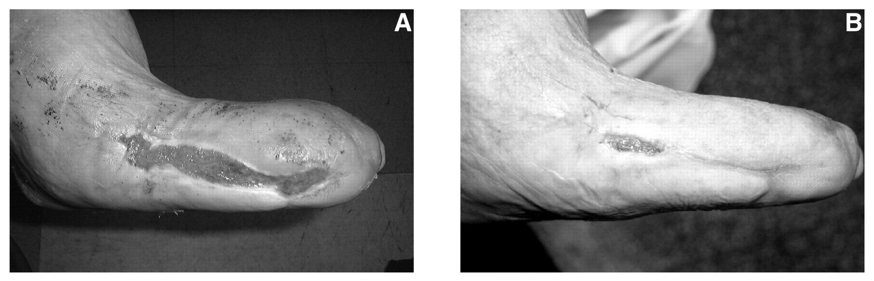
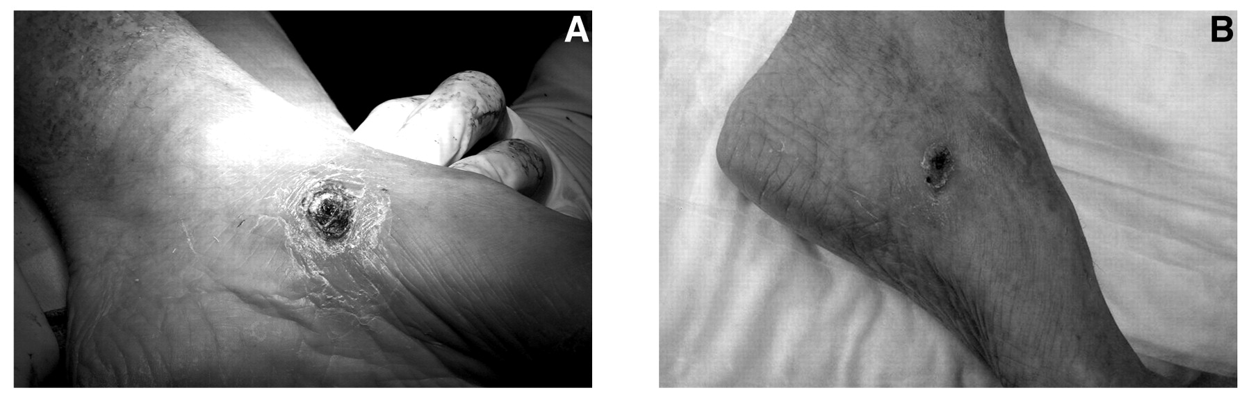
We studied the efficacy of topically applied concentrated autologous platelet-derived growth factors combined with sharp debridement as a healing modality for chronic lower-extremity wounds present in patients for at least 6 months and unresolved by traditional care methods. Treatment of this patient population using this modality resulted in a 50% or greater decrease in surface area in 26 wounds, approximately three-quarters of those treated (Fig. 9
). Twenty wounds underwent complete closure during the trial (Fig. 10
). This achievement is remarkable considering the etiologic diversity of the wounds.
The lower-extremity concentrated autologous growth factor technique is minimally invasive, is simple to perform, requires only local anesthesia, and may be performed in an office setting. The total time required from initial patient contact to patient discharge is approximately 25 min, the duration of a medium-length office visit. Application can be performed weekly or biweekly and requires little patient maintenance. Autologous growth factor material makes available a safe wound-healing medium with no risk of graft failure or rejection in the uninfected individual. The use of this modality, however, is not recommended in patients with active infection, gangrene, or neoplasm. The material would act as a bacterial growth medium and has the potential to stimulate further mitosis in neoplastic cell lines.
This method of wound closure depends on permanent correction of the underlying pathologic processes responsible for initial wound formation. Unless the causes are clearly identified and addressed, another ulceration is inevitable.
Further research is needed in the application of concentrated autologous platelet-derived growth factors. We propose a prospective, randomized, double-blind study evaluating time to resolution of chronic lower-extremity wounds, which to our knowledge has not been previously investigated.
Conclusion
The complete reepithelialization of 20 ulcerations that had been present for more than 6 months in this study population demonstrates that application of concentrated autologous platelet-derived growth factor facilitates the reduction and closure of chronic lower-extremity wounds. Healing was achieved in a patient population with a variety of underlying pathologic entities. The resolution of chronic wounds is paramount in diminishing amputation risk and controlling increasing medical costs. This study has demonstrated the effectiveness of this technique as a wound-healing treatment modality in a diverse patient population.
Figure 2.
Collection of 55 mL of venous blood, to which 5 mL of anticoagulant citrate dextrose solution A was added.
Figure 2.
Collection of 55 mL of venous blood, to which 5 mL of anticoagulant citrate dextrose solution A was added.
Figure 3.
The 60-mL solution of blood and anticoagulant citrate dextrose solution A was loaded into the Gravitational Platelet Separation System and centrifuged for 15 min at 3,200 rpm.
Figure 3.
The 60-mL solution of blood and anticoagulant citrate dextrose solution A was loaded into the Gravitational Platelet Separation System and centrifuged for 15 min at 3,200 rpm.
Figure 4.
The Gravitational Platelet Separation System after centrifugation.
Figure 4.
The Gravitational Platelet Separation System after centrifugation.
Figure 5.
Platelet-rich (dual syringe) and platelet-poor (single syringe) fractions obtained after centrifugation.
Figure 5.
Platelet-rich (dual syringe) and platelet-poor (single syringe) fractions obtained after centrifugation.
Figure 6.
A platelet-rich fraction containing concentrated autologous growth factors was combined with thrombin and 10% calcium chloride.
Figure 6.
A platelet-rich fraction containing concentrated autologous growth factors was combined with thrombin and 10% calcium chloride.
Figure 7.
A completed platelet graft ready for application.
Figure 7.
A completed platelet graft ready for application.
Figure 8.
Graft application to a debrided wound bed dressed with nonadhering sterile gauze and a sterile antimicrobial dressing.
Figure 8.
Graft application to a debrided wound bed dressed with nonadhering sterile gauze and a sterile antimicrobial dressing.
Figure 9.
Before (A) and after (B) photographs of a patient with greater than 50% wound closure.
Figure 9.
Before (A) and after (B) photographs of a patient with greater than 50% wound closure.
Figure 10.
Before (A) and after (B) photographs of a patient with complete wound closure.
Figure 10.
Before (A) and after (B) photographs of a patient with complete wound closure.