Next Article in Journal
Recognition of the Flexor Digitorum Accessorius Longus
Previous Article in Journal
A Prospective Epidemiologic Survey on the Prevalence of Foot Disease in Hong Kong
 
 
Journal of the American Podiatric Medical Association is published by MDPI from Volume 116 Issue 1 (2026). Previous articles were published by another publisher in Open Access under a CC-BY (or CC-BY-NC-ND) licence, and they are hosted by MDPI on mdpi.com as a courtesy and upon agreement with American Podiatric Medical Association.
Article

Diabetic Limb Salvage. A Team Approach at a Teaching Institution

by
Chad DeNamur
1,2 and
Guy Pupp
1
1
Kern Hospital and Medical Center, Warren, MI
2
5880 S New Berlin Rd #10, Hales Corners, WI 53130
J. Am. Podiatr. Med. Assoc. 2002, 92(8), 457-462; https://doi.org/10.7547/87507315-92-8-457
Published: 1 September 2002

Abstract

In this retrospective review, 19 diabetic patients with significant lower-extremity pathology were assessed to determine the success of limb salvage in cases of varying complexity. The patients were either scheduled or at risk for below-the-knee amputation before intervention. After the limb-salvage procedure, patients were followed for 4 months to 9 years. Eighteen patients went on to have successful procedures, avoiding below-the-knee amputation; one patient had an above-the-knee amputation. The results demonstrate the benefits of an aggressive team approach with limb salvage as a goal.

Foot disorders are the leading cause of hospitalization of diabetic patients in the United States.[1-4] Diabetic patients account for up to 83% of the nontraumatic lower-extremity amputations in the United States each year and are up to seven times more likely to undergo a major amputation than nondiabetic patients.[5,6] In the United States, the rate of nontraumatic lower-extremity amputations in diabetic patients approaches 8.6 per 1,000 per year.[5,7,8] This rate is even higher in certain minority groups, including blacks, Hispanics, and Native Americans.[9]
Treating diabetic patients with limb-threatening situations can be the most challenging part of a foot and ankle surgeon’s practice. When dealing with this patient population, one cannot rely on a “textbook presentation.” To make the appropriate decision, a great deal of diagnostic and prognostic information must be gathered.
Limb salvage is defined as survival without an amputation at the level of the leg or thigh.[5] Major amputations are either below or above the knee. Studies have shown that the 3-year survival rate after one major amputation is as high as 50%, while the 5-year survival rate is around 40%.[7,10] The argument for limb salvage is supported by these figures alone. Limb salvage has been shown to be cost effective as well.[11,12] One study suggests that the higher the level at which the amputation is done, the higher the cost when rehabilitation, prosthetic costs, and further morbidity are considered. It has been estimated that about 50% of amputations in diabetic patients are made necessary because of neglect and could have been prevented with early intervention, conservative routine foot care, education, aggressive surgical treatment (revascularization, limb salvage, or both), and appropriate footwear.[1] A multidisciplinary approach to diabetic foot problems has gained much favor during the past several years. The specialists involved include a vascular surgeon, a foot and ankle surgeon, an endocrinologist or diabetologist, an infectious disease specialist, wound-care nurses and educators, and a qualified pedorthist. Several institutions across the country employ the multidisciplinary approach and have experienced a significant decrease in the number of lower-extremity amputations.[4,5,13]

The Diabetic Foot

The pathophysiology of diabetic feet must be understood in order to treat them. Peripheral sensorimotor polyneuropathy is the most important contributing factor in the increased vulnerability to lower-extremity amputation of the diabetic foot. More than 80% of diabetic patients with foot ulcerations have peripheral neuropathy.[12,14] When this is combined with increased microtrauma to high-plantar-pressure points, the result is a recipe for ulceration. The risk is even higher when one considers that diabetic patients are more likely to have peripheral vascular disease, a compromised immune system, and Charcot’s osteoarthropathy.
Sensorimotor neuropathy is thought to be due to axonal degeneration and secondary demyelinization. Hyperglycemia leads to an intracellular accumulation of sorbitol and other metabolites, causing depletion of myoinositol, which is necessary for myelin formation.[15] Good glycemic control is the best way to prevent the progress of sensorimotor neuropathy.
Neuropathic osteoarthropathy (Charcot’s joint) is another problem that complicates the disease process. Neuropathy plays a key role in the development of Charcot’s osteoarthropathy. Current theory suggests that there is an increased blood flow to the extremity secondary to autonomic neuropathy and sympathetic denervation, which decreases the ability of arterioles to vasoconstrict. This, in turn, causes an increase in bone resorption and osteopenia, which may be coupled with increased stress in the area due to motor neuropathy and muscle imbalance, especially of the triceps surae. The loss of sensation and proprioception allows abnormal stresses to occur across joints without the patient’s awareness. Osseous and joint destruction are inevitable if the foot is not protected. Armstrong et al[16] studied 55 diabetic patients with acute Charcot’s osteoarthropathy who presented with locations described by the Sanders pattern classification: 1) forefoot, 3%; 2) tarsometatarsal joint, 48%; 3) Chopart’s joint, 34%; 4) ankle joint, 13%; 5) posterior calcaneus, 2%. A majority of patients develop osteoarthropathy in the midfoot arch region, which is a vulnerable area for arch collapse and chronic, nonhealing ulcer formation. When acute Charcot’s arthropathy develops, patients should be nonweightbearing until resolution is seen clinically and radiographically. At the authors’ center, skin temperatures are obtained weekly to monitor the process. These patients must be nonweightbearing until resolution of the acute phase to prevent residual deformity and thus limit the risk of chronic ulceration, infection, and possible amputation. The authors have found total-contact casting to be the most effective way to keep patients nonweightbearing. Armstrong et al[16] casted patients with total-contact casts for an average (±SD) of 18.5 ± 10.6 weeks for acute Charcot’s osteoarthropathy.
Peripheral vascular disease also must be taken into account when dealing with diabetic feet. The Framingham study[17] showed that diabetes is a significant risk factor for atherosclerotic and peripheral arterial disease. It is important for foot-care specialists to work closely with vascular surgeons when dealing with compromised patients. Patients with nonpalpable pedal pulses should have a vascular work-up to assess their vascular supply. Ankle-brachial indices are not reliable in diabetic patients because of their inability to compress calcified vessels. In this study, patients were referred to the vascular surgeon whenever their vascular status was questionable. Arteriograms were obtained when needed, and revascularization procedures were performed whenever necessary. An intact vascular supply is important when doing any local foot procedure, whether elective or salvage. Without an adequate blood supply, healing will be compromised and failure is probable. The exception is when emergency incision and drainage is pertinent. In one study, 35 of 42 (83%) extremities with patent bypass grafts achieved and maintained primary healing of the local foot procedure.[18] Clearly, ischemic patients with osteomyelitis do not automatically require below- or above-the-knee amputations. These patients can be managed with wound care, control of infection, revascularization, and limb salvage.
In consulting a vascular surgeon, limb salvage is the primary goal. What one surgeon may deem salvageable, another may see as in need of amputation. A vascular surgeon who believes in aggressive limb salvage should be consulted. He or she must be willing to bypass compromised limbs that may be infected. Appropriate blood flow is necessary for the healing of any local procedure. An infected limb with osteomyelitis may be maintained with incision and drainage, local wound care, and intravenous antibiotics until revascularization is successful. With the vascular surgeon’s intervention, local salvage procedures can then be performed with confidence that healing is likely to occur.

Results

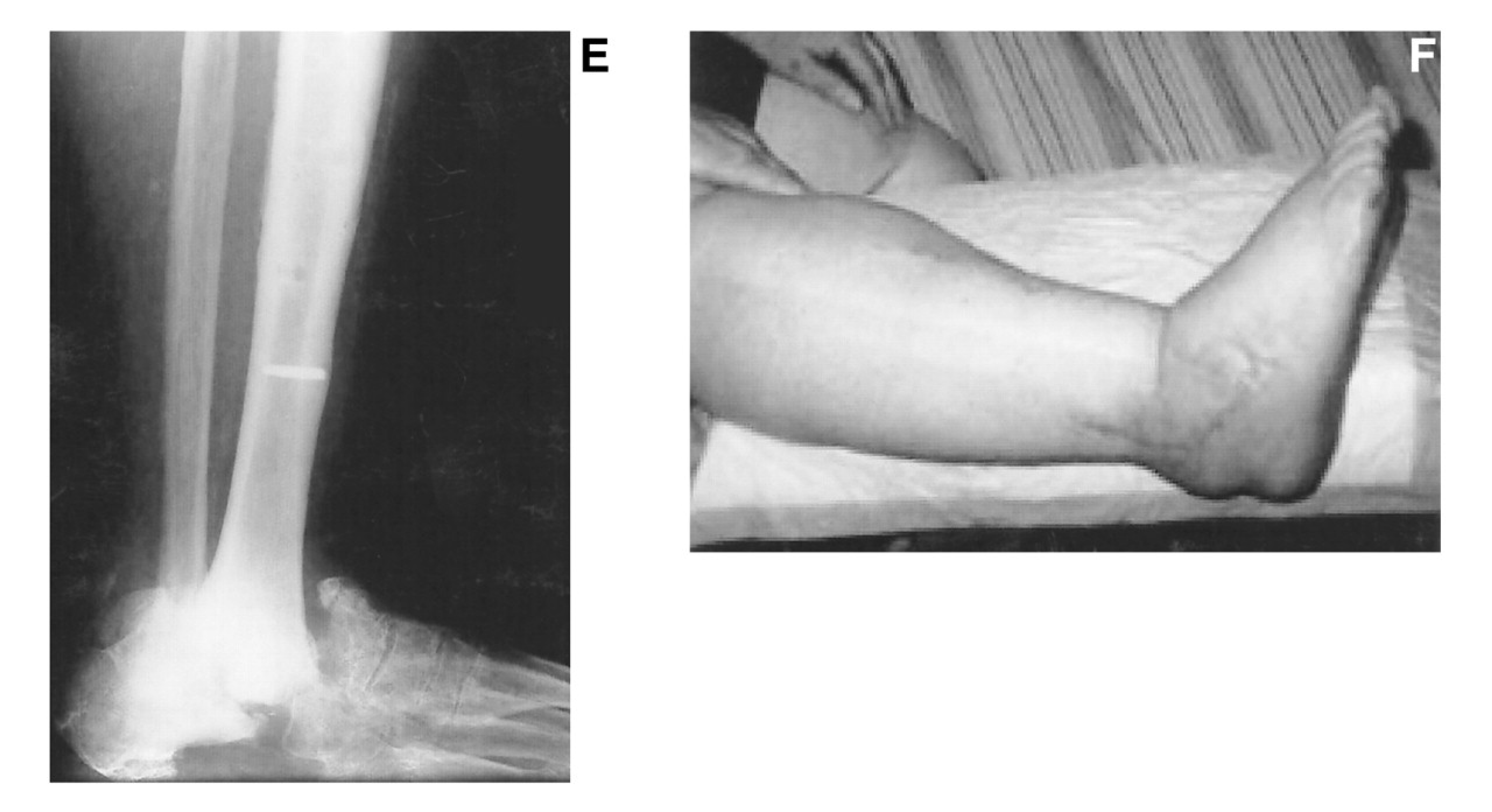
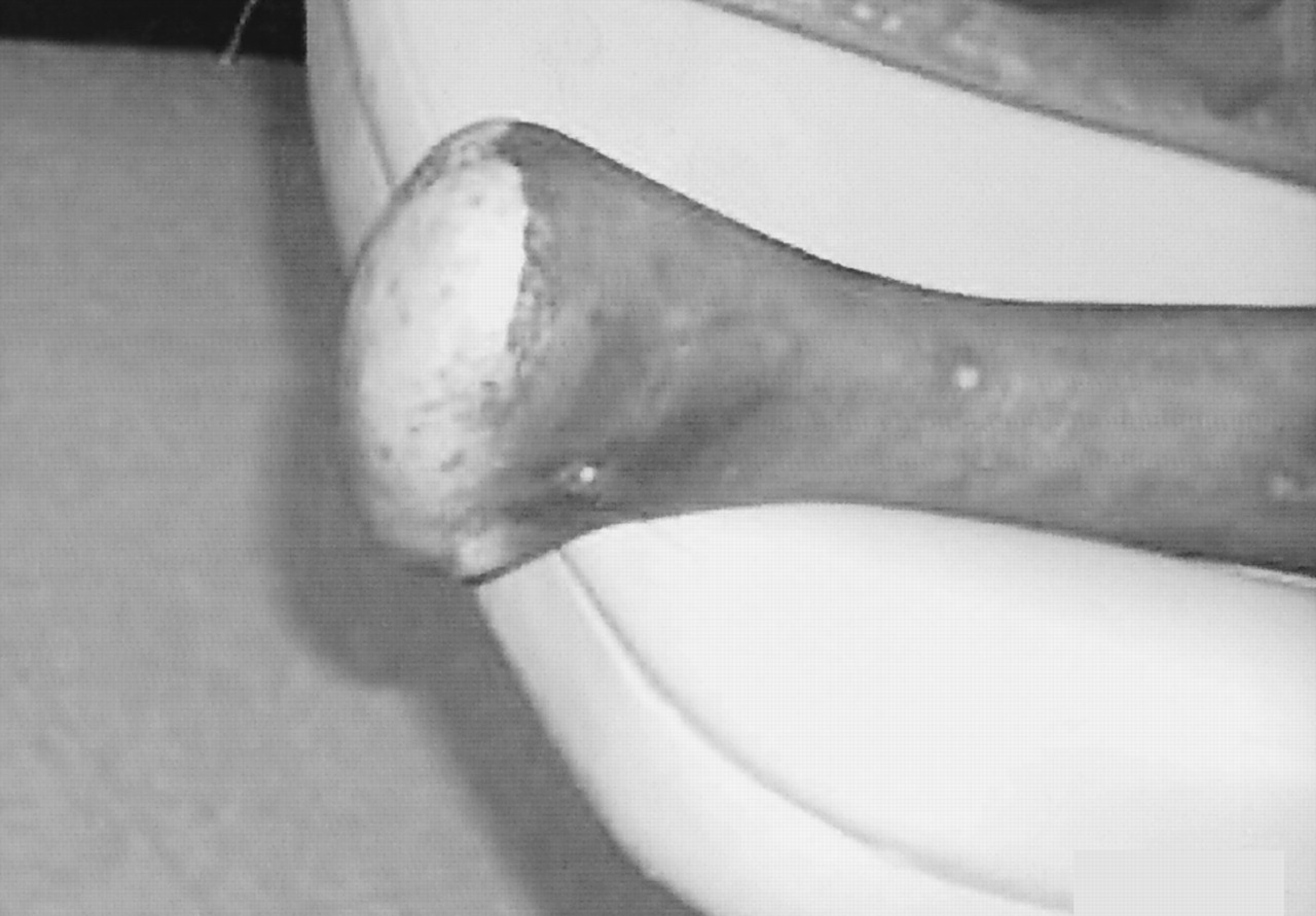
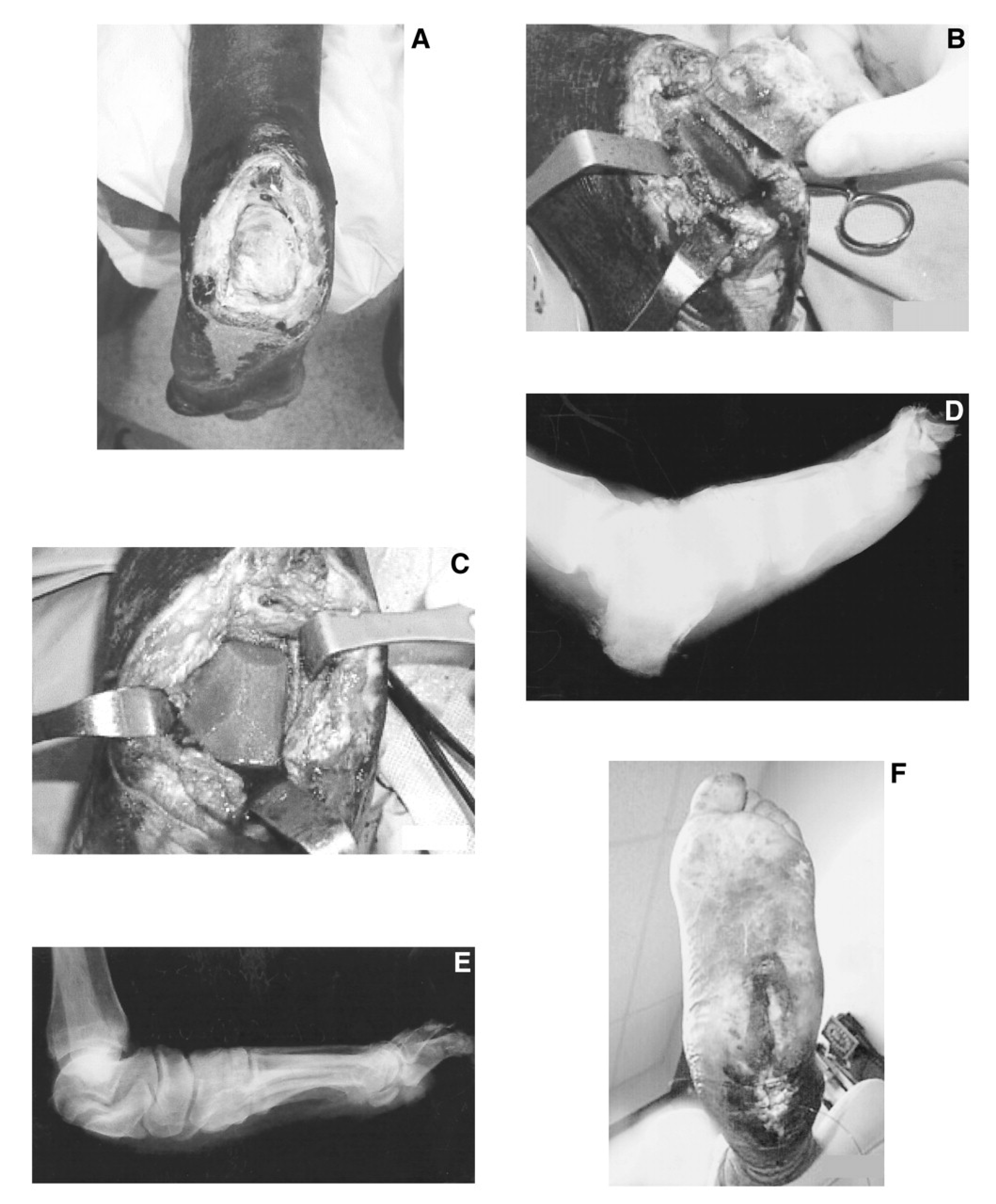
In this retrospective review, 19 diabetic patients with limb-threatening situations were reviewed to demonstrate the success of limb salvage in cases of varying complexity. A majority of these patients were considering below-the-knee amputations prior to presentation. The study consisted of 12 men and 7 women; the ages of the patients ranged from 34 to 65 years, with a mean age of 50.1 years. Eighteen of the 19 patients underwent successful limb-salvage procedures. Postoperative follow-up ranged from 4 months to 9 years. One patient proceeded to above-the-knee amputation without salvage. Four of the 18 patients who underwent successful limb salvage procedures are now deceased. Sixteen patients were diagnosed with contiguously spread osteomyelitis secondary to diabetic neuropathic ulcerations. Ten of the 18 patients with salvaged limbs received a diagnosis of Charcot’s osteoarthropathy, which complicated treatment and diagnosis. Six of these 10 patients had Charcot’s ankle joints with dislocation. If osteomyelitis was present, the affected bone was resected, and ankle fusion or stable pseudarthrosis was achieved. A stable pseudarthrosis is not considered failure because it has proved functional in several patients (Fig. 1). Four patients received Chopart’s amputations, and three underwent transmetatarsal amputations of the affected foot. All patients with foot amputations went on to complete healing (Fig. 2). One patient underwent a subtotal calcanectomy (Fig. 3). Three patients required revascularization of the affected limb prior to the salvage procedure. Five patients had previous below-the-knee amputations of the contralateral limb. Eight patients were on renal dialysis.
After adequate postoperative care, patients were evaluated by a certified pedorthist for appropriate footwear and ancillary devices to prevent further breakdown of the salvaged limb. Custom-molded shoes were used for almost every patient, and a patellar tendon–bearing device was used in nine patients.

Discussion

Dealing with diabetic patients who have major limb complications requires a great deal of patience. Patients tend to neglect what they cannot feel, and noncompliance can be a problem. It is important to instruct these patients to monitor their condition after healing to prevent further problems.
A team approach to diabetic limb salvage should be the standard of care. It is the responsibility of the health-care provider to arrange the appropriate consultations. If a foot-care provider does not feel comfortable dealing with these patients, he or she should refer the patient to someone who has the appropriate interest and experience.
This review reported on a series of diabetic limb-salvage cases and illustrated that even severely complicated limbs can be saved. The multidisciplinary approach can be very successful. However, the individuals on the team must be qualified. Treating this population is extremely challenging for foot and ankle surgeons. The goal should be to save limbs; therefore, amputations should be performed only when necessary.
  1. Frykberg RG: Team approach toward lower extremity amputation prevention in diabetes. JAPMA87: 305, 1997.
  2. Gibbons GW, Eliopoulos GM: “Infection of the Diabetic Foot,” in Management of Diabetic Foot Problems, 2nd Ed, ed by GP Kozak, DR Campbell, RG Frykberg, et al, WB Saunders, Philadelphia, 1995.
  3. Frykberg RG: “Diabetic Foot Ulcerations,” in The High Risk Foot in Diabetes Mellitus, ed by RG Frykberg, Churchill Livingstone, New York, 1991.
  4. Thomson FJ, Veves A, Ashe H, et al: A team approach to diabetic foot care: the Manchester experience. Foot2: 75, 1991.
  5. Van Gils CC, Wheeler LA, Mellstrom M, et al: Amputation prevention by vascular surgery and podiatry collaboration in high-risk diabetic and nondiabetic patients. Diabetes Care22: 678, 1999.
  6. Armstrong DG, Lavery LA, Harkless LB, et al: Amputation and reamputation of the diabetic foot. JAPMA87: 255, 1997.
  7. American Diabetes Association: Diabetes 1996 Vital Statistics, American Diabetes Association, Alexandria, VA, 1996.
  8. Centers for Disease Control and Prevention: Diabetes Surveillance, 1993, Centers for Disease Control and Prevention, Atlanta, 1993.
  9. Ashry HR, Lavery LA, Armstrong DG, et al: Cost of diabetes-related amputations in minorities. J Foot Ankle Surg37: 186, 1998.
  10. Lee JS, Lu M, Lee VS, et al: Lower extremity amputation: incidence, risk factors, and mortality in the Oklahoma Indian diabetes study. Diabetes42: 876, 1993.
  11. Ollendorf DA, Kotsanos JG, Wishner WJ, et al: Potential economic benefits of lower extremity amputation prevention strategies in diabetes. Diabetes Care21: 1240, 1998.
  12. Pinzur MS, Stuck R, Sage R, et al: Benchmark analysis on diabetics at high risk for lower extremity amputation. Foot Ankle17: 695, 1996.
  13. Gibbons GW, Marcaccio EJ, Burgess AM: Improved quality of diabetic foot care, 1984 versus 1990: reduced length of stay and costs, insufficient reimbursement. Arch Surg128: 576, 1993.
  14. Caputo GM, Cavanagh PR, Ulbrecht JS, et al: Assessment and management of foot disease in patients with diabetes. N Engl J Med331: 854, 1994.
  15. Greene DA, Lattimer SA, Sima AAF: Sorbitol, phosphoinositides, and sodium potassium-ATPase in the pathogenesis of diabetic complications. N Engl J Med316: 599, 1987.
  16. Armstrong DG, Todd WF, Lavery LA, et al: The natural history of acute Charcot’s arthropathy in a diabetic foot specialty clinic. JAPMA87: 272, 1997.
  17. Akbari CM, LoGerfo FW: Diabetes and peripheral vascular disease. J Vasc Surg30: 373, 1999.
  18. Rosenblum BI, Pomposelli FB, Giurini JM, et al: Maximizing foot salvage by a combined approach to foot ischemia and neuropathic ulceration in patients with diabetes: a 5-year experience. Diabetes Care17: 983, 1994.
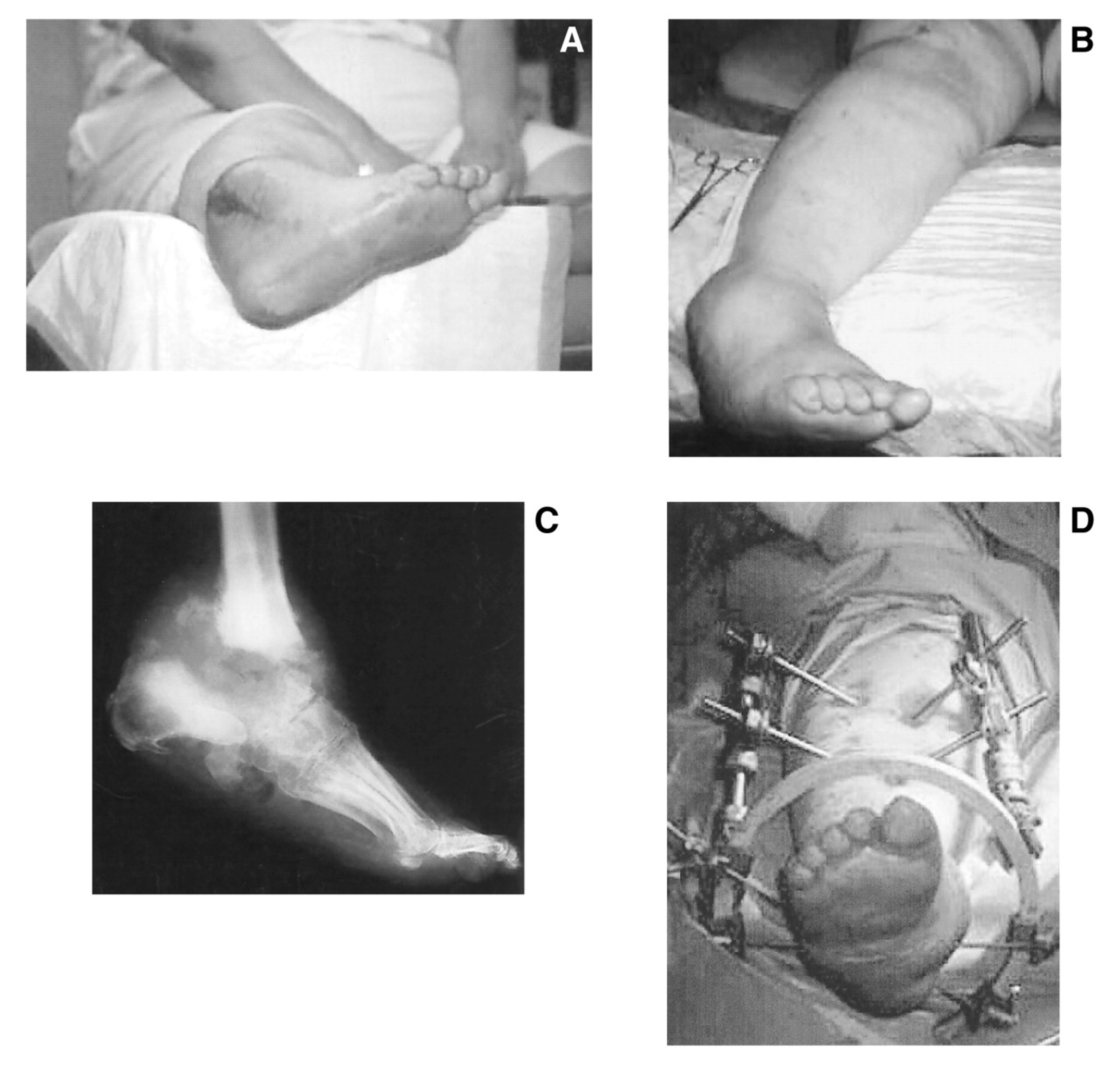
Figure 1. Patient 11. A, Preoperative photograph of Charcot’s ankle with an osteomyelitic talus; B, severe ankle varus deformity; C, preoperative lateral ankle radiograph; D, postoperative photograph with external Ilizarov frame intact. E, Postoperative lateral ankle radiograph; F, postoperative photograph of patient showing healing with good alignment.
Japma 92 00457 g001aJapma 92 00457 g001b
Figure 2. Patient 18. Postoperative view of Chopart’s amputation.
Japma 92 00457 g002
Figure 3. Patient 19. A, The patient presented with exposed osteomyelitic posterior calcaneus; B, intraoperative view of subtotal calcanectomy; C, intraoperative view of healthy bleeding calcaneus; D, preoperative lateral radiograph showing destruction of exposed posterior calcaneus; E, postoperative lateral radiograph after resection of infected bone; F, postoperative photograph showing healing by secondary intention after minimal wound dehiscence.
Japma 92 00457 g003