Introduction
Neuropathic osteoarthropathy, also known as Charcot’s arthropathy, is a common complication in patients with diabetes mellitus and severe neuropathy. It is characterized by pathologic fractures, joint dislocation and deformity [
1–
4]. Its prevalence ranges from 0.16% in a general population of patients with diabetes to 13% of patients presenting to a high-risk diabetic foot clinic [
3,
5,
6]. The etiology of the arthropathy is largely unknown, although several theories have been propagated. The major morbidity of a Charcot joint is a deformity. This can be devastating, because a bony protuberance and/or gross instability in the face of neuropathy can lead to repetitive ulcerations and subsequent amputation.
In this review we will discuss the etiology, diagnosis and therapy of Charcot’s arthropathy of the foot in persons with diabetes mellitus.
Etiology
Although the French neurologist Jean Martin Charcot (1825–1893) gave his name to neuropathic osteoarthropathy, he was not the first to describe this condition [
7]. In 1703, Musgrave described it as a complication of venereal disease [
8]. Charcot, a well-known member of the French school of physicians and neurologists, also linked the disease to venereal disease. Furthermore, in 1868 he described neuropathy as an essential etiological factor. In those days, neuropathic osteoarthropathy was a common complication of neurolues. Syphilis remained the disease associated with the arthropathy until 1936. In that year, Jordan [
9] was the first to concisely describe it as a complication of diabetes mellitus.
Due to the development of insulin and penicillin, diabetic complications have become more common while complications of syphilis have decreased (respectively). Ultimately, diabetes has far superseded syphilis as the most common preexisting etiological condition at the root of development of osteoarthropathy.
Charcot believed that osteoarthropathy was due to deficiencies in trophic centers in the spine. German physicians, most notably Virchow and Volkmann, opposed Charcot’s theory. They believed that Charcot’s theory had no solid scientific basis and was predicated solely on clinical observations. Their competing etiological theory was rooted in neurotraumata. A foot without proper protective sensation is subject to trauma. These traumata ultimately lead to fractures which then heal with exuberant bone formation. This theory was tested by Eloesser [
10] who sectioned posterior nerve roots in animals: following a period of activity he found bony changes in 71% of these animals. In more recent years, Finsterbush and Friedman [
11] repeated Eloesser’s experiments, but casted the animals after sectioning the posterior roots. The authors noted a difference in the response to immobilization between normal and denervated groups and concluded that trauma, though important, is not the primary factor leading to the deterioration of desensitized joints. Indirectly, support for the German theory was found by Armstrong and Lavery [
12] who identified increased pressures on the plantar aspect of the forefoot in acute Charcot’s arthropathy. This problem, when coupled with glycosylation of the Achilles tendon, creates tremendous strain in the midfoot, causing collapse. This is, incidentally, the most frequent location for Charcot’s arthropathy. Anatomical involvement of Lisfranc’s (tarsometatarsal) and Chopart’s (talonavicular/calcaneocuboid) joints account for more than 80% of incident episodes in the foot and ankle [
3,
12].
Finsterbush and Friedman’s work opened the way for further hypotheses. Subsequent investigators noted that osteopenia was sometimes present in patients with osteoarthropathy [
13,
14]. They postulated that increased blood flow causes resorption of bone leading to weakening of supporting structures. The hyperemia is caused by loss of sympathetic tone and constrictive control of the vessels. The osteopenic bone is susceptible to fractures caused by even minor trauma. In later studies, principally performed by Edmonds and co-workers [
15,
16], scintigraphy indeed demonstrated an increase in blood flow in the presence of neuropathy. Others have suggested that repetitive trauma by itself increases bone turnover. Indeed, osteoclast activity as measured by pyridinoline crosslinked carboxy-terminal telopeptide domain of type-I collagen and urinary deoxypyridinoline seems to be increased in patients with osteoarthropathy [
17–
19]. Bone formation, and with this bone turnover, also seems to be increased in these patients as measured by the increased levels of the carboxy-terminal propeptide of type-I collagen and bone-specific alkaline phosphatase [
18,
19]. These suggestions were confirmed by a study that found decreased bone mineral density in Charcot patients [
20].
Currently, it is common belief that the real etiology lies somewhere inbetween the aforementioned neurovascular and neurotraumatic theories. Autonomic neuropathy might cause osteopenia by an increase in blood flow to the extremity. Sensory neuropathy makes the patients unaware of the abnormal stress on the joint caused by motor neuropathy. Such abnormal stress can cause bone damage through osteoclast activity and can lead to fractures [
11,
13,
14,
17–
21].
Diagnosis and Monitoring
The diagnosis of Charcot’s arthropathy is usually made on clinical manifestations consisting of a painful, swollen and warm foot in the presence of neuropathy and often full or bounding pedal pulsations. Because these manifestations can be mild, especially at the onset of the symptoms, physicians should have a high index of suspicion.
Radiological examination is an important tool in detecting, staging and monitoring Charcot’s arthropathy. The clinical manifestations are typically less than would be expected of the degree of bone destruction [
22]. Radiological diagnosis is based on the presence of destruction, subluxation or dislocation. In the early phase of the disease, the x-ray is often normal or may demonstrate slight changes in early cases [
23]. It is advisable to repeat x-rays within a couple of weeks if there is a high clinical suspicion for osteoarthropathy. Subtle changes might become more obvious over time. More advanced cases show varying stages of fracture and healing. Even more advanced cases show joint instability, with trabecular bridging and sclerosis.
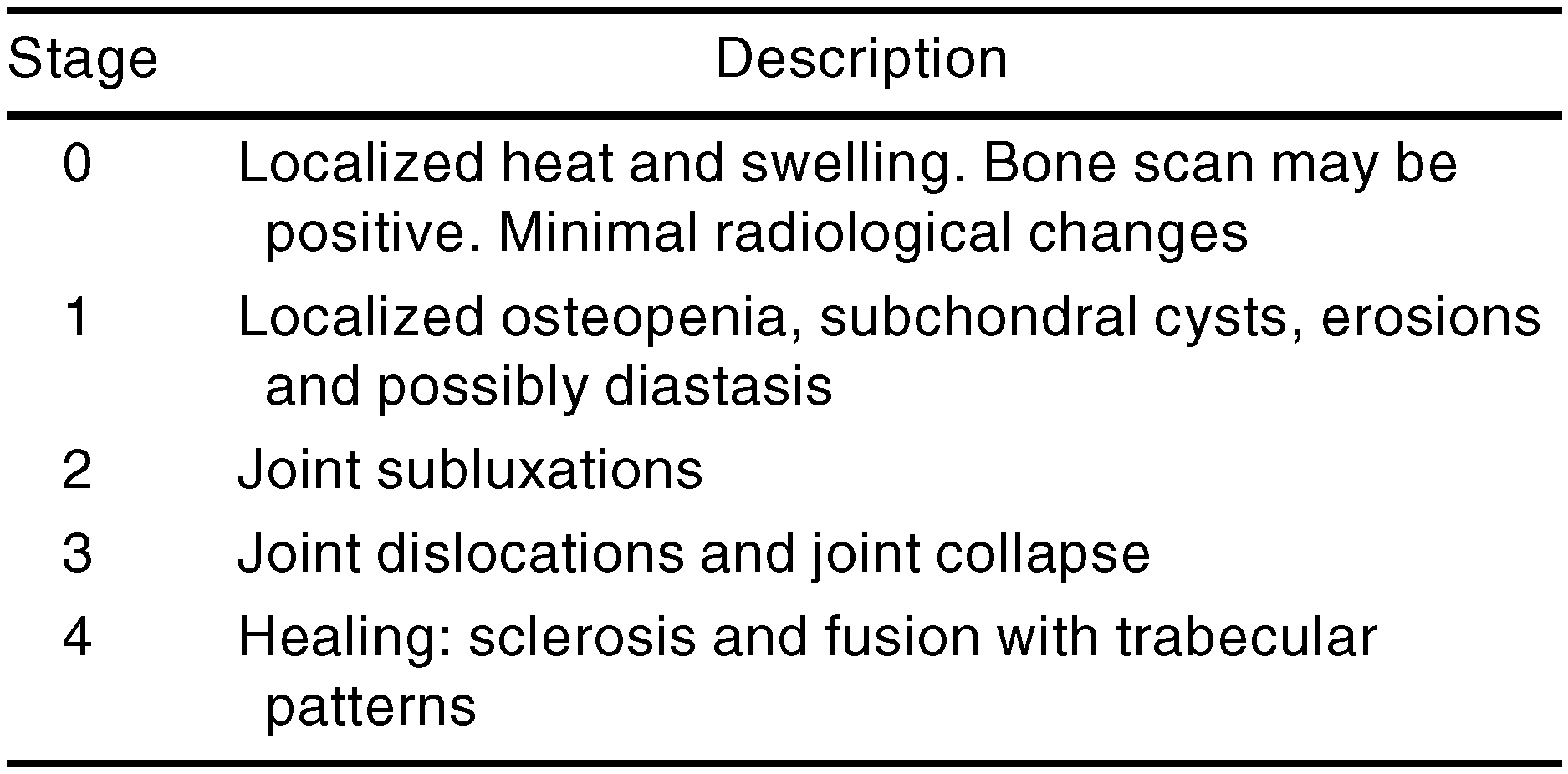
Several authors have staged Charcot’s arthropathy. In a landmark tome published in 1966, noted American orthopedist Eichenholtz [
24] described three anecdotal phases (
Table 1). A disadvantage of this system is that the early phases are absent. As mentioned earlier, radiological changes can be absent in the early stages of the disease. Therapy is likely to have its greatest effect in these early stages. More recently, Sella and Barrette [
25] have developed another Charcot classification system based on radiological findings, which does include the early phase (
Table 2). Armstrong and Lavery [
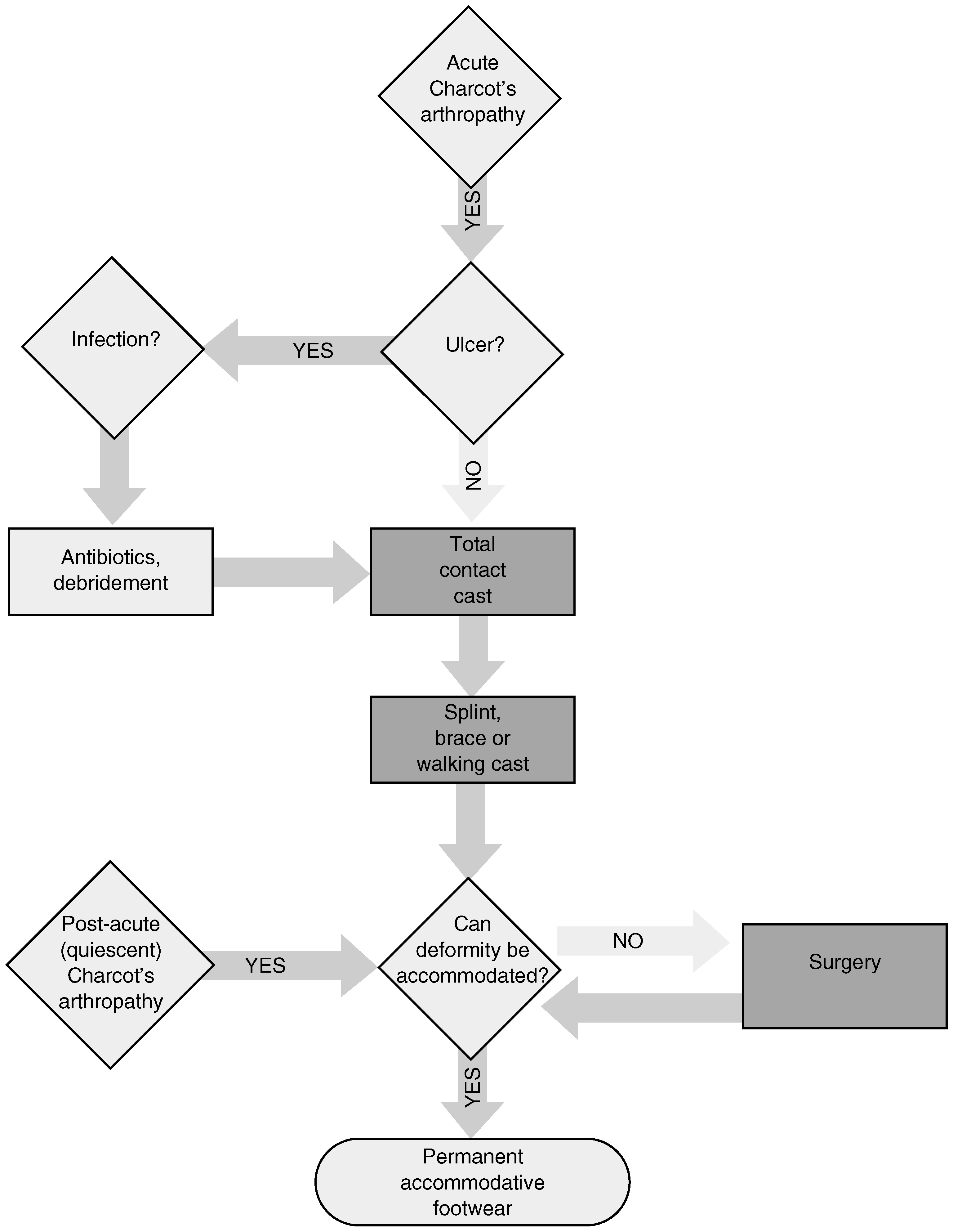
2] described a pragmatic, treatment-based, two-part staging system—indicating that the foot is either ‘acute’, implying that offloading, protection and stabilization are key components, or it is ‘post-acute (quiescent)’, meaning that the area in question is ready for progressive weightbearing and shoeing, as is feasible (
Fig. 1). Feet that are not able to be shoed may then be considered for reconstructive surgery or amputation.
Another way to use radiographs in Charcot feet is to describe the anatomical distribution pattern according to Sanders and Mrdjencovich [
26], who reported five different patterns ranging from the forefoot (pattern 1), to the Lisfranc’s joint (pattern 2), the lesser tarsus (pattern 3), the ankle (pattern 4) and the posterior calcaneus or the ‘posterior pillar’ (pattern 5) [
26,
27].
A Charcot joint is hotter than the joint in the contralateral foot. This heat becomes less intense when the disease settles. Dermal thermometry of the foot has become possible with the wide availability of handheld infrared thermometers. These temperature scanners are relatively inexpensive instruments that have been shown to be quite accurate and practical in following the progress of the disease during total contact casting [
28,
29].
Bone scanning can reveal perturbation of the small bones of the foot. Isotopic uptake at the area of bone destruction is elevated on average two- to three-fold compared with normal individuals [
16]. Edmonds and co-workers [
15,
16] suggested using the ratio of the uptake of the foot and a standard area on the ipsilateral tibia. However, another study suggested that a contralateral reference point showed more consistent trends over time [
28].
As blood flow is usually increased in a Charcot foot, the ‘dynamic phase’ of the scan can be used to see if there is increased appearance of radioactivity in the first 2 min following isotope injection. Dynamic phase uptake correlates closely with disease activity and decreases in later stages of the disease [
28]. Why this takes place is still uncertain, although it suggests that a functional rather than a structural abnormality may be the root cause of Charcot’s arthropathy [
23].
If the diagnosis of a swollen joint is uncertain, refuge can be sought in histological analysis of bone and synovium. The diagnosis is based on the finding of multiple shards of bone and soft tissue embedded in the deep layers of the synovium [
30]. An important differential diagnosis is osteomyelitis, which can be excluded by use of a sterile blunt probe. Osteomyelitis and certainly osteitis may be suspected in all cases where it is possible to ‘probe to bone’ [
31,
32]. Microbiological and histological biopsy can ultimately show the difference between Charcot’s arthropathy and osteomyelitis.
Therapy
Treatment can be divided into three different categories. These include offloading and casting, radiotherapy and pharmacological therapy. The oldest technique and most conventional technique used is casting. It is directed at uncompromised immobilization and offloading of the affected foot until the inflammation has resolved [
3]. As mentioned earlier, Finsterbush and Friedman [
11] reported good results in casting of rabbits in the 1970s. Later studies in humans have confirmed these findings. The method most commonly used is a total contact cast as described by Kominsky [
33]. It consists of an inner layer of plaster padded with felt. The malleoli are covered and the plaster reaches the tibial crest. The outer splint is made of fiberglass and a rubber sole is attached to the bottom. The results of total contact casting in the Charcot foot were studied by Armstrong et al. [
3]. In their study, they used a total contact cast in acute Charcot’s arthropathy. At quiescence, when the temperature differential between the affected foot and the contralateral foot was less than 1°C for at least 2 weeks, the patients were transferred to cast walkers. These are off-the-shelf removable casts made of a hard plastic skeleton, with felt and Velcro straps. When the temperature was about equal to that of the contralateral foot for 1 month, patients were transferred into prescription footwear. The patients’ feet became quiescent at a mean of 4 months and patients were transferred to orthopedic footwear at a mean of just over 6 months.
There has been a study into the efficacy of radiotherapy [
34]. In this clinical trial, conducted by Chantelau and Schnable, 14 patients were randomized to receive radiotherapy or sham therapy. Standard treatment consisted of offloading and bed rest. Patients who received radiotherapy healed faster than those who received placebo radiotherapy (5.5 vs. 7 months). Although this pilot study was underpowered, results of radiotherapy seem to have some potential promise and should be investigated in more robust trials.
As mentioned earlier, patients with osteoarthropathy exhibit increased levels of markers for bone turnover. It seems only logical to dampen this process by pharmacological intervention. Some studies have been carried out into medical treatment with bisphosphonates [
19,
35]. In both studies, patients received either a single intravenous infusion of 90 mg pamidronate or a saline placebo infusion. The study by Selby et al. [
19] incorporated six subjects. Patients who received pamidronate demonstrated a reduction in disease activity judged by improvement of symptoms, a decrease in temperature and a fall in the level of bone-specific alkaline phosphatase compared with baseline values. The other study from the same institution by Jude et al. [
35] was larger (39 patients) and was conducted with the important additions of more robust analysis of bone markers and a placebo arm. These authors found a significant reduction in skin temperature and bone turnover measured by a reduction of alkaline phosphatase activity and urinary deoxypyridinoline. However, the effect of the bisphosphonates was not sustained. Levels of markers for bone turnover returned to normal 6–12 months after infusion.
The authors concluded that a single dose of pamidronate leads to a transient reduction of bone turnover and disease activity in Charcot patients, but that the most effective dose, or doses, still needs to be studied and will perhaps ultimately be shown to be higher and more frequent.