The diagnosis of acute Charcot’s arthropathy relies on the clinician’s awareness of the condition and a high index of suspicion in susceptible patients. Clinical manifestations are sometimes mild, and diagnosis may be uncertain until a period of observation has taken place. In more obvious cases, it is necessary to check that treatment has been effective in preventing collapse of the foot structure. At some stage, a determination must be made that the underlying disease has entered into an inactive phase. Assessment of residual bony and biomechanical abnormalities is necessary at this point to determine the long-term management of the now chronic Charcot foot. Thus careful monitoring of the progress of Charcot’s arthropathy is required.
Conceptually, monitoring can be divided into monitoring disease activity and assessing the development of foot structural abnormalities and associated problems. This article discusses the roles of clinical assessment and radiographic techniques in monitoring the acute Charcot foot, as well as the more recently studied quantitative techniques of infrared dermal thermography and quantitative bone scanning.
Clinical Examination
The presentation of acute Charcot’s arthropathy is typically a warm, swollen foot that may be painful, but always less than would be expected from the degree of swelling and underlying bony destruction.[
1] There are signs of peripheral neuropathy, usually associated with full or bounding pedal pulses. The diagnosis of acute Charcot’s arthropathy is relatively easy if the clinician is aware of the condition. However, when the patient presents early or has mild disease, the swelling and heat may be low grade or localized to a small area. It is important in these cases to assess the patient regularly to detect any increase in disease activity. Clinically, this is best achieved by assessing the degree of heat and swelling to confirm the diagnosis. Feeling both feet in quick succession with the same part of the hand is the standard way to compare temperature. The shoes and socks must be removed from both feet for an adequate period of time before examination, as patients are naturally more inclined to expose the abnormal foot, giving it a lower temperature. Although the authors try to maximize objectivity by always measuring the circumference of the affected area, swelling is usually assessed only by visual inspection.
The development of deformity is a pivotal event, as the major morbidity of Charcot’s arthropathy is due to ulceration at the sites of bony protuberances. Patients are often unaware of deformity owing to a lack of pain and cannot be relied on to report its development. An important part of monitoring is to inspect the foot at regular intervals for any change in shape and to palpate along the medial and plantar aspects of the midfoot for bony protuberances. The presence of localized callus is also a clue that deformities are causing excessive pressure. The development of these abnormalities suggests that there is inadequate off-loading and immobilization of the feet, which requires intensification of treatment and increased vigilance in detecting ulceration at these pressure sites. This type of physical assessment is largely subjective, but it remains the mainstay of assessing disease activity of Charcot’s arthropathy.
Radiographic Techniques
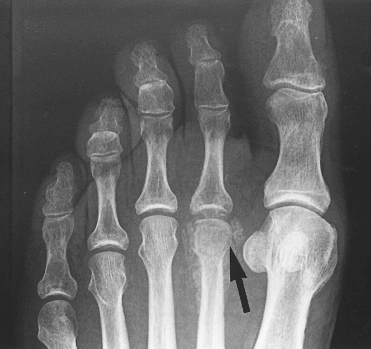
Radiographic examination is an important adjunct to clinical examination. X-rays of both feet should be taken as soon as possible after initial presentation. They may be useful in diagnosis and may serve as a baseline for ongoing comparison. In early stages, x-rays may be normal or demonstrate only subtle changes (
Fig. 1).[
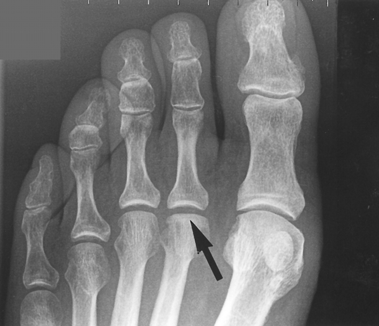
2] If clinical examination suggests underlying disease, further x-rays should be obtained within a few weeks because the fractures and healing may become more conspicuous during this period (
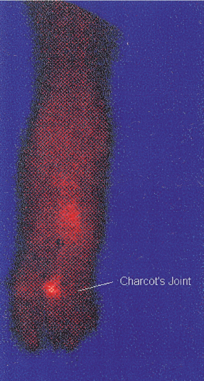
Fig. 2). Performing a bone scan is also worthwhile in this situation, as it is more sensitive and can confirm the presence of underlying pathology earlier (
Fig. 3). In more advanced cases, an x-ray at presentation is likely to reveal fractures at varying stages of development and healing. Radiographic diagnosis is based on the presence of bone destruction, subluxation, or dislocation. Follow-up x-rays of the affected foot every few months after diagnosis will show the progression of Charcot’s arthropathy. For example, once trabecular bridging and sclerosis are seen radiographically, the acute destructive phase characterized by joint instability is gradually giving way to the coalescent phase. There would be relatively little mobility of the affected joints at this stage. Recognizing this transition to healing is important because it helps to determine when immobilization could be gradually replaced by ambulation. On the basis of this principle, Eichenholtz et al[
3] subdivided Charcot’s arthropathy into three radiographically defined stages: stage 1, development, characterized by fragmentation of bone and dislocation; stage 2, coalescence, demonstrated by absorption of small fragments of bone and fusing of larger pieces to joint surfaces, causing ankylosis; and stage 3, remodeling, characterized by healing and new bone formation.
Figure 1.
The patient presented with slight discomfort of the foot. The x-ray shows only minor flattening of the second metatarsal head (arrow).
Figure 1.
The patient presented with slight discomfort of the foot. The x-ray shows only minor flattening of the second metatarsal head (arrow).
Figure 2.
X-ray of the foot taken 3 weeks after the initial x-ray shown in
Figure 1. Fragmentation of the second metatarsal head had become obvious (arrow).
Figure 2.
X-ray of the foot taken 3 weeks after the initial x-ray shown in
Figure 1. Fragmentation of the second metatarsal head had become obvious (arrow).
Figure 3.
Bone scan of the foot clearly showing an area of increased isotope uptake. The bone scan was performed at the same time as the x-ray shown in
Figure 1.
Figure 3.
Bone scan of the foot clearly showing an area of increased isotope uptake. The bone scan was performed at the same time as the x-ray shown in
Figure 1.
The Eichenholtz staging system does not include the earliest stages of the disease, when radiographic changes are not evident and treatment has the greatest chance of preventing deformity. More recently, Sella and Barrette[
4] applied another radiographically based staging system of Charcot’s arthropathy that includes the acute phase when clinical signs are present but x-ray changes are not. Their system is as follows: stage 0, localized heat and midfoot swelling (a bone scan may be positive at this stage, but only minimal changes may be present radiographically); stage 1, localized osteopenia, subchondral cysts, erosions, and possibly diastasis; stage 2, joint subluxations; stage 3, joint dislocations and joint collapse; and stage 4, sclerosis and fusions with trabecular patterns within bone to indicate healing.
The guidelines for staging Charcot’s arthropathy help to assign patients to certain categories. However, the numerical notations are not proportional to the extent of disease, and the categories are more a reflection of the phase than of the severity of the disease. McCrory et al[
5] attempted to improve on these guidelines by subdividing radiographic changes into “worsening” or “healing,” with numerical scores assigned to each by two independent observers. Worsening was characterized by joint dislocation and bone disruption, while healing was based on ankylosis of the bone fragments. Using such semiquantitative techniques, the authors were able to show that during contact casting, patients showed relatively little radiographic worsening and ongoing healing of the bone fragments. Even for such a carefully conducted study, the semiquantitative nature of the methods would make it difficult to subject the data to more stringent statistical analysis.
In addition to its use in staging Charcot’s arthropathy, x-ray monitoring also provides information on the degree and nature of structural changes that occur in the affected foot. These changes include the lowering of the calcaneal inclination angle, anterior and medial shifting of the talus, and the degree of plantar prominence. The occurrence of these and other structural changes is important to long-term prognosis and, together with clinical examination, guides surgery, bracing, orthotic, and footwear requirements.
Skin Temperature Measurement and Bone Scanning
Owing to the limitations of assessing Charcot’s arthropathy by clinical examination and radiographic techniques, more quantitative methods of defining its activity would be beneficial. These methods would allow better documentation of the natural history of Charcot’s arthropathy and more rapid testing of whether any treatment is having an impact on the underlying process of neuroarthropathy. Measurement of skin temperature and bone scanning of the feet are among the techniques that have been tried.
There is little doubt that acute Charcot’s arthropathy is characterized by increased temperature of the affected foot. Clinicians have known for a long time that the heat becomes less intense as the disease gradually evolves and settles. Eventually, although the affected foot may be deformed, it is no longer warmer than the contralateral foot. However, it has been difficult to express this knowledge in numerical terms. As a result, the acute response of the foot to treatment modalities such as rest and immobilization is also difficult to assess because it depends on small changes in temperature that cannot be confidently detected by clinical examination. The advent and ready availability of a relatively inexpensive handheld infrared temperature scanner has meant that much of this difficulty can be overcome. McGill et al[
6] used this device to document prospectively the clinical status of 17 diabetic subjects with acute Charcot’s arthropathy and follow their progress at regular intervals during treatment with contact casting. Adhering to carefully defined conditions for temperature measurements is important to ensure accuracy. In the aforementioned study, the scanner was held over the warmest part of the foot affected by Charcot’s arthropathy. The location of this spot was documented and subsequent measurements were taken at the same location. Care was also taken to ensure that patients were at rest for more than 10 min with their footwear or cast taken off before the temperature was taken. With these precautions, the coefficient of variation of temperature measurements taken on the same day was about 5%.
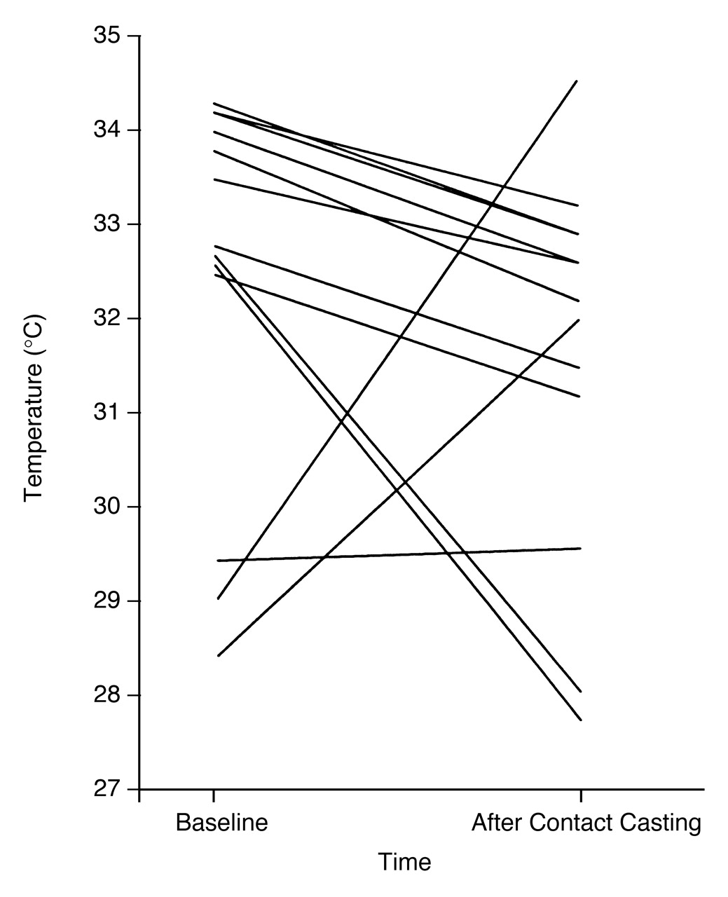
Owing to the variation in the stage and severity of the underlying disease and status of peripheral circulation, there was a considerable range of skin temperatures among the individual patients at presentation. The absolute temperature reading of the foot is not particularly useful in the diagnosis or monitoring of this condition. However, with minor exceptions, the skin temperature fell in most patients during contact casting (
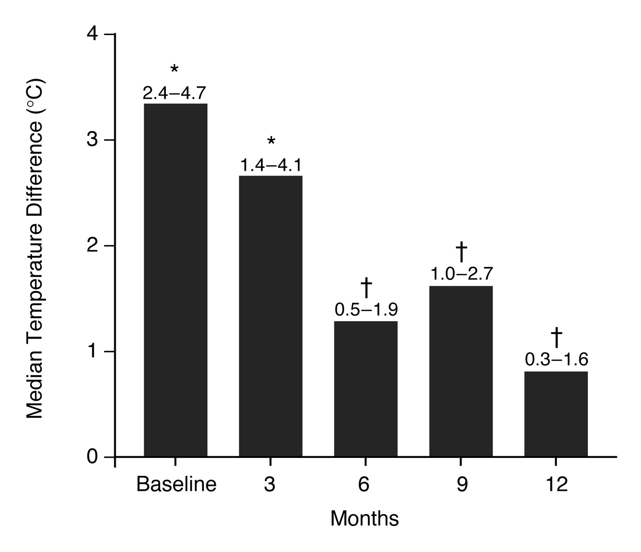
Fig. 4). This not only confirmed the response to this form of treatment but also objectively identified individuals whose atypical clinical course indicated that their treatment required further scrutiny. Factors such as inadequate fitting of the cast, inadequate rest, and coexisting infection can play a part. When all of the patients studied were considered as a group, the affected foot was initially 3.3°C (interquartile range, 2.4° to 4.7° C) warmer than the unaffected foot, and the temperature difference became progressively smaller with casting during the 12-month follow-up period (
Fig. 5). After 6 months, the temperature gradient between the feet was only 1.3°C (0.5° to 1.9° C), significantly less than at baseline, and by 12 months it was only 0.8°C. This type of quantitative data enables treatments to be compared objectively with one another.
Figure 4.
Skin temperature of the affected feet taken with a handheld infrared temperature scanner before and after contact casting.
Figure 4.
Skin temperature of the affected feet taken with a handheld infrared temperature scanner before and after contact casting.
Figure 5.
Median temperature difference between the affected and unaffected feet of the group of patients monitored during contact casting. Interquartile ranges are shown above the bars. *Different from normal foot (P < .006). †Different from baseline (P < .03).
Figure 5.
Median temperature difference between the affected and unaffected feet of the group of patients monitored during contact casting. Interquartile ranges are shown above the bars. *Different from normal foot (P < .006). †Different from baseline (P < .03).
Measurement of skin temperature is a useful technique for monitoring the progress of acute Charcot’s arthropathy. Even for a relatively small group of patients, temperature differences between the two feet can be used as an end point for research purposes. Others have used similar temperature measurement techniques to monitor the progress of Charcot’s arthropathy.[
7] In their comprehensive study of six patients, McCrory et al[
5] concluded that individual skin temperatures were too variable to be clinically useful. However, they took many measurements within a period of days, and the temperature changes obviously were too small to be quantified precisely, as their own calculations showed that temperature changed an average of only 0.022°C per day. In contrast, although skin temperature was measured at every follow-up visit in the study by McGill et al,[
6] only the results at three monthly intervals were evaluated. Thus the differences in the conclusions of the two studies may be more apparent than real.
Because the structural pathology of Charcot’s arthropathy is essentially fractures of the small bones of the foot, it is not surprising that the characteristic finding on a bone scan is intense isotopic uptake at the area of bony destruction. This is similar to what would be expected with any other cause of fractures. Edmonds et al[
8] were the first to exploit this phenomenon to produce quantitative data. They demonstrated that radioisotope uptake per unit of affected area was on average two to three times as high as in normal individuals without neuroarthropathy. The authors tried to further improve precision by expressing the ratio of the difference in radioisotope uptake between a standard area on the tibia of the ipsilateral side and the affected foot. The increased isotope uptake of Charcot’s arthropathy was again confirmed using this method.
The authors have further extended this concept by evaluating various methods of expressing numerical results of bone scans. The quantification of isotope uptake is well established in the investigation of thyroid disease, but it has been explored less in the study of Charcot’s arthropathy. In the study by McGill et al,[
6] bone scans were performed on the same group of 17 patients who had temperature measurement as described above. The uptake was expressed in the affected
versus the unaffected foot (the bilateral method), and its accuracy was evaluated against the ipsilateral method. Care was taken to ensure that a predefined area over the affected bones was always used for measurement. The isotope uptake in the affected foot at baseline was about 2% of the injected dose, and it fell gradually over the 12-month contact casting period. In the bilateral method, the affected side initially took up twice as much isotope, but this fell progressively, and by 12 months it was only about 30% to 40% higher. The ratio was also about twice as high in the affected bones when using the ipsilateral method, but it fell less steadily over the same observation period. Overall, the bilateral method showed a more consistent trend of normalization during the casting follow-up period. The bilateral method probably is also superior because it correlates better with the temperature gradient (
r = 0.9), whereas no such relationship could be demonstrated using the ipsilateral method. This showed that the bilateral approach is more comparable to the clinical method of comparing temperature between the two feet. However, it also implies that, properly performed, measurement of skin temperature is probably as reliable as bone scanning for quantifying acute Charcot’s arthropathy.[
6]
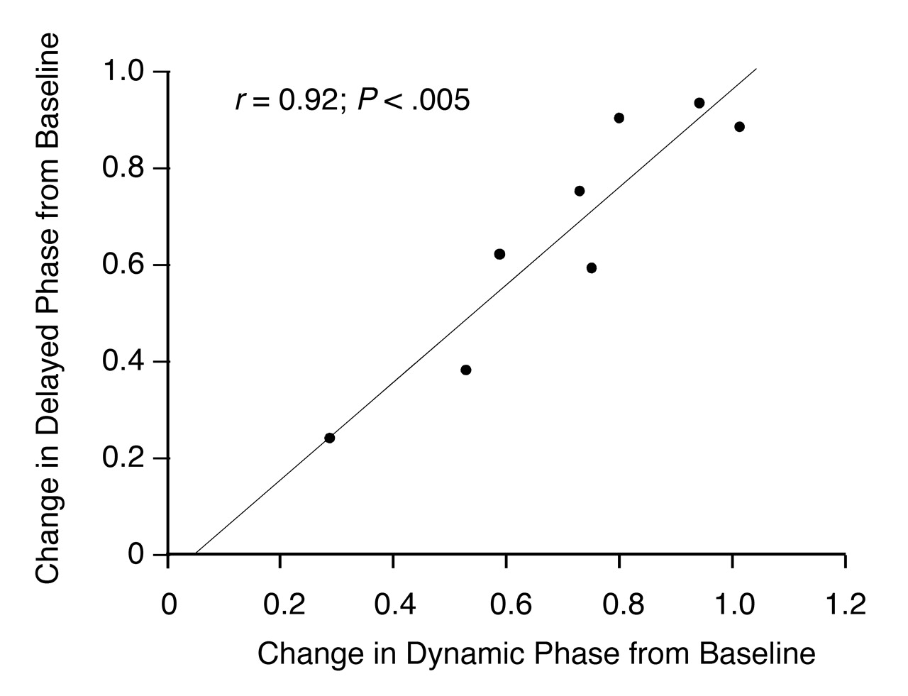
The use of bone scans in patients with Charcot’s arthropathy also provides an opportunity to study blood flow to the feet. Blood flow is estimated from the dynamic phase of the scan and is represented by the appearance of radioactivity in the first 2 min after injection of the isotope. Measured in this manner, it is evident that blood flow to the affected foot is considerably increased in patients with Charcot’s arthropathy. The presence of bounding pulses and high skin temperatures in the feet of affected patients are likely to be manifestations of this increase in blood flow. They may also be important factors underlying the pathogenesis of this condition, with the high blood flow creating an environment conducive to enhanced bone resorption. The dynamic phase isotope uptake correlates closely with the delayed phase (bone) isotope uptake, both at first presentation and as the Charcot’s arthropathy becomes quiescent with contact casting. Interestingly, in the study by McGill et al,[
6] uptake in both phases fell in parallel (
Fig. 6), suggesting that there is a strong link between blood flow and activity of the neuroarthropathy from a pathogenesis point of view. It is not possible at this stage to determine whether the reduction in blood flow causes a fall in activity of the Charcot’s arthropathy or if a more quiescent bone disease state creates less demand for blood flow. More frequent monitoring of the dynamic and delayed phase of the bone scan during therapeutic intervention may help answer this question. The reduction of blood flow over a period of several months in a Charcot foot is an interesting phenomenon. As discussed by Jeffcoate et al,[
2] it is difficult to understand why this should happen if the increased blood flow is the result of irreversible sympathetic denervation. The time scale is too short for it to be attributed to the onset of associated atheromatous disease. It suggests a functional, rather than a structural, abnormality and may be due to loss of a sympathetic vasodilatory response.
Figure 6.
Relationship between changes in the delayed and dynamic phases in the quantitative bone scanning from baseline to 12 months.
Figure 6.
Relationship between changes in the delayed and dynamic phases in the quantitative bone scanning from baseline to 12 months.