The term “hallux limitus,” as defined by Root et al,[
1] refers to a deformity in which the first proximal phalanx is plantarly subluxated on the first metatarsal head. As a result, there is inadequate dorsiflexion at the first metatarsophalangeal joint during the propulsive period of the gait cycle. Davies-Colley[
2] originally described this condition in 1887 using the term “hallux flexus.” It was named “hallux rigidus” by Cotterill[
3] in 1888. Since that time, various terms have been used in reference to the deformity, including “hallux dolorosus,” “dorsal bunion,” and the previously mentioned hallux flexus and hallux rigidus.[
2-
5] At present, the different nomenclature is generally thought to refer to different stages or symptoms of the same basic disease process.[
6]
The proposed etiologies of hallux limitus are numerous and include structural deformities such as an abnormally long or short first metatarsal bone, a hypermobile or elevated first ray, and an abnormally shaped metatarsal head.[
1,
7-
16] Osteochondritis dissecans, trauma, metabolic arthritides, neuromuscular disorders, and contracture of soft-tissue structures are also widely accepted etiologies.[
1,
2,
5,
10,
17-
19] Root et al[
1] concurred with several of these theories, attributing the deformity to the following six causative factors: hypermobility of the first ray (secondary to abnormal subtalar joint pronation), immobilization of the first ray, an excessively long ray, a dorsiflexed metatarsal, degenerative joint disease, and trauma. This discussion focuses primarily on hallux limitus secondary to pes planus.
An association between pes planus and hallux limitus was first recognized by Nilsonne[
7] in 1930. The pes planus foot type and conditions resulting in pronation are widely accepted etiologies of hallux limitus.[
1,
10,
15,
20-
23] As noted by Camasta,[
24] the aforementioned conditions resulting in pronation and thus hallux limitus include the presence of an os tibiale externum.
Root et al[
1] state that abnormal pronation of the foot results in hypermobility of the first ray, which is the most frequent etiology of hallux limitus. This occurs primarily in a rectus-type foot, which is everted. The ground reaction forces act to elevate the first ray. Subtalar joint pronation also diminishes the effective pull of the peroneus longus tendon, resulting in decreased plantarflexion of the first metatarsal and hypermobility. This plantarflexion of the first ray is necessary for normal dorsiflexion of the first proximal phalanx, which is required during the propulsive period of gait.[
1] Inadequate dorsiflexion of the proximal phalanx results in dorsal jamming, repetitive microtrauma, and marginal joint proliferations.[
15] The first proximal phalanx becomes plantarly subluxated on the first metatarsal. This subluxation is potentiated by the flexor tendons, which are plantarflexing in an attempt to stabilize the hallux against the ground.[
1] This process results in gradually increased pain, decreased range of motion, and a dorsal exostosis of the first metatarsophalangeal joint.
Hallux limitus is a progressive deformity that has been divided into various stages and subtypes. Functional hallux limitus refers to decreased range of motion of the first metatarsophalangeal joint only with weightbearing, whereas structural hallux limitus refers to decreased range of motion at all times.[
22]
Although pain and decreased motion of the first metatarsophalangeal joint are the predominant findings, other clinical symptoms may be present as well. Pain and compensatory hyperextension of the first interphalangeal joint with plantar medial hyperkeratotic tissue are often noted. Subungual exostosis, osteochondroma, and various nail deformities may result from trauma to the hyperextended distal phalanx. Gait abnormalities may also be observed as the propulsive period of gait is avoided because of the decreased motion and pain in the first metatarsophalangeal joint.[
1]
Classification and Pathomechanics of the Accessory Navicular
The os tibiale externum is a supernumerary bone located on the medial aspect of the navicular. This accessory ossicle was first described by Bauhin in 1605.[
25] It was further described and named the os tibiale externum by Pfitzner[
26] in 1896. The terms “accessory navicular,” “navicular secundum,” “prehallux,” “bifurcate navicular,” “divided navicular,” and “accessory tarsal scaphoid” have also been used. The overall prevalence of this accessory ossicle in the healthy population, as determined in various studies,[
26-
29] ranges from 10% to 15%. In comparison, a study conducted by Wood and Spencer[
30] found that the os tibiale externum is present in 19% of patients with pes planus. “This is an incidence of one-third greater than that found in the general population.”[
30]
In 1978, Veitch[
31] formulated a three-part classification system to categorize the anatomical variants of the accessory navicular. Type I represents a true sesamoid bone located within the posterior tibial tendon proximal to the navicular tuberosity. Type II is defined as a “true accessory scaphoid,” meaning that this is a separate bone arising from a secondary ossification center. This true accessory scaphoid has been shown to be adjoined to the navicular
via hyaline cartilage, fibrocartilage, a mixture of the two, or osseous tissue.[
32] Type III is enlargement of the navicular tuberosity itself.[
31] The focus of this study is the type II os tibiale externum.
Kidner,[
33] in 1929, was the first to theorize that the presence of an accessory navicular creates a fall in the longitudinal arch by disrupting the “pull” of the posterior tibial tendon. This disruption is thought to occur because the attachment of the tibialis posterior is displaced dorsomedially, creating an adductory force rather than a supinatory force on the foot. The support of the medial longitudinal arch by the tibialis posterior will be decreased. This adductory force will also cause soft-tissue structure impingement between the accessory navicular and the medial malleolus, resulting in pain. According to Kidner,[
33,
34] with the fall in the medial longitudinal arch height, the foot will abduct to alleviate this pain. With this pathologic mechanism in mind, Kidner[
33,
34] developed a surgical procedure involving removal of the os tibiale externum along with attachment of the posterior tibial tendon to the navicular at a more plantar lateral position.
Kidner’s theory and whether the posterior tibial tendon transfer improves medial longitudinal arch height have been widely debated. Many authors[
35,
36] are in agreement with Kidner’s theories and have found an increase in arch height after the previously mentioned procedure. Other authors[
31,
37-
40] believe that pes planus is an incidental finding occurring with the accessory navicular and that the lateral tendon transfer is not an advantageous procedure. It is generally accepted that the accessory navicular causes pain and discomfort, which is alleviated by its removal.
Other mechanisms correlating the accessory navicular with pes planus have been proposed. Wood and Spencer[
30] suggest that ligamentous structures, such as the plantar calcaneal navicular ligament and superficial fibers of the deltoid ligament, support the medial longitudinal arch along with the posterior tibial tendon. The presence of the accessory navicular results in abnormal insertion of not only the posterior tibial tendon but also these ligaments. This abnormal insertion diminishes the medial longitudinal arch support provided by these structures. Wood and Spencer[
30] also suggest that the altered function of the tendon modifies the function of the ligaments.
The accessory navicular is also present significantly more frequently in patients with posterior tibial tendon tears.[
41,
42] A study by Karasick and Schweitzer[
41] found that the accessory bone is present two to four times more frequently in patients with posterior tibial tendon tears. Klein[
42] had similar results, further stating that the posterior tibial tendon tears create pes planus by permitting the plantar calcaneal navicular ligament to stretch. In the study by Klein, “83% of abnormal plantar calcaneal navicular ligaments were associated with a torn posterior tibial tendon.”[
42]
An alternative pathogenic mechanism was proposed by Borrelli,[
43] who suggested that the primary function of the posterior tibial tendon is stabilization of the midtarsal joint, with the “distal slips preventing abduction and dorsiflexion of the oblique midtarsal joint axis.” Expounding on Kidner’s theories, Borrelli[
43] proposed that the presence of the accessory navicular and disruption of the pulley system create an unstable midtarsal joint. Thus the entire foot becomes unstable, resulting in a pes planus deformity predominantly occurring in either the transverse or the sagittal plane.
As discussed previously, the accessory navicular and hallux limitus are commonly associated with the flexible pes planus foot. The following case presentations discuss patients in whom both of these conditions, as well as a flexible pes planus deformity, were seen. Radiographic variables used for evaluation are discussed in the next section.
Materials and Methods
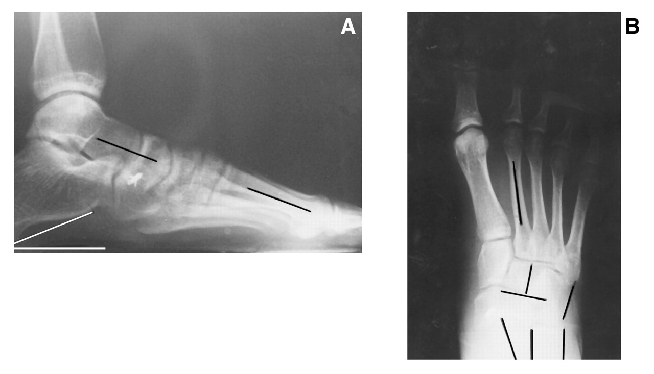
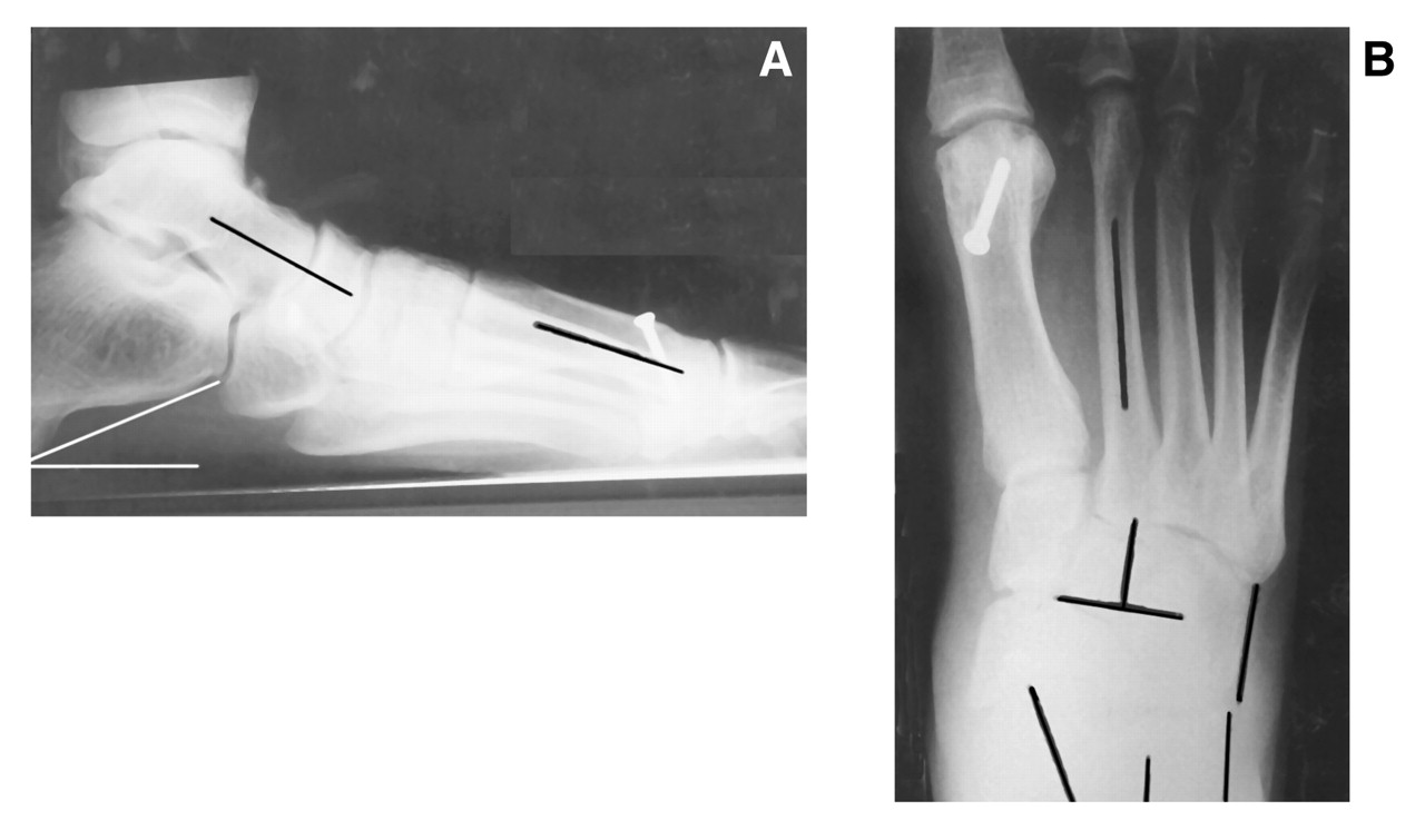
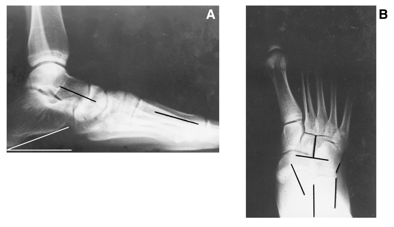
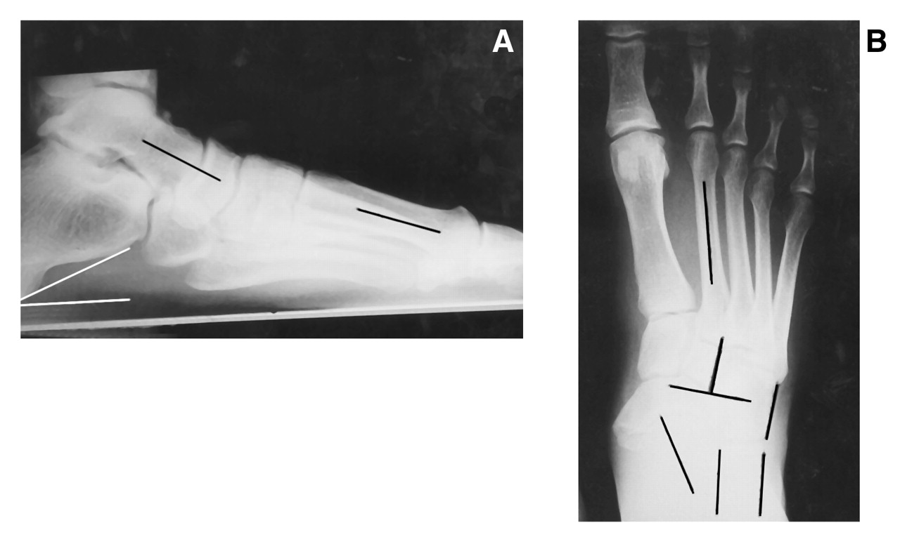
The cases of two patients with a symptomatic accessory navicular and hallux limitus occurring simultaneously are discussed in the following sections. Several radiographic variables were assessed preoperatively and postoperatively to evaluate the accessory navicular and hallux limitus, as well as a concomitant pes planus deformity. The forefoot adductus, metatarsus adductus, cuboid abductus, and talocalcaneal angles were measured on the anteroposterior view. The forefoot adductus angle was determined to be the angular relationship between the longitudinal bisector of the second metatarsal (to represent the forefoot) and the longitudinal axis of the rearfoot. The metatarsus adductus angle was determined to be the angular relationship between the longitudinal bisector of the second metatarsal (to represent the forefoot) and the longitudinal bisector of the midfoot. The cuboid abductus angle was determined to be the angle formed between the longitudinal axis of the rearfoot and the lateral aspect of the cuboid. The talocalcaneal angle was determined to be the angle formed between the longitudinal bisectors of the rearfoot and the talus (the line formed between the bisection of the head and neck of the talus).
The calcaneal inclination, lateral talocalcaneal, and talar first metatarsal angles were measured on the lateral view. The calcaneal inclination angle was determined to be the angle between a line formed along the inferior aspect of the calcaneus and the weightbearing surface. The lateral talocalcaneal angle was determined to be the angular relationship between the longitudinal bisector of the talar head and neck and the calcaneal inclination axis. The talar first metatarsal angle was determined to be the angular relationship between the longitudinal bisector of the head and neck of the talus and the longitudinal bisector of the first metatarsal. The results of the angular evaluations comparing preoperative and postoperative measurements for each patient are given in
Table 1.
Case 1
A 15-year-old girl presented to the podiatry clinic at Shelby County Medical Clinic in Harlan, Iowa, with arch pain in her right foot during exertional activities. Orthopedic examination revealed a decrease in the medial longitudinal arch height of the right foot during static stance. There was decreased right first metatarsophalangeal joint range of motion in the non-weightbearing position indicating functional hallux limitus. The Silfverskiold test[
44] was positive for a gastrocnemius equinus. Radiographs revealed an os tibiale externum of the right navicular (
Fig. 1). The accessory navicular was classified as type II. Conservative care was initiated for the upcoming year with a low-Dye strapping, followed by custom-molded orthoses. Approximately 6 months later, the patient presented with a painful right hallux and right medial arch pain. She requested definitive therapy to relieve the pain, as she was unable to participate in sporting activities. A modified Kidner procedure was performed using a bone anchor, which attached the remaining free posterior tibial tendon to the navicular tuberosity with the foot in a slightly inverted position. The area was tested by everting the foot, which revealed adequate strength. Postoperative radiographs were taken 5 weeks later (
Table 1,
Fig. 2). The patient resumed wearing the previously dispensed orthoses without further treatment for the functional hallux limitus. She is currently a very active and competitive cross-country athlete.
Case 2
A 36-year-old man presented to Tower Medical Clinic in Des Moines, Iowa, with pain in his right medial arch and first metatarsophalangeal joint of several years’ duration. The patient had worn orthotic devices for several years, which did not provide adequate relief of the pain. Orthopedic examination revealed a decrease in the medial longitudinal arch height of the right foot. Radiographic findings included osteophytic lipping of the dorsal aspect of the right first metatarsal head and an os tibiale externum of the right navicular (
Fig. 3). The accessory navicular was classified as type II. The patient was diagnosed as having painful hallux limitus and an accessory navicular. He was a very active person and wanted to continue running without discomfort. Because conservative methods had failed during the past few years, he consented to surgery. A modified Kidner procedure was performed on the navicular with an associated Youngswick Austin osteotomy performed on the first metatarsal. An anchor was placed into the posterior medial aspect of the patient’s right navicular. The free posterior tibial tendon was attached to the anchor, and immediate restoration of the patient’s medial arch was noted. Postoperative radiographs were taken 11 months later (
Table 1,
Fig. 4). The patient increased his running to 45 min at a time, an activity he had not been able to perform for more than 5 years.
Discussion
Clinical evaluation of both patients revealed unilateral planus foot types. Preoperative radiographic studies indicated that the pes planus was a transverse deformity with abnormal talocalcaneal, cuboid abductus, and metatarsus adductus angles. Correction of these angles to a more rectus foot type was noted on evaluation of postoperative radiographs. A decreased forefoot adductus angle is also associated with a transverse plane deformity. This angle was not decreased preoperatively and was not significantly changed postoperatively in either patient.
A decrease in the talometatarsal angle was also noted postoperatively in Case 1. This decrease indicates a less elevated first ray, which would alleviate the hallux limitus deformity. The amount of correction was increased compared with that in Case 2. This discrepancy may be due to the long-standing nature of the deformity in Case 2, with increased adaptive changes and more resistance to surgical correction. Preoperative radiographic evaluation did not indicate a sagittal plane deformity.
The primarily transverse plane correction is an interesting finding, as hallux limitus is considered to be a sagittal plane deformity. The authors cannot rule out investigative error as an explanation for this finding but suggest that the abnormal pull of the posterior tibial tendon caused by the accessory navicular created primarily a transverse plane deformity, which predisposes the individual to develop hallux limitus. Numerous pathogenic mechanisms have been proposed for various steps in this process. Whether the accessory navicular causes pes planus remains unknown; a causal relationship has not been proven. Similarly, the correlation between the accessory navicular and hallux limitus must be further researched by evaluating a greater subject volume; the authors have simply proposed a link between the two conditions.
Figure 1.
Preoperative lateral (A) and anteroposterior (B) radiographs of Case 1 showing an os tibiale externum of the right navicular.
Figure 1.
Preoperative lateral (A) and anteroposterior (B) radiographs of Case 1 showing an os tibiale externum of the right navicular.
Figure 2.
Postoperative lateral (A) and anteroposterior (B) radiographs of Case 1 taken at 5 weeks.
Figure 2.
Postoperative lateral (A) and anteroposterior (B) radiographs of Case 1 taken at 5 weeks.
Figure 3.
Preoperative lateral (A) and anteroposterior (B) radiographs of Case 2 reveal osteophytic lipping of the dorsal aspect of the right first metatarsal head and an os tibiale externum of the right navicular.
Figure 3.
Preoperative lateral (A) and anteroposterior (B) radiographs of Case 2 reveal osteophytic lipping of the dorsal aspect of the right first metatarsal head and an os tibiale externum of the right navicular.
Figure 4.
Postoperative lateral (A) and anteroposterior (B) radiographs of Case 2 taken at 11 months.
Figure 4.
Postoperative lateral (A) and anteroposterior (B) radiographs of Case 2 taken at 11 months.
Table 1.
Preoperative and Postoperative Angle Measurements
Table 1.
Preoperative and Postoperative Angle Measurements