Achilles tendon rupture, either traumatic or atraumatic, is a common clinical finding in podiatric practice. Atraumatic rupture is most prevalent in patients with rheumatoid arthritis, lupus erythematosus, gout, hyperparathyroidism, and chronic renal failure.[
1-
7] Spontaneous rupture may stem not only from the systemic effects of these diseases on the body, but also from the long-term use of oral steroids. In several cases of Achilles tendon rupture, oral steroids were used intermittently for asthma, chronic airway obstruction, rheumatic fever, and pneumonia.[
1-
8]
The Achilles tendon begins to show signs of normal degeneration after the third decade of life. Steroid therapy is thought to accelerate this degenerative process.[
9-
11] Histologic studies have shown a suppressed fibroblastic reaction and inhibition of growth under the influence of steroids.[
12,
13] In a study of rabbits by Unverferth and Olix[
14] in 1973, changes in the Achilles tendon were noted after direct steroid injection. Injected tendons demonstrated swelling, subcutaneous hemorrhage, disruption of collagen bundles, and hypertrophied paratenon.
The body also manufactures its own steroids to maintain the integrity of body tissues and to inhibit fibroblastic growth and migration.[
12] Increasing the amount of steroid in the body by oral therapy or direct injection may predispose patients to tendon rupture by suppressing the normal fibroblastic repair mechanisms. This rupture usually occurs 2 to 6 cm proximal to the Achilles tendon insertion, where a zone of avascularity exists. Lagergren and Lindholm[
10] proved the existence of this zone angiographically in 1958.
An evaluation of recent literature shows no correlation between dosage or duration of steroid use and injury. Dosages varied from 3 to 60 mg/day. The time from initiation of steroid therapy until rupture varied from only a few months to more than a decade. Prednisone was used most often, but ruptures were also seen with other steroids, such as betamethasone and triamcinolone.[
13,
15] A majority of these patients were in their sixth decade of life, and bilateral rupture was not uncommon.
Cases of spontaneous Achilles tendon rupture with local steroid injection have also been reported. Many patients were athletes in their twenties who had received multiple steroid injections into the Achilles tendon, often for treatment of tenosynovitis. These ruptures can occur as soon as 1 week after a single injection into the tendon, or not for months after a long series of injections.[
8,
10]
Regardless of the type of steroid therapy, patients usually report pain in the calf after minimal activity, such as a slight twist, standing on toes, climbing stairs, or walking. Occasionally, strenuous exercise, such as dancing or basketball, precipitates the injury. Difficulty in plantarflexing the foot or rising up onto the toes, warmth, edema, and a palpable defect are common presenting symptoms. Plain film x-rays and magnetic resonance imaging (MRI) are often used for diagnosis and to better determine pathology.
The decision to institute conservative
versus surgical treatment depends on patient age, activity level, general health, and physician discretion. Conservative treatment consists of immobilization and nonweightbearing for 3 to 8 weeks. When surgical intervention is performed, most tendons have thickened tendon sheaths, local signs of hemorrhage, and degeneration of the tendon.[
1,
4,
6,
8,
11,
13-
18] Continuing steroid therapy after tendon repair may cause it to rupture again during the patient’s lifetime.[
8]
Case Report
A 58-year-old woman presented with pain and swelling of her right ankle. The patient reported that mild pain and swelling in her right calf had begun about 1 month earlier. She also noticed difficulty standing up after working in her garden. The patient recalled no specific traumatic event, but she did remember stumbling a few times just before her symptoms began. She visited a general medical clinic six times during the next 3 weeks. The patient was initially diagnosed with a sprain and put into a splint. At subsequent visits, the patient reported no decrease in symptoms. She was put on antibiotics for suspected infection and sent for a Doppler study to rule out deep venous thrombosis. The Doppler study and plain film x-rays were negative. At this point, the patient complained of increased pain and swelling in her right calf with no relief of symptoms.
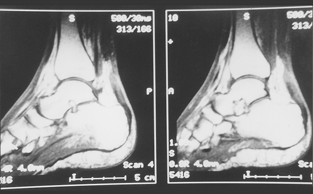
The patient was referred to a podiatric physician, who immediately diagnosed an Achilles tendon rupture. The diagnosis was based on a clinical examination that revealed an absence of plantarflexion with calf compression, an inability to rise up onto the toes, and pain along the Achilles tendon with a palpable gap noted along the medial side. The patient was scheduled for MRI, immobilized, and sent to the office of the second author (N.S.H.) for a surgical consultation. The MRI results revealed a complete tear of the right Achilles tendon centered approximately 4 to 5 cm proximal to the calcaneal insertion. A 1.5- to 2.0-cm defect and prominent thickening with interstitial degeneration and edema extending proximally and distally were also noted (
Fig. 1).
The patient’s medical history was significant for Addison’s disease, which was diagnosed in 1987. To treat this adrenocortical insufficiency, she was placed on a maintenance dose of 5 mg/day of prednisone for about 10 years; the dosage was increased to 7.5 mg/day for the next 3 years. The patient reported having bronchial infections and asthma attacks since she had been diagnosed with Addison’s disease. These secondary problems averaged 10 incidents each year. During these events, her prednisone dose was increased to 60 mg/day for several days, and then tapered slowly back to her maintenance dose. She had also been on a daily dose of 0.1 mg of fludrocortisone acetate since 1987.
Surgical Procedure
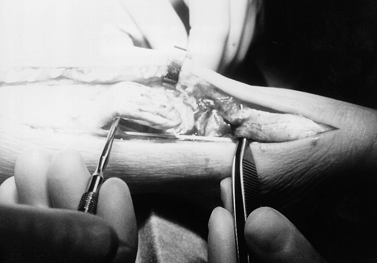
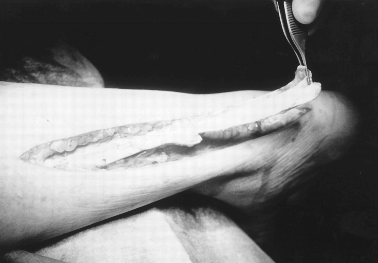
After administration of spinal anesthesia, the patient was placed in a prone position on the operating table. After the right lower leg was prepared and draped in the usual sterile manner, the foot was exsanguinated and a well-padded thigh cuff was inflated to 300 mm Hg for hemostasis. A linear skin incision was centered over the Achilles tendon, ending just proximal to its insertion site on the calcaneus. Blunt and sharp dissection was carried down to the paratenon, avoiding all neurovascular structures. All bleeders were cauterized to keep a majority of the venous system intact. The paratenon was then incised in a linear fashion and retracted. A 2-cm deficit was noted approximately 4 cm proximal to the Achilles tendon insertion. Both ends of the tendon were blunted, which is consistent with a chronic rupture (
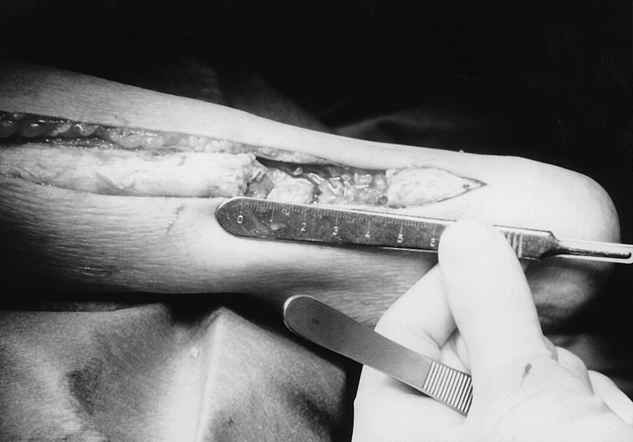
Fig. 2). The rest of the tendon was smaller than would be expected. After freshening up the edges to viable tendon, the size of the defect increased to 4.5 cm (
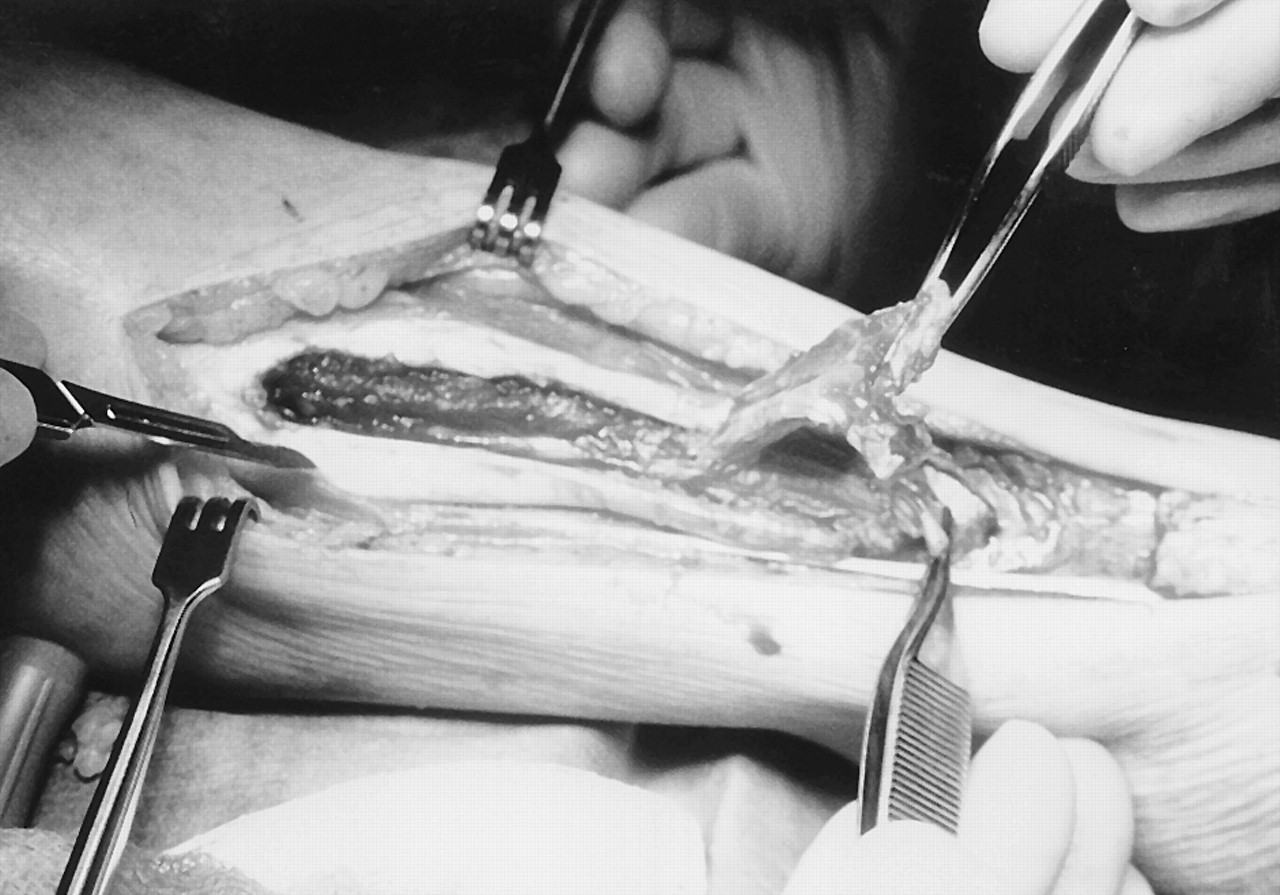
Fig. 3). Because of the size of the defect, it was decided to slide a portion of the central aspect of the gastrocnemius aponeurosis/Achilles tendon down to cover the gap. A 7-cm section of the gastrocnemius aponeurosis/Achilles tendon was then excised from its central portion, starting at the superior edge of the rupture site. This free graft represented about one-third of the total width of the tendon. The soleus muscle belly and a portion of the conjoined tendon were left intact. Because the graft’s superior edge was bulkier and stronger, the graft was rotated 180° so that the proximal, stronger portion of the graft would be placed at the distal aspect of the defect (
Fig. 4). The graft was placed with an overlap of 2 cm on the proximal and distal aspects of the good tendon edges. It was sutured in place using 2-0 Vicryl (Ethicon, Inc, Somerville, New Jersey). The defect where the graft was taken was also repaired using 2-0 Vicryl simple sutures to bury the grafted tendon between the two edges (
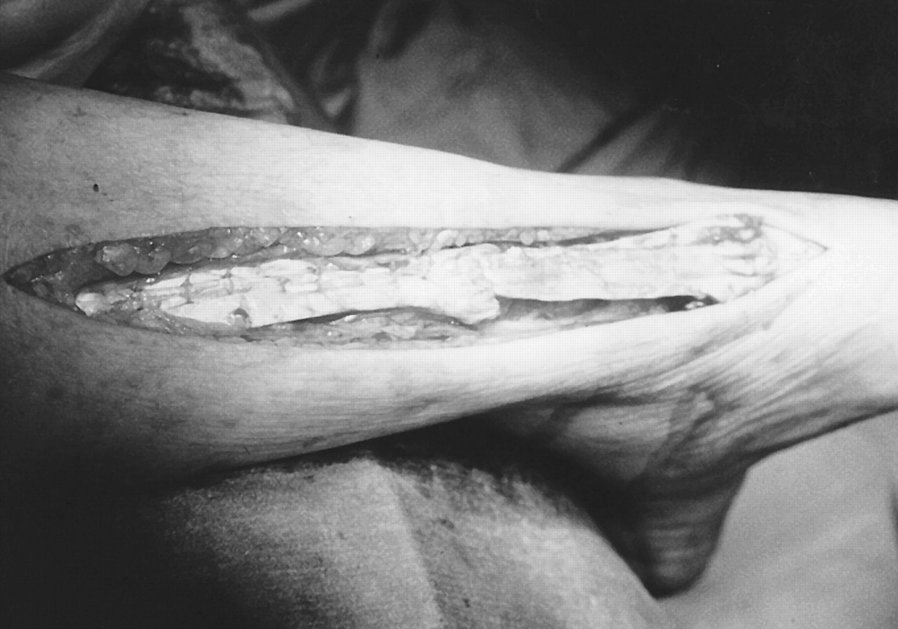
Fig. 5). The plantaris tendon was transected because of its degeneration. The foot was noted to reach a 90° position relative to the leg after the repair had been completed (
Fig. 6). The surgical site was irrigated and closed in anatomical layers, with care taken to reapproximate the paratenon. A postoperative injection of local anesthetic was infiltrated about the posterior leg. The surgical site was dressed with dry, sterile dressings and the cuff was released with good flow noted to all digits. The patient was placed in a posterior splint in a slightly plantarflexed position and instructed to remain strictly nonweightbearing.
The postoperative regimen included staple removal and 8 weeks of continued nonweightbearing. The patient began passive range-of-motion exercises at 6 weeks, and at 8 weeks she was sent for physical therapy to improve her muscle strength and increase her range of motion. She was also allowed weightbearing to tolerance at this time. The patient’s pain level and swelling decreased throughout the postoperative period, and her muscle strength and range of motion slowly increased. At 10 months after surgery, the patient stated that she was doing fine. She had some occasional soreness and swelling in her right calf, but her muscle strength was still improving. The patient reported that she was still unable to jump but could jog lightly.
Discussion
The case presented involved the repair of an Achilles tendon rupture with a large defect due to chronic oral steroid use. An abundance of cases of spontaneous Achilles tendon rupture have been reported in the literature.[
1,
6,
11,
13,
15-
18] No specific correlation can be made with the duration of steroid use, steroid dosage, or disease states involved with these ruptures. Some general statements, however, can be made about the patients. They have a systemic disease such as Addison’s disease, rheumatoid arthritis, or lupus erythematosus that necessitates chronic steroid therapy. These patients are often on maintenance doses of 5 to 10 mg/day of prednisone, with increased dosing as needed to control primary or secondary medical conditions. They are usually in their sixth decade of life and have taken oral steroids continuously for at least 3 months. Bilateral ruptures are common. Because of the chronic nature of these ruptures, the ends of the Achilles tendon are often degenerated and require debridement before repair can take place. As was true in the case reported here, this often leads to a large defect in the tendon, which makes the repair more challenging. In this case, the surgeon chose to do a slide graft, one of a number of different procedures that can be performed.
The literature also reflects the increased risk of Achilles tendon rupture with direct infiltration of steroid into the tendon area. This usually occurs after several injections over a short period of time, but it has been reported after a single injection. These patients are often younger and involved in a strenuous activity when rupture occurs.[
8,
14]
Spontaneous Achilles tendon rupture due to steroid use is not uncommon. Physicians need to be aware of this potential side effect of steroid use and take proper precautions to protect their patients.
Figure 1.
T1-weighted magnetic resonance image clearly showing a complete rupture of the Achilles tendon.
Figure 1.
T1-weighted magnetic resonance image clearly showing a complete rupture of the Achilles tendon.
Figure 2.
Achilles tendon rupture with a 2-cm defect. The tendon edges are fibrotic and nonviable.
Figure 2.
Achilles tendon rupture with a 2-cm defect. The tendon edges are fibrotic and nonviable.
Figure 3.
After freshening up the edges of the tendon, the defect increased to 4.5 cm.
Figure 3.
After freshening up the edges of the tendon, the defect increased to 4.5 cm.
Figure 4.
The middle third of the Achilles tendon was rotated 180° and slid distally to repair the defect.
Figure 4.
The middle third of the Achilles tendon was rotated 180° and slid distally to repair the defect.
Figure 5.
Tendon graft repair with 2-cm overlap proximally and distally. The gap where the tendon graft was taken was closed primarily.
Figure 5.
Tendon graft repair with 2-cm overlap proximally and distally. The gap where the tendon graft was taken was closed primarily.
Figure 6.
Completed repair of Achilles tendon rupture with graft.
Figure 6.
Completed repair of Achilles tendon rupture with graft.