Next Article in Journal
A Retrospective Analysis of JAPMA Publication Patterns, 1991–2000
Previous Article in Journal
Understanding Statistics. Putting P-Values into Perspective
 
 
Journal of the American Podiatric Medical Association is published by MDPI from Volume 116 Issue 1 (2026). Previous articles were published by another publisher in Open Access under a CC-BY (or CC-BY-NC-ND) licence, and they are hosted by MDPI on mdpi.com as a courtesy and upon agreement with American Podiatric Medical Association.
Article

Amelanotic Melanoma Masquerading as an Ingrown Toenail

by
Harvey Lemont
1 and
Jane Brady
2
1
Laboratory of Podiatric Pathology, and Professor, Section of Podiatric Medicine, Temple University School of Podiatric Medicine, Eighth at Race St, Philadelphia, PA 19107
2
Submitted during second-year surgical residency, Beth Israel Deaconess Medical Center, Boston, MA
J. Am. Podiatr. Med. Assoc. 2002, 92(5), 306-307; https://doi.org/10.7547/87507315-92-5-306

Published: 1 May 2002
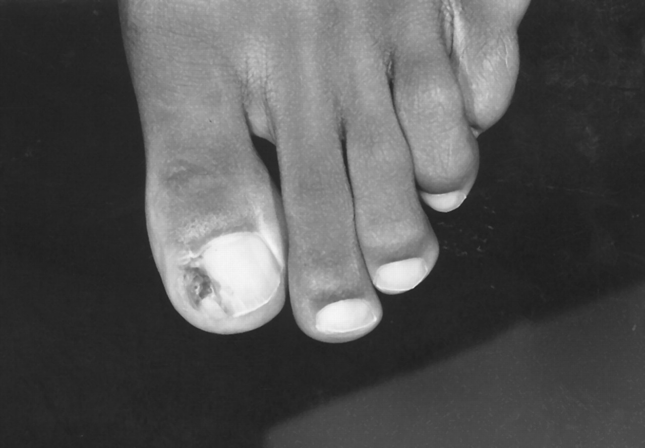
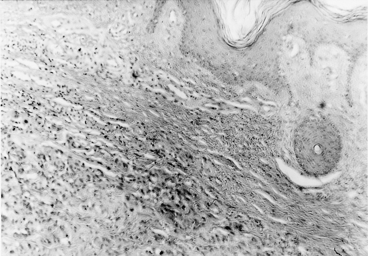
A 45-year-old black woman presented to the skin clinic of Temple University School of Podiatric Medicine with paronychia at the medial aspect of her right hallux. A mild serous to purulent discharge with mild odor was noted. A small, red, dome-shaped lesion consistent with a pyogenic granuloma was present at the margin of the nail plate and nail fold (Fig. 1). The patient had been seen previously on multiple occasions by another physician who had prescribed soaks and antibiotics and had twice removed the portion of the nail plate that was “ingrown.” The patient stated that following each surgery she had no pain and the nail returned to its normal appearance. Excisional biopsy of the soft tissue revealed a fairly unremarkable picture histologically. Although the overlying epidermis demonstrated increased pigmentation limited to the basal layer, it did not demonstrate any obvious increase in melanocytic proliferation. Scattered pigment-like material, however, was noted within the dermis (Fig. 2).
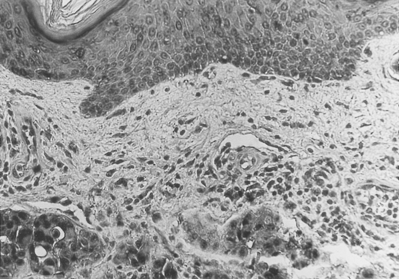
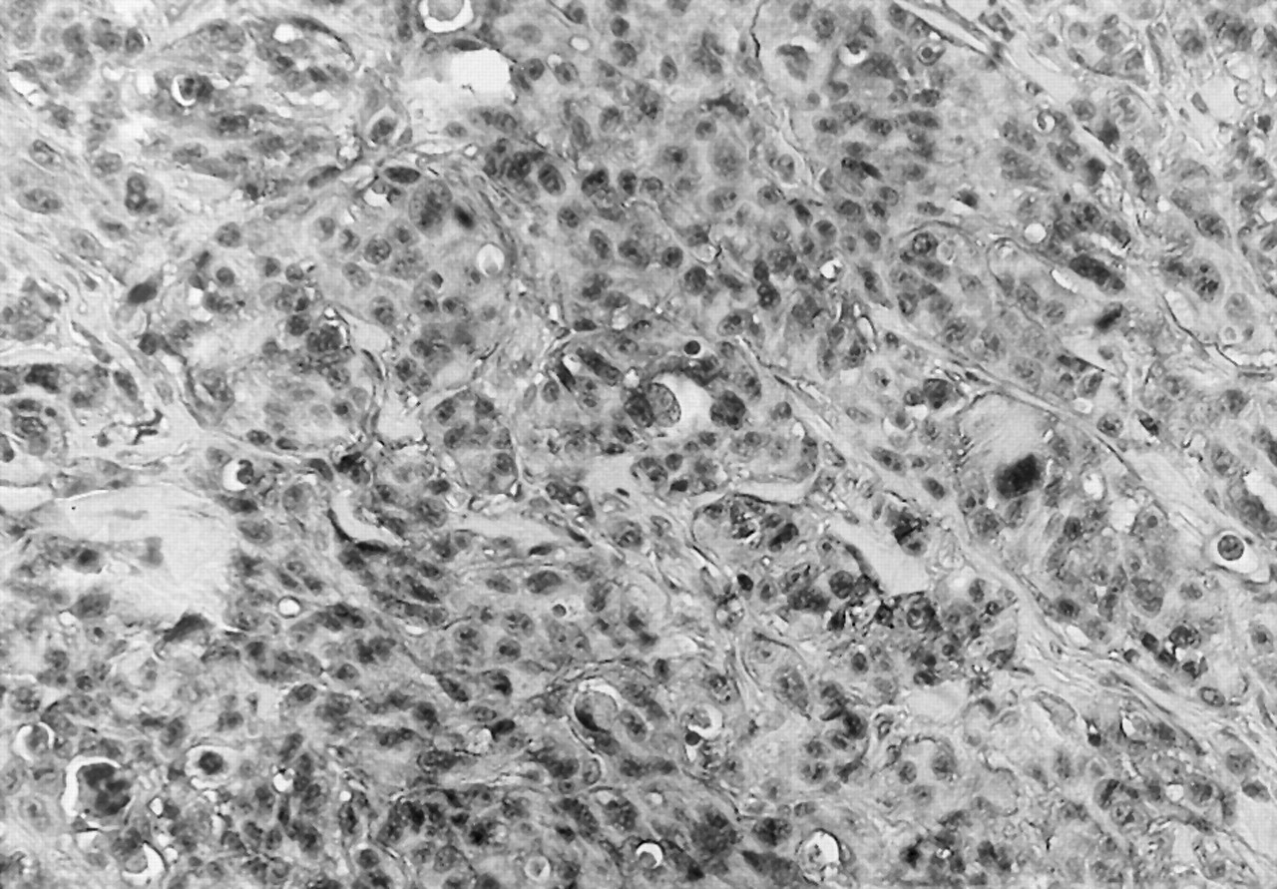
Use of S-100 immunoperoxidase staining techniques showed the pigment to be melanin. Melanin, in addition to being seen within the dermis, was also scattered throughout the epidermis (Fig. 3). The melanocytes were large and laden with melanin (Fig. 4). The melanocytic neoplasm measured 1.6 mm in depth and was referred for sentinel lymph node biopsy, which was positive using reverse transcriptase–polymerase chain reaction techniques. Histologic examination of the draining basin lymph nodes was negative for melanoma.

Comment

The difficulty in diagnosing melanoma of the nail unit can be attributed to its excellent masquerading abilities. It may appear as common benign nail unit problems, such as pyogenic granuloma, granulation tissue, or ingrown nail [1,2].
Papachristou and Fortner [3] found that 42% of 52 patients ultimately diagnosed with nail melanoma were inappropriately treated with procedures such as nail excision and cauterization. A common feature of paronychia due to melanoma is the patients’ initial presentation of chronic inflammation, draining, and loosening of the nail plate, which in the authors’ experience can last anywhere from 6 months to 3 years before a definitive diagnosis is established. The lack of pigmentation is a major factor in many physicians’ failure to consider melanoma as a possible diagnosis [4].
The literature supports the view that a physician’s initial approach to what appears to be an infected ingrown toenail with granulation tissue should be to prescribe antibiotics, antifungals, or even steroids [47]. While some investigators suggest that amelanotic lesions bleed less than pyogenic granulomas with manipulation, and that pyogenic granulomas show more rapid growth [6], these distinctions should not be relied on in making a diagnosis. In addition, the belief that subungual melanoma does not affect the young is not supported by the literature, with cases reported in patients as young as 16 years of age [5].
Since amelanotic melanoma most often cannot be diagnosed clinically, a physician should have a very low threshold for biopsy early in the presentation [3]. Unfortunately, while a biopsy can provide a definitive diagnosis, most patients presenting with granulation tissue are not biopsied or are inappropriately biopsied [6,7]. Even a punch biopsy has been demonstrated to provide a sufficient amount of tissue for diagnosis; smaller amounts, however, can yield inconclusive results [7]. In some cases, these lesions may persist for long periods of time without a diagnosis being established, resulting in extension to the underlying phalanx [6].
Although subungual malignant melanoma is rare, accounting for only 2% to 3% of all melanomas, the patient’s mean survival time is significantly shorter than in cases of melanoma of other body regions [1,2]. The high mortality rate can be attributed to a higher incidence of delayed diagnosis compared with lesions of the dorsum of the foot and ankle [2]. Amelanotic melanoma, often masquerading as the commonly seen infected ingrown toenail, accounts for 15% to 65% of malignant subungual lesions [1,7,8]. The study by Papachristou and Fortner [3] of 52 patients with subungual lesions noted that all seven who presented with amelanotic melanoma did not survive beyond the third year of diagnosis.

Figure 1. Amelanotic melanoma causing paronychia and “ingrown toenail.”
Japma 92 00306 g001
Figure 2. Biopsy of red, dome-shaped lesion reveals a nearly normal appearance under low power with only a hint of pigment-like material noted within the dermis (H&E, ×10).
Japma 92 00306 g002
Figure 3. Staining of the lesion with S-100 immunoperoxidase demonstrates that the pigment is melanin. Note clumps and islands of heavily pigmented cells within the dermis, with pigment also seen within all epidermal layers (×20).
Japma 92 00306 g003
Figure 4. Large atypical melanocytes exhibiting intense pigmentation with S-100 immunoperoxidase staining (×40).
Japma 92 00306 g004

References

  1. Dawber RP, Colver GB: The spectrum of malignant melanoma of the nail apparatus. Semin Dermatol10, 82, 1991.
  2. Fortin PT, Freiberg AA, Rees R, et al: Malignant melanoma of the foot and ankle. J Bone Joint Surg Am77, 1396, 1995.
  3. Papachristou DN, Fortner JG: Melanoma arising under the nail. J Surg Oncol21, 219, 1982.
  4. Winslet M, Tejan J: Subungual amelanotic melanoma: a diagnostic pitfall. Postgrad Med J66, 200, 1990.
  5. Rigby HS, Briggs JC: Subungual melanoma: a clinico-pathological study of 24 cases. Br J Plast Surg45, 275, 1992.
  6. Elmets CA, Ceilley RI: Amelanotic melanoma presenting as a pyogenic granuloma. Cutis25, 164, 1980.
  7. Koch SE, Lange JR: Amelanotic melanoma: the great masquerader. J Am Acad Dermatol42 (5 pt 1), 731, 2000.
  8. Levit EK, Kagen MH, Scher RK, et al: The ABC rule for clinical detection of subungual melanoma. J Am Acad Dermatol42 (2 pt 1), 269, 2000.