Surgical repair of a hammer toe by means of proximal interphalangeal joint arthroplasty alone, removing the head of the proximal phalanx, often leaves an unstable digit whose final position and alignment are easily affected by scar tissue and tendon balance. Stability is enhanced with use of proximal interphalangeal joint arthrodesis. In fact, studies have shown that proximal interphalangeal joint arthrodesis provides not only better stability but also better toe purchase and sagittal plane correction compared with arthroplasty alone. [
1] This procedure, when combined with metatarsophalangeal joint relocation, has been shown to reliably alleviate associated metatarsalgia. [
2] It also has a relatively high patient satisfaction rate. [
3]
Unfortunately, proximal interphalangeal joint arthrodesis usually requires a metal pin for appropriate fixation (
Fig. 1). The purpose of the pin is to provide the stability and apposition needed to allow the bone to unite. Use of a metal pin is not always practical, particularly in certain patients. Because the pin exits the skin for a protracted period, the site is subject to infection, pin trauma, pin bending, and accidental pin removal. In addition, the patient must keep the wound site dry for up to 6 weeks and must wear some type of protector over the pin. Finally, anticipation of pin removal can generate considerable anxiety, and the extraction itself may be painful. All of these factors can be frustrating for the patient and challenging for the surgeon.
It would be ideal if arthrodesis could be accomplished with internal fixation, especially using an absorbable bone graft, to obviate pin removal or the pin exiting through the skin. Internal fixation is best accomplished using the 2.4-mm-diameter cortical bone allograft pin. In the course of developing the procedure, it was determined that the 2.0-mm bone pin broke too easily to sustain the arthrodesis. Diameters larger than 2.4 mm did not fit easily within the intramedullary canals of the phalanges. The processing and sterilizing of cortical bone allograft pins have been well described by Reed. [
4]
Figure 1.
Use of a metal pin to fixate digital fusion.
Figure 1.
Use of a metal pin to fixate digital fusion.
Indications
Arthrodesis of the lesser toes using a cortical bone allograft pin is indicated for the correction of hammer toe deformity involving the proximal interphalangeal joint. The deformity can be flexible or partially ankylosed but must not involve severe hyperextension at the metatarsophalangeal joint (
Fig. 2). By itself, the procedure does not address the pathologic condition at the metatarsophalangeal joint, where temporary transarticular metal pin fixation may be required.
If there is a modest contracture of the soft tissues about the associated metatarsophalangeal joint, as demonstrated by the metatarsal push-up test, then a capsulotomy and collateral ligament release can be performed at the same time. It is usually not necessary to perform these procedures until bone pin arthrodesis has been accomplished. It is also usually necessary to lengthen the extensor tendons to ensure that all deforming forces are neutralized.
Figure 2.
Hammer toe deformity with minimal involvement of the metatarsophalangeal joint.
Figure 2.
Hammer toe deformity with minimal involvement of the metatarsophalangeal joint.
Materials and Methods
Patients with semiflexible and flexible hammer toes demonstrating moderate deformity were selected in sequence as they presented to the author’s office clinic. Inclusion criteria were manually reducible deformity using the Kelikian metatarsal push-up test, zero to moderate flexible contracture at the metatarsophalangeal joint, pain that had not resolved with conservative care, and little or no transverse plane deformity.
The goal was to correct the digital hammer toe deformity and stabilize the toe by means of internal proximal interphalangeal joint arthrodesis using a cortical bone allograft pin for fixation. The allograft used was cortical bone fashioned into a 2.4-mm-diameter pin (Allofix) prepared and supplied by the Musculoskeletal Transplant Foundation in Edison, New Jersey.
All of the bone donors had provided medical, social, and family histories. Blood samples from every donor were tested to identify infectious diseases such as hepatitis and syphilis and the human immunodeficiency virus (HIV), any of which would exclude the bone. The bone tissue was retrieved by trained medical personnel and was processed in Class 10 (static) clean rooms using aseptic techniques and processing methods aimed at further reducing the risk of contamination and disease transmission. Polymerase chain reaction, which has been found to be 99.6% sensitive and 99.9% specific, was used to determine the presence of HIV. Other assays included HIV-I and HIV-II antibodies, hepatitis B surface antigen, anti–hepatitis C virus, human T-cell lymphotrophic virus type I, and anti–hepatitis B core.
The author performed all of the surgeries using the same technique, with or without flexor digitorum longus tendon transfer for further stabilization. Because addition of the latter procedure seemed to enhance the outcome, it was used more frequently toward the end of the study.
Surgical Procedure
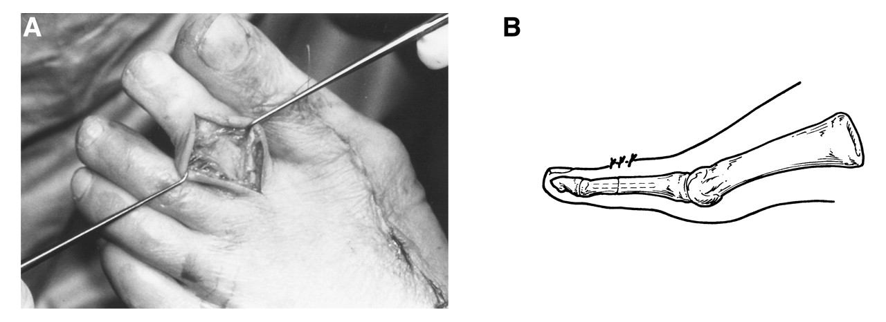
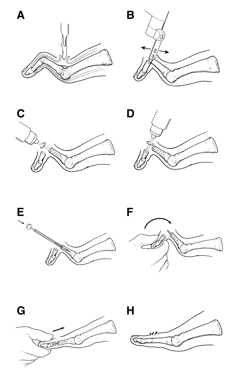
The steps for proximal interphalangeal joint arthrodesis using a cortical bone allograft pin are illustrated in
Figure 3. The soft-tissue dissection involves a longitudinal or serpentine incision from the metatarsophalangeal joint to the distal interphalangeal joint, followed by separation of the subcutaneous layer. The extensor tendons are retracted or separated for a Z-plasty lengthening, with sectioning of the hood apparatus as necessary. As the collateral ligaments are incised with periarticular capsulotomies, the joint surfaces are delivered into the wound.
Figure 3.
Steps performed in proximal interphalangeal joint arthrodesis. A, Release of the metatarsophalangeal joint; B, exposure of the proximal interphalangeal joint and denuding of cartilage; C, drilling of the proximal phalanx intramedullary canal; D, drilling of the middle phalanx intramedullary canal; E, insertion of a bone pin into the proximal phalanx medullary canal; F and G, maneuver for grasping the toe to insert the bone pin into the distal phalanx; H, the realigned digit with a cortical bone pin securing the arthrodesis site.
Figure 3.
Steps performed in proximal interphalangeal joint arthrodesis. A, Release of the metatarsophalangeal joint; B, exposure of the proximal interphalangeal joint and denuding of cartilage; C, drilling of the proximal phalanx intramedullary canal; D, drilling of the middle phalanx intramedullary canal; E, insertion of a bone pin into the proximal phalanx medullary canal; F and G, maneuver for grasping the toe to insert the bone pin into the distal phalanx; H, the realigned digit with a cortical bone pin securing the arthrodesis site.
A rongeur is used to remove the cartilage from both surfaces, and good cancellous bone is exposed with minimal sacrifice of length. At this point, the 2.4-mm smooth drill pin is driven into the shaft of both the proximal and middle phalanges, with care being taken not to perforate the subchondral bone into the next joint. The depth gauge is then used to determine the length of pin required to cross and sustain the arthrodesis site by measuring the depths of the proximal and middle phalanx intramedullary canals and totaling the two numbers. The desired length of the bone pin can then be marked and cut with a power saw. A rongeur should not be used because it may shatter the pin.
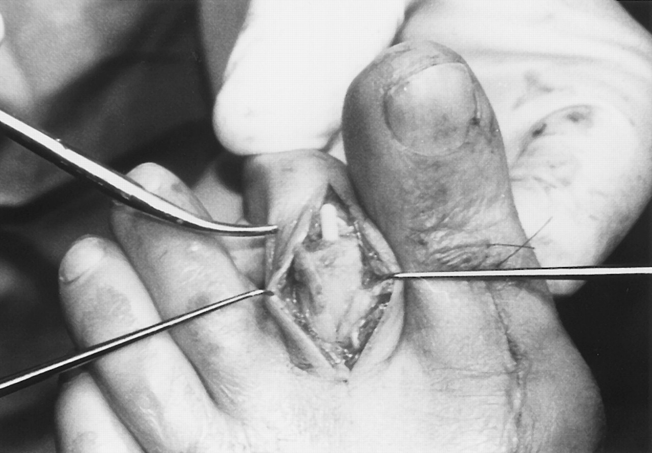
The pin is then inserted into the drilled intramedullary canal of the proximal phalanx shaft deep enough to almost reach the subchondral plate at the opposite end. This insertion will prevent a shallowly placed pin from slipping deeper into the canal when the middle phalanx is pushed onto the remaining exposed bone pin. Now the distal toe is firmly grasped and both distracted and plantarflexed at the same time (
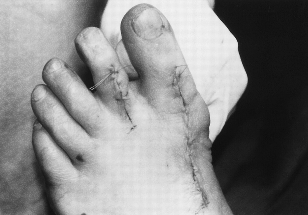
Fig. 4). Maintaining the distraction, the toe is then dorsiflexed to slip over the pin, which should slide into the middle phalanx. The toe should now be impacted along its straightened alignment using finger pressure to drive the phalanges together (
Fig. 5).
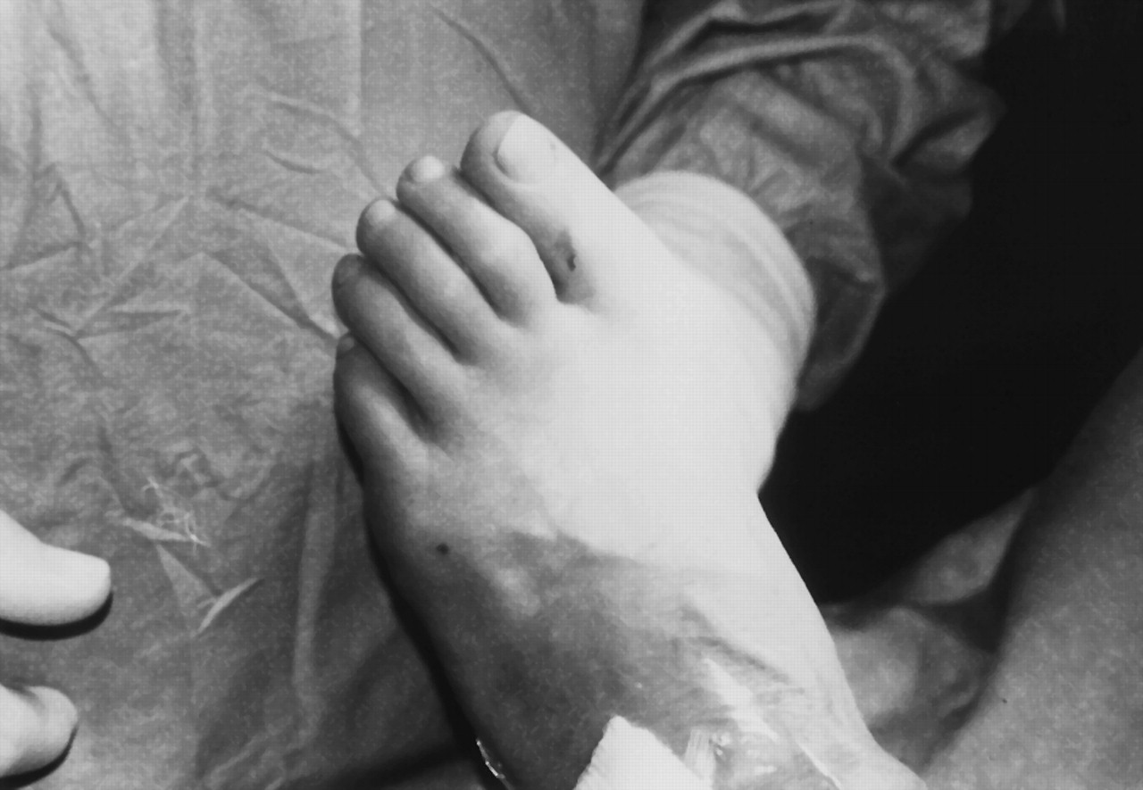
At this point, the foot is test-loaded with pressure under the metatarsal head (
Fig. 6). If the toe is modestly contracted at the metatarsophalangeal joint, appropriate soft-tissue releases can be performed.
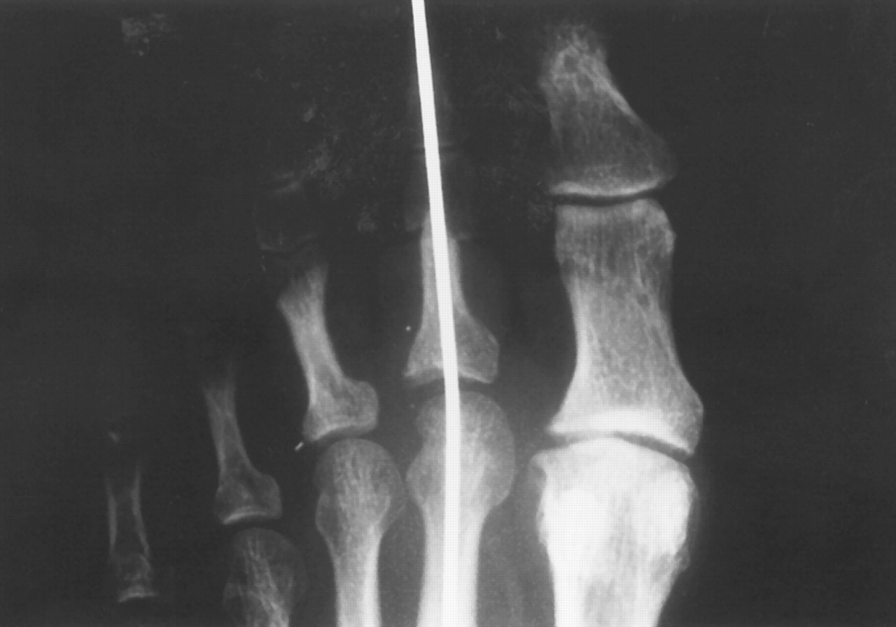
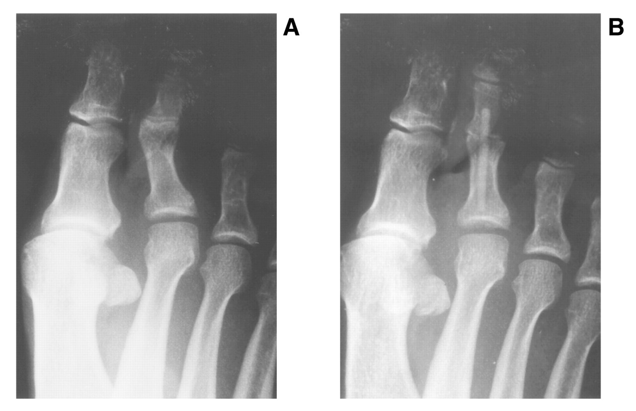
The tendons and incisions are then sutured for wound closure. A postoperative radiograph will confirm the position of the pin (
Fig. 7).
Figure 4.
Maneuvering of the distal toe onto the bone pin.
Figure 4.
Maneuvering of the distal toe onto the bone pin.
Figure 5 A and B.
Straightened distal toe with the arthrodesis fixation pin in place.
Figure 5 A and B.
Straightened distal toe with the arthrodesis fixation pin in place.
Figure 6.
Loading of the foot to check for reduction at the metatarsophalangeal joint.
Figure 6.
Loading of the foot to check for reduction at the metatarsophalangeal joint.
Figure 7.
A, Preoperative radiograph of a modest hammer toe deformity; B, postoperative radiograph of arthrodesis correction with an implanted cortical bone pin.
Figure 7.
A, Preoperative radiograph of a modest hammer toe deformity; B, postoperative radiograph of arthrodesis correction with an implanted cortical bone pin.
Results
Arthrodesis of the proximal interphalangeal joint of the lesser digits using the 2.4-mm cortical bone pin allograft was performed on 26 toes in 18 patients. There were 19 second toes, 5 third toes, and 2 fourth toes. Fifteen of the toes underwent the additional flexor digitorum longus tendon transfer procedure at the same time. The average age of the patients was 66 years (range, 43 to 82 years); 15 were women and 3 were men. The average length of follow-up was 15 months (range, 1 to 34 months).
Two toes developed flexion contractures at the distal interphalangeal joint postoperatively, and three toes became hyperextended at the metatarsophalangeal joint. Other toes showed a mild propensity for the same hyperextension but were splinted into rectus alignment. In these patients with subsequent deformity, no flexor digitorum longus tendon transfer had been done to help further stabilize the digit.
Partway through the bone-healing process, while the cortical bone pins were being resorbed and incorporated into new bone, the pin fractured in two toes. One toe went on to full arthrodesis and the other became a nonunion requiring revisional surgery.
One of the hyperextended toes was reduced with an extensor tenotomy/capsulotomy. Except for the two revisional surgeries, all of the toe fusions (n = 24) resulted in patient satisfaction. A total of 25 toes fused uneventfully, some managing to bridge gaps of 1 to 4 mm with the assistance of the cortical bone allograft pin.
Using serial radiographs, it was observed that the pins were fully resorbed and incorporated into the patient’s bone 6 to 8 months after the surgery. Sometimes remnants of the bone pins were visible on radiographs 1 year postoperatively (
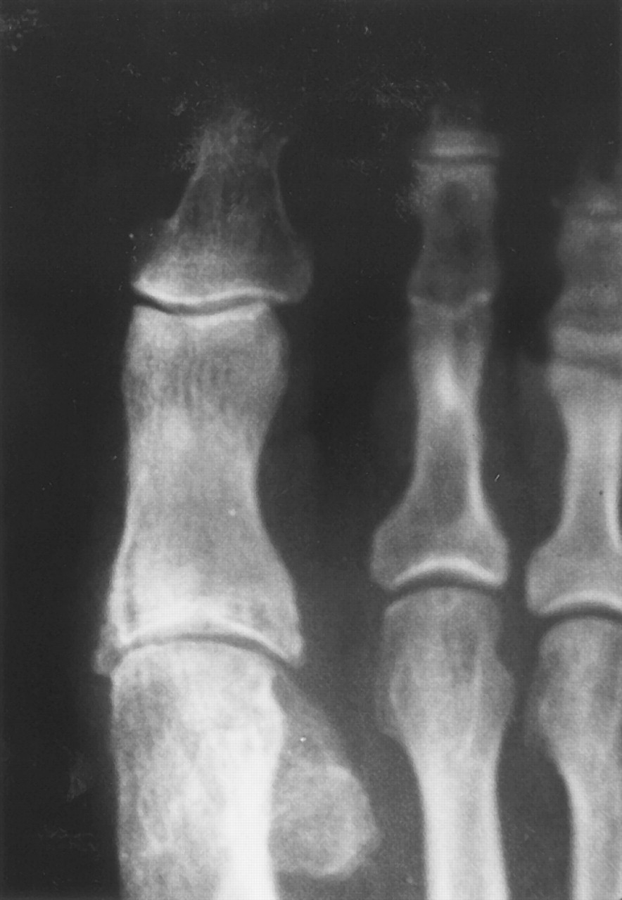
Fig. 8). This was of no clinical significance. A significant advantage of bone pins over synthetic pins is that bone pins are visible on serial radiographs as they are incorporated during the healing process.
Figure 8.
Radiograph of the cortical bone allograft pin 1 year postoperatively.
Figure 8.
Radiograph of the cortical bone allograft pin 1 year postoperatively.
Discussion
In 1990, Patton et al [
5] pioneered the use of absorbable pins for internal fixation of digital arthrodesis by using 1.3-mm-diameter synthetic pins made of poly-p-dioxanon. They reported favorable results from 58 digital fusions in 19 patients. It was also observed that the pins dissolved by hydrolysis within 6 months. Although there were no adverse reactions to the poly-p-dioxanon pins in the study by Patton et al, [
5] there have been several reported reactions to this material, such as osteolysis due to foreign body reactions. [
4]
Fixation devices such as staples, pins, and screws have been produced using poly-L-lactic acid. This material has the longest degradation time for synthetic implants in bone, with gross geometry still evident at 1 year and taking 2 to 3 years to absorb. [
6,
7] Still, it is the least reactive material and can provide effective bone stabilization in foot surgery procedures. [
8] Unfortunately, the long-term effects in the body are of some concern, and the pins and screws create holes that may not fill with bone, failing to restore integrity for 1 year or longer.
Internal fixation implants have been fashioned out of cortical allograft bone for successful use as bone plates and screws to fixate fractures [
9] and as Allofix bone pins to stabilize short Z-osteotomies for bunionectomies. [
4] The former devices regained three-quarters of the biomechanical strength of controls by participating in the healing process and forming external bone callus and were superior to the metal compression plates, which gained less than half of the strength of the controls. The cortical bone plate allografts eventually were incorporated into the host bone by being revascularized and remodeled into cancellous bone.
Allogeneic bone used as a fashioned transplanted bone graft is better termed an “allo-implant.” For other bone graft purposes, it is known as an “allograft.” Although autogenous bone has been shown conclusively to be the ideal bone graft, especially in higher-risk situations, freeze-dried allogeneic bone has been proven to be effective and physiologic for many bone implantation purposes, [
10–
12] even when specifically reviewed for foot and ankle surgery. [
13]
Studies of bone graft physiology have shown that bone allografts, usually cancellous, are resorbed and revascularized [
14] and are then replaced with host bone. [
15–
20] This also occurs with cortical bone grafts, [
21] as they are incorporated into the local bone through the physiologic principles of bone healing. [
22,
23] Freeze-drying, or lyophilization, tends to reduce the antigenicity and to stabilize the bone for transplantation and storage. Although the primary method of incorporation of the bone graft is creeping substitution
via osteoconduction, freeze-drying preserves enough of the bone morphogenic protein to stimulate osteoinduction, further enhancing its ability to consolidate into the local bone.
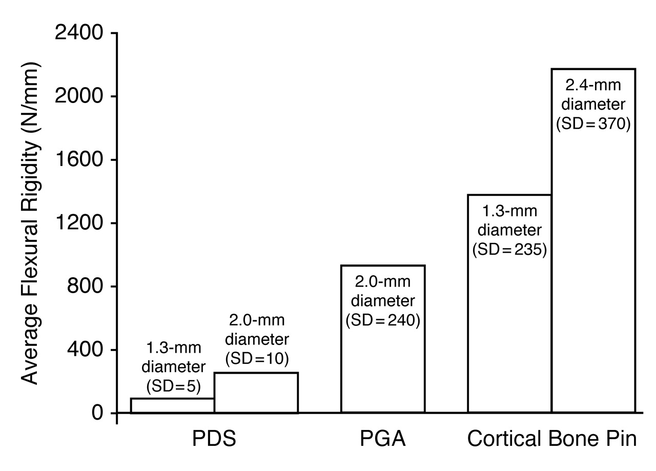
Cortical allograft bone pins provide flexural rigidity (
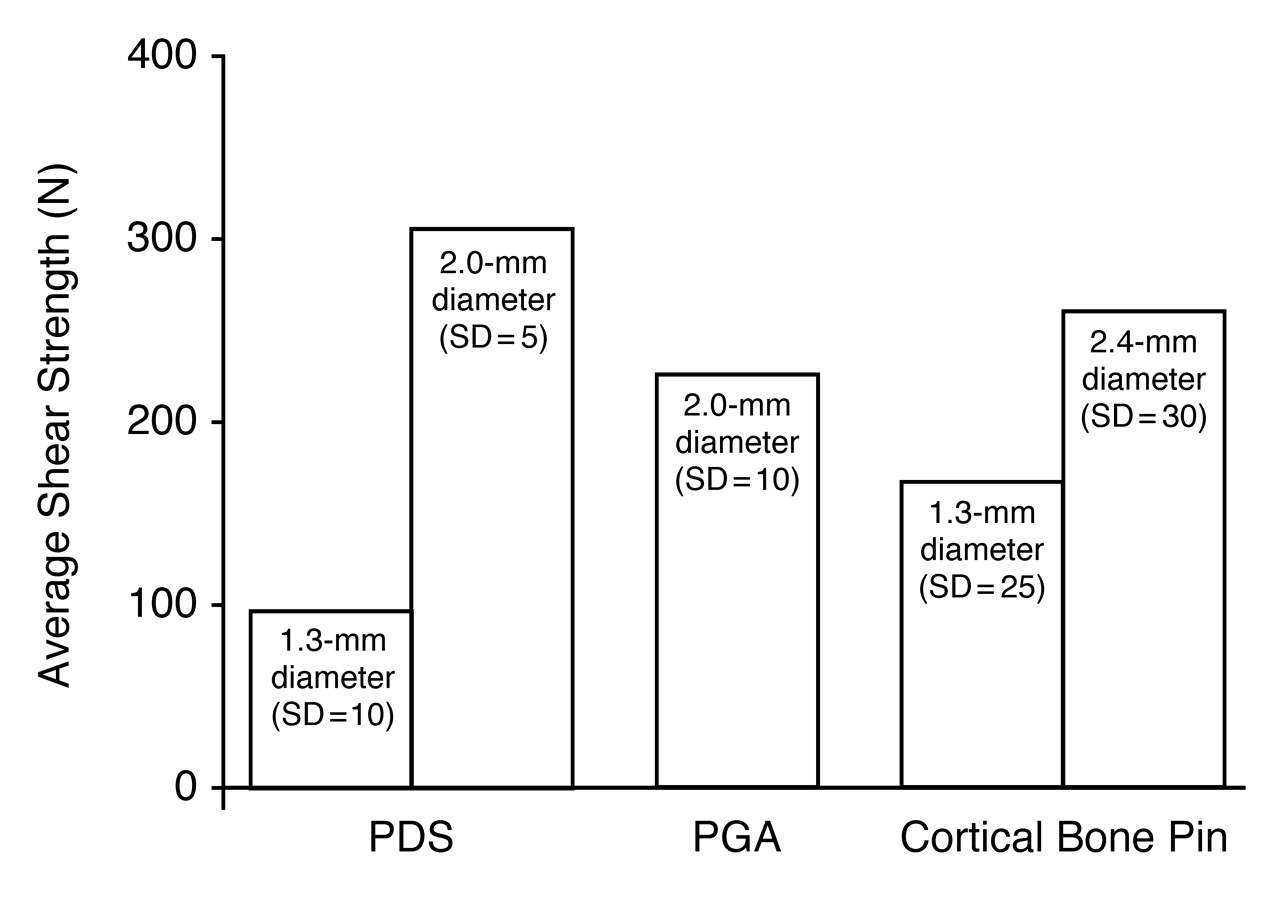
Fig. 9) and resist shearing forces (
Fig. 10); however, they lack strength against compressive and bending forces. [
24] Although preparation and freeze-drying tend to mildly reduce the bending and torsional strength of the bone pins, none of these modest limitations were observed to have any adverse effects on the success of the digital corrections, a finding consistent with use of the pins for fixation of distal first metatarsal osteotomies for bunionectomies. [
4]
Figure 9.
Average flexural rigidity of cortical bone pins versus synthetic pins (poly-p-dioxanon [PDS] and polyglycolic acid [PGA]). (Courtesy of the Musculoskeletal Transplant Foundation, Edison, New Jersey.)
Figure 9.
Average flexural rigidity of cortical bone pins versus synthetic pins (poly-p-dioxanon [PDS] and polyglycolic acid [PGA]). (Courtesy of the Musculoskeletal Transplant Foundation, Edison, New Jersey.)
Figure 10.
Average shear strength of cortical bone pins versus synthetic pins (poly-p-dioxanon [PDS] and polyglycolic acid [PGA]). (Courtesy of the Musculoskeletal Transplant Foundation, Edison, New Jersey.)
Figure 10.
Average shear strength of cortical bone pins versus synthetic pins (poly-p-dioxanon [PDS] and polyglycolic acid [PGA]). (Courtesy of the Musculoskeletal Transplant Foundation, Edison, New Jersey.)
Bone allografts, when carefully prepared under strict guidelines, have been shown to be safe, with an extremely low risk of disease transmission. [
24–
26] The risk of HIV infection has been estimated to be 1 in 1.6 million in properly screened allograft bone. [
27]
The results of this clinical study using 2.4-mm Allofix cortical bone pins for fixation and arthrodesis of digital fusions showed the pins to be effective (92% successful) while avoiding many of the potential complications and inconveniences of percutaneous pin fixation. There have been no reports to date of disease transmission after implantation of cortical bone allograft pins.
Flexion at the distal interphalangeal joint, or mallet toe formation, after proximal interphalangeal joint arthrodesis is a well-known possible complication that may require further corrective surgery. One study [
3] demonstrated an incidence of 44%. This potential problem and the difficulty with metatarsophalangeal joint hyperextension were solved by adding a flexor digitorum longus tendon transfer to the procedure for additional stability against the supporting surface. As the study progressed, this additional maneuver was used more frequently.
Conclusion
The results of this study support the use of Allofix freeze-dried cortical bone pins to successfully heal bone, [
4] specifically digital fusions, not only by providing stable internal fixation but also by participating in the bone-healing process, further enhancing the consolidation by incorporation. The pins were even able to bridge significant gaps. Complications and risks were shown to be minimal, with further stability enhanced by using the flexor digitorum longus tendon transfer procedure. Such pins, therefore, are an effective alternative for use in digital arthrodesis, offering many advantages over other devices used for fixation.