Discussion
Hallux abducto valgus comprises both osseous and soft-tissue deformities. Correction has generally been accomplished by both osteotomy and muscle-tendon rebalancing. This not only eliminates the deformity but also addresses the etiology, preventing recurrence. Traditional teaching identifies one of the main deforming forces as the intrinsic muscles, primarily the adductor hallucis tendon, acting through the sesamoid apparatus. McBride [
1] described his muscle-tendon balancing technique for correction of hallux valgus. He believed “one of the greatest mechanical factors” in hallux valgus to be the lateral intrinsic muscles. Root et al [
3] described four stages of hallux valgus, beginning with unlocking of the first metatarsophalangeal joint as a result of abnormal pronation. The adductor hallucis tendon and the lateral head of the short flexor tendon place increased lateral tension on the base of the proximal phalanx
via the fibular sesamoid, causing the phalanx to subluxate laterally. The subsequent retrograde force of the hallux on the first metatarsal will increase the first intermetatarsal angle. The theory that metatarsus primus varus is secondary to hallux abductus has been widely accepted by the podiatric and orthopedic professions. Appropriate correction is therefore based on removal of the deforming forces and relocation of the fibular sesamoid. Release of the adductor hallucis tendon and fibular sesamoidal ligament with or without transfer has been described by many authors, including Ruch et al, [
4] Wooster et al, [
5] Schoenhaus and Cohen, [
6] and Kempe and Singer. [
7] Lateral release has been emphasized to remove the deforming forces of hallux valgus and release the fibular sesamoid to allow lateral transposition of the metatarsal head.
Recently, several studies have been published that call into question this theory of the etiology and appropriate treatment of hallux valgus. Jahss [
8] has stated that sesamoid release is not necessary for reduction of the intermetatarsal angle. Judge et al [
9] demonstrated that the position of the sesamoids is unaffected by hallux valgus correction, which is supported by Mann and Coughlin, [
10] Jahss, [
8] and Alvarez et al. [
11] Tanaka et al [
12] recently published a radiographic analysis of the relationship of various osseous landmarks in normal feet and feet with symptomatic hallux valgus deformity. They demonstrated that the position of the base of the proximal phalanx does not move in relation to the second metatarsal. As the first metatarsal moves medially, the hallux stays in place rather than drifting with the metatarsal. Their conclusion was that hallux valgus is primarily a deformity of metatarsus primus varus and that correction of the first metatarsal by means of osteotomy should be given first consideration. It can be concluded from these studies that if the positions of the sesamoids and proximal phalanx remain unchanged in hallux valgus deformity, the primary etiology is metatarsus primus varus, which is what needs to be addressed.
It is also clear that bunions have both positional and structural components. The retrograde force of the hallux on the metatarsal
via the intrinsic muscle–sesamoid complex as described by Alvarez et al [
11] and, as Sanders et al [
13] noted, the deforming force of the extrinsic muscles will clearly worsen the deformity. Any effective release considered should therefore include the conjoined tendon and the long flexor and extensor tendons, not just the adductor tendon. However, it has not been proven that metatarsus primus varus is secondary to hallux valgus. The results presented here indicate that hallux valgus is secondary to metatarsus primus varus.
The results of the present study clearly demonstrate that the deformity of hallux valgus can be corrected and maintained solely by metatarsal osteotomy in appropriately selected patients. During an average follow-up period of 18.4 months, the corrected position and alignment of the hallux and metatarsophalangeal joint were maintained.
There are several advantages of not releasing the sesamoids. David et al [
14] documented the role that the sesamoids play in hallux stability and normal function during gait. Mann and Coughlin [
10] have demonstrated the importance of the deep transverse intermetatarsal ligament. This ligament helps bind the capsule of the first and second metatarsophalangeal joints. Preserving these intermetatarsal ligaments would seem useful in the correction of hallux valgus. Any structure, including the heads of the adductor tendon, that originates lateral to and attaches to the first metatarsal binds the metatarsal heads and should be augmented rather than transected. Moreover, without interspace release there is much less dissection and less postoperative edema. Anecdotally, these patients seem to recover more quickly and have greater range of motion. The surgical repair can also be performed through a medial incision for a superior cosmetic result.
Soft-tissue adaptation does occur in hallux valgus, especially in long-standing deformities with higher degrees of joint subluxation. The patients originally considered for inclusion in this study included several in whom the joints could not be manually reduced, requiring soft-tissue release to attain joint congruity. These cases were not included in the study group. In all cases, the joint adaptive contracture was between the base of the phalanx and the fibular sesamoid. In the cases omitted from the study, transection of the conjoined tendon was required to obtain a rectus alignment of the hallux.
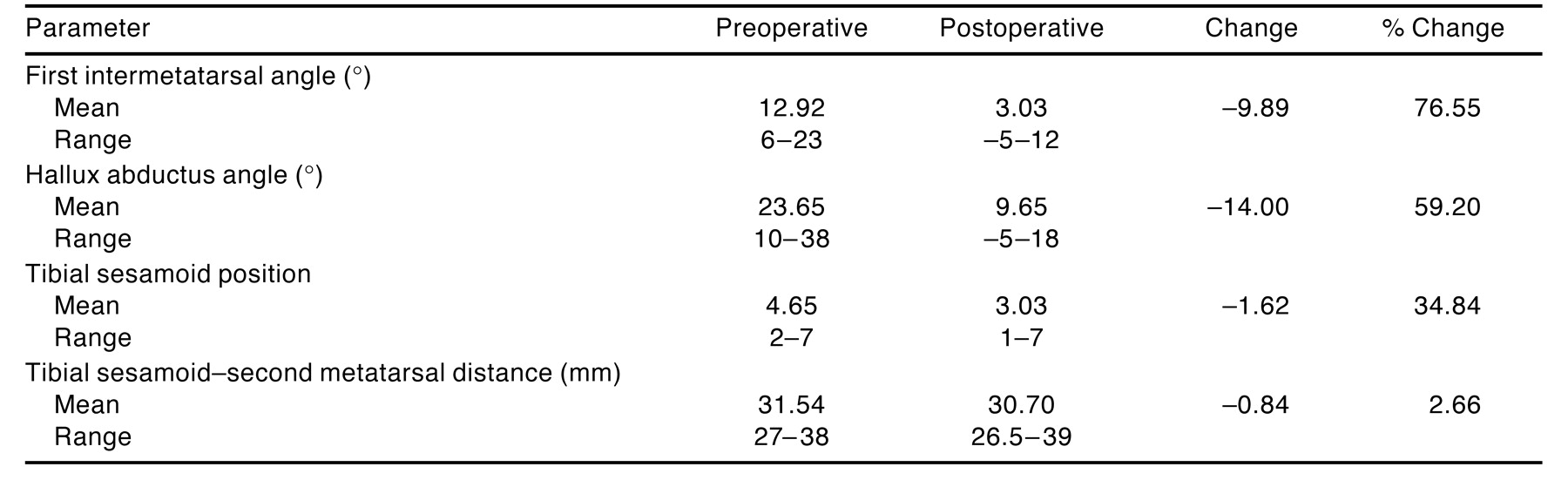
Reductions in all radiographic parameters were evident in all patients and correlated well with clinical improvement in position and alignment across the first metatarsophalangeal joint. These results compare favorably with those of a recently published study on hallux abducto valgus correction with interspace release (
Table 2). Without interspace release, the first intermetatarsal angle decreased by an average of 9.89° (to 3.03°), and the hallux abductus angle decreased by an average of 14.0° (to 9.65°). In contrast, in patients with interspace release, the first intermetatarsal angle decreased by an average of 7.93° (to 4.12°) and the hallux abductus angle decreased by an average of 17.14° (to 7.43°). The average change in first intermetatarsal angle was approximately 10% less in procedures performed with a traditional interspace release than in those with an isolated medial approach. However, the average change in the hallux abductus angle and tibial sesamoid position was greater with the lateral interspace release.
Table 2.
Radiographic Parameters Using Traditional Hallux Abducto Valgus Correction with Interspace Release
Table 2.
Radiographic Parameters Using Traditional Hallux Abducto Valgus Correction with Interspace Release
The greater change in the intermetatarsal angle in this study is believed to be partly due to the senior author’s tendency to perform aggressive translocation of the metatarsal head, often equal to 50% of the metatarsal head width. Also, by preserving all of the attachments between the lateral aspect of the metatarsal head and the rest of the foot, the soft tissues maintain the reduction of the intermetatarsal angle. Given that the lack of interspace release was associated with an average of 3.14° less correction in hallux abductus angle, the isolated medial approach procedure would seem best suited for deformities with only mildly or moderately elevated hallux abductus and first intermetatarsal angles. To address this, the authors evaluated those patients who had large first intermetatarsal and hallux abductus angles to determine whether the results differed for this subset of patients (
Table 3). Patients with first intermetatarsal angles greater than or equal to 15° (9 patients or 31.03%; 11 feet) had greater changes in hallux abductus angle, tibial sesamoid position, and tibial sesamoid–second metatarsal distance than those reported for the entire group (
Table 1). These findings suggest that a significant reduction of deformity can be obtained using the isolated medial approach even in cases of large first intermetatarsal angles. Patients with hallux abductus angles greater than 28° (10 patients or 34.48%; 13 feet) were evaluated separately and showed an overall reduction in hallux abductus angle of 19.09°. The flexibility or hypermobility of the first ray probably accounted for these results.
Table 3.
Radiographic Parameters for Patients with High First Intermetatarsal Angles (≥15°) Using the Isolated Medial Approach to Hallux Abducto Valgus Correction
Table 3.
Radiographic Parameters for Patients with High First Intermetatarsal Angles (≥15°) Using the Isolated Medial Approach to Hallux Abducto Valgus Correction
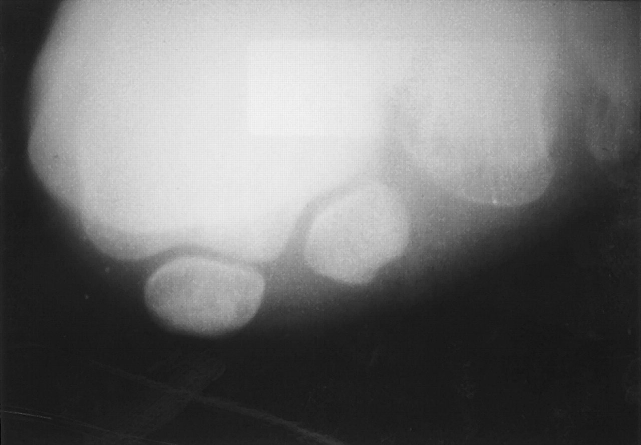
The tibial sesamoid position decreased by an average of 1.62 positions (on a 7-position scale) with the isolated medial approach. A general assessment of the tibial sesamoid position was made for patients who had sesamoidal axial radiographs taken before and after the operation (21 patients or 72.41%). The position of the tibial sesamoid was graded on the basis of location about the sesamoidal articular surfaces and cristae as described above. The position and alignment of the tibial sesamoid appeared essentially unchanged from the preoperative to the postoperative condition on the sesamoidal axial radiograph in 20 out of 21 patients (95.24%). As the average tibial sesamoid position decreased from 4.65 to 3.03, these findings based on transverse plane radiographs differed from those based on the sesamoidal axial views. The sesamoidal axial views confirmed that the sesamoid apparatus remained unchanged in position after surgery in the majority of cases (
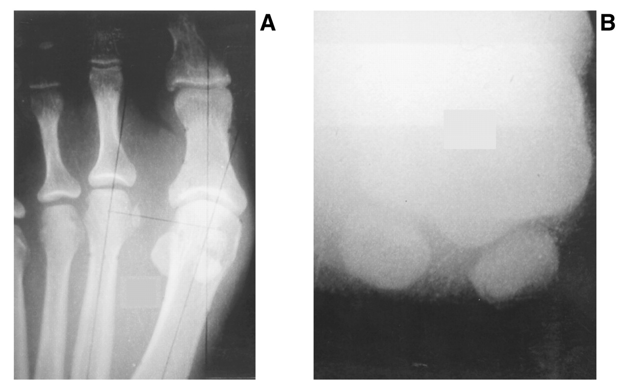
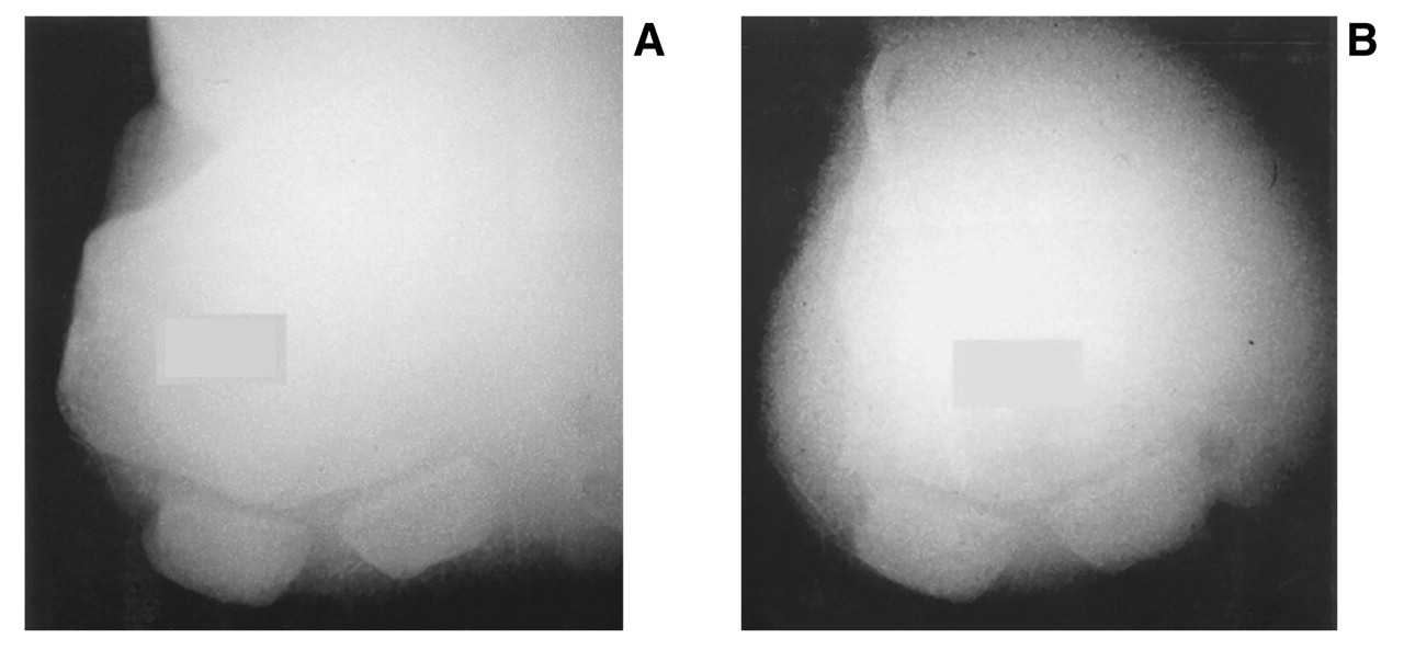
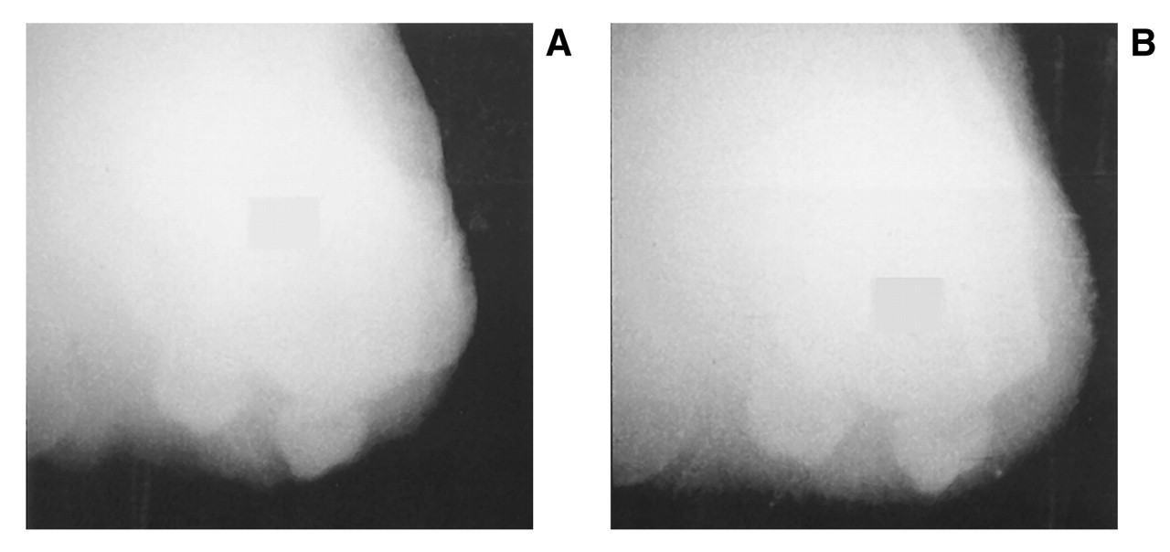
Figs. 1–
3). The tibial sesamoid can clearly appear deviated in the transverse plane on preoperative dorsoplantar radiographs while on the sesamoidal axial radiographs the joint appears completely congruous, with the sesamoids resting within their respective articular grooves (
Fig. 4). In each of the cases studied here, the preoperative dorsoplantar radiographs exhibited deviation of the sesamoid apparatus, while postoperatively these views uniformly suggested a change in that position. The findings on the sesamoidal axial view failed to corroborate the sesamoid deviation and subsequent change exhibited on the dorsoplantar views. This signifies that the apparent change in sesamoid position on a dorsoplantar radiographic view of a hallux abducto valgus deformity is actually the result of an oblique rotation of the metatarsal head and the sesamoid apparatus. In the same way that the plantar structures of the forefoot appear to be shifted laterally in a medial oblique radiograph, the oblique projection of the first metatarsophalangeal joint in hallux abducto valgus deformity results in the appearance of a lateral shift in the sesamoid apparatus when in fact it is fixed in its soft-tissue bed. This brings into question the significance of the tibial sesamoid position as measured on dorsoplantar radiographs. The authors recommend that sesamoidal axial radiographs be a standard preoperative tool used to determine the presence of any degree of sesamoid apparatus deviation. This will aid the surgeon in determining the need for a soft-tissue release.
The tibial sesamoid–second metatarsal distance decreased by an average of 0.84 mm after isolated medial approach correction, a statistically significant difference. This finding differs from that reported in the study of Judge et al, [
9] in which the change in sesamoid distance from the second metatarsal was 0.05 mm. This could mean that after the transposition of the metatarsal head, with the fibular sesamoidal ligament and adductor tendon left intact, the sesamoid apparatus is being marginally translocated laterally along with the metatarsal. The clinical significance of this phenomenon is unknown. Another possible explanation is that the tibial sesamoid–second metatarsal distance decreases as the head of the metatarsal and the associated soft-tissue attachments are translocated laterally owing to a rotational radiographic effect rather than a transverse plane deviation of the sesamoid.
The current study answered three pertinent questions regarding hallux abducto valgus surgery performed without the benefit of an interspace release: 1) Is the intermetatarsal space release integral for optimal outcome in hallux abducto valgus surgery? In patients with manually reducible metatarsophalangeal joints and mobile first ray segments, interspace release is not needed to achieve correction of the hallux abducto valgus deformity. 2) Is there an increased recurrence of hallux abducto valgus if an interspace release is not performed? The authors have found no evidence of early or delayed recurrence of deformity in this study, in which 44.83% of the patients were followed up for longer than 2 years. 3) Is the outcome of bunion surgery predictable for deformities with either moderately or severely elevated intermetatarsal angles when an interspace release is not performed? In this study, a small subset of 9 patients (31.03%; 11 feet) presented with first intermetatarsal angles of 15° or more. The hallux abductus angle and tibial sesamoid position correction achieved in all of these cases was greater than that obtained in cases with lower preoperative intermetatarsal angles (
Table 3).
Figure 1.
A, Preoperative dorsoplantar radiograph exaggerates a sesamoidal deviation; B, sesamoidal axial radiograph reveals a nearly congruous alignment.
Figure 1.
A, Preoperative dorsoplantar radiograph exaggerates a sesamoidal deviation; B, sesamoidal axial radiograph reveals a nearly congruous alignment.
Figure 2.
A, Preoperative sesamoidal axial radiograph shows a deviated sesamoid position; B, postoperative radiograph after medial eminence resection shows residual convexity with sesamoidal deviation unchanged.
Figure 2.
A, Preoperative sesamoidal axial radiograph shows a deviated sesamoid position; B, postoperative radiograph after medial eminence resection shows residual convexity with sesamoidal deviation unchanged.
Figure 3.
Preoperative (A) and postoperative (B) sesamoidal axial radiographs fail to corroborate a significant change after surgery.
Figure 3.
Preoperative (A) and postoperative (B) sesamoidal axial radiographs fail to corroborate a significant change after surgery.
Figure 4.
Sesamoidal axial radiograph reveals varus rotation of the metatarsal head with congruency of the sesamoids within their articular facets.
Figure 4.
Sesamoidal axial radiograph reveals varus rotation of the metatarsal head with congruency of the sesamoids within their articular facets.
Conclusion
Using an isolated medial approach V-osteotomy, clinical and radiographic correction of the hallux abducto valgus deformity was achieved without the benefit of an interspace release. This correction was maintained in all 29 study participants throughout an average follow-up period of 18.4 months, with no clinical or radiographic recurrence noted. It is evident that the key to successful bunion surgery is the clinical reduction of both the first intermetatarsal angle and the hallux abductus angle, which were achieved using this technique.
The authors believe that this procedure results in less soft-tissue disruption and therefore less occurrence of postoperative adhesion. This may result in less restriction of motion and a better functional outcome in appropriately selected patients. The authors recommend that the sesamoidal axial view be a standard radiographic view for the preoperative assessment of hallux abducto valgus deformity. Such a view will help determine whether the sesamoid apparatus is actually deviated, subluxated, or dislocated, which is an important factor in the decision regarding the suitability of an interspace release. When the sesamoid apparatus is radically deviated or dislocated, soft-tissue structure contractures are likely to prevent adequate repair of the hallux abducto valgus deformity. This finding is integral to developing the surgical plan to restore normal joint alignment.
The purpose of this study was to validate the utility and effectiveness of an isolated medial approach for hallux abducto valgus correction in selected patients who do not require the first interspace release. It is not the authors’ intent to recommend elimination of anatomical dissection and first interspace release from all hallux abducto valgus surgery; rather, it is suggested that the isolated medial approach without first interspace release can be employed in suitable patients with predictable results. Because of the limited soft-tissue dissection, postsurgical adhesion and capsulitis are reduced, thus improving functional outcomes. A prospective, long-term outcome study should be conducted to compare the isolated medial approach technique and the traditional procedure with interspace release in terms of meaningful clinical changes in joint position.