Plantar foot ulceration is a well known complication of diabetes mellitus that must be treated appropriately to minimize the risk of more serious complications, such as infection and amputation.[
1-
5] Foot ulceration is a major cause of approximately 85% of lower-extremity amputations in patients with diabetes.[
6-
9] Although partial-foot amputation is a limb-salvaging measure, it is enormously difficult for the patient and can lead to an increased risk of subsequent amputations.
Elevated plantar foot pressures are one of the most difficult, yet essential, factors that must be controlled in order to promote healing of plantar ulcers. In a prospective study of 86 patients with diabetes, Veves et al[
10] determined that elevated plantar pressures in these patients were strongly predictive of subsequent ulceration in neuropathic patients. Higher than normal plantar pressures develop under the metatarsal heads with weightbearing as a consequence of motor neuropathy. Motor neuropathy and intrinsic muscle atrophy can result in foot deformity in which the toes become clawed, the metatarsal heads become prominent, and the fibrofatty footpads migrate anteriorly.[
11-
14] Limited joint mobility is also a complication of long-term diabetes that further impedes the foot in redistributing high loads and contributes to the development of high plantar pressures. Limited joint mobility is caused by nonenzymatic glycosylation of collagen in the connective tissues surrounding the joints.[
15] The talocrural, subtalar, and first metatarsophalangeal joints are particularly important to patients with long-term diabetes.[
16,
17]
Autonomic neuropathy is an insidious and often unrecognized complication of long-term diabetes that can cause alterations in microvascular blood flow and eccrine gland activity.[
2,
5,
14] Anhidrosis with resultant dry skin is a common sequela of autonomic neuropathy. The combination of xerotic skin and high plantar pressures leads to callus formation, which, in turn, acts as a foreign body on the plantar foot surface. The insensate diabetic patient is predisposed to plantar ulceration from neglected calluses due to the development of high, underlying, plantar pressures during walking. Several studies have confirmed these findings, indicating that plantar calluses are strong predictors of the occurrence of foot ulcers. Reiber et al[
18] found that callus formation was a component cause in 30% of the pathways leading to ulceration. Murray et al[
19] found that ulcers recur only at the sites of callus formation. In a study of 225 age-matched patients, Lavery et al[
20] found that neuropathy, foot deformity, high plantar pressures, and a history of amputation were significantly associated with foot ulceration. Other studies have also confirmed the role of neuropathy and high plantar pressures in the etiology of diabetic foot ulceration.[
21-
23]
High plantar pressures play a pivotal role in the pathogenesis of plantar ulceration of the diabetic foot. The additional complications inherent in the diabetic population greatly magnify the damaging effect of high plantar pressures. Adequate offloading is paramount to the successful healing of diabetic plantar ulcers, and is, therefore, a widely accepted treatment principle.[
2,
24] Several common modalities, including total contact casting, surgical boots, felt padding, removable walking braces, and half shoes, accomplish offloading with varying degrees of success.[
2,
25] Two recent studies support the superior efficacy of total contact casting in the management of neuropathic plantar foot ulcers.[
26,
27] The cost and technical difficulty of applying weekly total contact casts, however, has led many clinicians to prefer alternative offloading devices. In a small study, one such alternative, the Rocker Insole (Langer Biomechanics Group, Inc, Deer Park, New York), was shown to offload forefoot plantar pressures effectively.[
28] The primary aim of the present study was to assess the Rocker Insole’s ability to reduce forefoot plantar pressures in a larger group of patients. Furthermore, the offloading capabilities in patients with and without diabetes were compared.
Research Design and Methods
The plantar foot pressures of 25 subjects with various foot types from the Des Moines University podiatric clinic were randomly measured for a period of 7 months. Patients with and without diabetes were enrolled in the study. The subjects signed an informed consent form prior to enrollment. The Health, Safety, and Human Use Committee for Clinical Investigation at Des Moines University approved the research proposal.
The F-Scan computerized insole sensor system (TekScan, Inc, South Boston, Massachusetts) was used to measure dynamic plantar foot pressures.[
29,
30] The F-Scan uses a flexible, thin insole that consists of 960 sensor units or elements in a grid configuration that provides high spatial resolution. While walking, vertical forces at the foot and shoe interface were measured by each of the individual 5 mm
2 elements through force sensitive resistor technology.[
29] The insole was then directly coupled to an ankle cuff amplifier unit connected by long cable to the hard card installed in a desktop computer running the F-Scan software. Once connected, displays of force and pressure were available for both feet simultaneously.
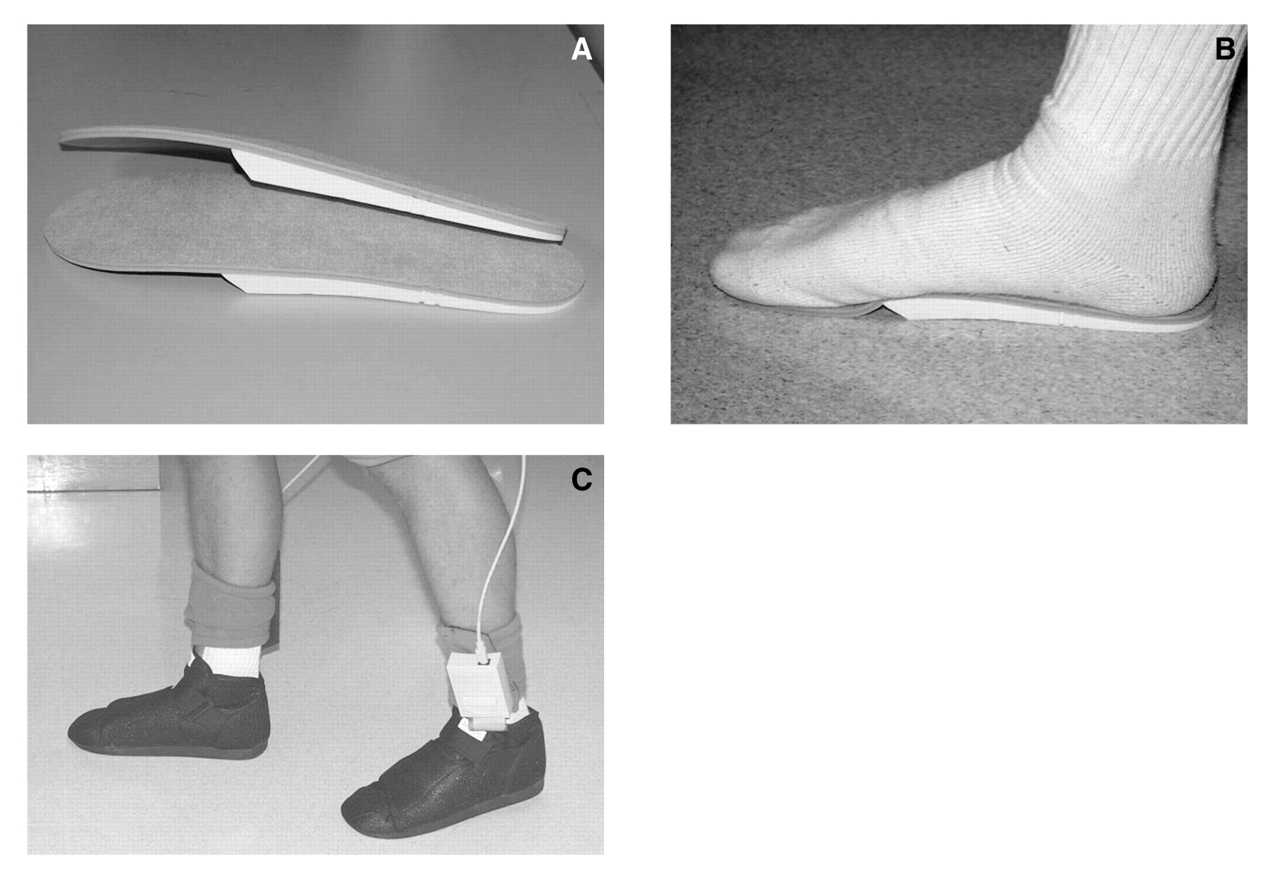
The F-Scan insole system measured plantar foot pressures in three different types of footwear: the patient’s own tennis or oxford shoe, a standard postoperative surgical boot, and that same surgical boot with the addition of a Rocker Insole. The Rocker Insole’s anterior wedge design, fabricated from dense, closed cell foam, effectively offloads the forefoot when fitted properly behind the metatarsal heads (
Figs. 1A-C). The insole can be trimmed as necessary to fit the foot and surgical shoe.
The dynamic plantar pressures in each shoe modality were measured twice for each foot. Three representative footsteps were then selected from the tracings and an average for each peak pressure was obtained. Each foot contributed its own set of measurements and constituted one observation. Therefore, the 25 patients contributed two observations each for a total of 50 distinct sets of data per shoe modality tested.
The patients all wore a standard nylon stocking to minimize variability. Measurements were calibrated for each patient according to manufacturer specifications. Before testing, patients walked for several minutes to become acclimated to the insole and shoe. Patients were asked to walk at their normal cadence and stride length across a linoleum floor. During gait, the F-Scan software defaulted to the area of maximal peak pressure under the forefoot and displayed an indicator of exact location as well as a numerical value in kg/cm2. The peak forefoot pressures recorded for each subject corresponded to the mean peak pressure of three representative footsteps for each foot in the three footwear conditions.
Statistical analysis was performed using nonparametric tests for continuous variables. Subjects served as their own control for both feet, which were measured independently. Wilcoxon rank sum tests were used to compare nonpaired groups and Wilcoxon signed-ranks tests were used to compare differences within each paired observation. Chi-square and Fisher’s exact tests were used to compare discrete variables between groups. Statistical significance was obtained at P < .05 (α = 0.05).
Results
Table 1 lists the characteristics of the study population. The group consisted of 25 subjects (13 men, 12 women), ranging in age from 23 to 67 years, with a mean age of 37 years. Ten patients (40%) had diabetes mellitus and one diabetic patient had an active ulcer. The average body mass index for the subjects with diabetes was 30.8
versus 25.3 for the subjects without diabetes (
P < .01), resulting in an average body mass index of 27.5 ± 5.7. The mean age of the subjects with diabetes was 49.7 years
versus 28 years for the subjects without diabetes (
P < .000) for a combined mean age of 36.7 years.
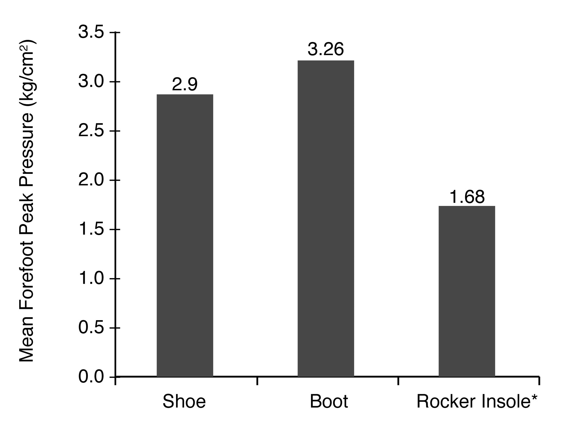
When combining foot pressure data for the entire group of observations (n = 50), the mean peak pressure in the forefoot while wearing the surgical boot was highest (3.26 ± 2.26 kg/cm
2), followed by the patient’s shoe alone (2.90 ± 1.5 kg/cm
2). The surgical boot with Rocker Insole (1.68 ±1.09 kg/cm
2) resulted in the lowest forefoot peak pressures (
P < .000 for Rocker Insole
versus shoe and boot) (
Fig. 2).
Table 2 demonstrates that the maximum peak pressure for each footwear modality was higher overall in the diabetic population when compared to the nondiabetic population (
P < .000). The highest mean peak pressure values were obtained for the surgical boot, regardless of whether the subject did or did not have diabetes (4.89 ± 2.65 kg/cm
2 versus 2.13 ± 0.85 kg/cm
2). The Rocker Insole placed in the surgical boot provided the lowest mean peak pressure values of all the footwear conditions in both patients with and without diabetes (2.50 ± 1.19 kg/cm
2 versus 1.13 ± 0.56 kg/cm
2).
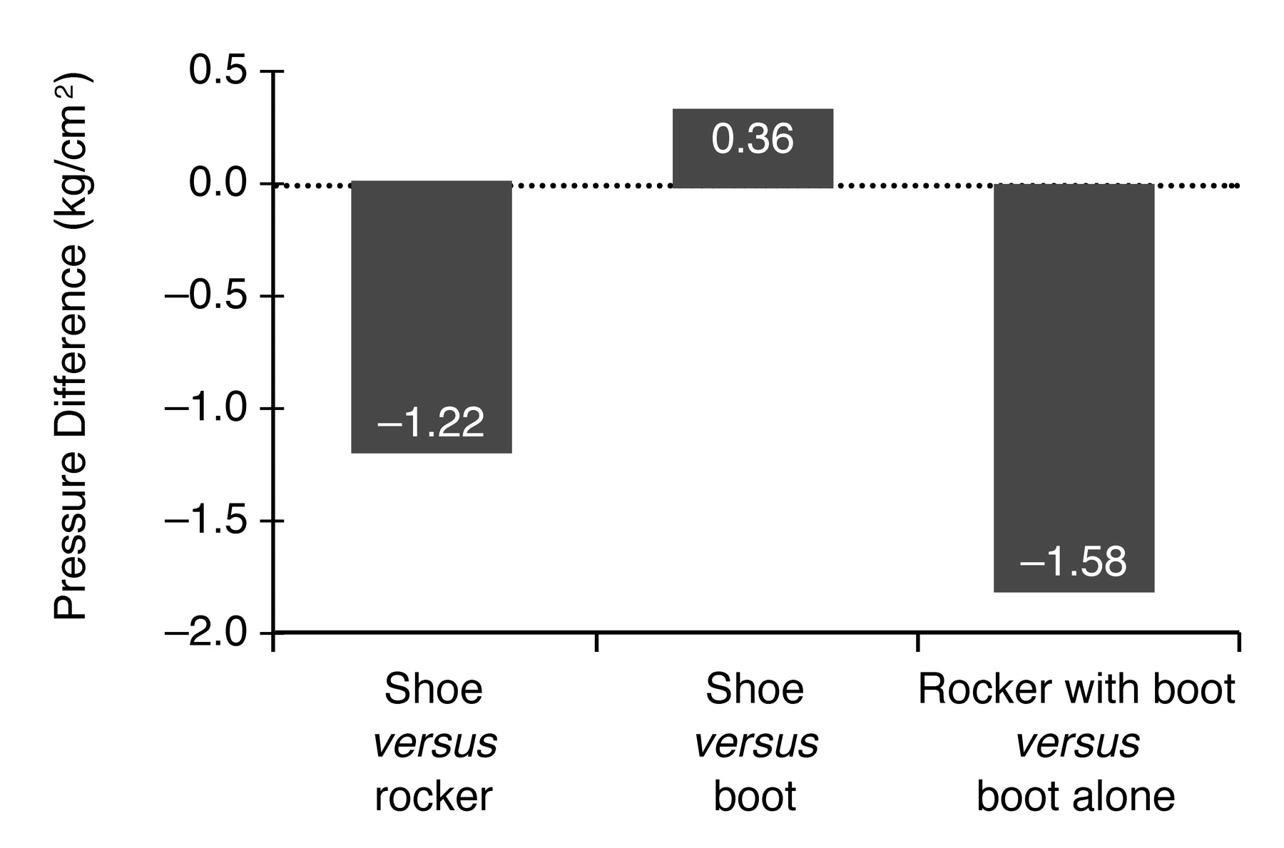
When the mean peak pressures of all subjects were combined, the surgical boot revealed an unexpected 12% increase in mean peak pressure (
P < .011) as compared to the patients’ regular tennis or oxford shoe (
Table 3). When inserted into the patients’ own footwear, the Rocker Insole decreased the forefoot pressures by 42% (
P = .000). The Rocker Insole inserted into the surgical boot reduced the forefoot peak pressure by 48% (P = .000) compared to the boot alone, which was the most effective modality tested for offloading this area of the foot. These differences are illustrated in
Figure 3. As hypothesized, therefore, the Rocker Insole did reduce the peak plantar metatarsal head pressures.
Discussion
The importance of offloading plantar ulcers in patients with diabetes has long been regarded as essential to successful healing and prevention of further complications.[
2,
5,
8,
12,
14] Total contact casting has proven quite effective as an offloading modality and is often considered the gold standard.[
25-
27,
30] However, these casts are time consuming to apply, costly, and must be changed frequently to avoid iatrogenic lesions.[
2,
30] Therefore, alternative methods, such as felt padding, postoperative shoes, half shoes, or removable walking braces, are often used to achieve offloading and promote ulcer healing.[
2] Although not considered to be quite as effective as the total contact cast, these modalities are safe, easily applied, and are significantly less expensive. The Rocker Insole is also an inexpensive, safe, convenient, and a readily available offloading device. The current study has shown that the Rocker Insole can significantly reduce maximum plantar pressures across the forefoot when it is worn in conjunction with a surgical boot. When the postoperative boot was worn alone, maximum peak pressures actually increased across the plantar metatarsal heads by 12%, as compared to the patients’ own casual footwear. This increase may be attributed to a poor fit of the foot to the boot, which would allow slippage of the heel, or may be due to the inflexibility of the sole without adequate rocking action over the forefoot. Both could conceivably increase forefoot pressures as compared to flexible walking shoes.
Although the current study was small, the patients with diabetes had significantly higher plantar pressures than the patients without diabetes. However, there was virtually no difference in the offloading ability of the Rocker Insole between the two groups. The Rocker Insole effectively provided offloading for the plantar metatarsal heads regardless of the subjects’ diabetic status. Thus, the neuropathy that is found in so many diabetic patients does not appear to decrease the efficacy of the Rocker Insole in reducing plantar pressures. A 48% reduction (P = .000) in plantar pressures compared to those in the surgical boot alone, or a 42% reduction (P = .000) as contrasted to initial shoe pressures, are both significant and support the use of the new Rocker Insole in treating plantar ulcers under the forefoot. It is important, however, for the Rocker Insole to be fit correctly so that the wedge ends just proximal to the metatarsal heads.
The mean peak pressures of the patients with diabetes had greater standard deviations than the patients without diabetes for all shoe modalities tested. This may initially be attributed to a smaller diabetic study population than the nondiabetic group in this study. This may also be the result of the difference in body mass index and average age of the two groups: the group with diabetes was more overweight (mean body mass index, 30.8) and older (mean age, 49.7 years) as compared with the group without diabetes (body mass index, 25.3; mean age, 28 years). Age, race, and obesity have been correlated with elevated plantar pressures in several studies.[
21,
22,
31-
33]
Conclusion
This study verified that the new Rocker Insole significantly provides offloading for the plantar metatarsal heads of patients with and without diabetes. Its use in the management of plantar forefoot ulcers may be warranted for patients with forefoot ulcers who are not appropriate candidates for total contact casting or other types of offloading devices. Further research is indicated to determine if a 48% reduction in mean peak pressure is sufficient to significantly improve outcomes in healing plantar forefoot ulcers. Comparative studies with other offloading modalities would also define the role of Rocker Insoles in the management of diabetic ulcers under the metatarsal heads.