Next Article in Journal
Offloading Properties of a Rocker Insole. A Preliminary Study
Previous Article in Journal
Cellular Senescence and Matrix Metalloproteinase Activity in Chronic Wounds. Relevance to Debridement and New Technologies
 
 
Journal of the American Podiatric Medical Association is published by MDPI from Volume 116 Issue 1 (2026). Previous articles were published by another publisher in Open Access under a CC-BY (or CC-BY-NC-ND) licence, and they are hosted by MDPI on mdpi.com as a courtesy and upon agreement with American Podiatric Medical Association.
Article

Use of Clinical Laboratory Parameters to Evaluate Wound Healing Potential in Diabetes Mellitus

by
Patricia L. Abu-Rumman
1,
David G. Armstrong
1,2 and
Brent P. Nixon
1
1
Department of Surgery, Podiatry Section, Southern Arizona Veterans Affairs Medical Center, Tucson, AZ
2
Department of Medicine, Manchester Royal Infirmary, Manchester, UK
J. Am. Podiatr. Med. Assoc. 2002, 92(1), 38-47; https://doi.org/10.7547/87507315-92-1-38

Published: 1 January 2002

Abstract

Clinicians caring for chronic wounds can easily overlook nutritional status. Patients with diabetes are at high risk for primary and secondary malnutrition. Although profiles exist defining the extent of the deficiency, the process of wound healing and the interactions of the macronutrients and micronutrients necessary to accomplish it must first be understood. In elderly patients with diabetes, additional factors such as liver and renal function, the interdependence of the immune system, and protein synthesis, also must be considered. This article provides a practical format to assist clinicians in better evaluating this often difficult-to-assess area of care.

Wound healing is accomplished through a delicate balance of four major conditions: decreased bacteria load, sufficient perfusion, protection from mechanical stress, and adequate nutrition. Clinical intervention may be necessary in all areas to promote healing. Decreasing bacterial load and perfusion are assayed relatively easily through laboratory and special studies. However, the nutritional status of the patient is not easily quantified or interpreted by standard methods due to the complex interactions of the individual nutrients and their dependence on adequate liver and renal function. Comorbidities, such as diabetes, advanced age, and poor renal and liver function, complicate the interpretation of laboratory values and reference ranges.[1]
It is important to assess malnutrition, identify patients at risk, and intervene as early as possible. Malnutrition has been reported to be as high as 50% in medical and surgical patients and 60% in nursing home patients.[2] While this rate is alarmingly high, proactive intervention appears to be effective. Mears[3] used prealbumin levels to identify patients nutritionally at risk within 48 hours of admission and then supplemented their diets. For patients whose diets were supplemented, hospital stays were shortened an average of 2 days for those identified as at moderate risk for malnutrition and up to 12 days for high-risk patients. Tucker and Miguel[4] similarly reported a shortening of 1 day for every 2 days of early dietary supplementation and noted that 56% of the at-risk patients accounted for 72.5% of the total hospital charges.

Malnutrition Classification and Assessment

Malnutrition can be divided into primary and secondary types. Primary malnutrition is related to inadequate intake and poor-quality food choices. This process develops in stages, according to Labbe and Veldee.[5] First, the quantity of nutrient is limited, which causes the stores to be depleted. Next, the anabolic processes are affected. Ultimately, these shortages are manifested in tissue damage.
Primary malnutrition is a staggering problem among the elderly because of multiple negative comorbidities, such as decreased mobility, dysgeusia, lack of dentition, multiple medications, alcohol and tobacco use, cognitive impairment, depression, social isolation, and socioeconomic status.[2], [6]-[8] Although daily energy requirements for the elderly may be about ⅓ less than the average adult, the individual nutrients required are the same.[7], [9]-[11]
Secondary malnutrition, also common in the elderly, occurs when a disease process or disability alters metabolism and the necessary daily requirements.[2] All diabetic patients can be placed in this category. A fasting-like state exists in diabetic patients. Low insulin levels detected by the body stimulate catabolic responses from the muscle and adipose tissue. The liver initializes gluconeogenesis from these catabolic processes and secretes increased ketones and free fatty acids. Energy-sensitive tissue, such as the brain and cardiac muscle, then use free fatty acids and ketones for energy.[12], [13]
Identifying the malnourished patient has been widely debated in the medical literature. Methods of identification include physical assessment, subjective tests, anthropometric tests, laboratory profiles, immune function studies, and global and multivariable studies. Several of these methods are part of a standard history and physical examination.
Physical assessment is a tool already used by clinicians. However, identification of malnutrition requires several key factors to be evaluated, including whether patients appear to look their stated age, and the condition of the nails, oral mucosa, conjunctiva, hair, and skin turgor.[14] Many of the key questions of the history have been incorporated into subjective panels that dieticians and clinicians use. The Subjective Global Assessment[15] is a questionnaire for protein-energy nutritional status that focuses on history of weight loss, anorexia, vomiting, edema, loss of subcutaneous fat, and muscle wasting. Another screening tool is the Determine Checklist, which consists of 10 social and dietary questions that are used to identify patients who are nutritionally at risk.[15], [16] The subjective test panels are limited by the bias and cognitive ability of those participating in the screening.[17] Anthropometrical measurements, such as height, weight, arm circumference, and the skin-fold thickness of the tricep muscles, are used to compare patients against standardized values.[18] The body mass index incorporates these measurements by dividing the weight (kg) of the patient by the square of the height (m) to determine body fat.[7], [19] Some of these measurements are not reliable in elderly patients since they tend to have curvature of the spine, increased intramuscular fat, and decreased lean mass.[17], [20]
Bannerman et al[18] conducted an anthropometrical evaluation that compared subjects from South Wales and Edinburgh. The authors demonstrated a distinct difference of skeletal size in the two populations and showed that the reference data were not representative of the two groups. In addition, most reference ranges do not specify height and weight for adults older than 65 years of age.[21] Anthropometric measurements do not provide clinicians with information on which micronutrients and macronutrients are deficient. This problem has prompted the development of multiple laboratory profiles. Many nutritional screening profiles are available, but most of them focus on protein-calories malnutrition. Ferguson et al[22] and Guenter et al[23] used albumin and prealbumin; Potter and Luxton[24] used prealbumin; and Spiekerman et al[25], [26] used a profile of prealbumin, total protein, albumin, cholesterol, and aspartate transaminase to screen for protein-calories malnutrition. The American Diabetes Association recommends initial evaluation of patients through fasting blood sugar, hemoglobin A1C, fasting lipid profile, creatinine, urinalysis, and thyroid-stimulating hormone level.[27] The University of Washington Medical Center has developed and implemented multiple laboratory screening profiles for malnutrition. Its nutritional screening panel consists of albumin; prealbumin; vitamins A, C, and E; folate; zinc; and the zinc protoporpyrine-to-heme ratio. The Medical Center also has a wound healing panel that consists of albumin, prealbumin, vitamins A and C, and zinc.[5] The profiles do not account for the immunologic component in wound healing, however. Delayed hypersensitivity and total lymphocyte counts have been used to assess the immune function of the patient. These tests are based on the decreased response to antigen exposure and decreased production of lymphocytes in malnourished individuals. Finally, global and multivariable nutrition profiles combine components from several methods.[28] The Law panel, proposed by Selzer, consists of total lymphocyte count, albumin, and weight change.[14] The Nutritional Risk Index is a calculated value that uses serum albumin and standardized weight.[29] Another assessment, developed by Mullen et al,[15] is the Prognostic Nutritional Index, which consists of albumin, transferrin, delayed cutaneous hypersensitivity, and anthropometry.[19] Tomaido[8] used albumin and total lymphocyte count to screen malnourished elderly patients. A profile of wound healing potential can be developed from the global assays.

Wound Healing

Wound healing occurs in three distinct phases. The first phase, inflammation, involves the time from injury to 6 days, when the fibrin clot forms in the wound site. Cells that respond during this time are neutrophils and monocytes. Platelets also release factors. The fibroblastic proliferation and angiogenesis phase begins after Day 1 through Day 4, and lasts until Day 14 through Day 21. During this time period, rapid epithelial cell growth and collagen synthesis take place. Finally, the wound remodeling phase begins with the development of myofibroblasts. Tensile strength of the tissue is increased during this time period.
Wound healing places stress on the patient.[2] Daily energy requirements must be increased by 1.5 when patients are in a stress condition.[21] Metabolically, the body responds to stress by producing a catabolic state and, if infection is present, possibly a hypermetabolic state.[2] In acute infections, both catabolic and anabolic reactions may be increased. This response is regulated by hormonal and inflammatory controls. Increases in oxygen consumption, proteinolysis, gluconeogenesis, lipolysis, and acute-phase reactants occur.[2], [30] In patients with diabetes, the inflammatory and proliferative phases of wound healing are defective.[31] Shi et al[32] studied the supplementation of arginine and nitric oxide, which results in increased wound tensile strength and collagen formation. This area of supplementation may prove promising in future human trials of wound healing.

Nutrients Necessary for Wound Healing

Nutrients required for wound healing can be divided into macronutrients and micronutrients. Macronutrients consist of carbohydrates, proteins, and lipids; and micronutrients encompass water, oxygen, vitamins, and minerals. A wound healing laboratory profile must identify deficiencies, follow the treatment of deficiencies, screen for liver and kidney function, and address the interaction of the immune system. The ideal laboratory test would measure an actual nutrient at a given point in time. This test would measure a metabolically stable end product with a short half-life and be limited to a single nutrient imbalance that reflected the status of the nutrient in the total body. Diurnal variation, immune system interactions, medications, and liver or kidney dysfunction would not affect the end product.[2], [5], [16], [33] However, this ideal situation does not exist. Each laboratory result must be considered a piece of the puzzle in which multiple factors are necessary for the clinician to visualize the whole situation. It is not practical or cost-effective to assay some of the nutrients necessary for wound healing.

Liver and Renal Function

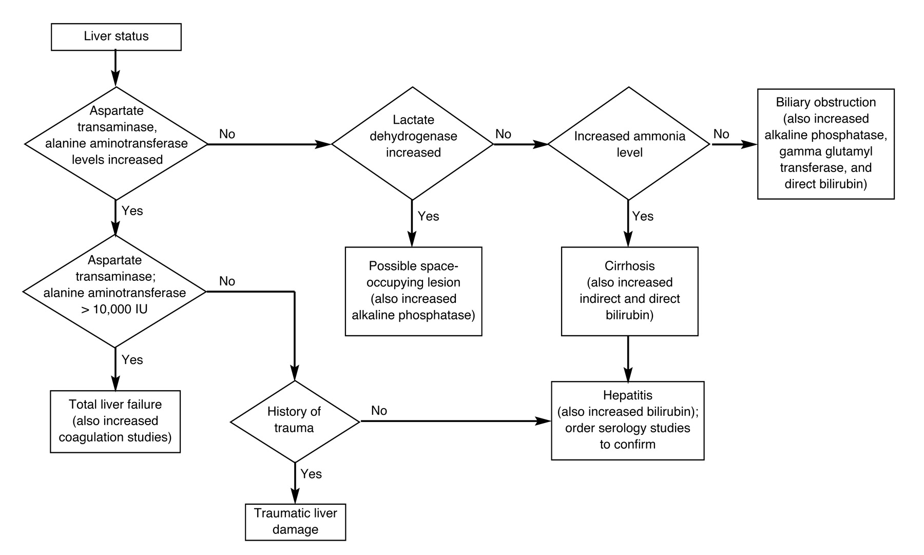
Hepatic and renal dysfunction affect the metabolism and retention of many analytes. The most sensitive assay for hepatic injury is alanine aminotransferase (serum glutamate-pyruvate transaminase); however, the ratio of aspartate aminotransferase (serum glutamic-oxaloacetic transaminase) to alanine aminotransferase is useful in identifying the origin of injury. A ratio greater than 1 is consistent with alcoholic hepatic damage; if, however, the ratio is greater than 400, then acute hepatitis should be suspected. Bilirubin, alkaline phosphatase, lactate dehydrogenase, and gamma glutamyl transferase are also used to assess liver function.[34 ] Figure 1 can be used to evaluate liver chemistries.
Figure 1. Evaluation of liver chemistries.
Japma 92 00038 g001
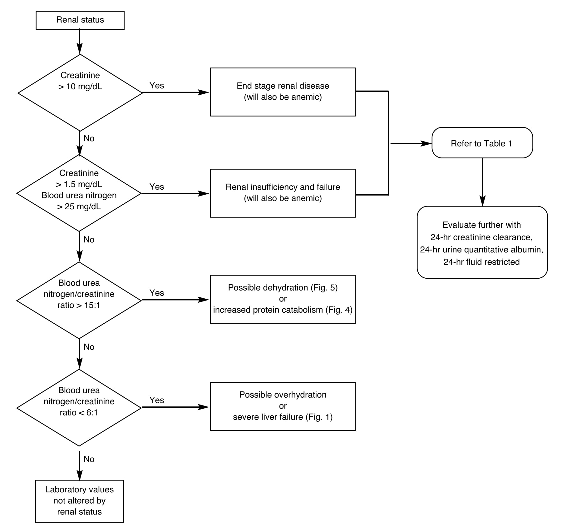
Abnormal liver function can decrease serum protein, albumin, prealbumin, transferrin, imunologic proteins, and globulins and increase prothrombin times.[6], [35], [36] Like hepatic dysfunction, renal dysfunction can be detected through clinical chemistries. Renal status may be grossly assessed by means of blood urea nitrogen, creatinine, the ratio of blood urea nitrogen to creatinine, or the estimated glomerular filtration rate[5], [34], [37], [38] (Fig. 2). Creatinine changes only by renal status and will not appear elevated until more than 25% of nephrons are dysfunctional.[39] Unlike blood urea nitrogen, creatinine is not affected by protein metabolism or dehydration.[37] Additional laboratory results that suggest the presence of renal dysfunction include cellular, granular, or waxy casts in the urine; proteinuria; hematuria; and changes in multiple serum chemistries. The following show increased serum values in renal failure: potassium, phosphorus, alkaline phosphatase, magnesium, uric acid, triglycerides, cholesterol, and very low density lipoprotein. The following show decreased serum values: sodium, calcium, albumin, and total protein.[40]-[42] If an abnormality in the renal system is suspected, it can be further investigated with a creatinine clearance study or a fluid-restricted osmolality study. Also, urine albumin levels greater than 300 mg/24 hr have been used to identify nephropathy.[43], [44]
Figure 2. Laboratory evaluation of renal status.
Japma 92 00038 g002

Macronutrients

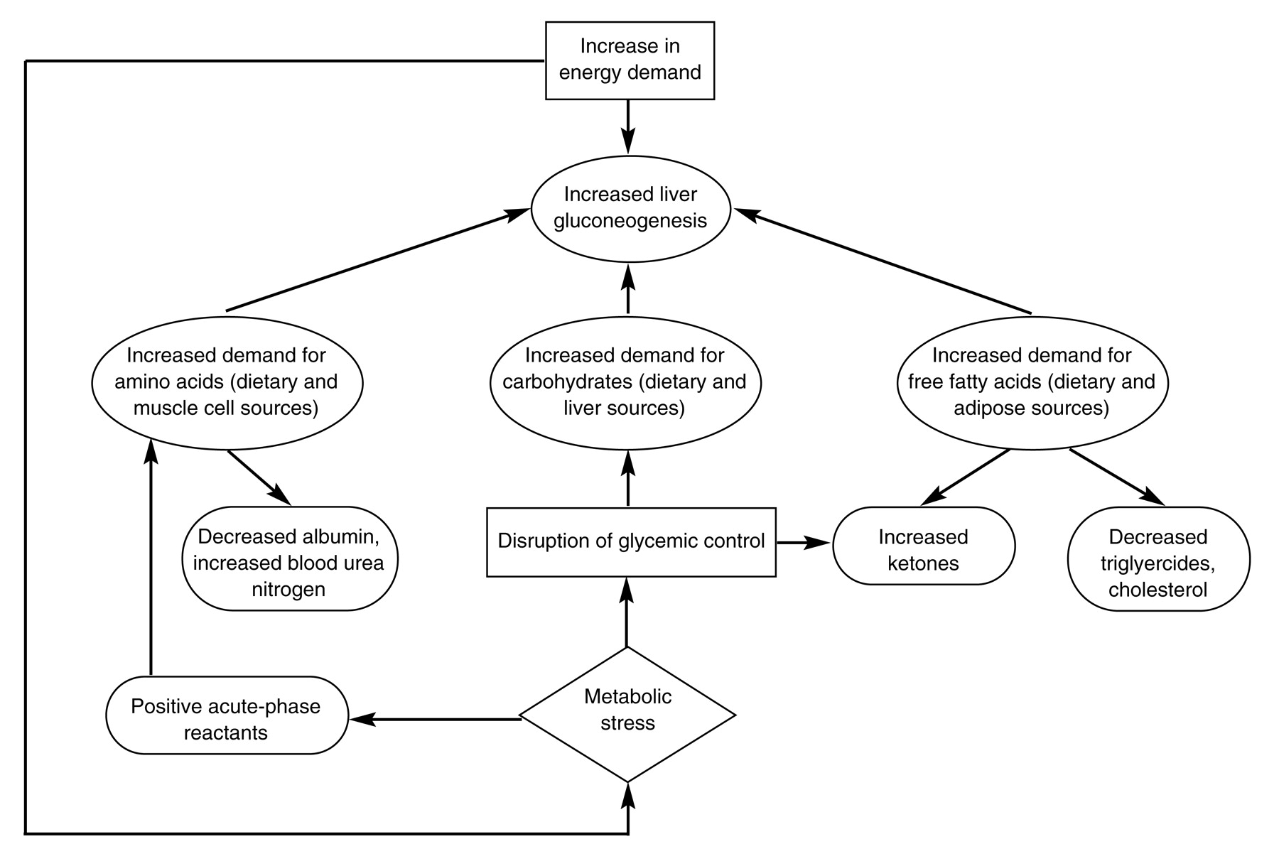
In patients with diabetes, it is difficult to separate the macronutrients as they are so closely linked in response to stress (Fig. 3). Metabolic stress or periods of increased metabolism can alter glycemic control.[27] It is important during this time to obtain a baseline hemoglobin A1C and daily fasting blood glucose.[45], [46] During times of increased cellular energy demand, carbohydrate metabolism may be altered and lipolysis may occur. Increased levels of acetone, beta-hydroxybutyric acid, and acetoacetic acid may appear in the urine or serum. These ketones may be analyzed to evaluate whether energy demands have surpassed the intake of dietary carbohydrates and whether endogenous lipids and proteins are being catabolized for energy needs. Initial serum triglyceride and cholesterol values in diabetic patients may be normal or slightly decreased.[13], [30] Triglyceride and cholesterol levels tend to be increased in poorly-controlled diabetes, chronic renal failure, and alcoholism; and are decreased in malnutrition, inflammation, and infection. Thus, the presence of a significantly depressed initial laboratory value for triglycerides reflects severe nutritional disturbances.
Figure 3. Interaction of macronutrients.
Japma 92 00038 g003
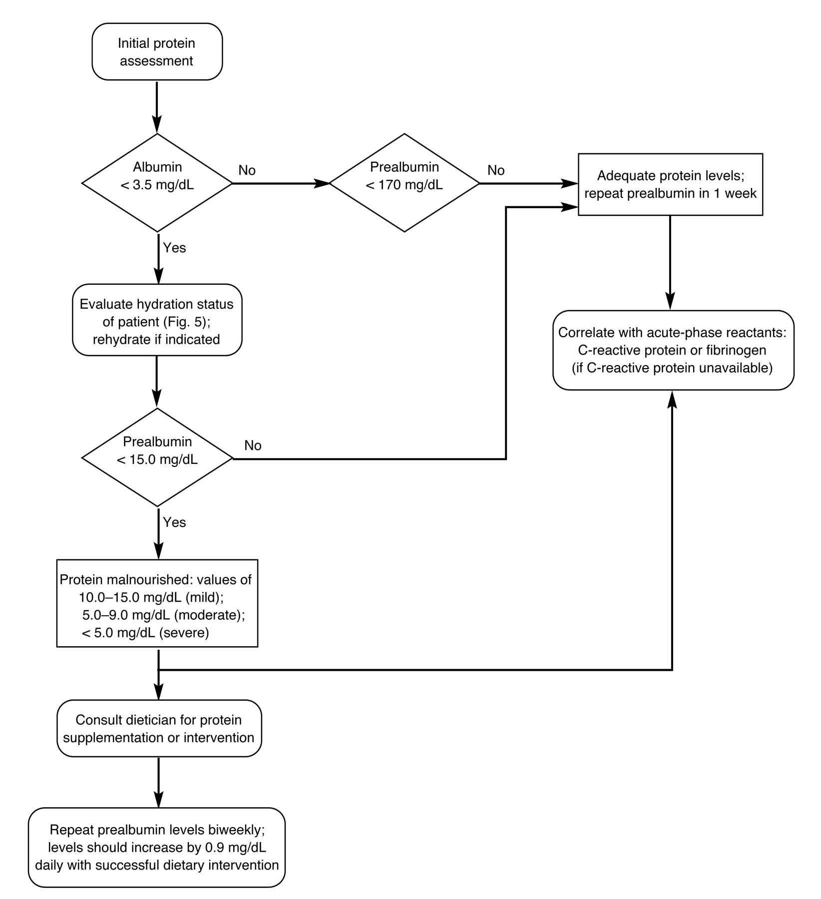
Protein assessments are more difficult than clinical laboratory measurements of carbohydrates and lipids. Protein synthesis has traditionally been evaluated with albumin, prealbumin, transferrin, and retinol-binding proteins.[47] Albumin, with a half-life of 20 days, is very sensitive to the patient’s hydration level. It has a large presence in the extravascular space, and laboratory values may be slow to respond to alterations.[25] Albumin is decreased in renal disease, liver disease, and dehydration.[14], [22], [33], [48]-[50] Albumin levels below 30 mg/dL have been associated with a fourfold increased morbidity and a six-times greater risk of mortality.[25], [51]-[53] As a result of the long half-life of albumin, it is useful for prognostic purposes only. Prealbumin (transthyretin) has a half-life of 2 days and is not significantly affected by the hydration status of the patient.[23], [24], [54]-[58] Levels below 30 mg/dL are accompanied by a 1.8 times greater risk of mortality.[58] The importance of prealbumin is directly related to its ability to follow the progress of dietary intervention. Prealbumin levels have been documented to respond to nutritional therapy within 7 days.[19]
Transferrin, another serum protein, has been used in the past as a nutrition-screening analyte. However, transferrin is an acute-phase reactant that is also influenced by hydration, anemia, antibiotics, and liver dysfunction.[7], [19], [25], [59], [60] These factors make it unsuitable as a nutritional screening tool. Retinol-binding protein is an acute-phase reactant that is also a carrier protein for vitamin A. It can be greatly increased in renal failure and liver failure; thus, it is not a reliable analyte in the diabetic patient.[25] The recommended assays for protein levels are albumin and prealbumin; however, as noted in Figure 4, values must be applied differently.
Figure 4. Evaluation of protein status.
Japma 92 00038 g004
In the infective or inflammatory state, acute-phase proteins will be produced preferentially, which will decrease the production of other serum proteins. Stress will activate the macrophages to release interleukins that will stimulate production of C-reactive protein and fibrinogen.[61], [62] This, in turn, will decrease production of albumin, prealbumin, retinol-binding protein, and transferrin.[60], [62] Stenvinkel et al[11], [63] correlated a positive C-reactive protein with the malnourished state. Thus, an immunologic component is necessary to accurately assess the protein status of the patient. Other immunologic studies, involving decreased delayed cutaneous hypersensitivity and total lymphocyte count, have been linked to protein malnutrition.[52], [64] However, the total lymphocyte count will be elevated if the patient has a history of alcoholism. If the patient is a chronic smoker, the white blood count and total lymphocyte count will be increased.[65] Immune parameters, such as metabolic ones, must be interpreted carefully.

Micronutrients

Water is an especially important micronutrient in the elderly diabetic population. Elderly patients have decreased amounts of osmoreceptors to detect thirst and inefficient renal antidiuretic hormone receptors. Physical barriers may also make it hard for them to access water, or they may drink increased amounts of alcohol or caffeine, which have a diuretic effect.[66] Many do not drink the minimum of 1,500 mL daily that is necessary.[50]
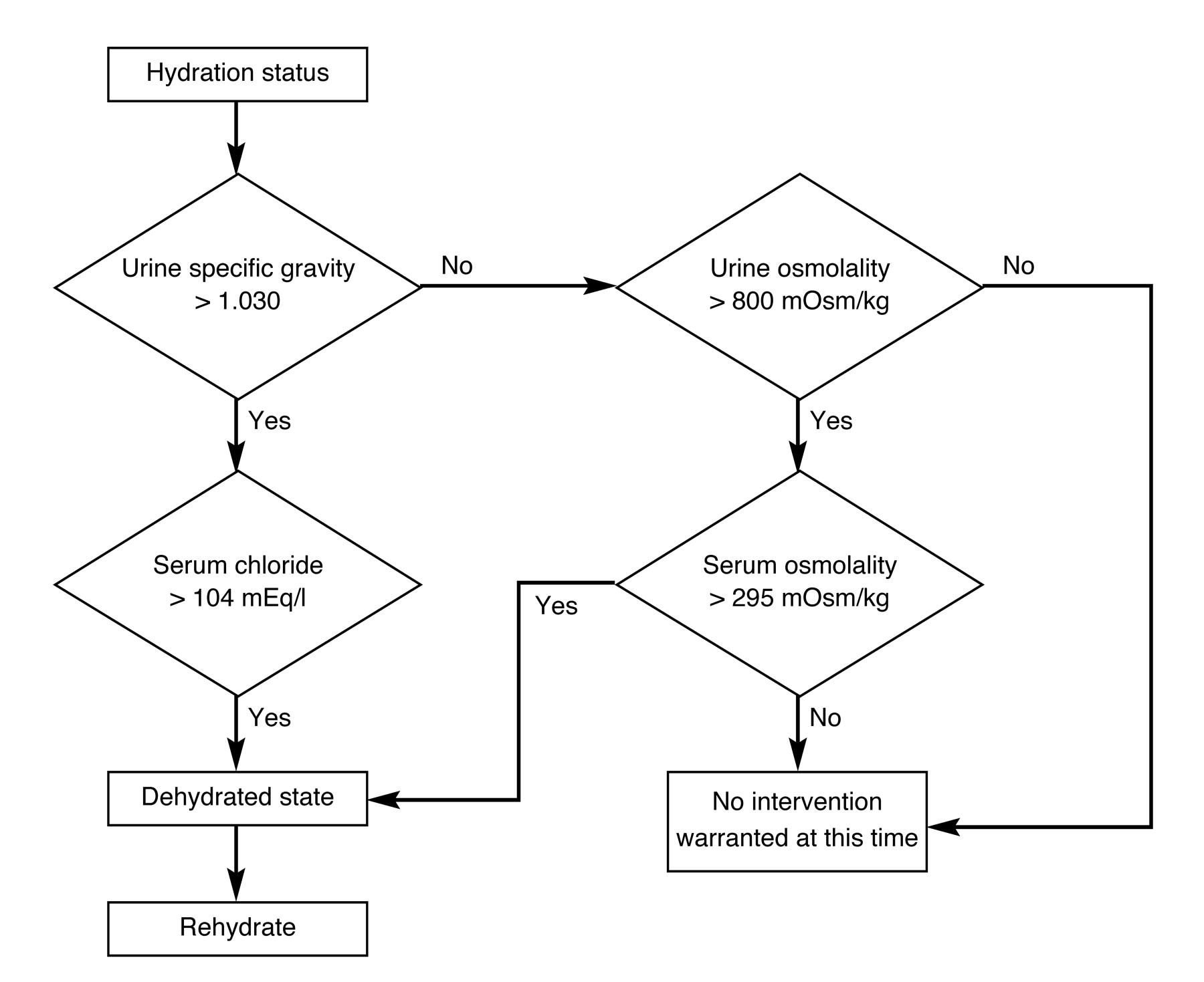
Clinical chemistries used to determine hydration status include urine specific gravity, urine osmolality, serum chloride, and serum osmolality[39] (Fig. 5). Urine specific gravity is the ratio of solution compared with the mass of an equal volume of water. Osmolality is the measure of dissolved particles in solution. Urine osmolality is more dependent on hydration than serum osmolality. Additional chemistries that can be considered are sodium, potassium, and hematocrit. The following also show increased values in dehydrated patients: hemoglobin, albumin, total protein, and blood urea nitrogen. Once identified, dehydration should be corrected by the clinician and monitored by serum electrolyte normalization.
Figure 5. Evaluation of hydration status.
Japma 92 00038 g005
Oxygen is an important element in many enzymatic and cellular metabolic reactions necessary in cellular viability, growth, and proliferation. The most common clinical laboratory assessment of oxygen is an indirect measure of the blood’s ability to transport oxygen to the cells. Hemoglobin and mean corpuscular hemoglobin concentrations are part of the complete blood count that measures the transport mechanism for oxygen. These values should be measured frequently to evaluate the need for transfusion in the severely anemic patient. These values do not represent the level of oxygen in the blood, however. Arterial blood gases, which should not be routinely evaluated due to the risks of the procedure, measure the actual levels of oxygen in the blood.[34]
Oxygen is an important factor in wound healing; its importance, however, lies in its ability to reach the wound site. Transcutaneous oxygen levels and studies of vascular status can properly analyze this. Perfusion studies, rather than clinical chemistries, are important for this micronutrient.
Vitamins and minerals are a vital link in the process of collagen formation, enzymatic function of the immune system, and, ultimately, wound healing (Table 1). Vitamin C and zinc are the most common micronutrients found to be deficient in the malnourished.[5] It would initially appear important to evaluate the representative value and availability of the clinical laboratory methods for these substances. Many laboratory tests base the micronutrient assay on the binding protein for the micronutrient. During the presence of an acute-phase protein, assays for vitamins A, C, and E; zinc; copper; and iron will not be an actual reflection of body stores because of the decrease of binding protein.[5], [31], [33], [60] In addition, some serum analytes, such as zinc, reflect less than 0.2% of total body content and assayed results are of little value.[33] Water-soluble vitamins also have a short half-life and must be supplemented on a regular basis. Individual vitamins and minerals could be assayed in a repetitive method to monitor for success of therapeutic intervention. This may not be valuable, however, while the wound is present. The clinician should supplement the necessary vitamins and minerals for wound healing rather than rely on laboratory values.[8]
Table 1. Micronutrients: Vitamins and Minerals
Japma 92 00038 i001

Conclusion

Wound healing depends on adequate macronutrients and micronutrients. Laboratory assessment of nutritional status is possible if each test is interpreted correctly. A profile can be established for admission purposes, and therapeutic intervention can be evaluated for macronutrients and two components of micronutrients. Liver and renal function are vital to the interpretation of laboratory data. Interactions of the immune system cannot be separated from the protein status of the patient and the need to be monitored. The assay of vitamins and minerals is time consuming, not cost-effective, and may not reflect the actual status of the body stores. However, this does not detract from the importance of supplementation. As the area of clinical and immunologic chemistries continues to evolve, the topic of the wound healing profile will need to be assessed periodically.

References

  1. Fields-Gardner C, Ayoob KT: Position of the American Dietetic Association and Dietitians of Canada: nutrition intervention in the care of persons with human immunodeficiency virus infection. J Am Diet Assoc100: 708, 2000.
  2. Simko MD: “Nutritional Assessment in Critical Care,” in Nutrition Assessment: A Comprehensive Guide for Planning Intervention, 2nd Ed, ed by MD Simko, C Cowell, JA Gilbride, p 285, Aspen Publishers, Rockville, MD, 1995.
  3. Mears E: Outcomes of continuous process improvement of a nutritional care program incorporating serum prealbumin measurements. Nutrition12: 479, 1996.
  4. Tucker HN, Miguel SG: Cost containment through nutritional intervention. Nutr Rev54: 111, 1996.
  5. Labbe RF, Veldee MS: Nutrition in the clinical laboratory. Clin Lab Med13: 313, 1993.
  6. Andersen H, Borre M, Jakobsen J, et al: Decreased muscle strength in patients with alcoholic liver cirrhosis in relation to nutritional status, alcohol abstinence, liver function, and neuropathy. Hepatology27: 1200, 1998.
  7. Lipshitz DA: Malnutrition in the elderly. Semin Dermatol10: 273, 1991.
  8. Tomaido PP: Malnutrition in the elderly: its recognition and treatment. Compr Ther11: 54, 1985.
  9. Ramaker J, Wood WG: Transthyretin: an explanation of “anomalous” serum thyroid hormone values in severe illness. ?J Clin Chem Clin Biochem28: 155, 1990.
  10. Stanley K: Assessing the nutritional needs of the geriatric patient with diabetes. Diabetes Educ24: 29, 1998.
  11. Stenvinkel P, Heimburger O, Tuck CH, et al: Apo(a)-isoform size, nutritional status and inflammatory markers in chronic renal failure. Kidney Int53: 1336, 1998.
  12. American Diabetes Association: Management of dyslipidemia in adults with diabetes (position statement). Diabetes Care23 (suppl 1): S57, 2000.
  13. Shils ME: Modern Nutrition in Health and Disease, 9th Ed, Williams & Wilkins, Baltimore, MD, 1998.
  14. Barrocas A, Belcher D, Champagne C, et al: Nutritional assessment practical approaches. Clin Geriatr Med11: 675, 1995.
  15. Mullen JL, Buzby GP, Waldman MT: Prediction of operative morbidity and preoperative nutritional assessment. Surg Forum30: 80, 1979.
  16. Boult C, Krinke UB, Urdangarin CF, et al: The validity of nutritional status as a marker for future disability and depressive symptoms among high-risk older adults. J Am Geriatr Soc23: 91, 1994.
  17. Roubenoff R, Giacoppe J, Richardson S, et al: Nutrition assessment in long-term care facilities. Nutr Rev54: S40, 1996.
  18. Bannerman E, Reilly JJ, MacLennan WJ, et al: Evaluation of validity of British anthropometric reference data for assessing nutritional state of elderly people in Edinburgh: cross sectional study. BMJ315: 338, 1997.
  19. Lipkin EW, Bell S: Assessment of nutritional status: the clinician’s perspective. Clin Lab Med13: 329, 1993.
  20. Tierney AJ: Undernutrition and elderly hospital patients: a review. J Adv Nurs23: 228, 1996.
  21. Zawada ET: Malnutrition in the elderly: is it simply a matter of not eating enough?Postgrad Med100: 207, 1996.
  22. Ferguson RP, O’Connor P, Crabtree B, et al: Serum albumin and prealbumin as predictors of clinical outcomes of hospitalized elderly nursing home residents. J Am Geriatr Soc41: 545, 1993.
  23. Guenter P, Malyszek R, Bliss DZ, et al: Survey of nutritional status in newly hospitalized patients with stage III or stage IV pressure ulcers. Adv Skin Wound Care13: 164, 2000.
  24. Potter MA, Luxton G: Prealbumin measurement as a screening tool for protein calorie malnutrition in emergency hospital admissions: a pilot study. Clin Invest Med22: 44, 1999.
  25. Spiekerman AM: Proteins used in nutritional assessment. Clin Lab Med13: 353, 1993.
  26. Spiekerman AM, Rudolph RA, Bernstein LH: Determination of malnutrition in hospitalized patients with the use of a group-based reference. Arch Pathol Lab Med117: 184, 1993.
  27. American Diabetes Association: Standards of medical care for patients with diabetes mellitus. Diabetes Care23 (suppl 1): S32, 2000.
  28. Gallagher-Allred CR, Voss AC, Finn SC, et al: Malnutrition and clinical outcomes: the case for medical nutritional therapy. J Am Diet Assoc96: 361, 1996.
  29. Naber THJ, DeBree A, Schermer T, et al: Specificity of indexes of malnutrition when applied to apparently healthy people: the effect of age. Am J Clin Nutr65: 1721, 1997.
  30. Kinney JM: Metabolic responses of the critically ill patient. Crit Care Clin11: 569, 1995.
  31. Konstantinides NN, Lehmann S: The impact of nutrition on wound healing. Crit Care Nurse13: 25, 1993.
  32. Shi HP, Efron DT, Most D, et al: Supplemental dietary arginine enhances wound healing in normal but not inducible nitric oxide synthase knockout mice. Surgery128: 374, 2000.
  33. Gershwin ME: “Application and Interpretation of Commonly Used Nutritional Assessment Techniques,” in Nutrition and Immunology: Principles and Practice, ed by ME Gershwin, p 3, Humana Press, Totowa, NJ, 1999.
  34. Sauberlich HE: Laboratory Tests for the Assessment of Nutritional Status, 2nd Ed, CRC Press, Boca Raton, FL, 1999.
  35. Lieber CS: Alcohol and the liver: metabolism of alcohol and its role in hepatic and extrahepatic diseases. Mt Sinai J Med67: 84, 2000.
  36. Walsh K, Alexander G: Alcoholic liver disease. Postgrad Med J76: 280, 2000.
  37. Avram MM, Goldwasser P, Erroa M, et al: Predictors of survival in continuous ambulatory peritoneal dialysis patients: the importance of prealbumin and other nutritional and metabolic markers. Am J Kidney Dis23: 91, 1994.
  38. McCarthy JT: A practical approach to the management of patients with chronic renal failure. Mayo Clin Proc74: 269, 1999.
  39. Corbett JV: “Renal Function Tests,” in Laboratory Tests and Diagnostic Procedures with Nursing Diagnoses, 5th Ed, p 90, Prentice Hall Health, Upper Saddle River, NJ, 2000.
  40. Molina MF, Riella MC: Nutritional support in the patient with renal failure. Crit Care Clin11: 685, 1995.
  41. Nannipieri M, Penno G, Loredana R, et al: Transcapillary escape rate of albumin in type II diabetic patients. Diabetes Care20: 1019, 1997.
  42. Sacher RA, McPherson RA, Campos JM: “Laboratory Assessment of Body Fluids,” in Widmann’s Clinical Interpretation of Laboratory Tests, 11th Ed, ed by RA Sacher, RA McPherson, JM Campos, FA Davis, Philadelphia, 2000.
  43. American Diabetes Association: Diabetic nephropathy. Diabetes Care23 (suppl 1): S69, 2000.
  44. Taft JL, Nolan CJ, Yeung SP, et al: Clinical and histological correlations of decline in renal function in diabetic patients with proteinuria. Diabetes43: 1046, 1994.
  45. American Diabetes Association: Hospital admission guidelines of diabetes mellitus. Diabetes Care23 (suppl 1): S83, 2000.
  46. American Diabetes Association: Nutrition recommendations and principles for people with diabetes mellitus. Diabetes Care23 (suppl 1): S43, 2000.
  47. Shears GJ, Deutschman CS: Common nutritional issues in pediatric and adult critical care medicine. Crit Care Clin13: 669, 1997.
  48. American Diabetes Association: Translation of the diabetic nutrition recommendations for health care institutions (position statement). Diabetes Care23 (suppl 1): S47, 2000.
  49. Borish L, King MS, Mascali JJ, et al: Transthyretin is an inhibitor of monocyte and endothelial cell interleukin-1 production. Inflammation16: 471, 1992.
  50. Chernoff R: Effects of age on nutrient requirements. Clin Geriatr Med11: 641, 1995.
  51. Bernstein L, Pleban W: Prealbumin in nutrition evaluation. Nutrition12: 255, 1996.
  52. Henderson RA, Talusan K, Hutton N, et al: Serum and plasma markers of nutritional status in children infected with the human immunodeficiency virus. J Am Dietetic Assoc97: 1377, 1997.
  53. Kaplan LA, Minkowitz G, Rakower D: Serum prealbumin in institutionalized populations. Nutrition15: 51, 1999.
  54. Bernstein LH, Leukhardt-Fairfield CJ, Pleban W, et al: Usefulness of data on albumin and prealbumin concentrations in determining effectiveness of nutritional support. Clin Chem35: 271, 1989.
  55. Brose L: Prealbumin as a marker of nutritional status. J Burn Care Rehabil11: 372, 1990.
  56. Charet JC, Watine J, Lepretre A, et al: Orosomucoid: prealbumin ratio in the monitoring of lung cancer. Clin Biochem29: 287, 1996.
  57. Ingenbleek Y, Young V: Transthyretin (prealbumin) in health and disease: nutritional implications. Annu Rev Nutr14: 495, 1994.
  58. Sreedhara R, Avram MM, Blanco M, et al: Prealbumin is the best nutritional predictor of survival in hemodialysis and peritoneal dialysis. Am J Kidney Dis28: 937, 1996.
  59. Bernstein L, Bachman TE, Meguid M, et al: Measurement of visceral protein status in assessing protein and energy malnutrition: standard of care. Nutrition11: 169, 1995.
  60. Manning EMC, Shenkin A: Nutritional assessment in the critically ill. Crit Care Clin11: 603, 1995.
  61. Bourguignat A, Ferad G, Jenny J, et al: Diagnostic value of C-reactive protein and transthyretin in bone infections of the lower limb. Clin Chim Acta235: 27, 1996.
  62. Kaysen GA, Stevenson FT, Depner TA: Determinants of albumin concentration in hemodialysis patients. Am J Kidney Dis29: 658, 1997.
  63. Stenvinkel P, Heimburger O, Paultre F, et al: Strong association between malnutrition, inflammation, and atherosclerosis in chronic renal failure. Kidney Int55: 1899, 1999.
  64. Henry JB: “Interpreting Laboratory Results: Reference Values and Decision Making,” in Clinical Diagnosis and Management by Laboratory Methods, 19th Ed, ed by JB Henry, p 1450, WB Saunders, Philadelphia, 1996.
  65. Parry H, Cohen S, Schlarb JE, et al: Smoking, alcohol consumption, and leukocyte counts. Am J Clin Pathol107: 64, 1997.
  66. Flanigan KH: Nutritional aspects of wound healing. Adv Wound Care10: 48, 1997.
  67. Demling RH, DeBiasse MA: Micronutrients in critical illness. Crit Care Clin11: 651, 1995.
  68. Gecelter GR, Comer GM: Nutritional support during liver failure. Crit Care Clin11: 675, 1995.
  69. Mooradian AD, McLaughlin S, Boyer CC, et al: Diabetes care for the older adult. Diabetes Spectrum11: 70, 1999.
  70. Rojas AI, Phillips TJ: Patients with chronic leg ulcers show diminished levels of vitamins A and E, carotenes, and zinc. Dermatol Surg25: 601, 1999.