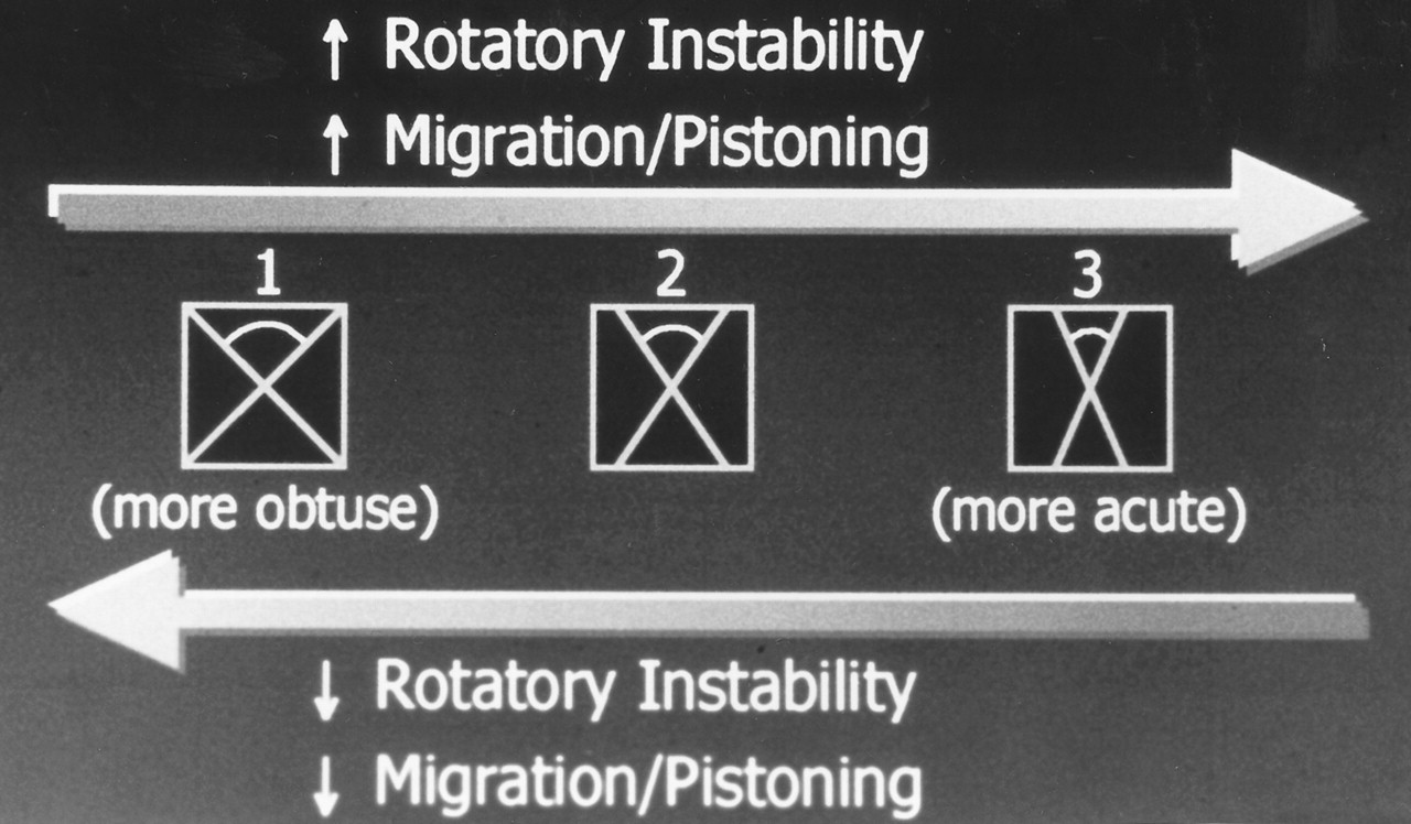
Table 1.
Indications for Arthrodesis of the Hallucal Interphalangeal Joint
The authors present a very simple technique that has been found to be extremely effective in avoiding virtually all of the common problems associated with crossing Kirschner wires to achieve arthrodesis of the hallucal interphalangeal joint. It is hoped that this technique will encourage surgeons to reconsider the use of Kirschner wires as a primary technique for fusion of this joint whenever, in the surgeon’s judgment, it appears that screw fixation is likely to result in less-than-optimal rigid internal-compression fixation.
Although this technique does not provide the compression at the fusion site that can be achieved with the use of cancellous screws, it does provide very effective and predictable stable fixation for an indefinite time. An advantage of this technique is that the fixation devices may be removed without another surgical procedure. Also, frontal-plane rotation, which is known to occur with axial compression of one screw, is virtually eliminated. Finally, the authors have found this technique to be faster and less invasive than techniques employing screw fixation.
Surgical Technique
Any type of skin incision that has been described for fusion of the interphalangeal joint may be employed in this technique. The technique may be begun via a longitudinal linear, transverse semielliptical, or curvilinear incision. The procedure can be performed from either a dorsal or a plantar approach, such as when performing a concomitant excision of a plantar ossicle or an associated lesion or ulcer.
Following the incision, the extensor tendon is reflected and the interphalangeal joint is disarticulated. The articular surfaces are removed to achieve correction of frontal, sagittal, and transverse plane deformities.
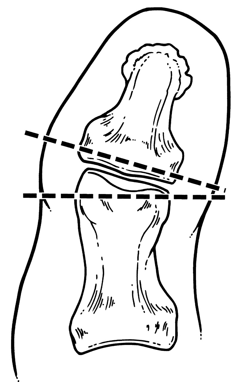
A variety of techniques may be employed for resection of the bone. Most of the bone should be removed from the proximal phalanx to achieve correction of one or more planal deformities (
Fig. 1). It is usually necessary to remove only a minimum of bone from the distal phalangeal base. Resection of large segments of bone from the distal phalanx does not achieve angular correction and should be avoided. Removal of an excessive amount of bone from the distal phalanx compromises fixation because of the size and configuration of the bone. The amount of bone that should be removed will depend on the extent of the deformity.
Figure 1.
Proper resection of bone to achieve angular correction when performing hallucal interphalangeal joint arthrodesis on a patient with abductus interphalangeal joint deformity. Angular correction is achieved by appropriate resection of bone from the proximal phalangeal head.
Figure 1.
Proper resection of bone to achieve angular correction when performing hallucal interphalangeal joint arthrodesis on a patient with abductus interphalangeal joint deformity. Angular correction is achieved by appropriate resection of bone from the proximal phalangeal head.
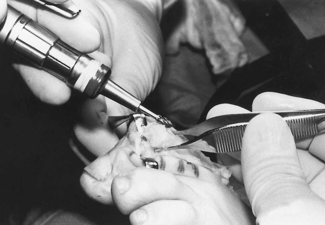
Hand instruments, such as rongeurs or bone-cutting forceps, are recommended for removal and resection of bone from the proximal phalangeal head. Power instruments may be used, but the use of hand instruments will result in a surface that is “splintered,” or rough, which will allow increased friction between the fusion interfaces. Surfaces with better friction will be less likely to rotate. Smoother interfaces are more likely to undergo undesirable rotation. The peripheral cortical rim of bone from the proximal and distal phalanx is then smoothed with a small power bur to enhance apposition of the cancellous portion of the fusion interfaces (
Fig. 2).
Figure 2.
Remodeling of the peripheral cortex following resection of the proximal phalangeal head. A small oval bur is used to decrease prominence of the cortical rim and enhance apposition of the cancellous portion of the bone.
Figure 2.
Remodeling of the peripheral cortex following resection of the proximal phalangeal head. A small oval bur is used to decrease prominence of the cortical rim and enhance apposition of the cancellous portion of the bone.
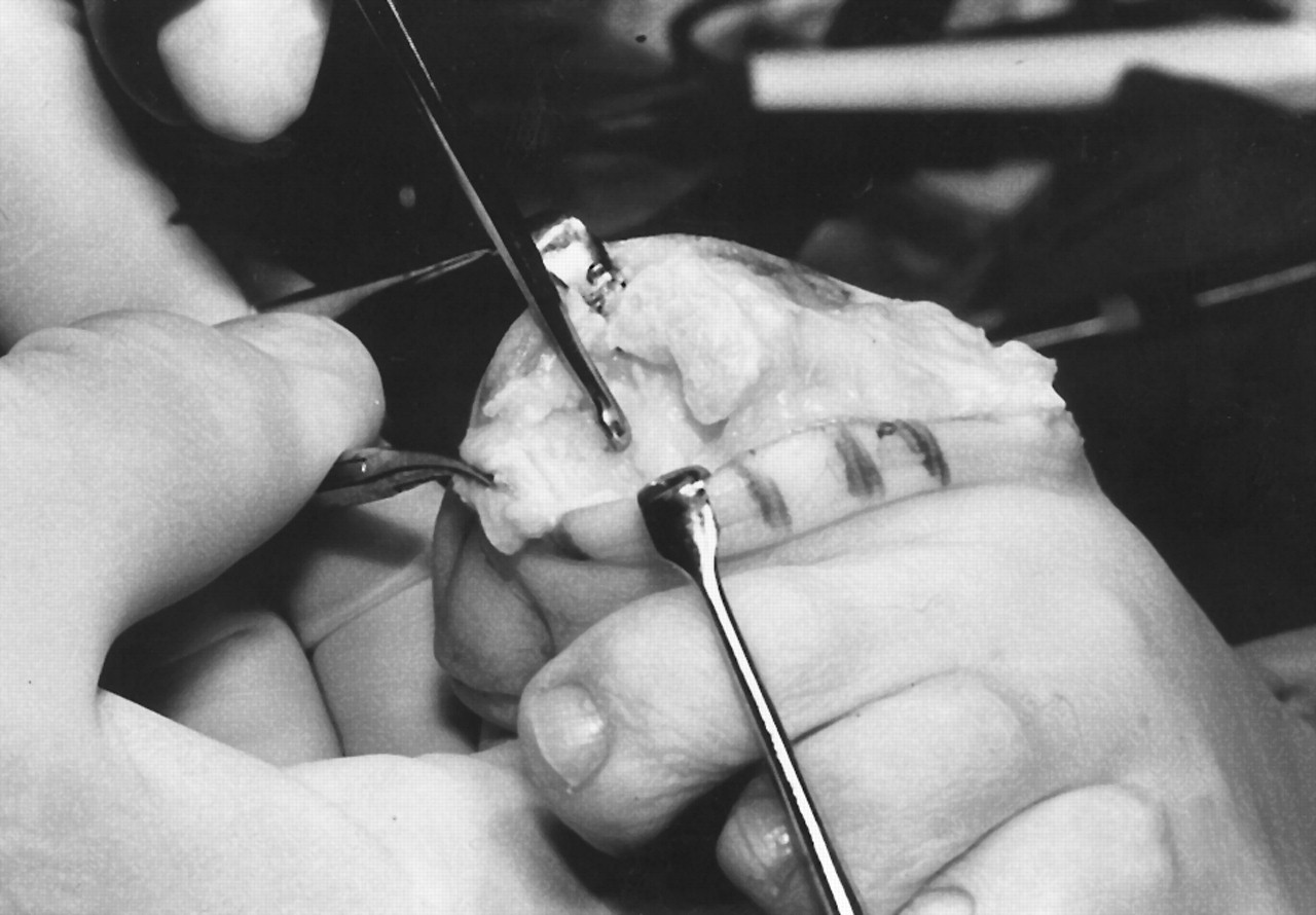
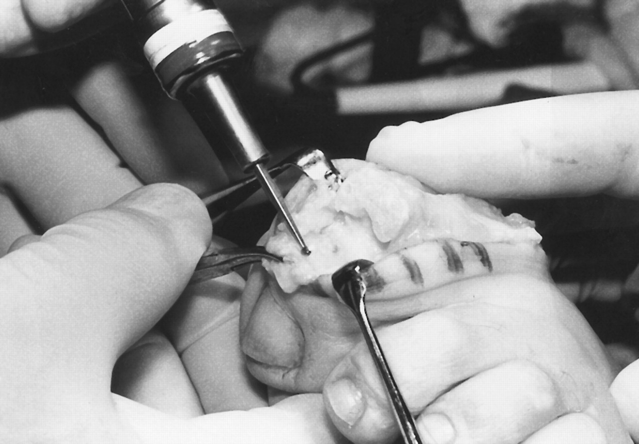
The authors frequently remove only the cartilage from the base of the distal phalanx, preserving the subchondral bone. This is accomplished by using small-bone curettes, a surgical knife, and a power bur (
Figs. 3 and
4). A series of 1-mm holes are drilled through the subchondral bone surface at the base of the distal phalanx. Drilling of the proximal phalanx is neither recommended nor necessary because resecting the head of the proximal phalanx provides exposure to a cancellous bone surface. In the authors’ opinion, drilling the subchondral bone surface of the distal phalanx enhances revascularization and allows arthrodesis that can be confirmed clinically and radiographically.
Figure 3.
Removal of the cartilage from the base of the distal phalanx with the use of a small-bone curette. All of the cartilage must be removed to the level of the subchondral bone.
Figure 3.
Removal of the cartilage from the base of the distal phalanx with the use of a small-bone curette. All of the cartilage must be removed to the level of the subchondral bone.
Figure 4.
Burring of the subchondral bone and cortical rim of the base of the distal phalanx to enhance apposition with the proximal phalangeal component.
Figure 4.
Burring of the subchondral bone and cortical rim of the base of the distal phalanx to enhance apposition with the proximal phalangeal component.
Following preparation of the bone, the hallux is placed in its clinically desired position, and apposition of the fusion surfaces is confirmed. Any adjustments for additional angular or position corrections are made. There should be maximum contact between the central surfaces of the fusion site. Contact is less critical for the peripheral edge of the fusion interfaces than for the more central portion of the fusion site.
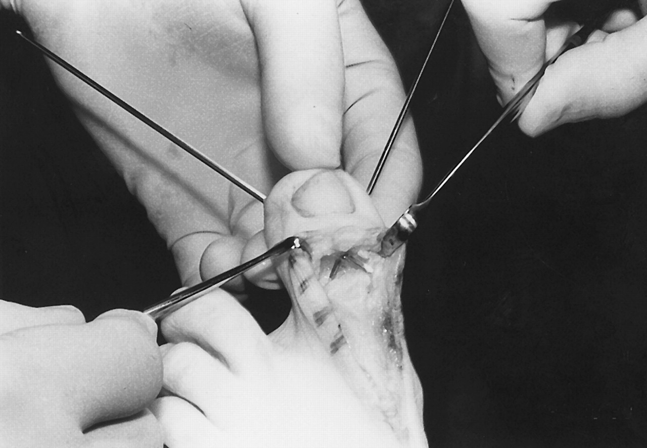
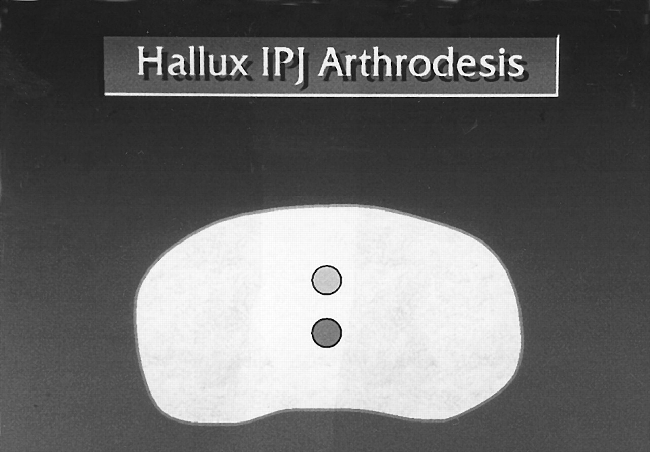
The interphalangeal joint is then stabilized by means of the standard retrograde technique with two 0.062 Kirschner wires in a crossing fashion. The Kirschner wires are typically driven distally through the distal phalanx, exiting the medial and lateral borders of the hallux. The point of penetration of the Kirschner wires in the base of the distal phalanx is central, with one Kirschner wire entering slightly superiorly and the other slightly inferiorly (
Fig. 5). This is done to avoid collision of the Kirschner wires as they are retrograded out to the tip of the toe and back into the proximal phalanx (
Fig. 6).
Figure 5.
Penetration points of Kirschner wires through the base of the distal phalanx. The surgeon should offset one wire superiorly and one inferiorly to avoid pin contact and reduce rotation.
Figure 5.
Penetration points of Kirschner wires through the base of the distal phalanx. The surgeon should offset one wire superiorly and one inferiorly to avoid pin contact and reduce rotation.
Figure 6.
Standard retrograde technique for insertion of two 0.062 Kirschner wires. Both pins have been retrograded distally to the tip of the toe. Only the tips of the pins should be showing in the fusion site at the time of apposition of the opposing surfaces.
Figure 6.
Standard retrograde technique for insertion of two 0.062 Kirschner wires. Both pins have been retrograded distally to the tip of the toe. Only the tips of the pins should be showing in the fusion site at the time of apposition of the opposing surfaces.
The authors have found that the type of tip on the Kirschner wire also plays a role in the ease and placement of the Kirschner wires. A Kirschner wire with a bayonet-type point is more difficult to handle than one with a diamond-shaped point. A diamond- or trocar-shaped point at the tip of the Kirschner wire allows the most accurate and precise placement. The Kirschner wires are then retrieved distally and retrograded so that they are flush with the surface of the distal phalangeal base. A double-ended Kirschner wire with trocar-shaped tips allows the most accurate and precise placement.
The next step in this technique is placement of the distal phalanx in its desired position against the proximal phalangeal surface. This step is critical to ensure proper clinical alignment and proper apposition of the fusion interfaces. The Kirschner wires are then driven proximally into the proximal phalanx and, preferably, exit the medial and lateral cortices of the diaphyseal area of the proximal phalanx. In some cases, the Kirschner wires have been driven more proximally into the subchondral bone area. The Kirschner wires should not be positioned or left within the medullary canal of the proximal phalanx, as this will significantly compromise stability of the arthrodesis site. Penetration of the subchondral bone plate or, preferably, the medial and lateral cortices, enhances the long-term stability of the Kirschner wires.
In some cases, the Kirschner wires are driven in such a way as to exit the distal pulp of the toe at its medial and lateral borders rather than exiting the toe along its medial and lateral sides. The more distal the point of exit, the more likely the Kirschner wires will cross proximal to the interphalangeal joint fusion site. The Kirschner wires that exit the medial and lateral borders of the toe are more likely to cross each other within the fusion site itself. Although the latter technique may seem to be more sound academically, it is a moot point clinically as long as the arthrodesis site has been maintained in a compressed state as the Kirschner wires are driven proximally. The specific placement of the Kirschner wires will depend on the nature of the deformity and the personal experiences and preferences of the surgeon.
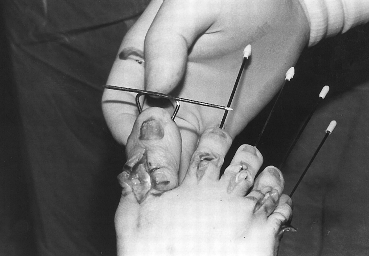
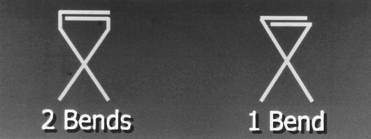
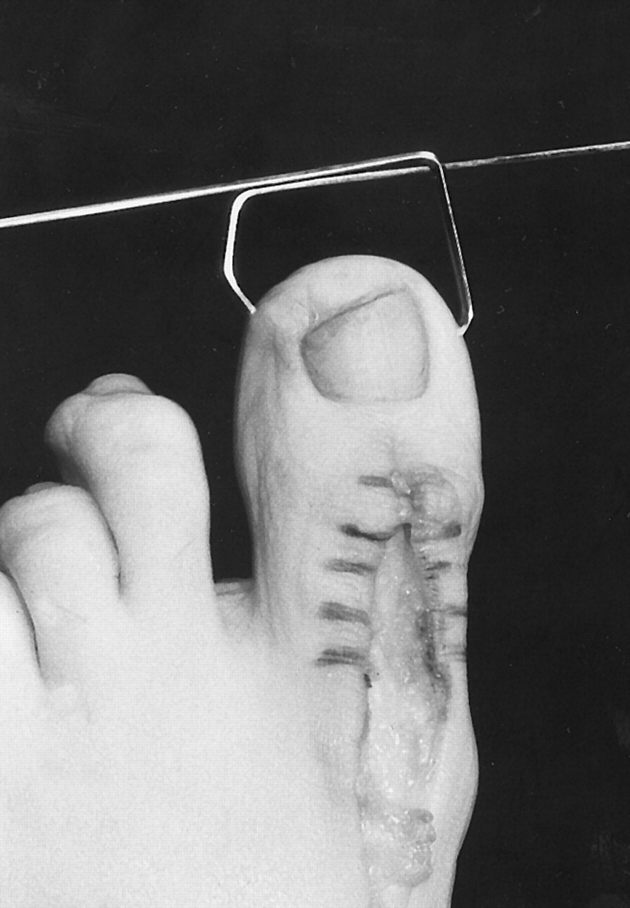
The external portions of the Kirschner wires are bent so that they cross transversely at the distal tuft of the toe and are parallel to each other. The point where the Kirschner wires exit the hallux will determine the number of bends required in each Kirschner wire. In cases where the Kirschner wires exit the distal pulp of the toe at its medial and lateral borders, only one bend is necessary for each Kirschner wire (
Fig. 7). In cases where the Kirschner wires exit the medial and lateral aspects of the hallux proximal to the distal tuft, two bends are required. The first bend will place the external portion of the Kirschner wire parallel to the long axis of the hallux, and the second bend will be at the distal tuft of the toe. This will be a 90° bend, which places the distal aspect of the external portion of the Kirschner wire parallel to the distal tuft of the toe (
Figs. 8 and
9).
Figure 7.
Diagram of two techniques to achieve the same functional outcome. When the Kirschner wires have exited the medial and lateral aspects of the toe, two bends will be required. When both Kirschner wires have exited the distal tuft of the toe, only one bend will be required. In either case, the two Kirschner wires will function as a single unit.
Figure 7.
Diagram of two techniques to achieve the same functional outcome. When the Kirschner wires have exited the medial and lateral aspects of the toe, two bends will be required. When both Kirschner wires have exited the distal tuft of the toe, only one bend will be required. In either case, the two Kirschner wires will function as a single unit.
Figure 8.
Intraoperative appearance of the toe following insertion of the Kirschner wires into the proximal phalanx. In this case, two bends have been required to achieve a parallel relationship at the tip of the toe.
Figure 8.
Intraoperative appearance of the toe following insertion of the Kirschner wires into the proximal phalanx. In this case, two bends have been required to achieve a parallel relationship at the tip of the toe.
Figure 9.
Intraoperative appearance of a patient in whom the crossing Kirschner wires have been driven more distally toward the pulp of the toe. In this case, only one bend is required to join the two wires at the tip of the toe.
Figure 9.
Intraoperative appearance of a patient in whom the crossing Kirschner wires have been driven more distally toward the pulp of the toe. In this case, only one bend is required to join the two wires at the tip of the toe.
A pair of heavy, short, needle-nosed pliers and a wire bender are indispensable to achieving accurate bends in the Kirschner wires. The tip of a typical surgical suction unit also may be used to facilitate bending. The Kirschner wires should not be bent indiscriminately, as the precise placement and angle of each bend will determine the final stability of the wires.
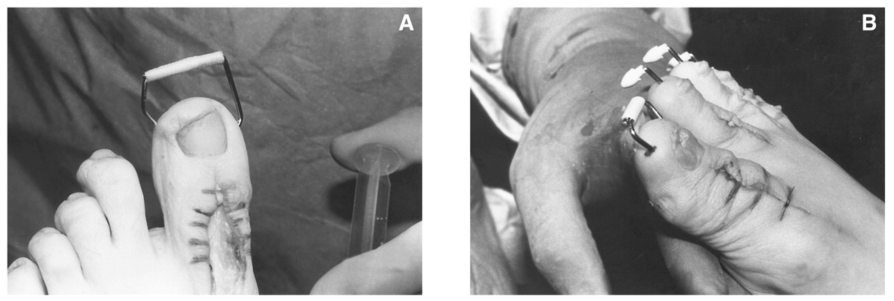
Finally, the excess portions of the Kirschner wires are cut. The two Kirschner wires, which have now been bent and positioned parallel to each other in the distal tuft of the toe, are fastened together with paper tape or wound-closure strips (
Fig. 10). If the fastening material is applied during the surgery itself, 0.5-inch wound-closure strips are recommended. If the fastening is performed after the closing of the surgical site and dressing of the toe, nonsterile paper tape is adequate.
Figure 10.
A, Appearance of the Kirschner wires after the excess has been cut and discarded. The two pins have been joined together by the use of an adhesive wound-closure strip. B, Final appearance of the patient shown in
Figure 8. Again, the two wires effectively have been joined together to function as a single unit. Digital arthrodesis of the lesser toes was also performed.
Figure 10.
A, Appearance of the Kirschner wires after the excess has been cut and discarded. The two pins have been joined together by the use of an adhesive wound-closure strip. B, Final appearance of the patient shown in
Figure 8. Again, the two wires effectively have been joined together to function as a single unit. Digital arthrodesis of the lesser toes was also performed.
The surgical wound is irrigated with normal sterile saline. The fusion site is inspected to ensure a good reciprocal fit between the adjacent surfaces. Intraoperative x-rays are obtained, if necessary, to confirm the alignment and positioning of the Kirschner wires. It may be desirable to obtain the intraoperative x-rays prior to bending the wires. Careful digital palpation also will confirm accurate placement. In some cases, the exit sites of the Kirschner wires in the proximal phalanx can be visualized; in other cases, it will be necessary to confirm their position by digital palpation. Range of motion of the first metatarsophalangeal joint should be checked to ensure that the Kirschner wires have not penetrated or crossed this joint.
The extensor tendon, the subcutaneous tissues, and the skin are reapproximated. The suture material used for closure will depend on the preferences of the surgeon; 3-0 and 4-0 absorbable multifilament synthetic sutures typically are used for deep closure, and the skin usually is closed with a monofilament absorbable or nonabsorbable synthetic suture material, such as polypropylene, nylon, or polydioxanone.
Postoperative Care
The surgical dressing is changed 5 to 7 days postoperatively, and the surgical site is inspected to identify any potential complications. A dry sterile dressing is reapplied. Two weeks postoperatively, the hallux typically is protected by the use of tube foam, which usually is cut slightly longer than the external portion of the Kirschner wires in order to provide additional protection. In some cases, the Kirschner wires are stabilized further by the use of paper tape or a large sterile strip that is attached to the nail dorsally and the tuft of the toe plantarly.
If swelling becomes a problem, it is best managed with a compression wrap of the hallux applied below, or preferably above, the tube foam. Caution should be exercised when applying such compression bandages to the hallux to avoid creating a tourniquet or a restrictive band that will result in increased edema distally.
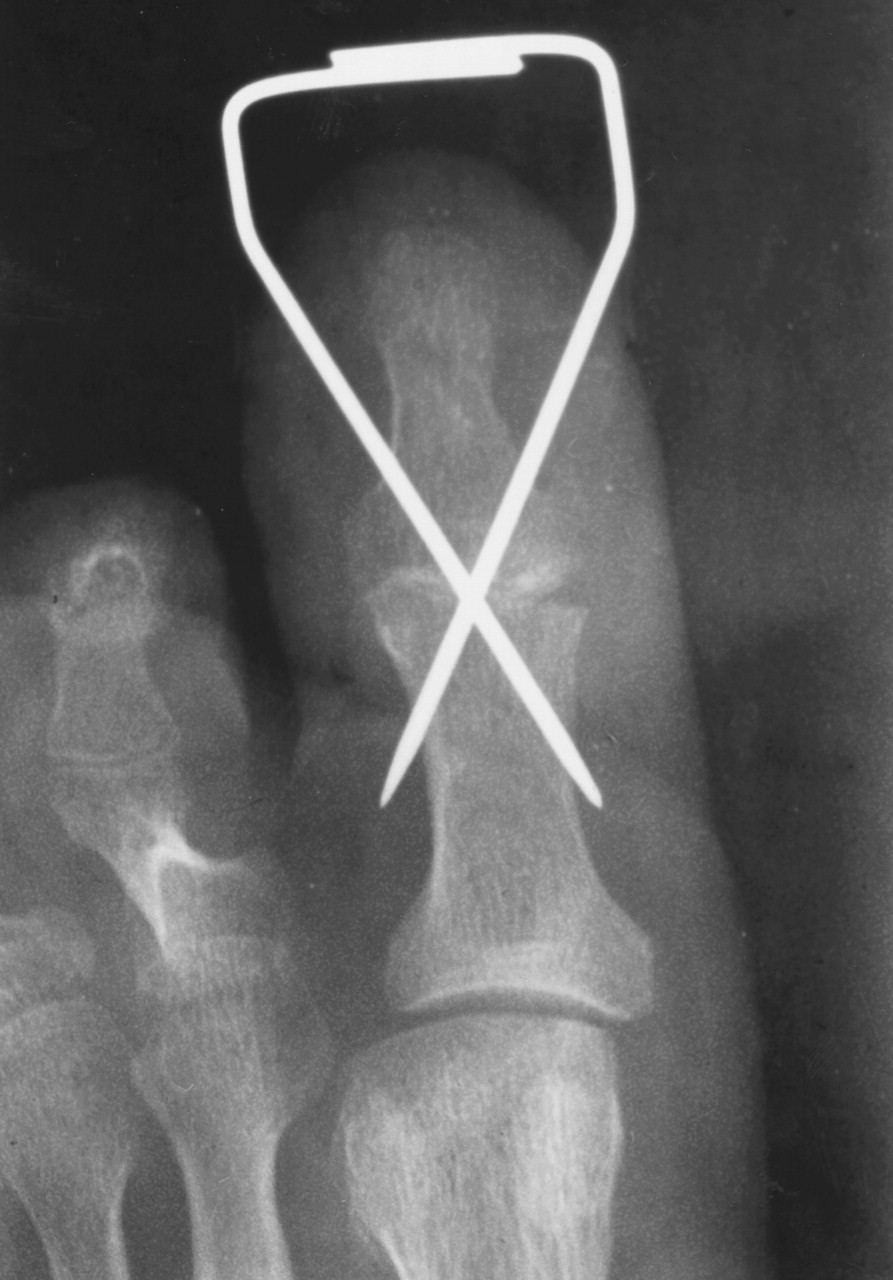
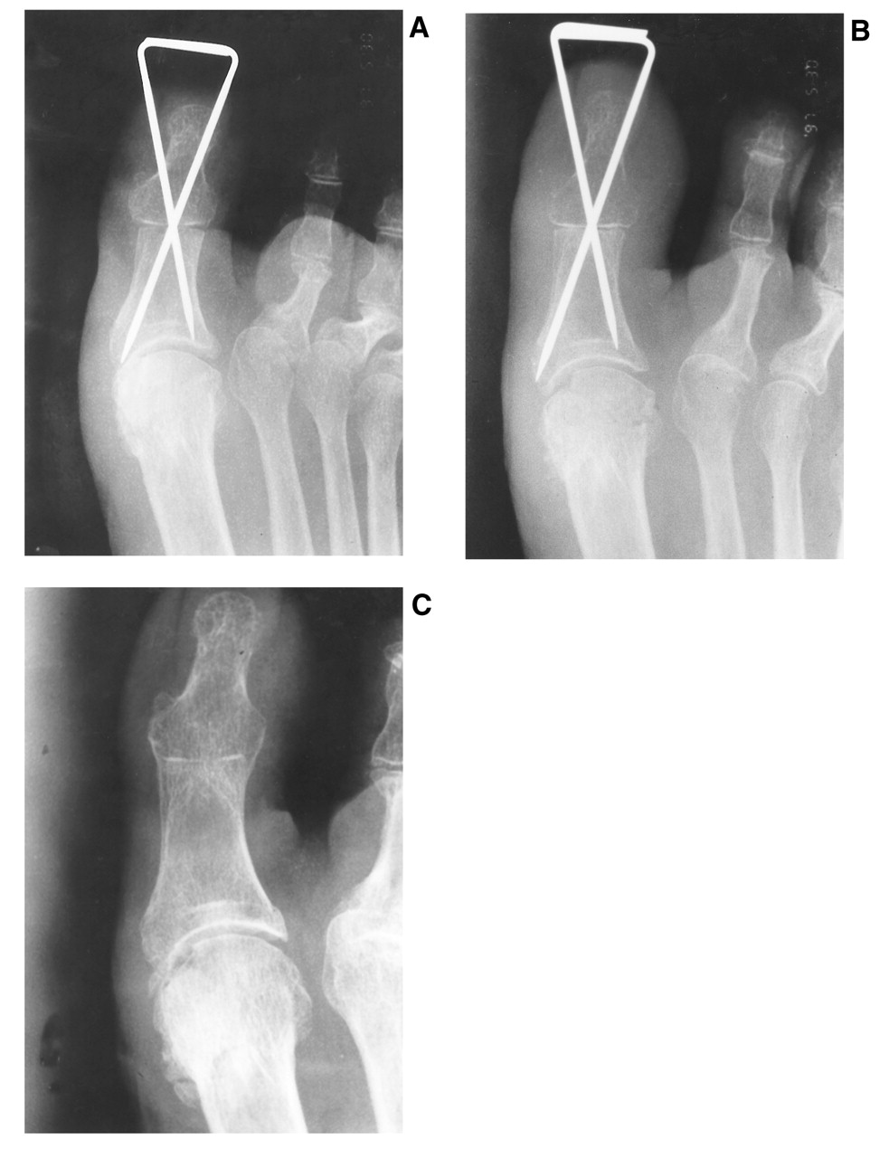
Serial x-rays are obtained to confirm consolidation at the arthrodesis site at 3, 6, and 9 weeks postoperatively. A minimum of two views is recommended. When solid arthrodesis is seen radiographically, the Kirschner wires may be removed (
Fig. 11 and
Fig. 12). The Kirschner wires are easily removed after separating the tips of their joined portions distally. This is accomplished with the use of a Number 15 or smaller blade to cut through the adhesive strip between the two Kirschner wires. The wires are then rotated and easily retrieved. Pliers may be used to facilitate removal. There is no need for a local anesthetic block when removing the wires. Compression should be maintained at the fusion site when removing the Kirschner wires to avoid undesirable and inadvertent dislodging of a possibly tenuous or incomplete arthrodesis.
Figure 11.
Postoperative x-ray following hallux interphalangeal joint arthrodesis using two crossing 0.062 Kirschner wires. Both of the Kirschner wires have been driven through the cortex of the proximal phalanx to enhance stability.
Figure 11.
Postoperative x-ray following hallux interphalangeal joint arthrodesis using two crossing 0.062 Kirschner wires. Both of the Kirschner wires have been driven through the cortex of the proximal phalanx to enhance stability.
Figure 12.
A, Postoperative x-ray 1 week after interphalangeal joint arthrodesis using two 0.062 Kirschner wires. In this case, the Kirschner wires have been driven to the base of the proximal phalanx without penetrating into the joint. B, Dorsoplantar x-ray of the same patient approximately 7 weeks postoperatively. Although radiographic arthrodesis is not seen, the fusion site healed uneventfully. The Kirschner wires maintained good stability throughout the healing process. C, Long-term follow-up of the same patient showing excellent radiographic arthrodesis. The patient had complete resolution of clinical symptoms. Degenerative changes of the metatarsophalangeal joint were not symptomatic.
Figure 12.
A, Postoperative x-ray 1 week after interphalangeal joint arthrodesis using two 0.062 Kirschner wires. In this case, the Kirschner wires have been driven to the base of the proximal phalanx without penetrating into the joint. B, Dorsoplantar x-ray of the same patient approximately 7 weeks postoperatively. Although radiographic arthrodesis is not seen, the fusion site healed uneventfully. The Kirschner wires maintained good stability throughout the healing process. C, Long-term follow-up of the same patient showing excellent radiographic arthrodesis. The patient had complete resolution of clinical symptoms. Degenerative changes of the metatarsophalangeal joint were not symptomatic.