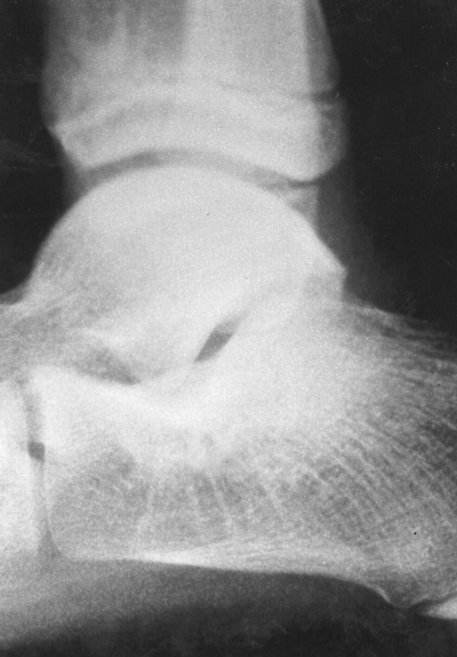
In 1983, Cowell and Elsner [
1] defined tarsal coalition as an abnormal union of two or more tarsal bones. This union leads to restriction or limitation of the subtalar joint, which may result in primary or secondary signs of coalition that may be seen on radiographs (
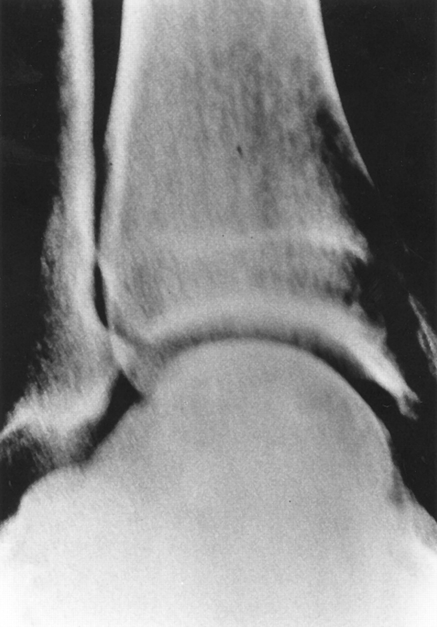
Fig. 1). [
2] Buckholz (unpublished data, 1975) used the terms “bar” and “bridge” to describe coalitions: a bar is a union between extra-articular bones and a bridge is a union between intra-articular bones. The exact frequency of tarsal coalitions is unknown, but the medical literature reports a range of 1% to 2%. [
3,
4] Ninety percent of tarsal coalitions affect the talocalcaneal or calcaneonavicular bones. [
5]
Figure 1.
Ball-and-socket ankle joint is the result of compensation for talocalcaneal coalition.
Figure 1.
Ball-and-socket ankle joint is the result of compensation for talocalcaneal coalition.
Tarsal coalitions have been associated with symphalangia, metatarsal anomalies, cuneiform coalitions, carpal coalitions, ball-and-socket ankle joint, middle ear ossicle fusion, Apert syndrome, small patella syndrome, and congenital vertical talus. [
6-
10]
Historically, coalitions have been classified according to numerous systems based on tissue type, etiology, anatomy, completeness of the coalition, and surgical type or location. It is not known if tarsal coalitions have a sex-based predilection, as there are conflicting reports pertaining to male/female ratios. [
11] Rankin and Baker [
12] have determined that there is no predilection for a particular race.
Clinically, tarsal coalitions may be symptomatic or asymptomatic. People who are initially asymptomatic usually develop symptoms during adolescence as a result of abnormal pedal biomechanics. The development of pain is usually insidious, but pain is often worse after physical activity. Ankle strain or sprain, increased body weight, and walking on uneven terrain may lead to symptoms, especially in young people as the union begins to ossify. Talocalcaneal coalitions typically begin to ossify when patients are approximately 12 to 16 years old. [
13] The related pain is relieved by rest or limitation (restriction) of subtalar and midtarsal joint motion. Muscle spasms (eg, of peroneus brevis, peroneus longus, tibialis anterior, or tibialis posterior), sinus tarsi syndrome, medial or lateral ankle pain, ankle joint instability, and tarsal tunnel pain are possible initial or long-term symptoms of tarsal coalition.
Physical examination usually reveals restricted subtalar joint pronation or supination. When peroneal muscle spasm is not found, passive subtalar joint motion may elicit pain. The foot may be flexible or rigid; flatfoot is not always present. The physical examination should be performed while the patient is nonweightbearing for assessment of passive range of motion, and while the patient is weightbearing to evaluate active subtalar motion and for gait analysis. Limited inversion may be accentuated by peroneal muscle spasms. The muscular tonicity on the peroneal muscles affords a protective maneuver by decreasing painful tarsal motion. [
14,
15]
Initial radiographs should include dorsoplantar and lateral views taken with the patient in the normal angle and base of gait, which will represent the functional angles and positions. [
16,
17] This standard approach may be adequate to reveal calcaneonavicular, talonavicular, and calcaneocuboid coalitions. However, these views may be inadequate for determining the individual involvement of the posterior and middle facets of the talocalcaneal joint. Further radiographic analysis for talocalcaneal coalitions may require Harris-Beath views [
3,
18] and Isherwood [
19] medial and lateral oblique views.
The extent of articular involvement in adolescents with talocalcaneal coalitions may be determined by computed tomography (CT). However, nonosseous unions may be difficult to view on CT scans; therefore, CT scans may not provide adequate information regarding articular involvement. The best imaging method for observing nonosseous unions is magnetic resonance imaging (MRI). This technology may become the standard for confirming the diagnosis of talocalcaneal and other coalitions.
Many conservative therapeutic modalities and surgical techniques are available for management of talocalcaneal coalition. Conservative modalities include anti-inflammatory drugs, casting, intra-articular injections (local anesthetic and cortisone injections), orthotic devices, padding, physical therapy, and shoe modification. When conservative measures fail to relieve symptoms, surgery should be considered. In adolescents who have coalitions without arthritic or secondary osseous changes, the best treatment option may be a surgical procedure. Surgical options include one or a combination of the following: resection with or without soft-tissue interposition, arthroereisis, isolated arthrodesis, and triple arthrodesis. Surgical resection is indicated in cases with less than 50% involvement of the articular surface and when adjacent osseous changes that would preclude range of motion free of pain (if motion were restored) are not present.
Case Report
A healthy 11-year-old girl who was an active participant in sports presented to the office of one of the authors (R.L.) complaining of pain in her feet and ankles. The pain was reported as ranging from a dull aching sensation to a sharp pain that was inferior and posterior to the medial malleolus. The left foot was more symptomatic than the right foot. The onset of pain was gradual each time with exercise and the development of symptoms was insidious. The patient experienced relief from her discomfort after periods of rest. Conservative treatment with functional orthoses had initially relieved her symptoms, but in the year prior to presentation her left foot had become painful again.
Physical examination revealed that the patient’s neurovascular status was intact bilaterally. There were no Tinel’s sign, Valleix’s points, or neuroma symptoms about the medial aspect of the left foot. Manual muscle testing revealed normal strength bilaterally. Tenderness was noted along the posterior tibial tendon just inferior to the medial malleolus of the left foot. There was no pain around the navicular tuberosity. A tender palpable lump that was immobile and nonfluctuant was noted inferior and posterior to the medial malleolus of the left foot.
Biomechanical examination revealed a subtalar joint range of motion of 27° for the right foot and 19° for the left foot. There was restricted supination of the left subtalar joint, with inversion noted only to the perpendicular. The forefoot was inverted relative to the rearfoot in the subtalar joint neutral position with the midtarsal joint maximally pronated. Severe pes planovalgus compensation was noted bilaterally. Gait analysis revealed an exaggerated depression of the medial longitudinal arch, significant forefoot abduction, and excessive calcaneal eversion bilaterally. The patient’s foot remained pronated throughout the gait cycle.
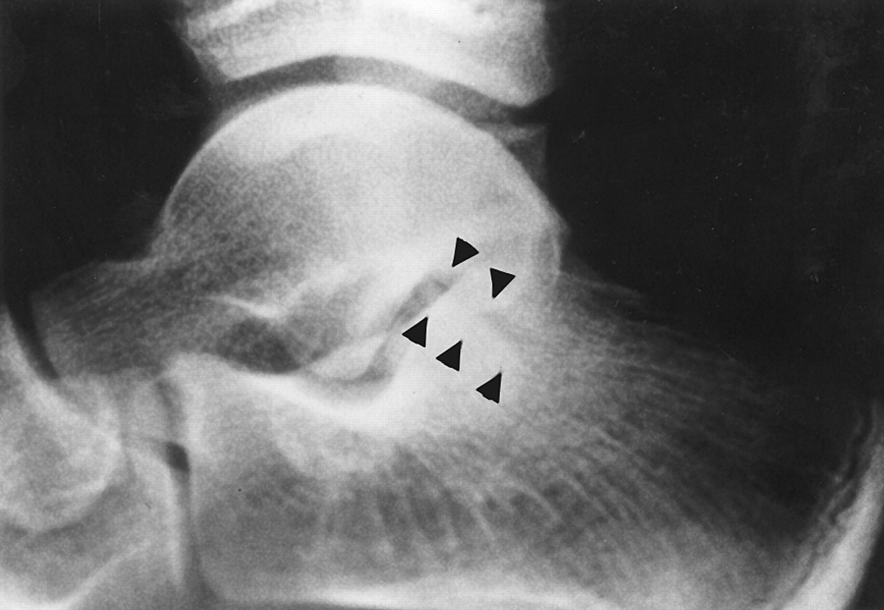
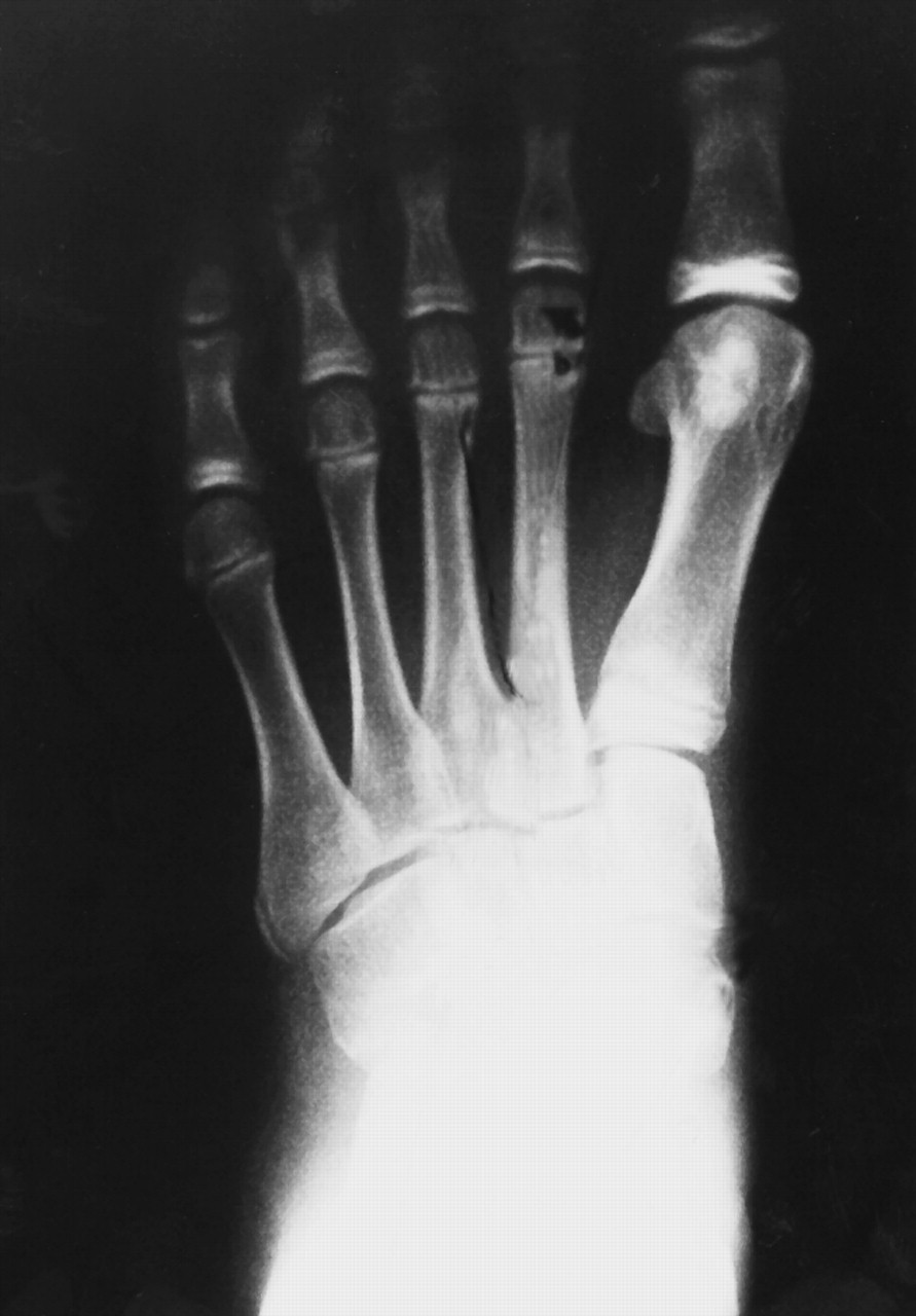
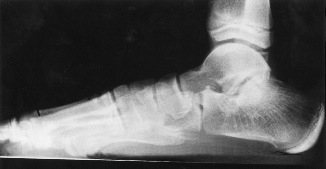
Standard radiographs revealed open growth plates with severe pronation, blunting of the lateral process of the talus, and an oblique-oriented lucency at the posterior facet of the subtalar joint (
Figs. 2–
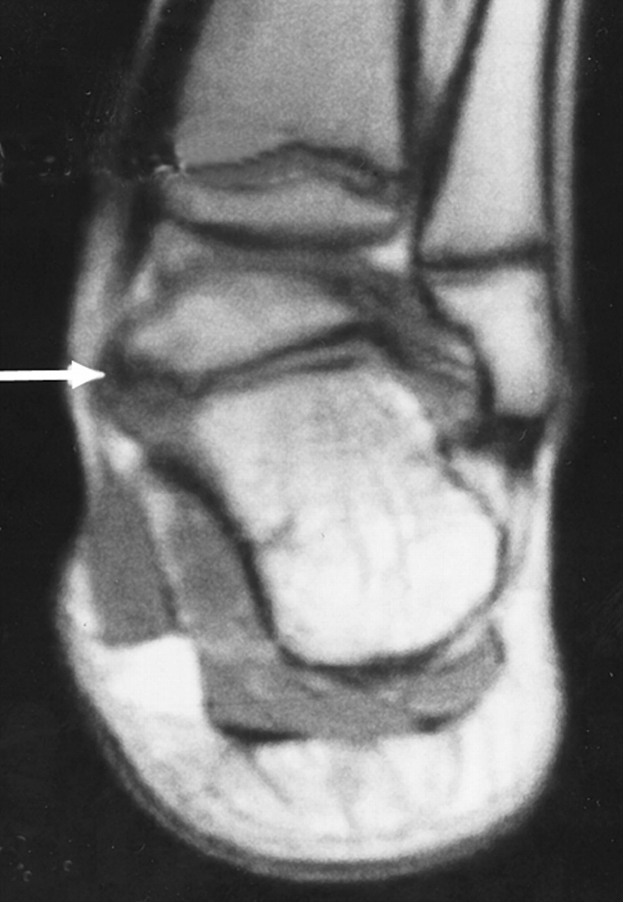
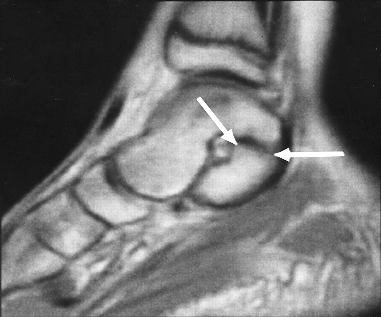
4). There was no evidence of arthritic changes in the subtalar or midtarsal joints. The MRI study revealed an incomplete union at the medial aspect of the posterior facet of the subtalar joint (
Figs. 5 and
6). Since the patient continued to experience pain with exercise despite orthotic therapy, and since she had only a partial coalition and no other significant osseous changes, surgical resection was advised.
Figure 2.
Dorsoplantar radiograph showing severe pes planovalgus.
Figure 2.
Dorsoplantar radiograph showing severe pes planovalgus.
Figure 3.
Lateral radiograph showing severe pronation and increased density at the subtalar joint posterior facet.
Figure 3.
Lateral radiograph showing severe pronation and increased density at the subtalar joint posterior facet.
Figure 4.
Magnified lateral radiograph showing oblique-oriented radiolucency at the subtalar joint posterior facet (arrowheads).
Figure 4.
Magnified lateral radiograph showing oblique-oriented radiolucency at the subtalar joint posterior facet (arrowheads).
Figure 5.
An MRI T1-weighted coronal plane image showing incomplete union at the medial aspect of the posterior facet (arrow). (Courtesy of Radiology Medical Group, Inc, San Diego, California.)
Figure 5.
An MRI T1-weighted coronal plane image showing incomplete union at the medial aspect of the posterior facet (arrow). (Courtesy of Radiology Medical Group, Inc, San Diego, California.)
Figure 6.
An MRI T1-weighted sagittal plane image demonstrating incomplete union at the posterior facet (arrows). (Courtesy of Radiology Medical Group, Inc, San Diego, California.)
Figure 6.
An MRI T1-weighted sagittal plane image demonstrating incomplete union at the posterior facet (arrows). (Courtesy of Radiology Medical Group, Inc, San Diego, California.)
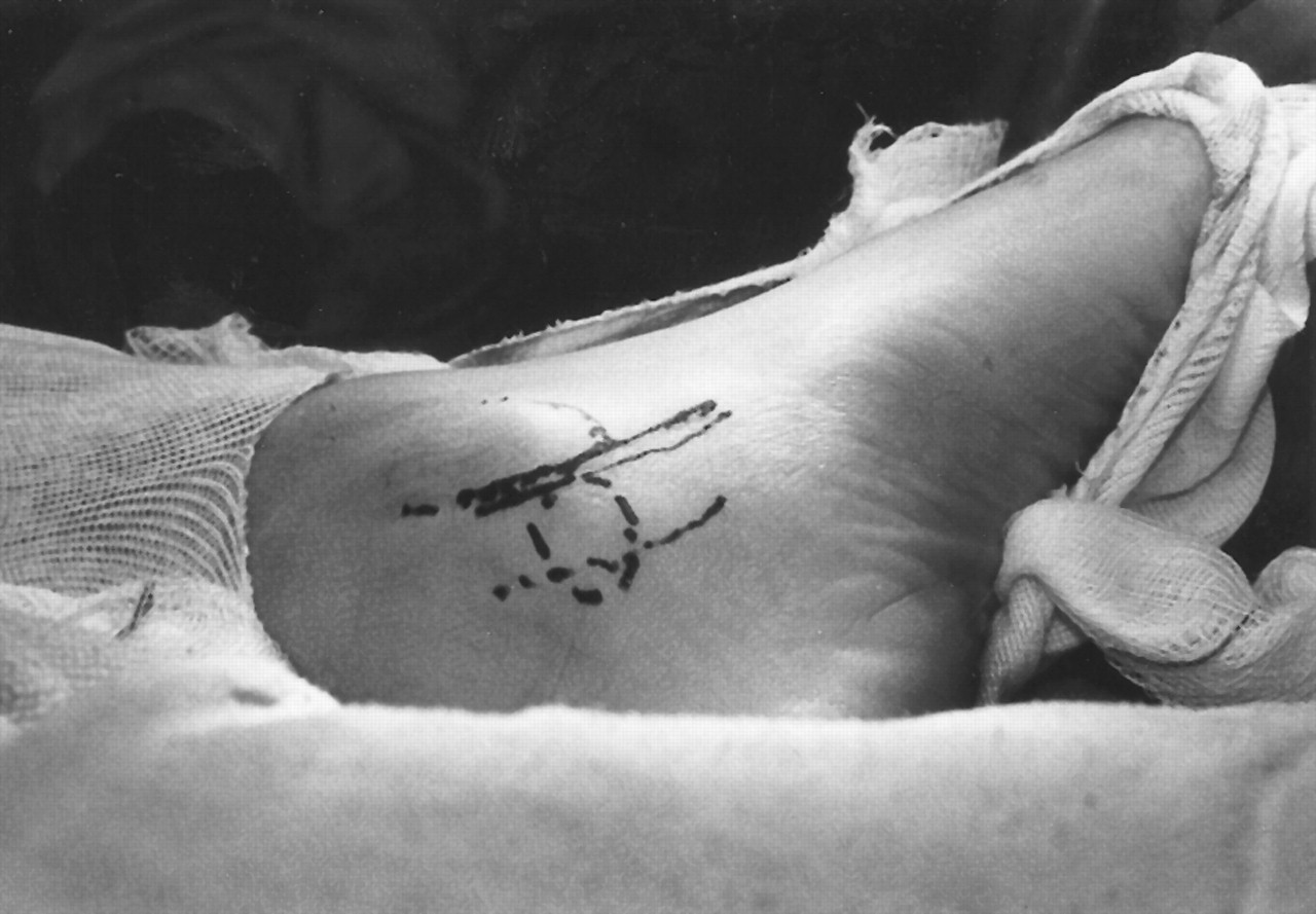
Surgical resection of the coalition was performed through a curvilinear incision that was posterior and inferior to the medial malleous (
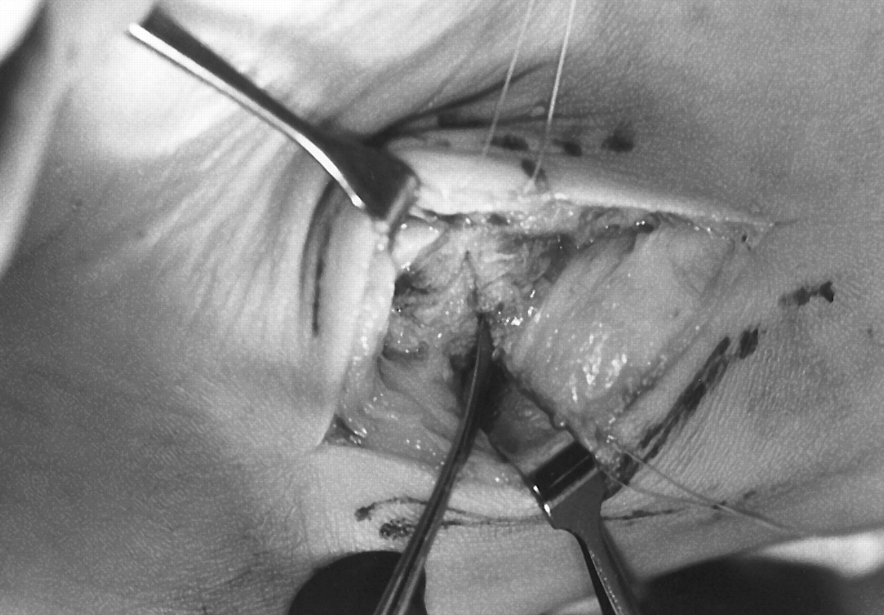
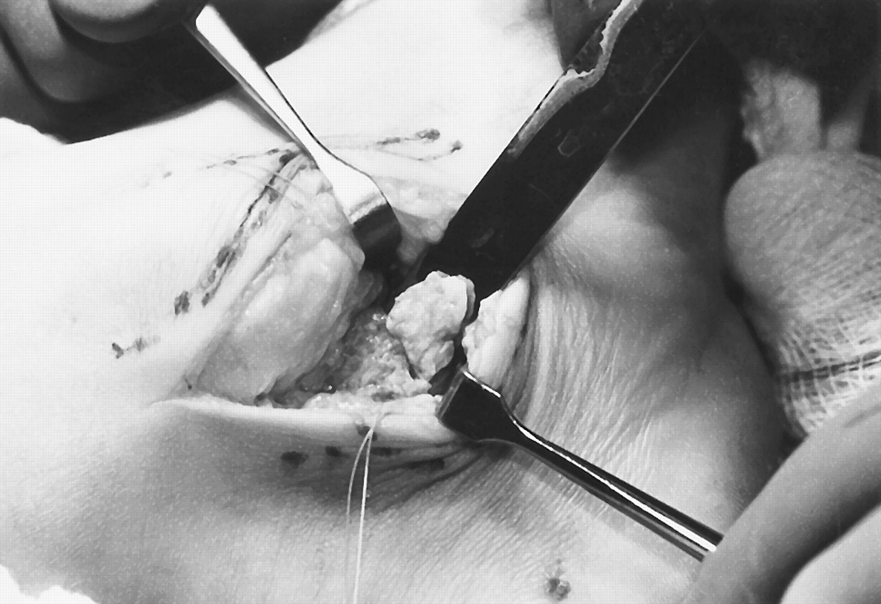
Fig. 7). The periosteum was excised and reflected, exposing the medial prominence of the posterior facet (
Fig. 8). The osseous prominence was resected with an osteotome, creating a natural separation at the fibrocartilaginous union (
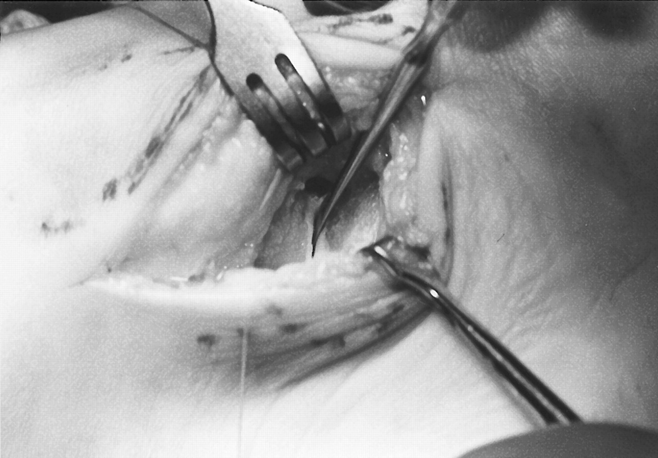
Fig. 9). This area was further resected with a rongeur until a more normal-appearing cartilaginous joint space was visible (
Fig. 10). As a result, intraoperative passive supination motion was increased. The wound was closed in layers over a TLS drain (Porox Technologies Corp, Fairburn, Georgia) and a short-leg removable cast was applied.
The cast was removed 2 weeks postoperatively; by the 4th week, the patient had no symptoms and was continuing to increase her level of activity. Postoperative radiographs at that point no longer demonstrated the pronounced set of oblique lines noted proximal to the sustentaculum tali that were indicative of this coalition (
Fig. 11). The patient continued to wear her orthoses and began soccer practice 4 months after surgery. However, functional orthotic therapy was continued because of the continued significant bilateral pes planovalgus. Additional surgical stabilization may be necessary in the future if significant uncontrollable symptoms associated with the severe pes planovalgus occur. At 10 months postoperatively, the patient was participating in volleyball and soccer and had no significant complaints.
Figure 7.
Lines indicate the incisional approach for surgical treatment.
Figure 7.
Lines indicate the incisional approach for surgical treatment.
Figure 8.
Exposure of osseous prominence medial to the posterior facet.
Figure 8.
Exposure of osseous prominence medial to the posterior facet.
Figure 9.
Resection of the osseous prominence with an osteotome.
Figure 9.
Resection of the osseous prominence with an osteotome.
Figure 10.
Further resection reveals a normal-appearing posterior facet with visible hyaline cartilage.
Figure 10.
Further resection reveals a normal-appearing posterior facet with visible hyaline cartilage.
Figure 11.
Immediate postoperative radiograph demonstrating a normal appearance of the posterior subtalar joint.
Figure 11.
Immediate postoperative radiograph demonstrating a normal appearance of the posterior subtalar joint.
Discussion
Posterior facet talocalcaneal coalitions, although rare, have been widely discussed in the literature. Surgical treatment of intra-articular coalitions typically involves extensive soft-tissue dissection and loss of cartilage or joint function, all of which increase postoperative sequelae. The case presented here is unique because of the restriction of the incomplete union to the medial edge of the posterior subtalar joint rather than across the entire articular surface. Moreover, surgical resection of the partial coalition likely will result in full restoration of function with minimal sequelae.
Conclusion
In this case report, radiography and MRI confirmed the diagnosis of an incomplete coalition of the talocalcaneal joint. Close examination of the posterior facet on radiographic and MRI studies allowed better visualization of the medial aspect of the joint. The medial joint was more oblique than the remainder of the joint in direction and irregular in appearance. In normal radiographic presentations, this area is difficult to visualize because of the superimposed lateral aspect of the talus and calcaneus.
Surgical resection has been shown to be effective in cases of talocalcaneal coalition involving less than 50% of the articular surface. [
20] Successful resection allows continued motion of the involved joint and eliminates pain. Because resection is effective only if there are no significant secondary osseous changes, it is mainly performed in young patients. Arthrodesis is the preferred surgical procedure when more than 50% of the joint is involved in the union, or in cases in which arthritic or significant structural changes have occurred.