Lower-extremity amputation affects approximately 60,000 people with diabetes every year, at an average cost of
$25,000 per amputation. [
1] As many as 51% of all nontraumatic amputations are performed on people with diabetes in the United States. Forty percent of these are toe amputations; 15% are foot and ankle; 25% are below the knee, and 25% are above the knee. [
2] Cutaneous ulceration in the diabetic foot is one of the most common pathologic events leading to lower-extremity amputation. [
3-
9] Pecoraro et al [
5] emphasized that most diabetic lower-extremity amputations do not result from a single cause but from several component causes. That study identified numerous component causes of amputation, including sensory neuropathy, ischemia, infection, minor trauma, skin ulceration, faulty wound healing, and gangrene. A critical triad of component causes was recognized in 72% of the cases, which included “an initial episode of minor trauma, resulting in cutaneous ulceration and subsequent failure to heal.” [
5] In most of these cases, minor trauma was the pivotal event that initiated the pathway to amputation. Furthermore, repetitive pressure was the most common form of minor trauma preceding ulceration.
Other studies have also recognized an association between repetitive pressure, associated callus or keratosis formation, and diabetic foot ulceration. [
10-
14] Young et al [
14] demonstrated that debridement of hyperkeratotic lesions, or calluses caused by repetitive pressure, reduces peak plantar pressures by 26%. This finding suggests that simple debridement of focal pressure keratosis may decrease the risk of foot ulceration. The main objectives of the present investigation were 1) to identify cases of diabetic foot ulceration preceded by focal pressure keratosis, 2) to assess the comorbidities at the time of ulceration, and 3) to examine the preventive treatments received prior to ulceration. On the basis of the results of previous studies, it was hypothesized that focal pressure keratosis would precede a majority of foot ulcerations in any diabetic population, that these patients would have systemic comorbidities that might increase their risk of ulceration, and that ulcer severity would be higher in cases in which little outpatient foot care had been received preceding the ulceration.
Research Designs and Methods
In this retrospective study, patients admitted to the Loyola University Medical Center and the Edward Hines Veterans Administration Hospital for inpatient or outpatient care of a diabetic foot ulcer between 1990 and 1997 were identified. Patients with a history of ulceration preceded by minor trauma were further studied. From this population, the inclusion criterion was a history of minor trauma due to focal pressure keratosis (callus formation) preceding ulcer formation. This was determined by identifying documentation in the charts of a hyperkeratotic lesion in the area of the ulcer preceding ulceration. For each case, data were collected on the basis of the time when an ulcer of at least a grade 1 (according to the Wagner Classification system) was first presented to the designated hospital or clinic. [
15] Ulceration cases were considered independently of patients; therefore, repetitive cases were weighted equally. Patients with grade 5 ulcerations were not included in this review.
In order to identify ulceration risk factors and comorbidities, data collection included age, duration of diabetes at presentation, peripheral vascular disease (diagnosis or absent pulses), chronic renal failure (diagnosis or creatinine greater than 1.5 mg/dL and blood urea nitrogen greater than 22), dialysis, lower-extremity bypass, peripheral neuropathy (decreased sensation documented in the foot), and hypertension. Patients who exhibited ischemic ulcerations with pain, eschar formation, and pulseless extremities were not counted as neuropathic. Other conditions noted were coronary artery disease, congestive heart failure, and renal transplant. Foot deformity included hallux valgus, contracted digits, Charcot’s foot, pes planus, equinovarus, pes cavus, hallux rigidus, prominent metatarsal heads, limited range of motion, footdrop, gastrocnemius and soleus equinus, onychomycosis, onychauxis, incurvated nails, and avulsion.
The following variables were quantified to evaluate management preceding ulcer formation: the number of visits to a foot clinic or hospital in the year prior to the event, the total duration of treatment, and the percentage of cases that had never been seen before in the designated clinic or hospital. Treatments prior to ulceration were documented; these included orthoses, prescription shoes, casting or posterior splints, debridement of calluses, education, surgical correction of a foot deformity, limited weightbearing, and accommodative padding. If any of the above information was unavailable from the charts, it was noted.
Each clinic visit involved a history that identified any foot complaints, as well as a systems review noting significant comorbidities. Both feet were examined for the presence or absence of pedal pulses and protective sensation. Deformities of the feet and nails were identified, including the presence of chronic focal pressure keratosis and any areas of recent injury.
Overgrown or dystrophic nails were reduced by means of topical alcohol preparation. Hyperkeratotic lesions were pared to healthy tissue with a sterile small scalpel; epidermal structures were left intact unless ulceration was found beneath the keratotic lesion. In those cases, the ulcers were thoroughly debrided and appropriate wound care was instituted, including treatment for infection, vascular evaluation, medical evaluation, and off-loading.
Orthoses, prescription shoes, or appropriate commercial shoes were recommended to patients depending on the severity of their deformity and the perceived risk of ulceration. Patients were counseled about foot care, and were also encouraged to optimize glucose control and avoid tobacco use. Follow-up appointments were recommended in 1, 2, or 3 months, depending on the severity of foot deformities, keratosis, nail problems, and comorbidities contributing to ulceration risk. Ulcerated patients were instructed in wound care and given a follow-up appointment 1 to 2 weeks later, unless the condition was severe enough to require hospitalization.
Parametric statistical methods were used for data analysis. Continuous variables were described by means of sample means with standard deviations. Differences between ulcer grades (1 or 2 versus 3 or 4) were identified with the chi-square test for homogeneity and the Student’s t-test for categorical and continuous variables, respectively. Trends by individual ulcer grade (1 to 4) were detected with the Mantel-Haenszel chi-square test; these are specifically noted where presented. In all cases, a two-sided α level of .05 was considered statistically significant.
Results
Of the 233 cases of diabetic foot ulceration preceded by minor trauma, 82.4% (192/233) were preceded by focal pressure keratosis and therefore met the criteria of the study. The average (± SD) age of the patients was 58.2 ± 12.7 years; the average duration of diabetes was 19.45 ± 11.9 years; and the average ulcer grade when each patient first presented to the designated foot clinic or hospital with the ulcer was 1.8 according to the Wagner Classification. Of the ulcers, 72% were grade 1 (102/192) or 2 (37/192) at initial presentation, with the remaining 28% being grade 3 (43/192) or 4 (10/192). The average number of visits in the year prior to the first time the patient presented to the designated foot clinic or hospital with the ulcer was 7.3 ± 10.2; the average amount of time that the patient had been treated in the designated foot clinic or hospital was 2.1 ± 2.8 years; and 46.9% of all cases had no documentation of previous visits to the designated foot clinic or hospital (
Table 1). Of the cases that contained documentation of previous visits, the most frequent interventions prior to ulceration were debridement of focal pressure keratosis (58.3%) and patient education (64.6%). Prevention measures that were associated with decreased ulcer grade when grades 1 and 2 were compared with grades 3 and 4 included protective shoes (
P = .004), debridement (
P = .001), education (
P = .001), and limited weightbearing (
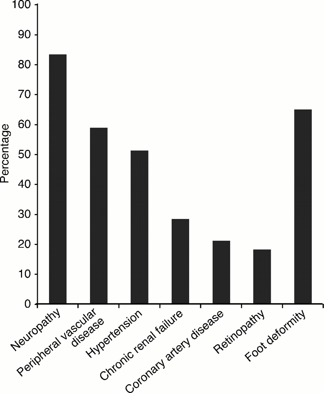
P = .03). Ulcerations ended in the following outcomes: 54.2% (104/192) were hospitalized and required surgical treatment; 22.9% (44/192) were hospitalized without requiring surgical treatment; 18.8% (36/192) required outpatient care only; 3.1% (6/192) were not yet closed at the time of this investigation; and 1.0% (2/192) were lost to follow-up. Of the comorbidities, the most prevalent were peripheral neuropathy (83.3%), the presence of a foot deformity (65.1%), peripheral vascular disease (58.9%), and hypertension (52.1%) (
Fig. 1). In comparisons of the presence of risk factors with ulcer severity, only the presence of chronic renal failure was associated with more severe ulcer formation (
P = .002). In contrast, the presence of neuropathy (
P = .068) and a foot deformity (
P = .001) were associated with lower-grade ulcers; this is likely to be related to improved care among patients previously identified with these risk factors.
Table 1.
Ulceration Characteristics at First Presentation to the Designated Foot Clinic or Hospital
Table 1.
Ulceration Characteristics at First Presentation to the Designated Foot Clinic or Hospital
Figure 1.
Prevalence of specific risk factors in cases of ulceration.
Figure 1.
Prevalence of specific risk factors in cases of ulceration.
A multivariate analysis was completed in which all of the factors were considered as potential predictors of ulcer severity. The results suggested that the presence of a foot deformity (P = .004; relative risk [RR] = .05) and education (P = .036; RR = .040) are the two most important factors associated with lower-grade ulcerations (grade 1 or 2). The presence of deformity may prompt greater awareness of the patient’s risk of ulceration and lead to more attention to the foot, explaining the relationship of deformity to lower-grade ulceration.
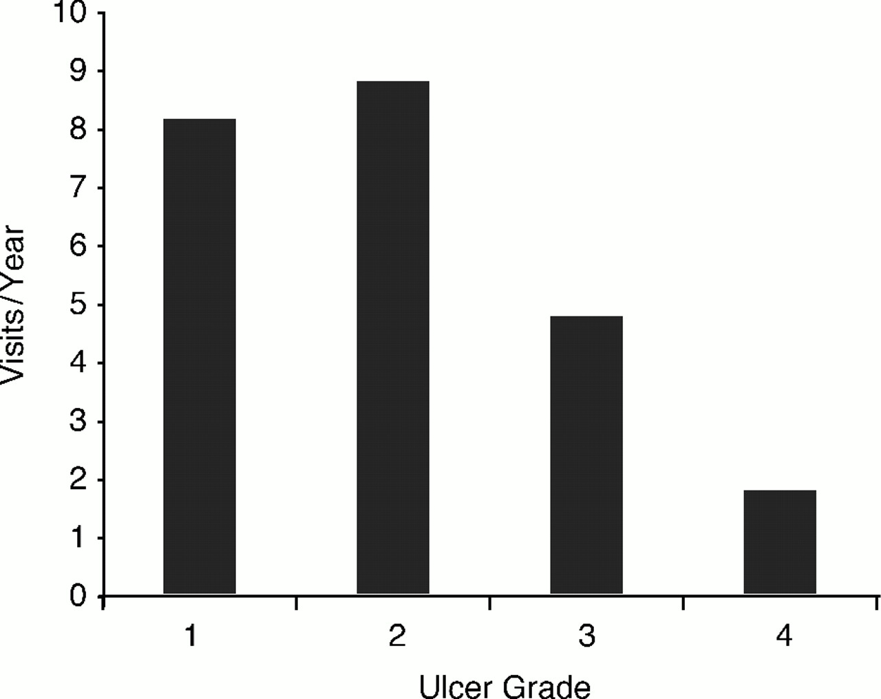
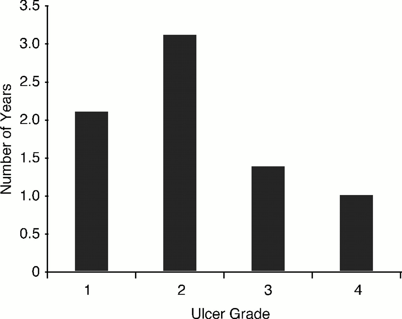
There were many clear indications in this study that the quality of premorbid treatment is associated with the severity of the ulcer and the required treatment. The number of visits per year was significantly higher in cases of lower-grade ulcers (
P = .03) (
Fig. 2); likewise, the duration of treatment was significantly higher among those who presented with lower-grade ulcers (
P = .04) (
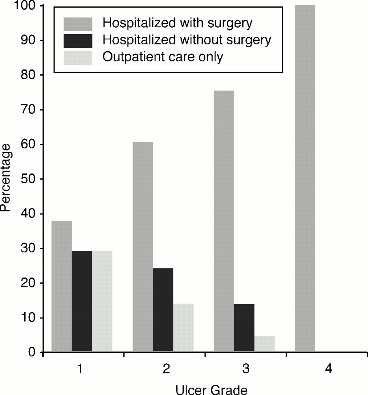
Fig. 3). There was a highly significant association between increasing care requirements and higher-grade ulcers, and the frequency of surgery required in treatment increased significantly with increasing ulcer grade (
P = .001) (
Fig. 4). Finally, ulcers that lacked documentation of care prior to ulcer presentation tended to be of higher grade (
P = .001).
Figure 2.
Mean visits per year in the year prior to ulcer formation, by ulcer grade at presentation. There is a significant difference between grades 1 and 2 versus grades 3 and 4 (P = .03).
Figure 2.
Mean visits per year in the year prior to ulcer formation, by ulcer grade at presentation. There is a significant difference between grades 1 and 2 versus grades 3 and 4 (P = .03).
Figure 3.
Mean duration of treatment in years by ulcer grade. There is a significant difference between grades 1 and 2 versus grades 3 and 4 (P = .04).
Figure 3.
Mean duration of treatment in years by ulcer grade. There is a significant difference between grades 1 and 2 versus grades 3 and 4 (P = .04).
Figure 4.
Required treatment by ulcer grade. There is a significant association between required treatment and ulcer grade (P = .001).
Figure 4.
Required treatment by ulcer grade. There is a significant association between required treatment and ulcer grade (P = .001).
Discussion
Much attention is given to the medical and surgical management of diabetic ulceration and infection. Few studies have focused on outpatient, low-technical interventions to prevent ulceration or reduce morbidity. Although the results of this review may seem intuitive, they demonstrate an important statistically significant relation between clinical foot care and ulcer severity. Simply put, more frequent clinical visits were associated with less severe ulcers, fewer hospitalizations, and less need for surgery in this patient group.
This patient analysis clearly identified chronic focal pressure keratosis, which includes intractable plantar keratosis, heloma durum, pinch callus, and heloma molle as common sources of minor trauma leading to ulceration. Patients exhibiting these various forms of focal pressure keratosis and other risk factors for ulceration, such as neuropathy and vascular disease, are less likely to ulcerate, require hospitalization, or undergo surgery for their foot condition if they have regular, frequent foot clinic visits. These visits should include risk evaluation, debridement of keratotic lesions, prescription of appropriate shoes, and patient education. If the cost of such a podiatric office visit for a patient is estimated at $60, even monthly care costs total only $720 per year, compared with the estimated cost of $25,000 for a lower-extremity amputation. Although compliance with the recommended frequency of visits was not measured in this study, a frequent trend observed by the senior author (R.A.S.) was that patients with ulcers who fail to come to their appointments frequently require admission for more severe, infected ulcerations.
The results of this study confirm that diabetic patients should be screened for risk factors leading to foot ulceration, including sensory deficits, vascular disease, and, in particular, evidence of chronic focal pressure keratosis, such as common corns and calluses. Those who present such risk factors should be directed into a foot clinic program that will provide the services previously described. This is especially true for patients with a history of previous ulceration or infection associated with keratosis. Routine implementation of such a protocol may produce substantial savings in hospitalization and surgical treatment for large groups of diabetic patients. The quality-of-life savings achieved by significant reduction in morbidity for these high-risk patients is inestimable.
Conclusion
This study identified focal pressure keratosis as the minor trauma preceding diabetic foot ulceration in 82.4% of cases. The preventive treatments received by the patients prior to ulceration and the comorbidities of the patient at the time of ulceration were noted. The results indicated that patients seen more frequently in the foot clinic had less severe ulcerations and were less likely to undergo surgery than those with infrequent clinical care. The most frequent treatments prior to ulceration were debridement and patient education. The most prevalent comorbidities of the patients in this study were peripheral neuropathy, peripheral vascular disease, hypertension, and foot deformity. These results indicate that aggressive outpatient care is effective in decreasing hospitalization, surgery, and amputation in patients with diabetes who are at risk for foot ulceration owing to chronic focal pressure keratosis.