References to foot radiographs are common in the orthopedic and podiatric literature. Assumptions are often made that the radiographs are taken in standardized positions, and that measurements from the radiographs are reliable and reproducible. The angle of gait describes the angle from the mid-sagittal plane assumed by each foot in the midstance of walking and the base of gait as the transverse separation of the feet in walking.[
1] This position would seem to represent a standardized method for establishing a natural stance position, as opposed to a contrived stance position, for the purposes of radiographic examination, which may indeed give a false impression of the bony relationships of the foot. However, the literature has not adequately reported if specific positioning of the feet, to simulate natural stance, is necessary to obtain reliable and reproducible radiographic measurements of the foot.
Podiatric and orthopedic literature are replete with radiographic interpretation taken from weightbearing foot radiographs.[
2-
4] There is also widespread reference to standardized radiographs for the quantification of foot abnormalities and the description of baseline values prior to treatment, particularly when referring to surgical intervention.[
5,
6] Furthermore, the reported outcomes of such intervention often depend upon measurements obtained from radiographs.
The importance of taking weightbearing x-rays of the foot has been discussed in the literature.[
1,
7,
8] The podiatric literature appears to support the concept that radiographic examination should be performed in the natural standing position that simulates the mid-stance phase of gait.[
1,
8,
9] Radiographs taken in the weightbearing position demonstrate the foot in a locked and static yet simultaneously kinetic and functional situation, providing a truer impression of the bony and soft tissue complex under stress.[
10] Perlman et al[
11] found that static lateral weightbearing radiographs taken in the patient’s angle and base of gait position are clinically similar to the findings seen during dynamic gait and captured in the midstance position.
Weightbearing lateral and dorsoplantar radiographs taken in a standardized manner with the feet in their angle and base of gait position have since developed as the accepted standard for much of the podiatric literature. Gamble and Yale[
1] maintain that the structural and functional integrity of the foot is reflected in the radiographic appearance of the mature foot in stance. Furthermore, bones of strong shape are aligned together in proper positions for structural stability and function in the normal foot. Radiographic evaluation, based on a mathematical modeling approach, has been developing in podiatric medicine, leading to better clinical diagnosis and treatments.[
7]
The most common radiographic views are dorsoplantar and lateral views of the foot that are obtained to delineate normal and pathologic osseous and soft tissue anatomy. Numerous authors have recommended the use of weightbearing, as opposed to non-weightbearing, views to provide more accurate information of the foot, believing that this approximates a functional position.[
1,
8,
9,
11,
12] Yet, standardization of the positioning by radiographers and podiatrists taking foot radiographs still remains variable. Shereff et al[
13] performed a study to compare standardized measurements of the foot in nonweightbearing and weightbearing radiographs. In their discussion, the authors reported that the data did not support the expected variations in the parameters measured on the weightbearing dorsoplantar views compared to the same parameters measured on the nonweightbearing views. They reported that differences of measurement did not occur in a consistent fashion, suggesting that their observations may be due to subtle deviation in radiographic technique, or variation in ligamentous laxity stability, or other anatomic factors from one individual to another.
The contributing effects of distortion and magnification to the sources of error on radiographic films are related to the positioning of the foot and its relationship to the central ray and the cassette. The importance of standardization was explored as early as 1945, when it was proposed that accumulative errors in radiographic images may be produced by varying the x-ray distance and the angle of the central beam to the foot.[
14] Errors of measurement associated with magnification problems have been further investigated using cadaveric metatarsals, where the magnification error was quantified to be 10% to 15%.[
15]
The purposes of this study were to ascertain: 1) if the positioning of the feet for radiographic assessment produces significant variations in measured angles, and 2) if radiographs taken on two separate occasions are reproducible. These are both important in podiatric medicine and surgery when assessing the effectiveness of biomechanical or surgical intervention on the foot. The study hypothesized that there are no significant differences in selected measurements between weightbearing radiographs taken with feet together and straight–ahead, and those taken in the subject’s angle and base of gait position. Furthermore, the study hypothesized that there is no significant difference between the selected measurements of radiographs taken on two separate occasions.
Methods
Subjects
To avoid gender bias, ten volunteer subjects, all male adult podiatrists, were chosen because of their extensive knowledge of the clinical uses and potential harmful nature of ionizing radiation. Subjects had an age range of 24 to 44 years, with a mean age of 31 years. All of the participants were provided with an explanation of the nature and purpose of the study with the approval of the Human Research Ethics Committee of Curtin University of Technology. It was explained that the films would be coded so they could not be identified by name. Each subject signed a consent form and a routine clinical gait assessment of the angle and base of gait of each subject was conducted and recorded for positioning purposes.
Inclusion criteria of the subjects required that both feet showed no clinical signs of foot pathology or abnormal function. Persons with conditions such as hallux valgus or digital deformities, excessive foot pronation or supination, extreme angle of gait, or other obvious structural anomaly were excluded from the study. In addition, any history of previous bone or joint disease, injury or surgery, that may have allowed the foot to be easily identified on the radiographs, was included in the criteria for exclusion.
A repeated measures study was conducted, consisting of the radiographic evaluation of plain radiographs of the feet of the ten subjects taken in two different weightbearing positions: 1) feet together and straight-ahead and 2) angle and base of gait positions. The radiographs were repeated on two separate occasions. For statistical purposes, each radiographed foot was regarded as a single subject, resulting in a total of 20 subjects. Comparisons of six radiographic measurements were then made from the sets of radiographic films produced on each occasion.
To standardize technique, dorsoplantar and lateral views in both positions were taken by the same experienced radiographer, using the same equipment in a standardized manner. The same procedure was repeated 2 weeks later. In order to assess the reliability of the examiner’s measurement, three randomly selected radiographic films were taken from the study and the six measurements were repeated on three separate occasions. After each set of measurements was taken and recorded from each radiograph, the film was wiped clean of all markings and re-examined on a different day.
Appropriate protective lead shielding was used to protect the upper body. Given that 16 views were taken for each subject, the total skin entrance exposure dose per subject was 2,560–3,392 microgray. This is the equivalent of 2.5–3.4 millisievert and falls well below the recommended yearly effective dose equivalent of 5 millisievert for volunteer studies as defined by the National Health and Research Council in Australia.[
16]
An orthoposer was used to facilitate taking the weightbearing views. Elevated from the ground, it enables the x-ray tube to be positioned at the required level to allow the central ray to be directed perpendicular to the film.[
17,
18] For the dorsoplantar view, each foot is imaged separately; the subject stands on the orthoposer on top of the film cassette with the central ray directed at the head of the talus at an angle of 15° to the vertical and approximately 100 cm above the feet. For the lateral view, each foot is imaged separately. The subject stands on the orthoposer with the film cassette placed vertically in the slot and the medial border of the foot against the film cassette with the central ray directed at the calcaneocuboid joint at 90° to the cassette at a distance of approximately 100 cm.
A china graph marking pencil was used to mark the films. A standard tractograph with a central hole to visualize the intersecting lines when measuring angles was used.
Radiographic Measurements
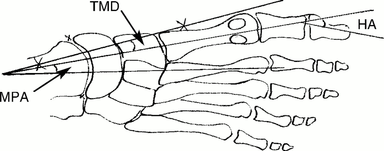
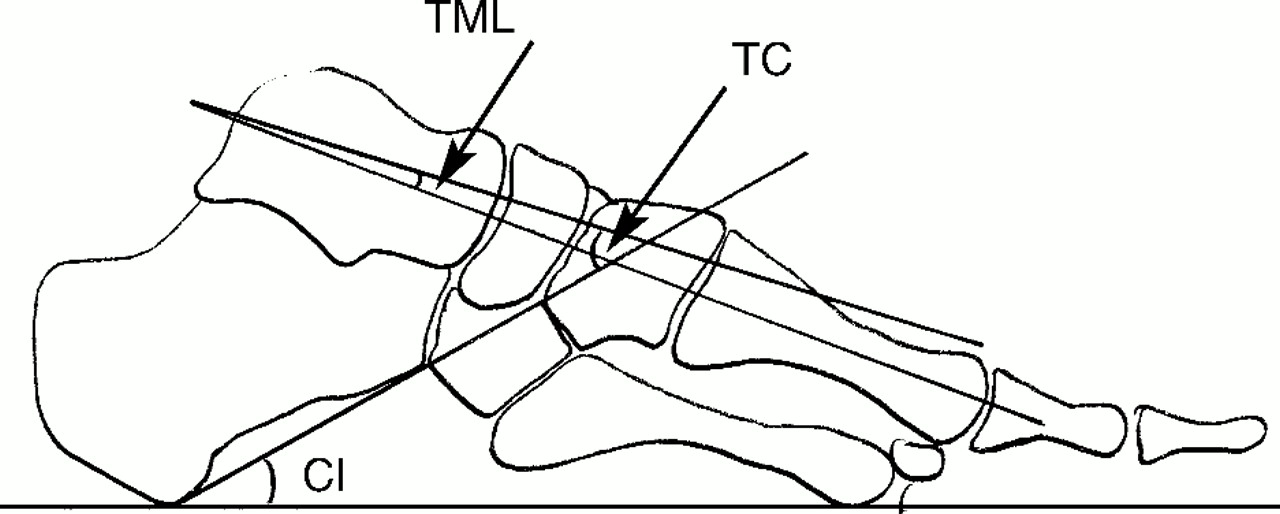
The six measurements (three each from the dorsoplantar and lateral views) used in this study have been used previously to describe radiographic foot morphology.[
1,
9,
19-
22] Measurements were then transferred to a data spreadsheet for later statistical analysis by SPSSX (SPSS Science, Chicago, Illinois).
Statistical Analyses
Twenty sets of raw data were collected. Each of the ten subjects provided two data sets, one from each foot. These represent independent data sets for the purposes of statistical analysis.
Each subject generated eight dorsoplantar and eight lateral radiographs, producing 480 different measurements for the cohort that was used in the analysis of measurements.
Figure 1 shows the angles measured from the dorsoplantar films and
Figure 2 shows the angles measured from the lateral films.
A repeated measures analysis of variance (ANOVA) was produced to test for differences between measurements in each radiographic position and at each episode. Intrarater reliability for all six measurements was tested by a repeated measures ANOVA that resulted in the calculation of intraclass correlation coefficients (ICCs), which were used to test for differences between the repeated measures and assess the reliability of the author’s measurements. A probability level of P < .05 was used to indicate significant differences for all statistical tests.
Results
This study was designed to investigate two sources of variations in weightbearing foot radiographs: differences of foot positioning and differences when radiographs are taken on two separate occasions. Descriptive subject data, including age, angle of gait, and base of gait measurements, are shown in
Table 1.
To test the reliability of the measurer (intrarater), three randomly selected sets of measurements were taken from the study and the six measurements were repeated three times. The calcaneal inclination and talar–first metatarsal angle measurements showed excellent correlations (above .90) and the hallux abductus angle showed good reliability (above .75).[
23] Poor reliability was shown for the talometatarsus angle (below .75). A correlation coefficient could not be calculated for the metatarsus primus adductus because it did not show enough variance. In contrast, the talocalcaneal angle showed great variations and therefore did not result in a valid ICC. This analysis showed that the F values were small, indicating agreement of scores, and the
P values were all greater than .05, indicating that the rater effect is not significant, as shown in
Table 2.
To test the null hypothesis that there are no significant differences in selected measurements between weightbearing radiographs taken in the feet together and straight-ahead position, and those taken in the angle and base of gait position, the mean, standard deviation, standard error, and a correlation matrix were calculated for each measurement. The ANOVA showed that all
P values were greater than .05, thereby allowing for acceptance of the null hypotheses that there are no statistical differences in radiographic measurements taken from films of feet in two different positions.
Table 3 summarizes the analysis of variance results.
The results from the repeated measures ANOVA (within-subjects design) show that for every dependent variable (radiographic measurement), the F values were smaller than the F critical values (
a = .05) and the
P values greater than .05, and hence, are not significant. The mean, standard deviation, and percentage differences suggest little variation exists between measurements of the first and second exposures for both feet together and straight-ahead and angle and base of gait positions detailed in
Table 4.
Measurements of weightbearing foot radiographs taken in two stance positions and repeated on two occasions were examined for differences. No statistically significant differences were demonstrated between any radiographic measurement evaluated in either stance position or exposure session.
Discussion
This study did not demonstrate significant differences of measurement of radiographic parameters between two specific foot-positioning methods, both of which are currently described in the literature and employed clinically. Radiographers often use the feet together and straight-ahead position when practitioners request weightbearing foot radiographs without providing specific instructions for foot positioning. The angle and base of gait position is widely used and requested by podiatrists in the belief that it simulates the midstance phase of gait. Both methods of positioning of the foot attempt to standardize procedure that helps ensure reproducibility.
The data collected and analyzed in this study were obtained from a reasonably homogeneous group of male subjects presenting with no foot pathology. They did not demonstrate wide variations in either their angle or base of gait. In this group of subjects, the range of angle of gait was 0° to 15°, with a mean of 8.2°. The range of base of gait was 5 cm to 15 cm, with a mean of 6.1 cm. These measurements were less than previously reported by Saltzman et al,[
24] who recorded an angle of gait ranging from 7° to 22°, and McIlroy and Maki,[
25] who reported 13° to 52° angle of gait and 6 cm to 28 cm base of gait. Hlavac[
26] considers the normal angle of gait to be 7.5° to 10° abducted from the midline, which is a result of normal ontogeny of femoral and tibial torsion. The limited range of angle and base of gait estimated for the individuals in the sample group may have been too close to the together and straight-ahead position to show statistical differences, particularly when most of the angular measurements were small.
The results of this study, therefore, may not accurately represent the variation of these values as seen in clinical practice. There may well be greater differences between positions due to the presence of pathology, such as hallux valgus, pes valgus or pes cavus, or with larger variations of angle and base of gait.
When studying the interobserver reliability of standard foot radiographic measurements, Saltzman et al[
24] found that the lateral talocalcaneal angle showed a large variability. There was some difficulty in identifying the appropriate bony landmarks on the complex shape of the talus required to measure talocalcanel and talometatarsus angles. However, most of the radiographic measurements selected demonstrated good to excellent intrarater reliability.
A further limitation of the study may relate to the age range of the subjects. No subject was under the age of 23 years or over 41 years, which may have restricted the outcome. Another possible limitation in the design of the study was the fact that all subjects were male, and there may be gender-based differences that may deserve further attention.
Due to the ethical constraints inherent in this study, a conscious effort was made to limit the number of subjects to a minimum. Obviously, the study would have greater validity had it been possible to involve a significantly large number of subjects, with and without foot pathology. A test of radiographer positioning reproducibility may also help to further validate the study. Radiographer accuracy and reproducibility were assumed for the purposes of this study.
Conclusion
The results of the study suggest that in this sample population, no differences in radiographic measurements were demonstrated with respect to placement of the feet together and straight-ahead position, or in angle and base of gait position, or with respect to time. A strong correlation of the repeated measures suggests that weightbearing foot radiographs can be reliably reproduced and thus valid comparisons can be made, over time, provided standardized radiographic techniques are employed.
This study, therefore, does not support the notion that weightbearing angle and base of gait radiographs more accurately reflect foot structure than standard weightbearing radiographs. Therefore, the widely held belief by podiatrists that angle and base of gait radiographs are superior to simple weightbearing radiographs must be questioned. The author recommends that a similar study be conducted using subjects with common foot pathologies, and therefore potentially different radiographic findings, which may influence the outcome of the results.