Chronically painful sesamoid disorders and fractures of sesamoids are traditionally treated with surgical excision of the fragments or of the entire sesamoid.[
1-
9] Although sesamoid excision is a successful procedure, removing the sesamoids of the first metatarsal can lead to mechanical instability of the first metatarsophalangeal joint[
10] and may predispose patients to joint deformities such as hallux abductovalgus and hallux flexus.[
1,
4,
11]
Traumatic first metatarsophalangeal joint dislocations are thought to be caused by hyperextension of the joint. First metatarsophalangeal joint dislocations are classified based on their anatomic pathology.[
12] Type I dislocations have an intact intersesamoidal ligament and sesamoids, and they are very difficult to close reduce. Type II-A dislocations have a ruptured intersesamoidal ligament and the intact sesamoids are typically widely separated medially to laterally. Type II-B dislocations usually have a transverse fracture of one sesamoid with an intact intersesamoidal ligament. Type II dislocations can usually be close reduced.
The procedure described in the following case report was performed in an attempt to preserve the anatomy and function of the first metatarsophalangeal joint. A successful internal fixation technique for a displaced tibial sesamoid fracture is demonstrated.
Case Report
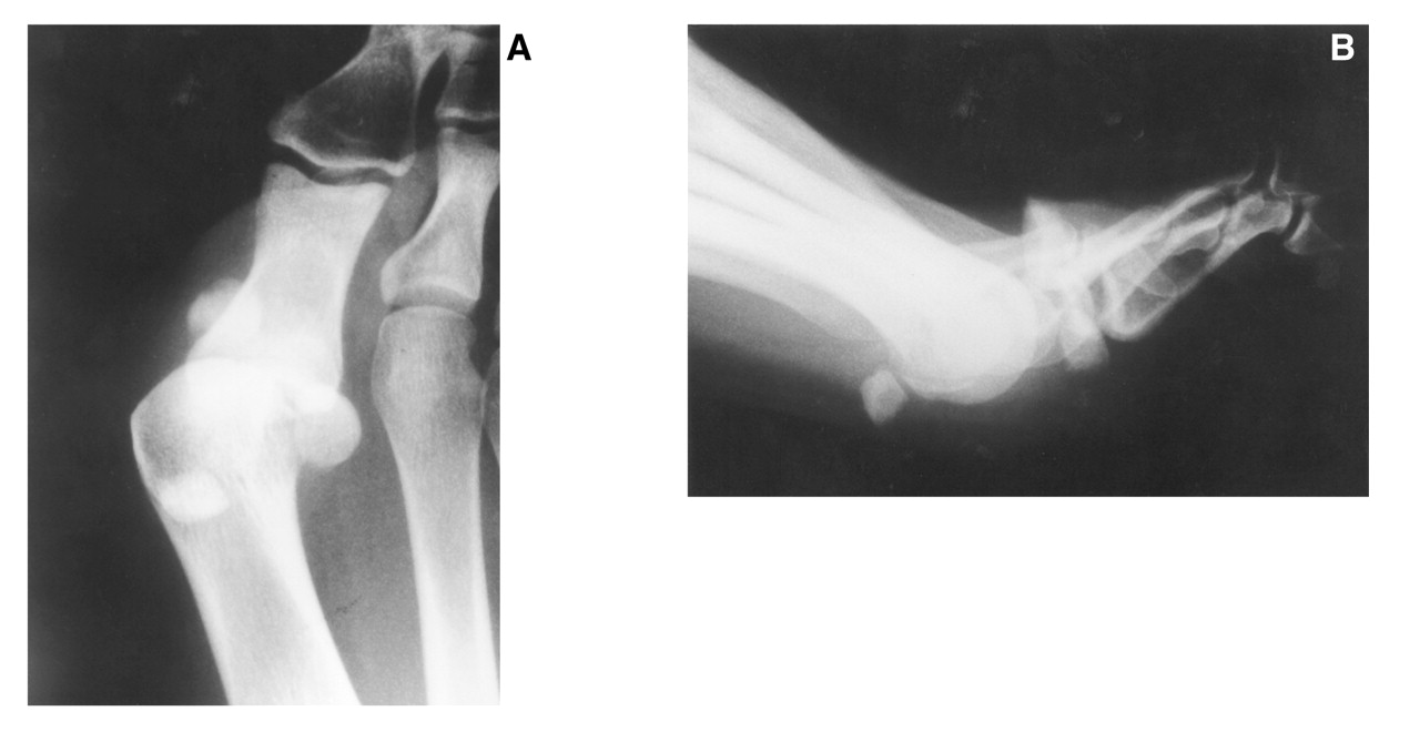
A 17-year-old adolescent girl sustained a type II-B dorsal proximal dislocation of the first metatarsophalangeal joint in a motor-vehicle accident. Prereduction radiographs demonstrated a 2-cm displacement of the two tibial sesamoid fragments (
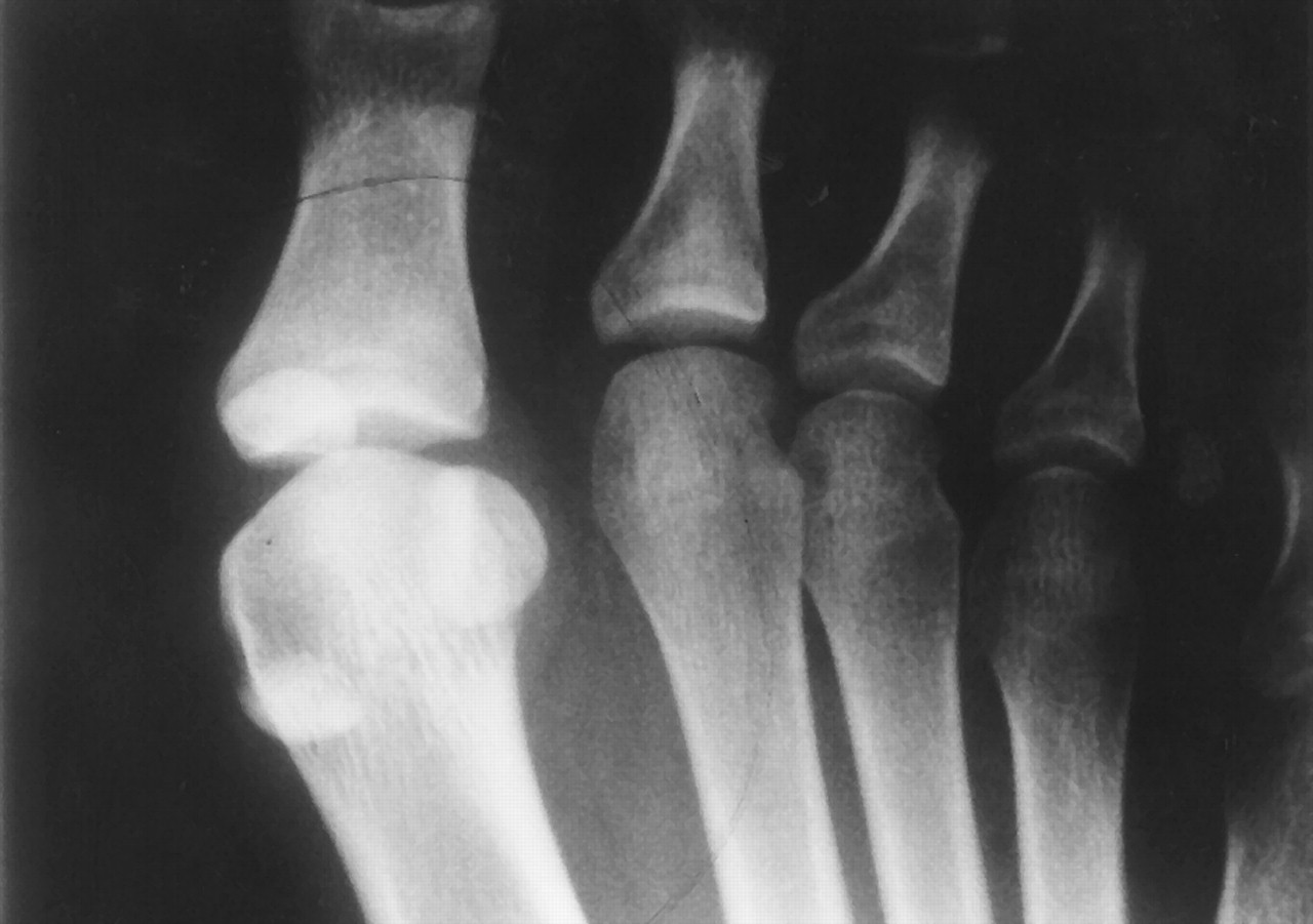
Fig. 1). After closed reduction, the tibial sesamoid fragments showed no significant reduction in displacement (
Fig. 2). Due to the continued displacement of the tibial sesamoid fracture and the subsequent likelihood of instability of the first metatarsophalangeal joint, a surgical approach was attempted to repair the tibial sesamoid.
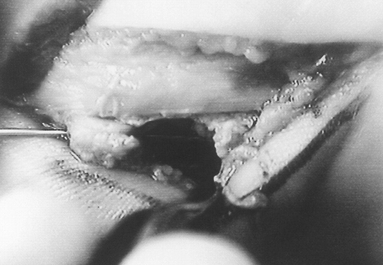
A medial approach to the first metatarsophalangeal joint was used and the fracture fragments were identified (
Fig. 3). A 0.045 Kirschner wire was used to drill a hole through the two sesamoid fragments (
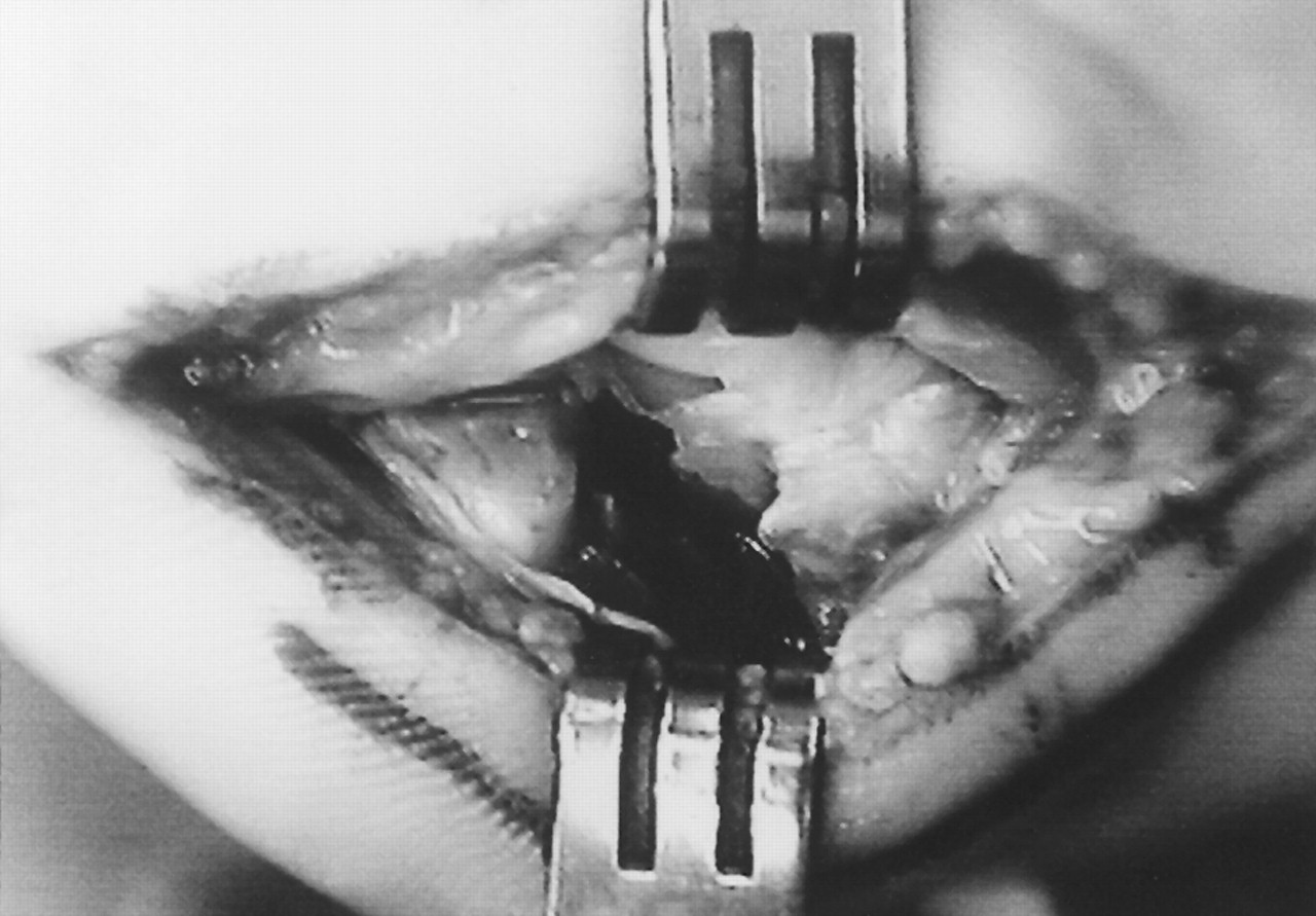
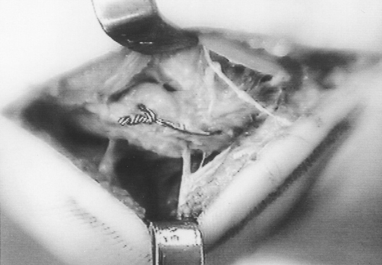
Fig. 4). A straight needle was used to thread a 0.028 monofilament wire through the holes and the two fragments were opposed with the articular surface aligned (
Fig. 5). The monofilament wire was cinched down and the knot was oriented plantar, distal, and medial to minimize interference with the first metatarsophalangeal joint and for easy access if surgical removal became necessary.
Postoperative care consisted of a plaster forefoot cast with an immobilization splint of the first metatarsophalangeal joint for 6 weeks. Physical therapy and limited full weightbearing in an athletic shoe continued for an additional 4 weeks. The patient returned to normal daily activity at 10 weeks with a limitation on sports involving ballistic movements off the ball of the foot. At 14 weeks after surgery, the patient returned to full activities with no limitations. At 36 months follow-up, she had no pain or limitation of activities.
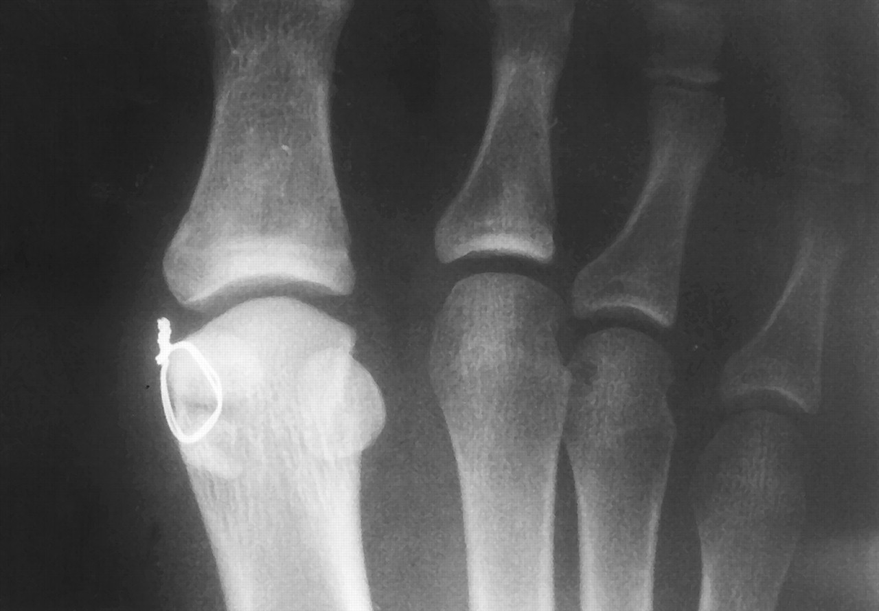
At 1 week after surgery, the radiograph demonstrated fair alignment of the fracture fragments with a lucent region at the lateral aspect of the sesamoid (
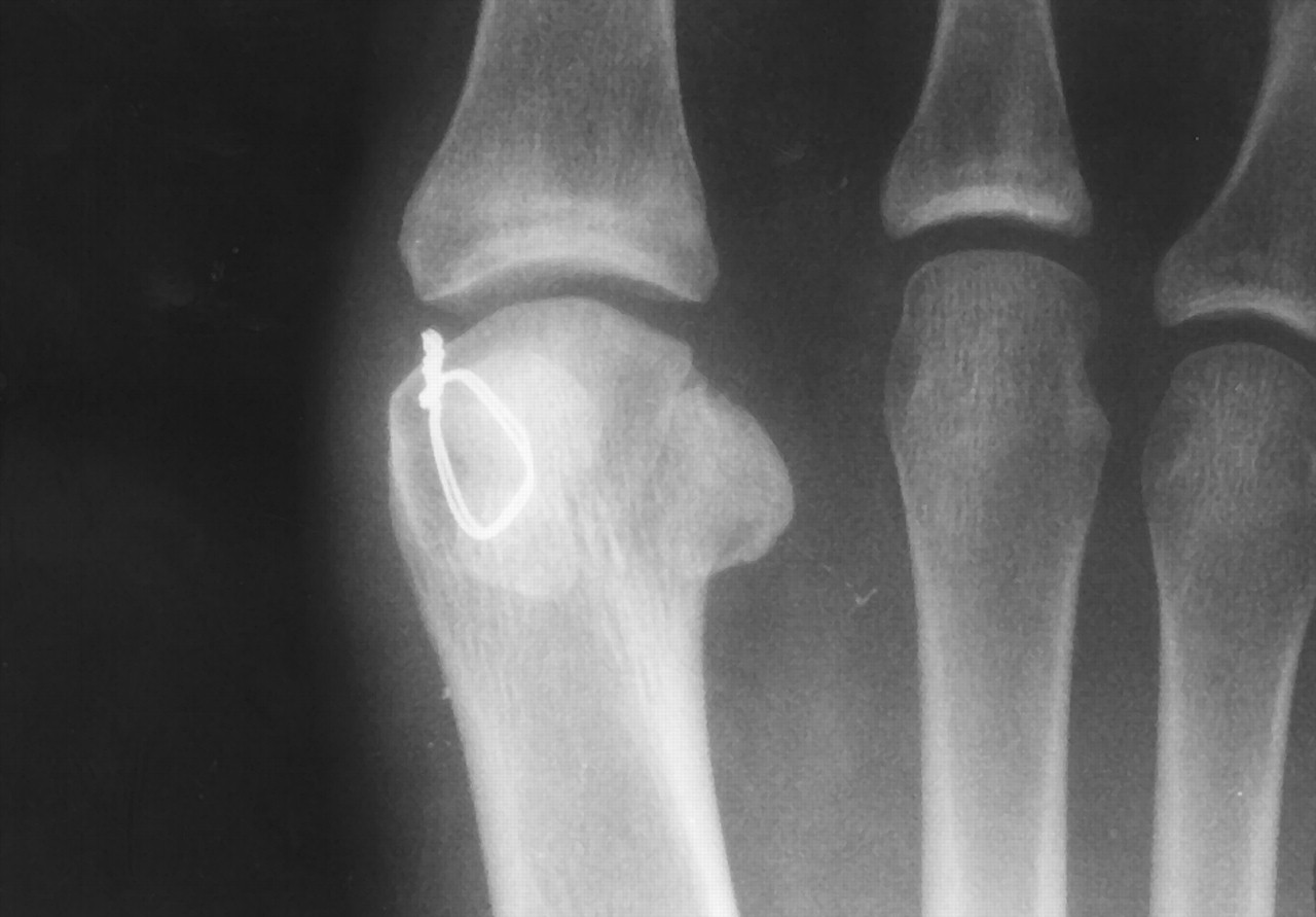
Fig. 6). At 6 weeks postoperative, consolidation of the fracture fragments was evident from trabeculation across the fracture site. At 14 weeks postoperative, complete fusion of the fracture site in the tibial sesamoid had occurred (
Fig. 7).
Figure 1.
A, Anteroposterior radiograph demonstrating the dislocation and 2-cm displacement of the tibial sesamoid fracture; B, lateral radiograph showing the dorsal dislocation of the first metatarsophalangeal joint with displacement of the tibial sesamoid fracture fragments.
Figure 1.
A, Anteroposterior radiograph demonstrating the dislocation and 2-cm displacement of the tibial sesamoid fracture; B, lateral radiograph showing the dorsal dislocation of the first metatarsophalangeal joint with displacement of the tibial sesamoid fracture fragments.
Figure 2.
Postreduction radiograph demonstrating reduction of the first metatarsophalangeal joint with no significant reduction of the tibial sesamoid fragments.
Figure 2.
Postreduction radiograph demonstrating reduction of the first metatarsophalangeal joint with no significant reduction of the tibial sesamoid fragments.
Figure 3.
Medial approach to the first metatarsophalangeal joint demonstrating the displaced tibial sesamoid fragments with rupture of the capsule and loss of the extensor digitorum brevis attachment to the distal fracture fragment.
Figure 3.
Medial approach to the first metatarsophalangeal joint demonstrating the displaced tibial sesamoid fragments with rupture of the capsule and loss of the extensor digitorum brevis attachment to the distal fracture fragment.
Figure 4.
Drilling of the tibial sesamoid fracture fragments with a 0.045 Kirschner wire parallel to the articular cartilage.
Figure 4.
Drilling of the tibial sesamoid fracture fragments with a 0.045 Kirschner wire parallel to the articular cartilage.
Figure 5.
Reduction of the tibial sesamoid fracture fragment with a 0.028 circlage wire with the knot oriented plantar distal medial. Extensor digitorum brevis and capsule are realigned.
Figure 5.
Reduction of the tibial sesamoid fracture fragment with a 0.028 circlage wire with the knot oriented plantar distal medial. Extensor digitorum brevis and capsule are realigned.
Figure 6.
Postoperative radiograph at 1 week demonstrating realignment of the tibial sesamoid fracture fragments with a lucent region at the lateral aspect of the tibial sesamoid.
Figure 6.
Postoperative radiograph at 1 week demonstrating realignment of the tibial sesamoid fracture fragments with a lucent region at the lateral aspect of the tibial sesamoid.
Figure 7.
Postoperative radiograph at 14 weeks demonstrating a solid union of the tibial sesamoid fracture site.
Figure 7.
Postoperative radiograph at 14 weeks demonstrating a solid union of the tibial sesamoid fracture site.
Discussion
The tibial sesamoid plays an integral role in the function of the first metatarsophalangeal joint.[
1,
3,
11] Although tibial sesamoid excision is a successful procedure, restoration of the functional anatomy with the avoidance of possible complications associated with tibial sesamoid excision is a more desirable approach. This case report demonstrates that it is possible to salvage the tibial sesamoid after an acute displaced fracture.
The use of the monofilament wire for one point of fixation across the fracture site allowed adequate stability for healing. Intraoperatively, the stability of the fixation was tested and was deemed sufficient to hold the reduction with adequate stability. If the stability had been insufficient, additional dissection with two points of fixation would have been attempted. The use of two points of fixation would have provided additional compression and stability across the fracture site. At 36 months follow-up, the patient had no complications. However, the monofilament wire may have to be removed in the future if any irritation develops.
The procedure described in this article may have significant implications for the treatment of stress fractures of the tibial sesamoid. Anderson and McBryde[
11] demonstrated that recalcitrant sesamoid fractures can be treated successfully with autogenous bone grafting. However, patients with excessive motion in the sesamoid, consistent with a fracture, were a contraindication for the grafting procedure and underwent excision of the sesamoid. Using the monofilament wire fixation technique for stabilization of the tibial sesamoid may allow these patients to receive the autogenous bone graft without undergoing excision of the tibial sesamoid.
Conclusion
A successful repair of a displaced tibial sesamoid fracture was demonstrated in this case report. The authors recommend that internal fixation should be attempted, particularly in active patients, prior to surgical excision of the tibial sesamoid. Furthermore, this procedure may be used in acute stress fractures and in conjunction with autogenous bone grafting for unstable nonunion or delayed union stress fractures.