The effort to optimize foot care for patients with diabetes led to the American Diabetes Association consensus statement on foot care, which recommended that the cutaneous pressure threshold be measured at least once a year. [
1] This has been the guideline since 1993. The goal of this recommendation is to reduce the risk of ulceration, infection, and amputation due to sensory loss that can occur through progressive neuropathy. Although screening for loss of protective sensation with the Semmes-Weinstein nylon monofilament has been increasingly reported, [
2-
11] a recent review [
12] has suggested that it is not clear which filament number is the most appropriate one to use, the 4.17, the 4.21, or the 5.07. For example, one study found a sensitivity of 84% and a specificity of 96% when the 4.21 filament was used to identify foot ulceration, [
3] while the most recent study of the 5.07 filament found that the sensitivity and specificity varied according to the number of sites tested on the bottom of the foot. [
11] This study found that with the 5.07 filament, the sensitivity and specificity varied from 40% and 98%, respectively, if just one site on the bottom of the foot was tested, to 83% and 77%, respectively, if five sites were tested. [
11] The most recent recommendations are to use the 5.07 filament. [
5-
11]
Filament numbers are the logarithm of the applied force (in tenths of a milligram) at a single spot on the skin surface, and they must be divided by the cross-sectional area of the filament to determine the applied pressure. [
13,
14] Since each filament has a different diameter, the stimulus presented to the patient differs not only in force, but also in the area of skin contacted, a situation suggesting that the psychophysical interpretation of the stimulus is not uniform. Interestingly, there are no published age-adjusted lower-extremity normative data for the Semmes-Weinstein monofilament, and there is no known relationship between abnormal Semmes-Weinstein monofilament data and neural degeneration. [
12,
15] Since there are reports of patients with foot ulceration who can still perceive the 5.07 filament, [
4] which represents 10 g of applied force or almost 100 g/mm
2 of pressure, it may be argued that measurement of the one-point static touch threshold is not the most appropriate test of sensibility to predict sensory loss in the foot. However, would the best alternative be for physicians to examine each patient with a full set of 20 filaments, with just the thinner filaments, or with a device that could continuously measure the cutaneous pressure threshold over a wide range? Such approaches might identify the patient at a stage at which novel medical treatment could reduce symptoms [
16] or in which surgical decompression of peripheral nerves might restore sensation. [
17-
20]
In 1992, an instrument that could measure the cutaneous pressure threshold for both one-point and two-point static touch was described. [
21] This computer-assisted instrument, the Pressure-Specified Sensory Device (Sensory Management Services, LLC, Baltimore, Maryland), measures the pressure required to perceive the number of metal prongs, one or two, that are touching the skin. Each metal prong is 1 mm in diameter, and its projected hemispherical contact surface area is 0.5 mm
2. The distance in millimeters between the prongs is related to the innervation density of that piece of skin. Axonal loss, whether related to neuropathy or chronic nerve compression, is correlated with abnormal (increased) two-point discrimination distance. [
15] Normative data for the lower extremity have been obtained with the Pressure-Specified Sensory Device, [
22] and this device has been used in a cross-sectional study of diabetic patients with and without foot ulceration. [
23] Regression analysis of Pressure-Specified Sensory Device data for patients with carpal and cubital tunnel syndrome demonstrated that the pressure required to distinguish one from two static points is the first parameter to become abnormal in chronic nerve compression. This occurs when the one-point static touch measurement is still normal and the distance between the two prongs is still normal. [
24]
Since the Pressure-Specified Sensory Device makes a measurement from 0.1 to 100 g/mm2, it approximates a Semmes-Weinstein monofilament set with an infinite number of filaments, all of the same cross-sectional diameter. When a test with a single filament, such as the 5.07, is done, the examiner only learns that the cutaneous pressure threshold is either above or below the value obtained when that marking number is converted into a pressure (g/mm2). A useful range can be obtained by determining the filament that defines the lower limit of stimulus perception when a finite set of filaments (set of five) is used, but it is still not a true threshold. The true threshold can be obtained only by using an infinite set of filaments, which is what the computer-assisted sensory testing approach does. The purpose of the present study is to use the Pressure-Specified Sensory Device to evaluate the relationship between one-point and two-point static touch cutaneous pressure thresholds in patients with peripheral nerve problems related either to diabetic neuropathy or chronic nerve compression, prior to the stage of ulceration.
Methods
A consecutive series of patients with lower-extremity complaints were tested with the Pressure-Specified Sensory Device from October 1999 to March 2000. Each test was performed by the same examiner (J.C.) and included measurement of the medial plantar nerve, medial calcaneal nerve, and the deep peroneal nerve bilaterally. The study consisted of 29 diabetic patients with symptoms of neuropathy. Since diabetic neuropathy is a bilaterally symmetrical disease, the measurements for the left foot were chosen for analysis in the study. The patients without diabetes were undergoing evaluation for tarsal tunnel syndrome. There were 35 patients in this category, 12 of whom had bilateral disease. Therefore, 47 feet were included for evaluation in the nondiabetic tarsal tunnel syndrome group.
Neurosensory testing was done with the Pressure-Specified Sensory Device as previously described. [
15,
21] In brief, this device employs two prongs attached by a force transducer to a hand-held instrument. Increasing force is applied to the surface of the skin that is being tested. Either one or both of the prongs can be applied, thus generating one- or two-point static touch. The one-point static touch stimulus is recorded in units of pressure (g/mm
2). When the Pressure-Specified Sensory Device records the pressure at which a one-point stimulus is distinguished from a two-point stimulus, the observation is recorded in terms of both pressure (g/mm
2) and distance (mm). When patients perceive the stimulus, they push a button with their hand, thereby creating a signal to the computer to stop recording increases in force. The recorded force is divided by the cross-sectional area of the prong to give the value in units of pressure, g/mm
2.
The relationship of the Semmes-Weinstein monofilament to the cutaneous pressure thresholds measured with the Pressure-Specified Sensory Device was mathematically obtained by reference to published calculations of the pressure applied by filaments of known diameter. [
13]
Comparison of the one-point with the two-point static touch measurements for the big toe (medial plantar), heel (medial calcaneal), and dorsum of the foot (deep peroneal) nerve were done for the pressure threshold measurements of both groups of patients. The range of pressure threshold measurements for the one-point static touch was 0.1 to 100 g/mm
2. For two-point static touch, the range was subdivided into patients with normal and those with abnormal innervation density. Normal innervation density is age related; at 45 years of age or younger, the 99% confidence limit for static two-point discrimination was 6 mm, and at ages older than 45 years, it was 8 mm. [
22] For the purposes of calculation of sensitivity and specificity, true disease was defined as the presence of axonal loss. Axonal loss exists if the two-point discrimination distance is greater than the 99% confidence limit for age. This is consistent with the lowest value at which a diabetic patient had a foot ulceration in a cross-sectional study of diabetic patients with and without foot ulceration: two-point static touch of 32.9 g/mm
2 at a distance of 9 mm. [
23] Therefore, absence of disease was defined as no axonal loss or normal innervation density. The two-point static touch pressure threshold measurement in the absence of disease was 0.1 to 100 g/mm
2. Presence of disease, or axonal loss, was obtained by adding 100 to the measured pressure threshold whenever axonal loss was present as determined by a two-point discrimination distance greater than 6 mm for age younger than 45 years and greater than 8 mm for age older than 45 years. This yields a range of 100.01 to 200 g/mm
2 for two-point static touch, creating a break point for patients with or without “true disease” at 100 g/mm
2. Analysis of the relationship between one-point and two-point static touch was done for each nerve in each group by means of the Pearson product-moment correlation coefficient with SigmaStat software, version 2.03 (SPSS Science, Chicago, Illinois).
For the purposes of this article, it is recognized that values do not currently exist for the nylon monofilaments that would permit the use of the 99% confidence limit as the upper limit of normal for the foot.
Results
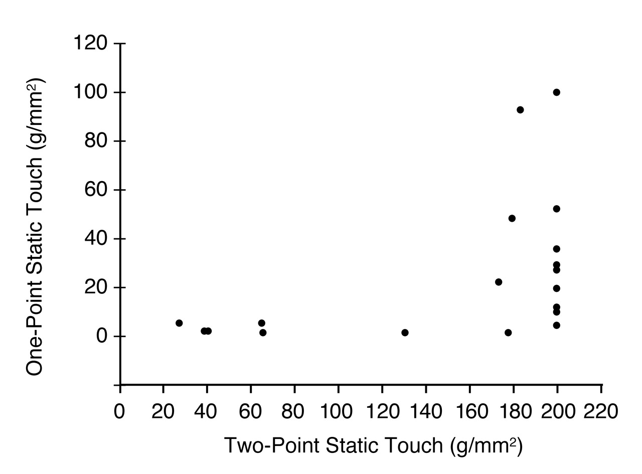
There was a poor correlation between one-point and two-point static touch measurements in patients with symptoms of diabetic neuropathy. This is illustrated for the medial plantar nerve in
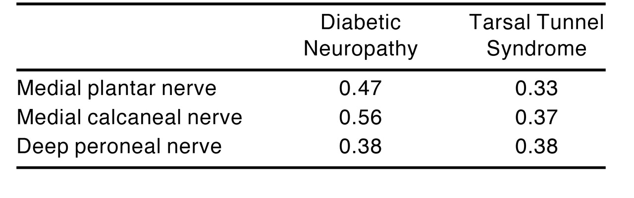
Figure 1; the correlation coefficients for all nerves tested are listed in
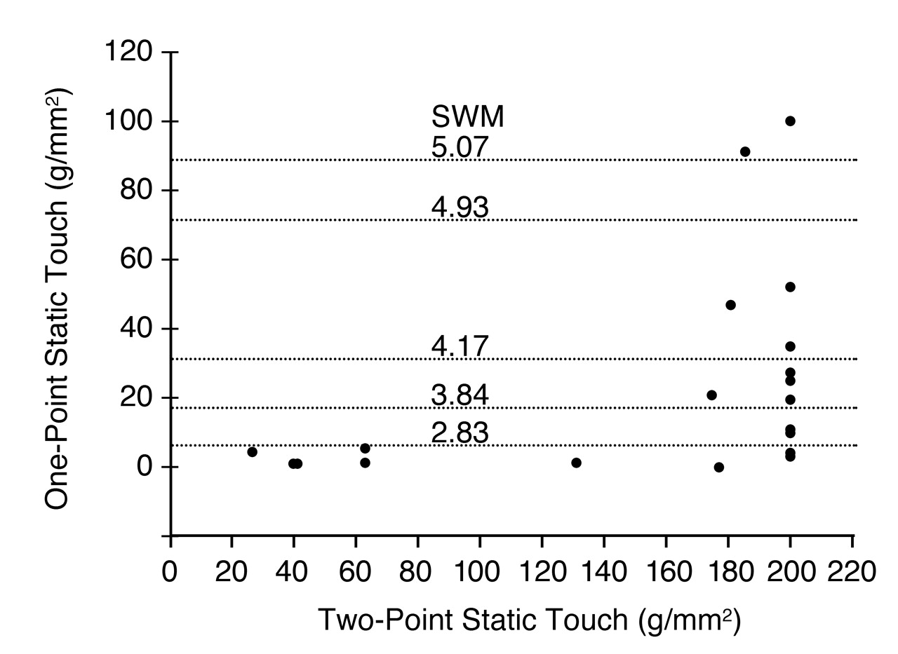
Table 1. The progressive increase in abnormal measurements found with two-point static touch was in contrast to the one-point static touch, which did not become abnormal until measurements with the two-point static touch demonstrated axonal loss. This finding was similar for measurements made over the heel and the dorsum of the foot. In patients with tarsal tunnel syndrome, there was also a poor correlation between one-point and two-point static touch measurements. Only in the most advanced cases of tarsal tunnel syndrome, with complete loss of two-point discrimination, did the one-point static touch measurement become abnormal. This is illustrated for the medial plantar nerve in this group of patients in
Figure 2. The correlation coefficients are given in
Table 1.
Figure 1. .
Relationship of the cutaneous pressure thresholds for one- and two-point static touch over the plantar aspect of the big toe (medial plantar nerve) in patients with symptomatic diabetic neuropathy who had no ulcerations. Measurements above the 100 g/mm2 level along the two-point static touch axis indicated abnormal two-point discrimination and axonal loss. Note that the one-point static touch threshold did not become elevated until an advanced level of axonal degeneration, as determined by the two-point static touch threshold. The correlation coefficient for this relationship is 0.47.
Figure 1. .
Relationship of the cutaneous pressure thresholds for one- and two-point static touch over the plantar aspect of the big toe (medial plantar nerve) in patients with symptomatic diabetic neuropathy who had no ulcerations. Measurements above the 100 g/mm2 level along the two-point static touch axis indicated abnormal two-point discrimination and axonal loss. Note that the one-point static touch threshold did not become elevated until an advanced level of axonal degeneration, as determined by the two-point static touch threshold. The correlation coefficient for this relationship is 0.47.
Figure 2.
Relationship of the cutaneous pressure thresholds for one-point and two-point static touch over the plantar aspect of the big toe (medial plantar nerve) in patients with tarsal tunnel syndrome. Measurements above the 100 g/mm2 level along the two-point static touch axis indicate abnormal two-point discrimination and axonal loss. Note that the one-point static touch threshold does not become elevated until a very advanced level of axonal degeneration, as determined by the two-point static touch threshold. The correlation coefficient for this relationship is 0.33. Abbreviation: SWM, Semmes-Weinstein monofilament.
Figure 2.
Relationship of the cutaneous pressure thresholds for one-point and two-point static touch over the plantar aspect of the big toe (medial plantar nerve) in patients with tarsal tunnel syndrome. Measurements above the 100 g/mm2 level along the two-point static touch axis indicate abnormal two-point discrimination and axonal loss. Note that the one-point static touch threshold does not become elevated until a very advanced level of axonal degeneration, as determined by the two-point static touch threshold. The correlation coefficient for this relationship is 0.33. Abbreviation: SWM, Semmes-Weinstein monofilament.
Table 1.
Pearson Correlation Coefficients for One-Point and Two-Point Static Touch Cutaneous Pressure Thresholds in the Foot.
Table 1.
Pearson Correlation Coefficients for One-Point and Two-Point Static Touch Cutaneous Pressure Thresholds in the Foot.
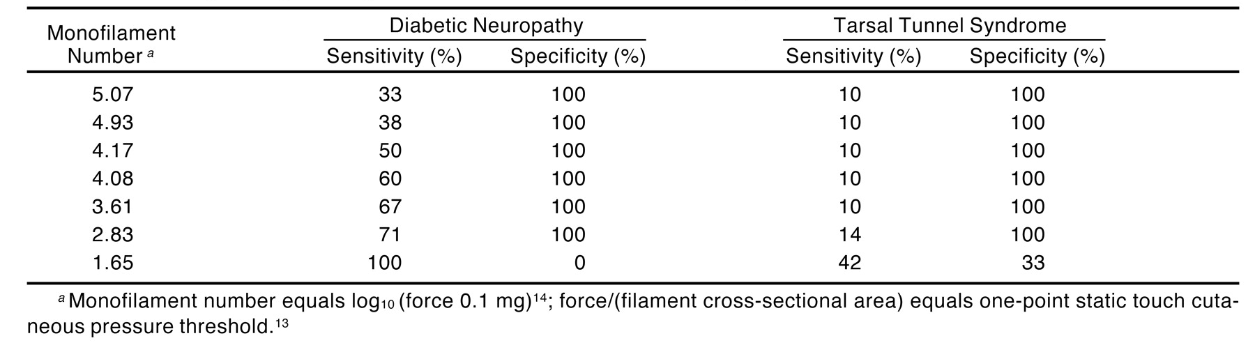
The sensitivity and specificity results are listed in
Table 2. There was relatively poor sensitivity when the cutoff for identification of disease was taken as the 5.07 monofilament equivalent: sensitivity was 30% for patients with diabetic neuropathy and 10% for those with tarsal tunnel syndrome. In
Figure 3, when the calculations are done for monofilaments with what would be lower numerical values (theoretically thinner filaments), the sensitivity does increase, but does not exceed 60% in diabetic patients with neuropathy or 10% in patients with tarsal tunnel syndrome until the one-point static touch threshold approaches what is probably at or below the normal threshold for the foot (the 3.61 filament, which might not be perceived by the normal foot). When the filaments that could probably be perceived by the normal foot are used for the calculations, the specificity dropped to 0% for the diabetic patients and 33% for the tarsal tunnel syndrome patients.
Table 2.
Sensitivity and Specificity of One-Point Static Touch to Identify Axonal Loss in Absence of Ulceration.
Table 2.
Sensitivity and Specificity of One-Point Static Touch to Identify Axonal Loss in Absence of Ulceration.
Figure 3.
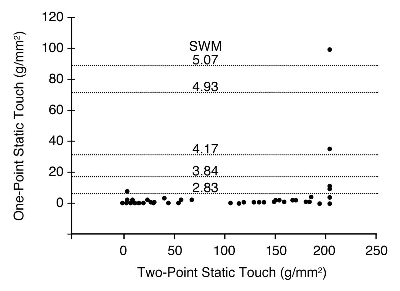
The same data set as in
Figure 1 but with horizontal lines drawn at levels representing markings of the Semmes-Weinstein monofilament set. Note that regardless of how low a filament number was chosen, there were still patients who could perceive this filament’s force of application but who already had axonal loss as determined by abnormal two-point discrimination, a location along the x-axis that is greater than 100 g/mm
2. For the 4.93 monofilament, the sensitivity for identifying axonal loss on this graph would be 38%, with a specificity of 100%. Abbreviation: SWM, Semmes-Weinstein monofilament.
Figure 3.
The same data set as in
Figure 1 but with horizontal lines drawn at levels representing markings of the Semmes-Weinstein monofilament set. Note that regardless of how low a filament number was chosen, there were still patients who could perceive this filament’s force of application but who already had axonal loss as determined by abnormal two-point discrimination, a location along the x-axis that is greater than 100 g/mm
2. For the 4.93 monofilament, the sensitivity for identifying axonal loss on this graph would be 38%, with a specificity of 100%. Abbreviation: SWM, Semmes-Weinstein monofilament.
Discussion
As large-fiber neuropathy or chronic nerve compression progresses, there is an increase in the two-point static touch threshold while the distance for that two-point discrimination remains normal. Because the monofilaments test only one-point perception, it is reasonable that this study demonstrated only weak correlation between one-point and two-point static touch for both diabetic neuropathy and tarsal tunnel syndrome for each of the three foot skin surfaces tested (
Table 1).
As in most diseases, if patients are identified earlier in the course of their disease, treatment of the complications of diabetic neuropathy can improve. If patients with the disease are to receive treatment prior to foot ulceration, sensory measurement techniques must become more sensitive. When screening has been done to identify foot ulceration, the use of monofilaments has been demonstrated to be efficacious in discriminating between groups of diabetic patients with and without foot ulceration, [
2-
8,
10] and monofilaments are therefore widely recommended. [
1,
9] While biomechanical and vascular problems are clearly risk factors for foot ulceration, it must be recognized that ulceration due to loss of protective sensation is the end result of neuropathy and represents severe large- and small-fiber axonal loss. When screening for this end result of neuropathy, it has not been relevant that there are no normative data for these filaments, or that recent studies demonstrate inherent technical problems with the nylon filaments themselves. [
11,
25] For example, in a group of 10 new 5.07 filaments, the force of application varied from 9.4 g to 10.4 g, and after 100 uses, the mean decrease in applied force was 1.2 g. [
25]
It may be asked whether thinner monofilaments, those that would apply less force and therefore, theoretically, be able to identify a peripheral nerve problem earlier, might be of value in screening for diabetic neuropathy or nerve compression in the foot. The present study used an experimental design that substituted the single metal prong of the Pressure-Specified Sensory Device for the multitude of cross-sectional diameters present in the traditional set of 20 nylon monofilaments. This single-diameter hemispherical metal prong has been previously demonstrated as avoiding the psychophysical problem presented by the set of filaments, which requires the subject to discriminate both a change in force and a change in surface area tested from the same stimulus. [
26] The Pressure-Specified Sensory Device is able to measure the continuous spectrum of thresholds from 0.1 to 100 g/mm
2. These one-point static touch values can be chosen for analysis of sensitivity and specificity to investigate whether lower one-point static touch values can identify stages of axonal loss prior to the presence of foot ulceration. The results of the present study demonstrate that lower values of one-point static touch, which is what would be evaluated with thinner Semmes-Weinstein monofilaments, are relatively insensitive to changes in sensation in the foot at stages where there is axonal loss, as measured by abnormal two-point discrimination, until the final stage at which there is complete loss of two-point discrimination. At this point, the lower one-point static touch values, which mathematically are similar to the 3.61, 2.83, and 1.65 markings of the Semmes-Weinstein monofilament, might be subthreshold (not perceivable) for the foot (
Figure 3;
Table 2). It is not known whether the 3.61 nylon monofilament would be within the 99% confidence limit for perception in the normal foot. Clearly, if these very thin nylon monofilaments cannot be perceived by the normal foot, they could not be used in clinical practice to identify early stages of axonal loss in diabetic neuropathy or chronic nerve compression.
In order to understand the results of this study, which suggest that thinner monofilaments would not be able to identify the early stage of diabetic neuropathy or of chronic nerve compression in tarsal tunnel syndrome, it is instructive to understand why nylon monofilaments were introduced in the late 1950s. Semmes, Weinstein, and their colleagues, clinical psychologists at Bellevue Hospital Center at New York University, New York, New York, were interested in identifying which part of the brain had to be injured to cause a loss of stereognosis. [
27] They used monofilaments because their wide range of force application enabled them to investigate “gross sensory loss” in the hand. The wide range of filament diameters and the force of application required them to use logarithmic values. Hands rarely become ulcerated, but loss of stereognosis for the hand may be the equivalent of advanced sensory loss in the foot sufficient to permit ulceration. When a test is necessary, however, to identify a medical problem early, the testing instrument must be able to identify changes throughout the course of the disease, have a normative database, and show a clear distinction between normality and disease. Measurement of the two-point static touch with the Pressure-Specified Sensory Device has demonstrated this for chronic nerve compression in the hand, [
24] and the normative database exists for the lower extremity. [
22]
If neurosensory testing is used to identify patients when their diabetic neuropathy is most amenable to diabetes education, or to the use of special footwear, or when the patient is a candidate for restoring sensation to the foot, [
17-
20] then measurement techniques that are more sensitive than monofilaments to earlier stages of peripheral nerve problems must be used. In a recent review, neurosensory testing with the Pressure-Specified Sensory Device has been demonstrated as able to identify both neural degeneration and neural regeneration for both the upper and lower extremity. [
28] The present study extends the understanding of the pathophysiology of lower-extremity peripheral nerve problems by identifying the pressure that two-point static touch, with normal two-point discrimination, will reach before the distance at which one point can no longer be distinguished from two points exceeds the 99% confidence limit. This point is noted in
Figure 1 for diabetic neuropathy and in
Figure 2 for tarsal tunnel syndrome at about 70 g/mm
2 on the x-axis. Measurement of the two-point static touch thresholds with the Pressure-Specified Sensory Device, therefore, enables identification of the entire spectrum of pathophysiology from early threshold change in the presence of normal two-point discrimination through the stage of loss of two-point discrimination with preservation of protective sensation. Throughout this range, one that precedes foot ulceration, the one-point static touch threshold remains essentially unchanged and is unable to describe these clinically significant changes.
Conclusion
One-point static touch threshold testing correlates poorly with measurement of two-point static touch threshold in the foot of diabetic patients with neuropathy and nondiabetic patients with tarsal tunnel syndrome. One-point static touch testing has a high percentage of false-negative results in identifying patients with axonal loss, a stage prior to that of foot ulceration. This early stage can be identified with the Pressure-Specified Sensory Device.