Clear cell sarcoma was originally described by Enzinger [
1] in 1965 as a slow-growing malignant tumor of the tendons and the aponeuroses. It later became apparent that the tumor was capable of producing melanin, giving rise to the name “malignant melanoma of soft parts.” However, clear cell sarcoma is different from classic malignant melanoma in several respects. Clear cell sarcoma is typically confined to deep softtissue planes, is associated with tendons and aponeuroses, and does not have epidermal or epidermal-dermal junctional changes; these factors are in sharp contrast to the superficial involvement of malignant melanoma. On histochemical analysis, clear cell sarcoma shows a pale-staining, homogeneous growth pattern with spindle-shaped cells arranged in an organoid or alveolar architecture. Classic clear cell sarcoma usually does not appear in bone; only a few cases of this tumor with osseous involvement have been documented in the literature. One case reported by Raynor et al [
2] in 1979 describes the complete destruction of the distal phalanx of the thumb. In another case, reported by Morishita et al [
3] in 1987, a tumor is reported arising from the juxtacortical region of the proximal tibia that secondarily invaded bone. Yokoyama et al [
4] in 1996 claim to have reported the first case of primary clear cell sarcoma of bone in a 33-year-old woman.
Clinical Presentation, Diagnosis, and Treatment
Clear cell sarcoma occurs most commonly in adults between the ages of 20 and 40. The original study of 21 cases by Enzinger showed a predilection for females. Other accounts, however, have not confirmed a sex-related prevalence [
3,
5,
6]. Clinically, the tumor presents as a slow-growing mass that is firm and mobile. It appears most often in the extremities, notably the ankle and foot.
When bone is involved, routine radiographs show a nonspecific, well-demarcated osteolytic lesion that is usually confined to the medullary cavity. On gross examination, clear cell sarcoma appears as a wellcircumscribed, unencapsulated solid, often lobulated, mass associated with a nearby tendon or muscle aponeurosis. There is usually no involvement of the skin. The size varies, but most tumors are between 2 and 6 cm in diameter.
Immunohistochemical analysis demonstrates the presence of S-100 protein antigens, reflecting the ability of the tumor cell to produce melanin. Immunoreactivity for S-100 protein and focal reactivity for HMB-45 are considered diagnostic features of clear cell sarcoma [
7].
Fibrosarcoma can be confused with clear cell sarcoma even though it has a dissimilar cellular structure and is not immunoreactive to the S-100 protein, which is a distinct characteristic in most cases of clear cell sarcoma. Unlike clear cell sarcoma, fibrosarcoma usually has osseous involvement. Malignant melanoma is a dermal lesion that may extend to the subcutaneous level, but except for metastasis, it is not as deep-seated as clear cell sarcoma. Clear cell sarcoma involves the deeper tissue and rarely involves the skin. Moreover, clear cell sarcoma does not cause hyperpigmentation of the overlying skin, a characteristic that is usually the initial observation in malignant melanoma.
Various modes of therapy have been used in the treatment of clear cell sarcoma, including radiation, chemotherapy, arterial limb perfusion, perilesional infusion of interferon alfa-2b, wide excision, amputation, and a combination of these treatments. Chemotherapy alone is of little use. However, bleomycin and vincristine, combined with radiation, have resulted in apparent cures [
8]. The use of intra-arterial chemotherapy has had some success in the treatment of soft-tissue sarcomas in the extremities. However, this treatment has a high risk of toxicity and its value in treating clear cell sarcoma is not well documented [
9,
10,
11].
Wide excision or amputation is the treatment of choice for clear cell sarcoma, according to the current literature [
12,
13]. If there is lymph node metastasis, treatment should also include resection of affected nodes. There is still a high rate of recurrence and/or metastasis of clear cell sarcoma after noninvasive treatment. Typically, the tumor will recur locally approximately 2.5 years after treatment is initiated and will metastasize after 3 to 4 years [
14,
15]. Metastasis, however, has been seen up to 30 years after the initial diagnosis [
14,
15].
Case Report
A 62-year-old woman presented with pain on the dorsum of her right foot. She recalled having her right foot stepped on 2.5 months prior to presentation with resultant pain, swelling, and ecchymosis. Prior to this, she walked 10 miles a week for exercise but was forced to stop this activity after the injury. After 3 weeks, the pain subsided and she resumed her normal activities. Six weeks after the injury, she again developed pain that was constant, aggravated with ambulation, and localized to the dorsomedial aspect of her right foot.
Her medical history was significant for multiple lesions of basal cell carcinoma, which were treated by surgical excision. In 1991, she underwent a limited mastectomy for stage I adenocarcinoma of her left breast. She received postoperative radiation and tamoxifen therapy for 3 years. There was no family history of cancer.
Physical examination revealed tenderness over the area of the medial cuneiform and first metatarsal base of the right foot. There were no palpable soft-tissue masses and there was no skin discoloration. The neurovascular status of the lower extremity was intact, although some edema was noted with dependency (positioning of the foot below the rest of the body). The patient could ambulate normally without apparent discomfort.
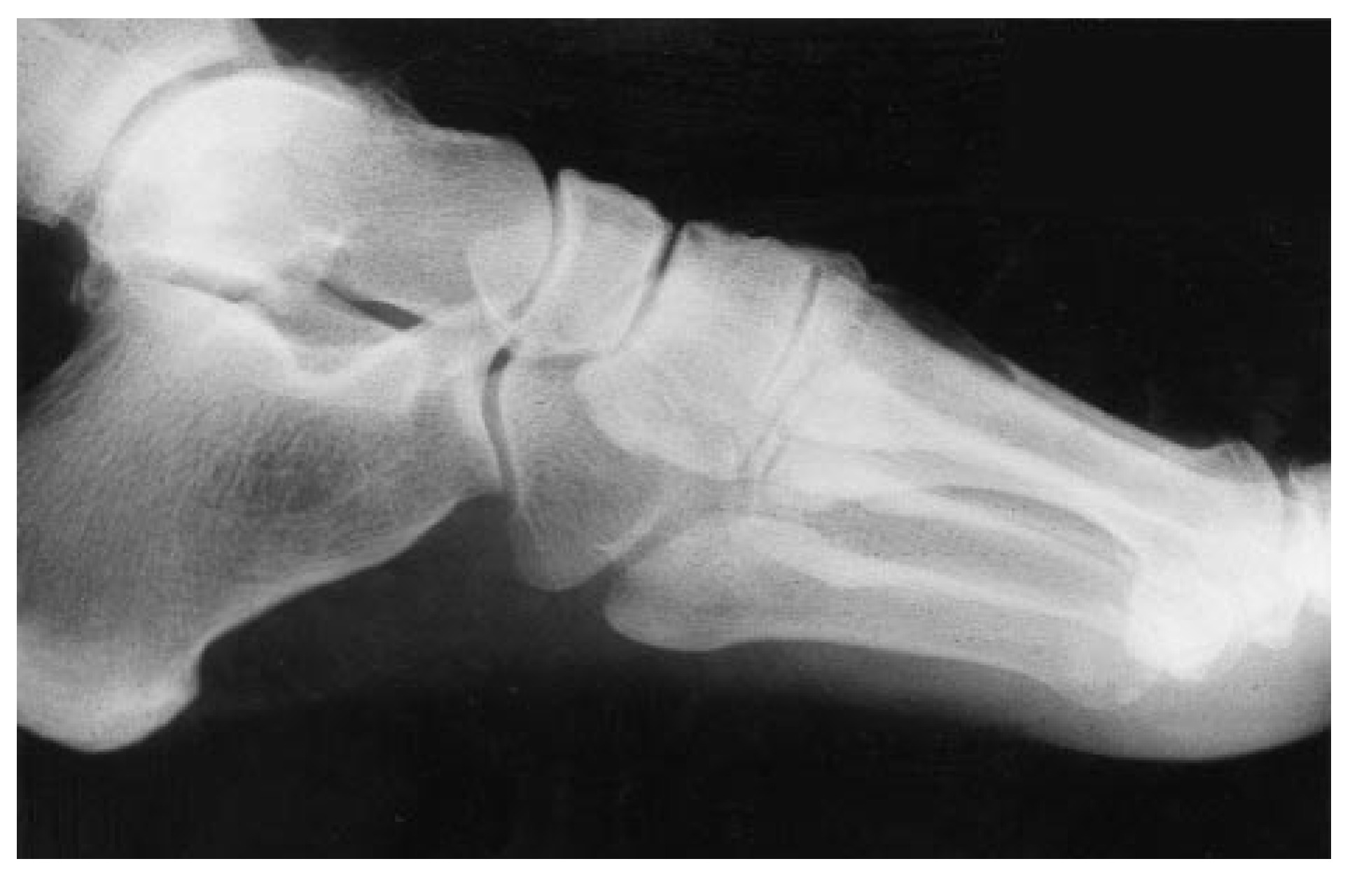
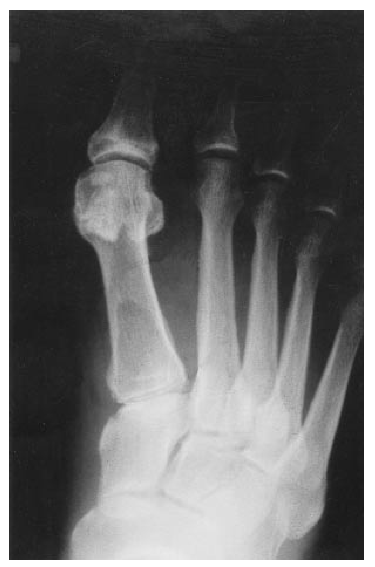
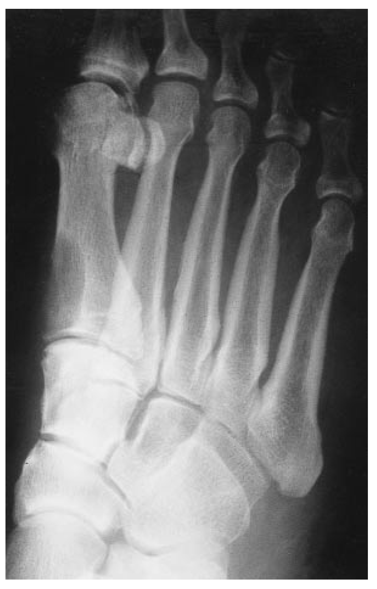
Radiographic examination revealed a well-circumscribed, homogeneous, ovoid-shaped, lytic lesion in the dorsoproximal aspect of the first metatarsal measuring 1 × 2 cm (
Figure 1,
Figure 2 and
Figure 3). The lesion appeared to be confined to the medullary cavity and had a thin but intact dorsomedial cortex.
A technetium-99 bone scan revealed intense focal uptake at the base of the first metatarsal in the delayed images, which corresponded to the lytic lesion on plain films. A fluoroscopically guided needle biopsy of the lesion was performed but was nondiagnostic. The specimen contained fragments of bony tissue; one minuscule fragment appeared to contain some associated spindle cells, which were considered to be artifacts. An open excisional biopsy with bone grafting was recommended, as it was believed that the lesion was a benign neoplasm.
Figure 1.
Dorsoplantar radiograph showing radiolucent lesion at first metatarsal base.
Figure 1.
Dorsoplantar radiograph showing radiolucent lesion at first metatarsal base.
Figure 2.
Lateral radiograph shows well-demarcated boundaries on the lesion.
Figure 2.
Lateral radiograph shows well-demarcated boundaries on the lesion.
Figure 3.
Medial oblique radiograph shows dorsomedial cortical thinning.
Figure 3.
Medial oblique radiograph shows dorsomedial cortical thinning.
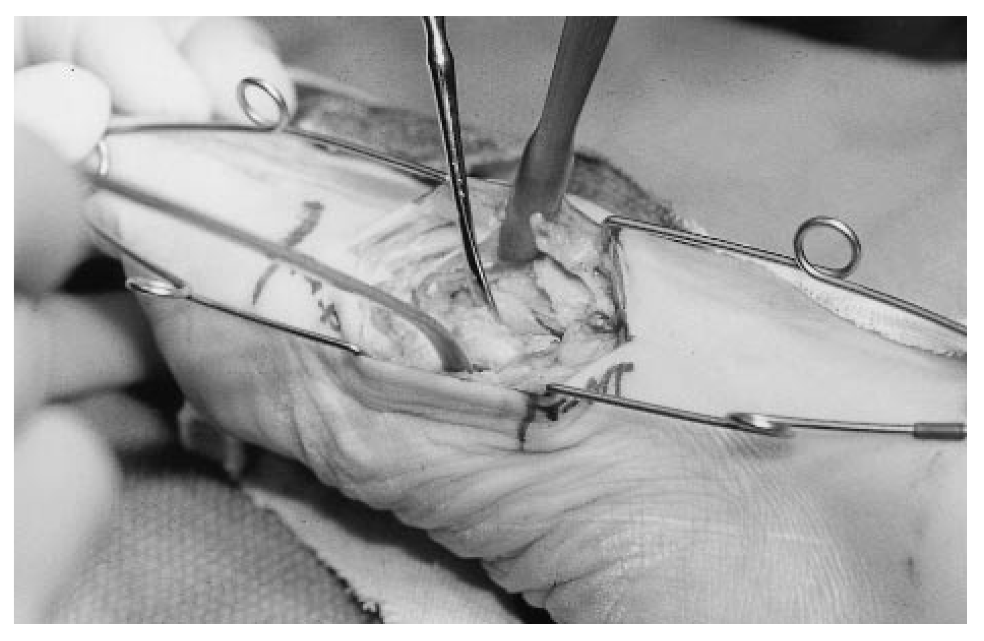
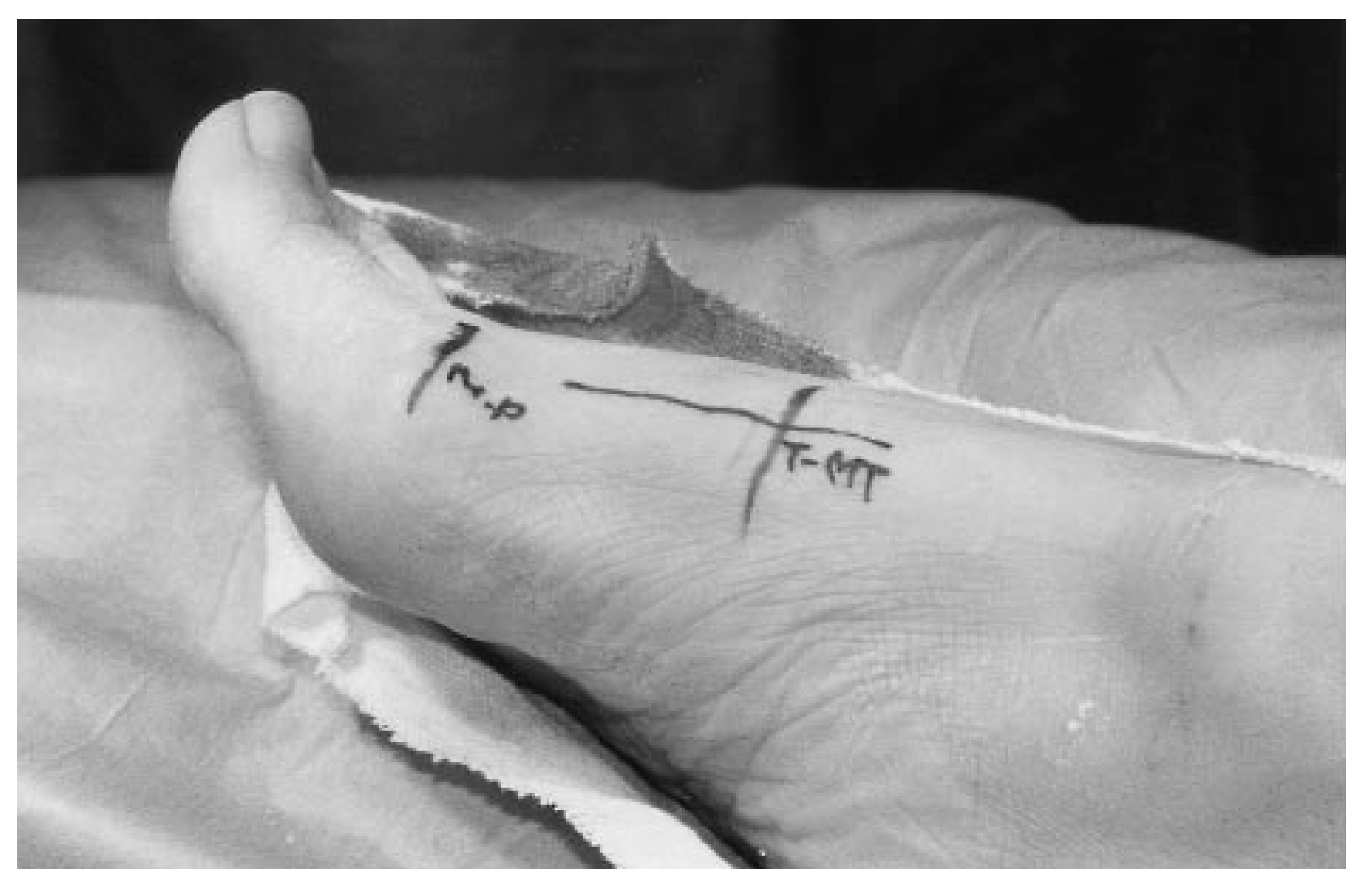
The patient consented to having an excisional biopsy. The dorsal cortex of the proximal first metatarsal was seen through a 4.5-cm linear incision (
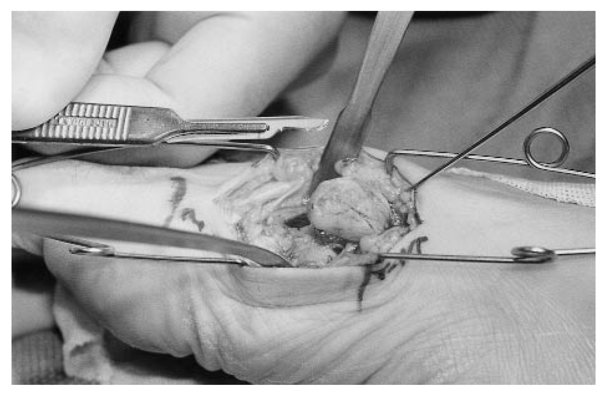
Figure 4). A thin dorsal cortex, seemingly covered only with periosteum, was seen dorsomedially. Exploration revealed a firm, grayish-white, intracortical mass (
Figure 5) that was enucleated without difficulty using a small retractor (
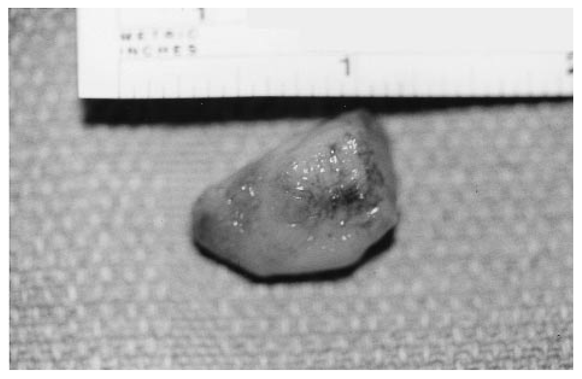
Figure 6). The homogeneous mass was firm, had a rubbery consistency, and measured 2.5 × 1.5 × 1 cm (
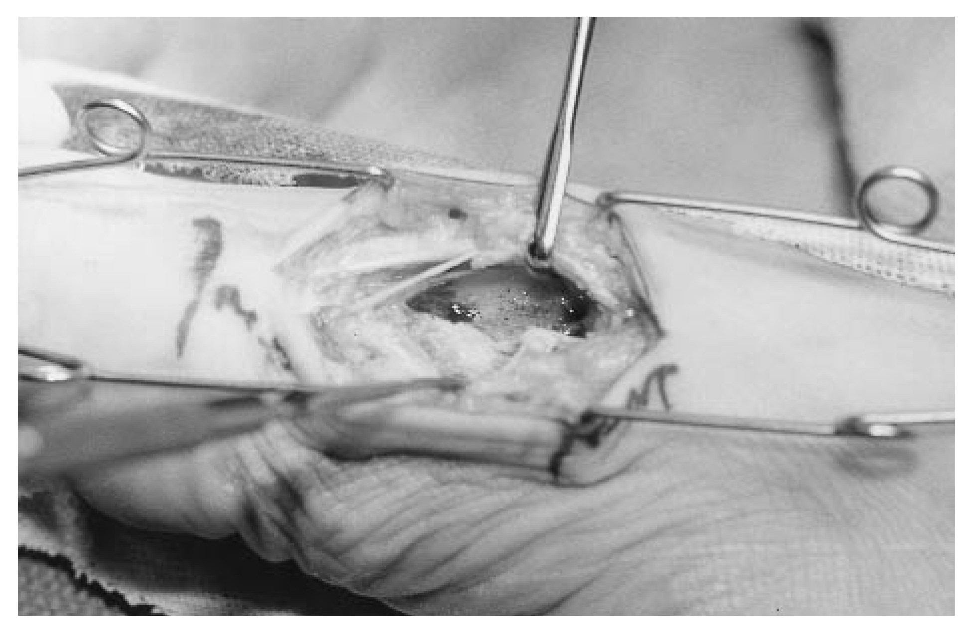
Figure 7). The specimen was sent to the pathology department for a frozen section, but no definitive diagnosis could be made. The defect was explored and the surrounding bone appeared intact (
Figure 8). A bone graft was not used and the incision was closed in layers.
Figure 4.
Incisional approach for biopsy.
Figure 4.
Incisional approach for biopsy.
Figure 5.
The dorsal surface of the tumor exposed at end of Freer elevator.
Figure 5.
The dorsal surface of the tumor exposed at end of Freer elevator.
Figure 6.
Intraoperative photograph shows the tumor being shelled out with a retractor.
Figure 6.
Intraoperative photograph shows the tumor being shelled out with a retractor.
Figure 7.
Gross appearance of the extracted tumor.
Figure 7.
Gross appearance of the extracted tumor.
Figure 8.
The defect in the first metatarsal base after removal of the tumor.
Figure 8.
The defect in the first metatarsal base after removal of the tumor.
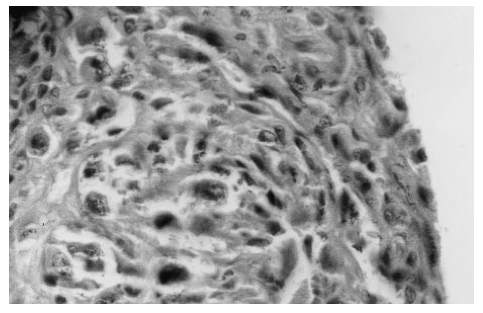
The report from the hospital pathologist described a cellular tumor with hyperchromatic, spindle-shaped cells of various size and areas of organoid or alveolar architecture, separated by hyaline intercellular stroma (
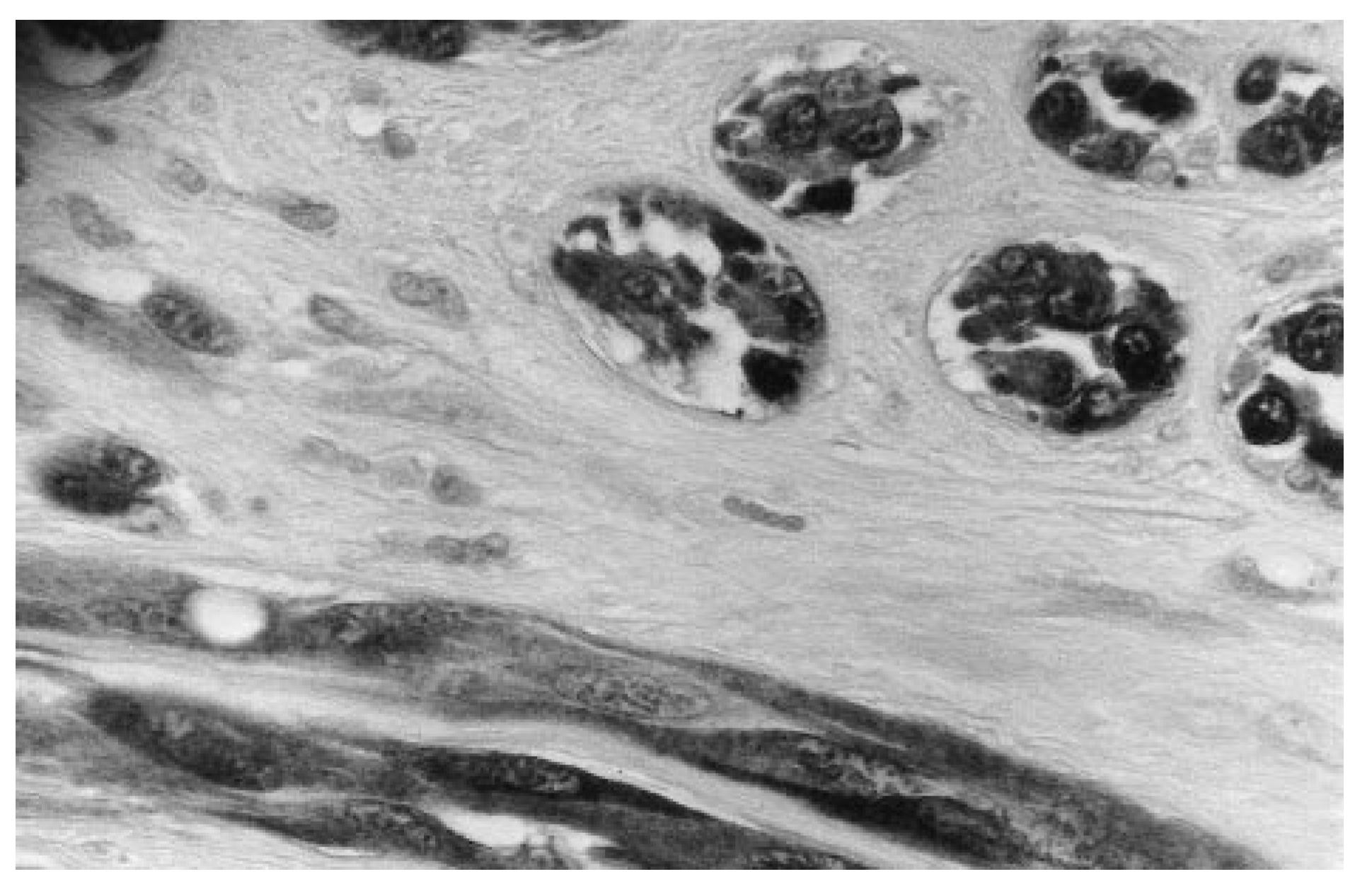
Figure 9). Immunohistochemical staining was positive for vimentin and negative for cytokeratins (AE1/AE2). The preliminary conclusion of the pathologist was probable malignant neoplasm of mesenchymal origin. The specimen was sent to the Mayo Clinic in Rochester, Minnesota, for further evaluation. The specimen stained positive for S-100 protein (
Figure 10) and negative for HMB-45. This result, along with the microscopic findings, supported a diagnosis of clear cell sarcoma.
Figure 9.
Photomicrograph of tissue showing spindle-shaped, hyperchromatic cells (H&E, ×400).
Figure 9.
Photomicrograph of tissue showing spindle-shaped, hyperchromatic cells (H&E, ×400).
Figure 10.
Photomicrograph of tissue with immunohistochemical stain, which was found positive for S-100 protein (×200).
Figure 10.
Photomicrograph of tissue with immunohistochemical stain, which was found positive for S-100 protein (×200).
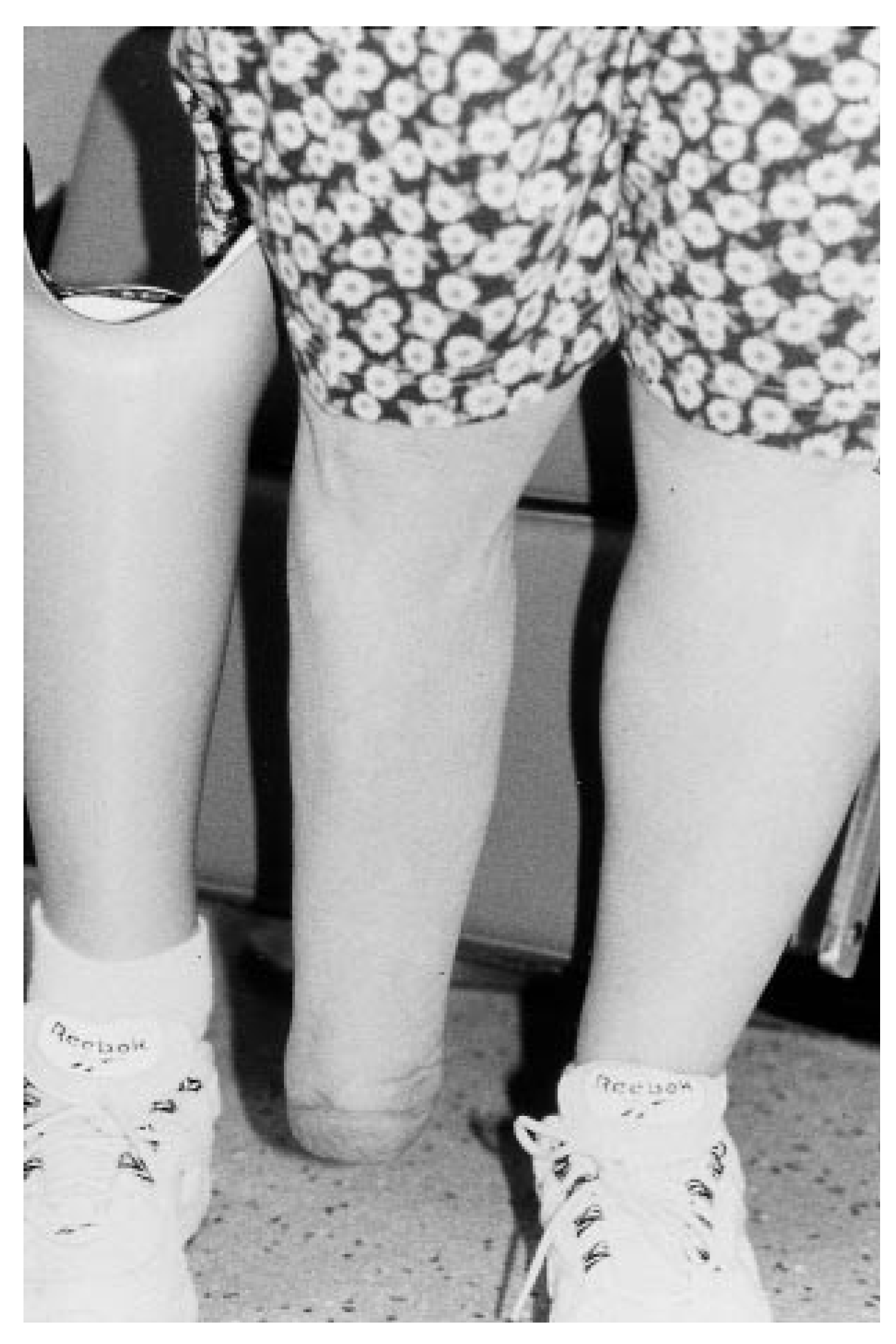
After the definitive diagnosis was made, the patient was referred to an orthopedic oncologist, who recommended a Syme’s amputation, which was performed. Postoperatively, amputation specimens from the original surgical site and the proximal margin were analyzed. The pathology report described focal infiltration with residual islands of clear cell sarcoma in the area of the excisional biopsy site of the first metatarsal. The proximal margin of the amputation site was free of tumor cell infiltration.
The patient’s postoperative recovery was uneventful and the amputation stump healed successfully. She was later fitted with a prosthesis. The patient was last seen 15 months postoperatively and there was no detectable tumor on radiograph (
Figure 11).
Figure 11.
The healed stump following Syme’s amputation, and prosthesis, 15 months postoperatively.
Figure 11.
The healed stump following Syme’s amputation, and prosthesis, 15 months postoperatively.