In December 1965, a US C-123 cargo airplane carrying Army of the Republic of Viet Nam (ARVN) troops crashed into a mountainside in Tuy Hoa, South Vietnam. Although no official flight manifest exists, 4 US crewmen and 81 male ARVN members were reportedly on board the aircraft. Enemy fire on the ground and adverse weather conditions precluded search and-rescue efforts in the area of the crash. In June 1974, a Vietnamese team recovered the commingled remains from the crash site and turned them over to US officials in Vietnam. Owing to the presence of unexploded ordnance, the search team did not enter the plane’s fuselage. As a result, the recovered remains were limited to individuals who were thrown clear of the aircraft. The remains were received at the US Army Central Identification Laboratory, Hawaii (CILHI), in May 1976 and the four American crew members were identified and laid to rest on US soil. The remaining skeletal and dental elements were held at the CILHI pending political developments between the United States and Vietnam.
The CILHI, located on Hickam Air Force Base and adjacent to Pearl Harbor, is unique in two respects:
First, it has the largest staff of forensic anthropologists (in addition to two forensic dentists) in the world. Second, it is the only laboratory of its type with responsibility for finding, recovering, and identifying the more than 90,000 US soldiers and civilians listed as missing in action (MIA) from all past wars. In this capacity, the CILHI deploys 12-person recovery teams worldwide to such diverse places as China, Japan, New Guinea, North Korea, Vietnam, and the Yuri Islands. These teams use standard archaeological techniques to recover the remains of American MIAs from aircraft crash sites and graves.[
1,
2,
3] Unfortunately, time and environmental variables, including acidic soils, bacteria, and roots that invade and split bones and teeth, reduce the normal adult skeleton of 206 bones to tiny fragments and, ultimately, dust. The present case provides scientists with anatomic information on perimortem (ie, occurring at the time of death) trauma to the calcaneus and talus sustained by at least 37 men who died in a single air crash in Vietnam.
Materials and Methods
Dry-bone specimens representing a minimum of 37 individuals were grossly examined by anthropologists at the CILHI. Talar fractures were classified, in a system modified from that of Wechsler et al,[
4] according to their location in the head, neck, dome, and posterior facet. Calcaneal fractures were classified, in a system modified from that of Sneppen and colleagues,[
5] according to their location in the calcaneocuboid facet; in the anterior, medial, and posterior facets; and through the posterior half of the body (resulting in anterior and posterior halves). Some tali and calcanei had multiple fractures and were scored accordingly (
Table 1). The bones were examined with the unaided eye and fragments were reassociated and glued together when possible. Because the majority of the skeletal elements could not be reliably articulated, each talus and calcaneus was treated and examined independently of all others. The bones were examined for perimortem fractures and postmortem breakage due to infiltration by roots and erosion by soil. Bones too fragmented or severely altered by soil and root invasion were excluded from study. The pattern of perimortem fractures was noted on skeletal diagrams. Bones exhibiting unusual or distinguishing perimortem fractures were photographed.
Results
Seven tali and eight calcanei were eliminated from the study because they were either too fragmented or too eroded (post mortem) for the authors to reliably distinguish perimortem fractures from postmortem damage (eg, from roots). Of the 63 calcanei suitable for study, 15 right (24%) and 22 left (35%) calcanei exhibited perimortem fractures ranging from minor breaks in one or more facets to avulsion of the sustentaculum tali (
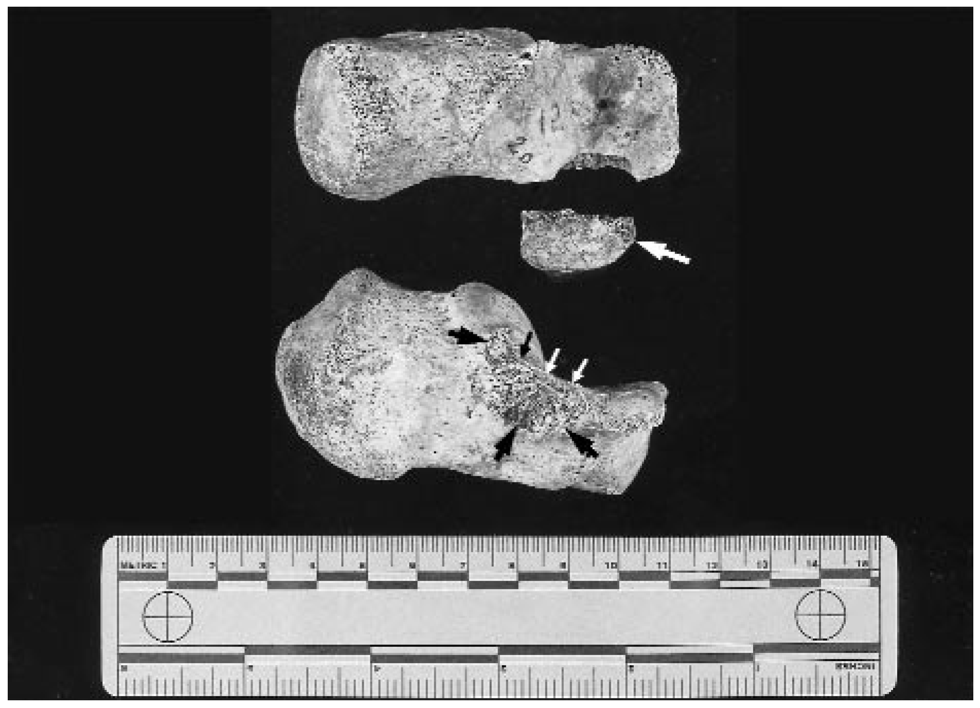
Fig. 1 and
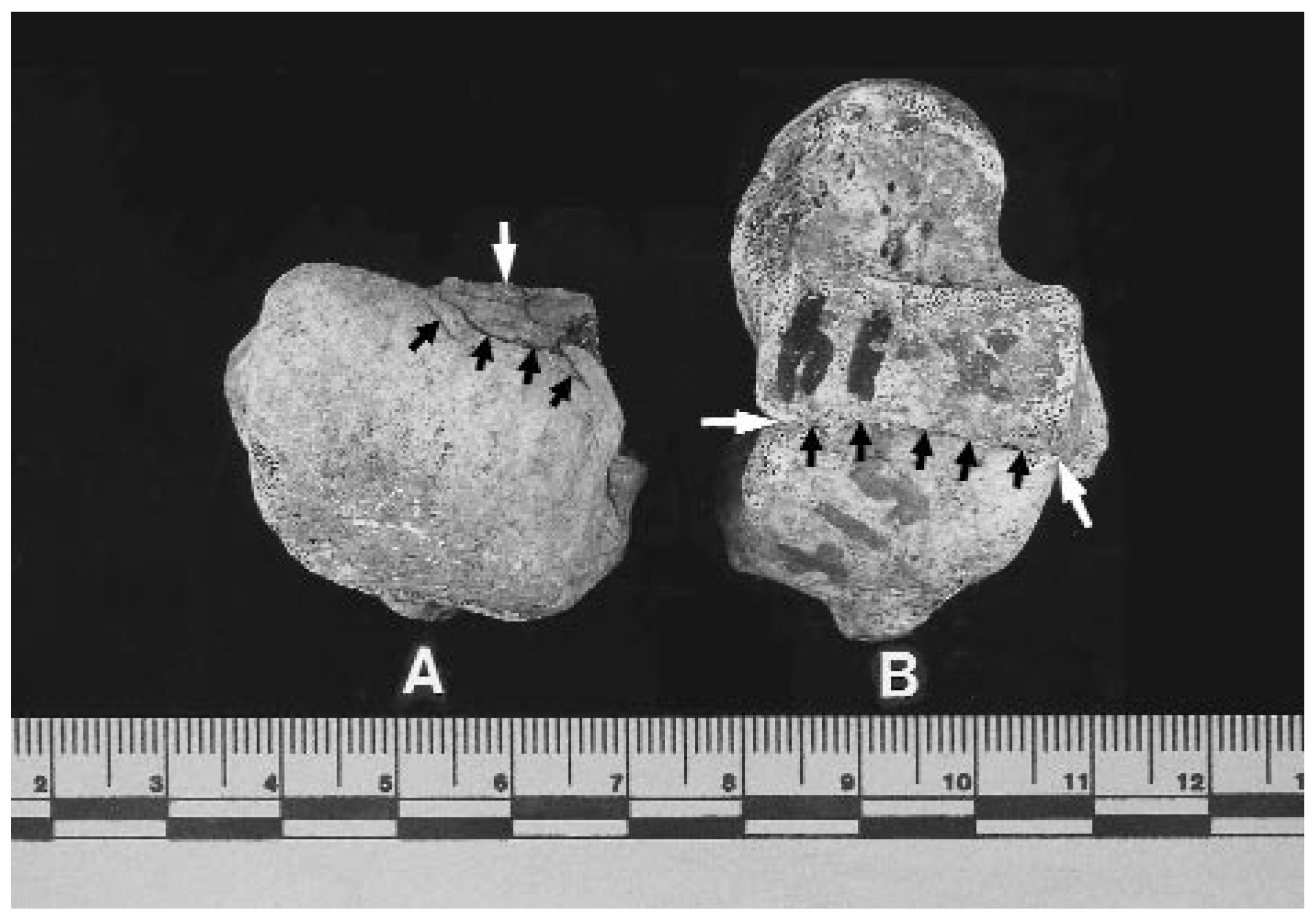
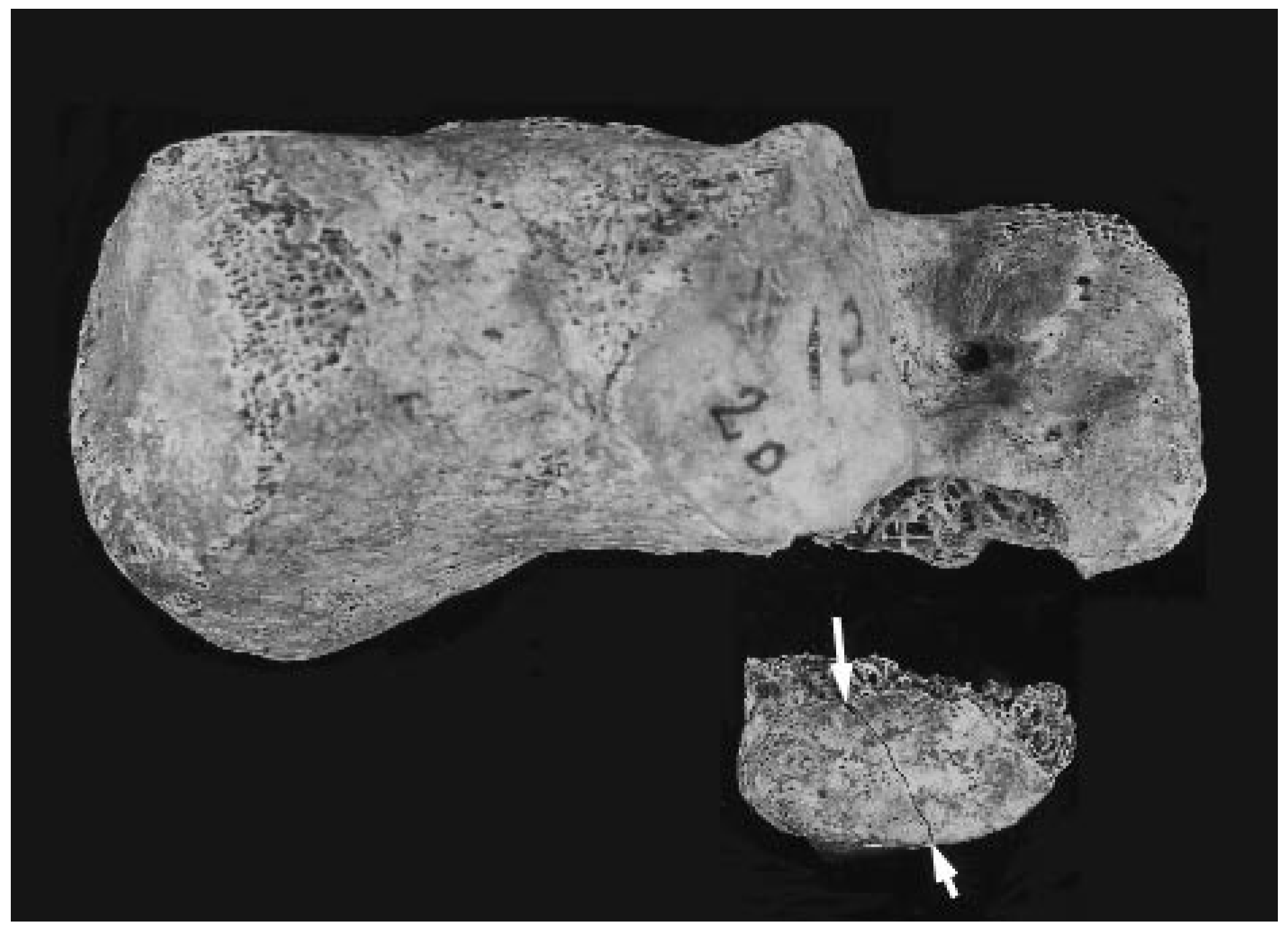
Fig. 2) to separation of the body into anterior and posterior halves. In comparison, of the 63 tali suitable for study, 9 right (14%) and 17 left (27%) tali exhibited perimortem fractures including minor subchondral breaks and areas of depressed bone in the head and dome, fragmentation and separation into anterior and posterior halves through the neck and dome (“aviator’s astragalus”[
6]) (
Fig. 3), coronal fractures completely through the dome, and fractures of the posterior facet (
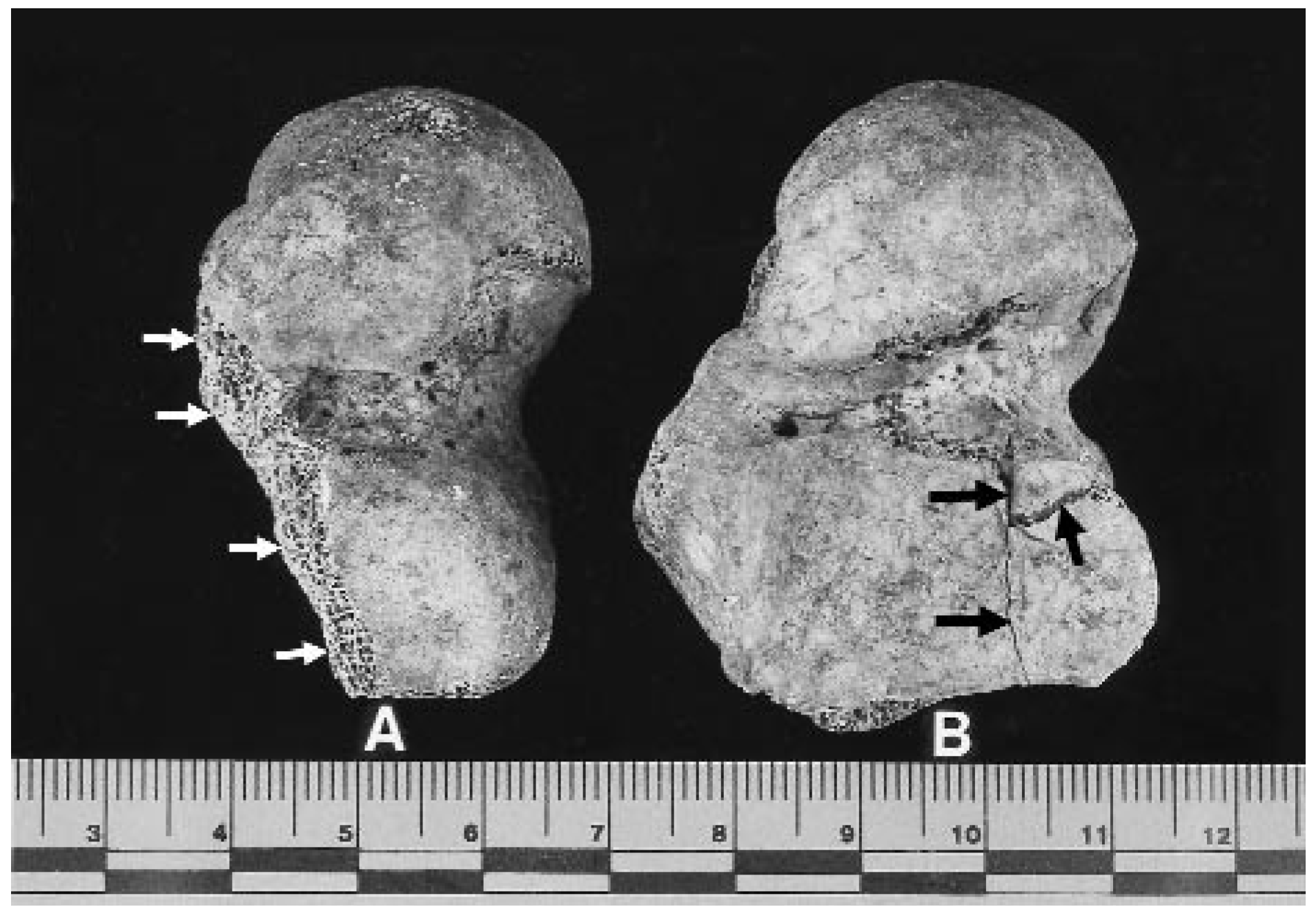
Fig. 4).
Figure 1.
Two left calcanei exhibiting complete separation of the sustentaculum tali (large arrow). The lower calcaneus reveals the extent of bony avulsion of the sustentaculum tali and exposed trabeculae.
Figure 1.
Two left calcanei exhibiting complete separation of the sustentaculum tali (large arrow). The lower calcaneus reveals the extent of bony avulsion of the sustentaculum tali and exposed trabeculae.
Figure 2.
Close-up of a left calcaneus (same bone as in the upper portion of
Fig. 1) and separate sustentaculum tali and minor fracture (arrows).
Figure 2.
Close-up of a left calcaneus (same bone as in the upper portion of
Fig. 1) and separate sustentaculum tali and minor fracture (arrows).
Figure 3.
Superior view of left (A) and right (B) tali. A, Complete separation through the neck (“aviator’s astragalus”) (large arrow) with concurrent compression of the dome (small arrows). B, At the time of the crash, the right talus (glued together in the laboratory) was broken coronally into anterior and posterior pieces along the fracture line (arrows). (The numbers are laboratory identifiers.).
Figure 3.
Superior view of left (A) and right (B) tali. A, Complete separation through the neck (“aviator’s astragalus”) (large arrow) with concurrent compression of the dome (small arrows). B, At the time of the crash, the right talus (glued together in the laboratory) was broken coronally into anterior and posterior pieces along the fracture line (arrows). (The numbers are laboratory identifiers.).
Figure 4.
Two left tali showing complete separation through the posterior facet and sinus tarsi (A, arrows) and incomplete fracture and small fragment of depressed bone (B, arrows).
Figure 4.
Two left tali showing complete separation through the posterior facet and sinus tarsi (A, arrows) and incomplete fracture and small fragment of depressed bone (B, arrows).
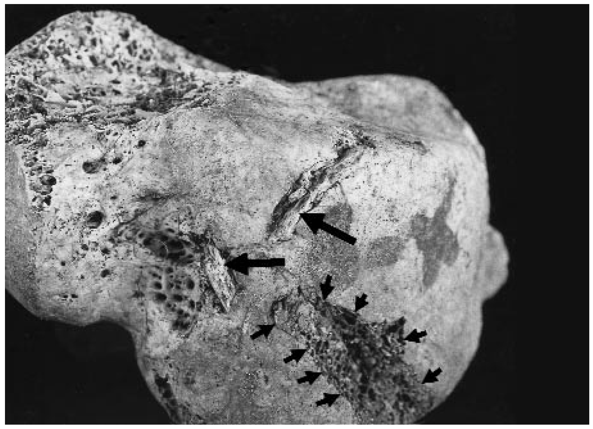
The most dramatic fractures resulting from this high-speed–impact incident were represented by complete separation of the sustentaculum tali in seven calcanei (11%), impacted bone fragments in one talar dome (
Fig. 5), and avulsion of the peroneal tubercle in one calcaneus. Surprisingly, 26 calcanei (41%) and 37 tali (59%) exhibited no evidence of perimortem trauma. The most common types of fracture in the calcaneus and talus, respectively, were separation into anterior and posterior halves through the calcaneal body (18/63 or 29%) and coronal separation into halves through the talar dome (8/63 or 13%).
Figure 5.
Left talus with gouge (small arrows) and two pieces of bone (probably from the gouge) that became embedded in the dome (large arrows) during the crash. Embedded bone such as this, regardless of its cause, is an extremely rare finding in archaeologically derived bones.
Figure 5.
Left talus with gouge (small arrows) and two pieces of bone (probably from the gouge) that became embedded in the dome (large arrows) during the crash. Embedded bone such as this, regardless of its cause, is an extremely rare finding in archaeologically derived bones.
Discussion
Fractures of the talus and calcaneus are commonly encountered clinically and radiographically [
7] and can result from a variety of insults and activities, including motor-vehicle accidents, sports injuries, lawnmower accidents (shearing), or falls from heights of 6 feet or more [
8]. Other than small chip and avulsion fractures, the most common reported site of talar fracture associated with violent injury is through the neck [
9,
10], while the most common site of calcaneal fracture is through the posterior facet [
11]. The socalled aviator’s astragalus is usually attributed to hyperdorsiflexion of the foot, causing the talar neck to become wedged and crushed between the calcaneus and the anterior lip of the tibia.
A review of the literature revealed a number of studies for comparison with the findings of the present report. Coltart [
12], for example, reported on 228 talar injuries (nearly 1%) of 25,000 fractures and dislocations in 218 members of the Royal Air Force. He found that injuries to the talus accounted for 106 (nearly 70%) of the most severe injuries and, in his opinion, truly justified Anderson’s 1919 title of “aviator’s astragalus.” Contrary to Anderson’s earlier belief, however, not all of the fractures noted by Coltart were the result of aviators having the soles of their feet resting on the rudder bar when their aircraft crashed (ie, some talar fractures were not associated with flying). The study revealed 56/114 (49%) chip and avulsion fractures, 6 (5%) compression fractures of the head, 15 (13%) fractures of the body, and 37 (32%) fractures of the neck (
Table 2).
Another study of 40 cases (
Table 2) of violent injury to the talus, by Mindell et al [
13], revealed 3/40 (8%) fractures to the body, 8 (20%) to the neck, and 1 (3%) to the posterior lip. These fractures represent trauma sustained by individuals resulting from motor-vehicle accidents, falls from heights of 6 to 20 feet, a fall from a chair, a crushing injury from a 750-pound mold, and being dragged by a horse. For comparison, the present study yielded 20/63 tali (32%) with fractures to the body, 7 (11%) to the neck, and 5 (8%) to the head. Surprisingly, the soldiers in this C-123 airplane crash sustained fewer (11%) “aviator’s fractures” of the talar neck than the 40 individuals injured in what should have been less severe and less life-threatening accidents (ie, involving lower energy) and approximately one-third as many neck fractures as found in Coltart’s Royal Air Force study. Judging from the appearance of tibiae and fibulae (the topic of a future report) of the soldiers in the C-123, the authors would have expected more vertical talar neck fractures (“aviator’s astragalus”), detached sustentaculum tali, and crushing injuries to the talar dome.
Variables affecting the pattern, distribution, and severity of talar and calcaneal fractures in the C-123 soldiers include the angle and speed of the airplane at impact and the location and position of each soldier on board the aircraft when it crashed. Unfortunately, there are no known survivors of the crash and no known witnesses who could provide more information on the incident.
Perimortem trauma associated with airplane and helicopter crashes, as with most types of high-speed– impact incidents, is usually severe and results in extensive trauma to human bones and teeth. Most physicians see foot and ankle trauma resulting from motor-vehicle accidents, sporting events, leisure activities, and falls from heights. The trauma sustained primarily in military (wartime) air crashes presents a pattern and distribution of skeletal fractures associated with unusually high-speed events. For example, skeletal trauma sustained in many helicopter crashes and the vast majority of jet crashes consists of bilateral and symmetrical comminuted fractures of nearly every bone in the body. With such force, the bones of the right side of the body typically break in a manner similar to those of the left, resulting in mirror images. There are, therefore, some skeletal fractures that the CILHI scientists have come to expect as a result of high-speed incidents. These include multiple fractures to the skull and mandible, the acetabulum (hip socket), the cervical neck of the femur, tubular portions of the arms and legs, the sacrum, and the lateral and medial malleoli (sometimes the malleoli are forced through the sides of leather flight boots), and separation of the sustentaculum tali. Some unexpected sites of trauma associated with air crashes include fractures of the thumb (eg, as a result of holding a flight-control stick), the patella as it is driven up against the femur, and spinous processes of the vertebrae.
This report is intended to provide physicians with general information on the expected types of trauma to the calcaneus and talus, as well as specific information on some unusual fractures resulting from high-speed incidents that might go unnoticed with conventional radiography or external physical examination. As some of these fractures have been found in association with relatively low-speed incidents (eg, involving troop helicopters), physicians may expect to find similar fractures in association with motor-vehicle accidents, sporting and leisure activities, and falls from heights.