Plantar verrucae are caused by infection with human papillomavirus (HPV). There are more than 75 types of HPV, each with a predilection for specific tissues in the body [
1]. Most studies of HPV in patients with human immunodeficiency virus (HIV) have dealt with HPV-16 and HPV-18, the causative agents of anogenital warts. These HPV types have been linked to genital carcinoma [
2,
3,
4]. As HIV continues to be better managed and controlled, and as HIV
+ patients survive longer, more attention can be devoted to the treatment of less-devastating infections, such as plantar verrucae, and clinical skill in this area has become more important.
There are three types of HPV that have been associated with the development of plantar verrucae, each differing in its clinical presentation. HPV-1 causes deep plantar warts, or myrmecia. These warts have a rough, horny surface that may project slightly above the skin. They are well circumscribed and contain capillary loops, so they may demonstrate punctate bleeding during debridement. They are usually found singularly, but can occur in groups. The high viral content of these warts makes them very contagious, but they are short-lived and respond well to treatment in the immunocompetent patient. HPV-2 causes mosaic warts, also known as superficial spreading warts. These are relatively painless unless they become large and keratotic with fissures. They demonstrate longevity and are more resistant to treatment in general. HPV-4 causes punctate or endophytic warts. These warts are small with a horny wall surrounding a central depression. They are usually painless and occur in groups [
5]. Punctate bleeding is usually not observed during debridement.
A recent epidemiologic survey addressed the issue of plantar verrucae in HIV
+ individuals. The study showed that HIV
+ individuals have a significantly higher incidence of plantar verrucae (17%) than do HIV
− individuals (2%) [
6]. Given this finding, it is necessary to compare clinical characteristics of plantar verrucae present in HIV
+ and HIV
− populations. An understanding of the characteristics of the plantar verrucae found in HIV
+ patients may lead to a better understanding of the pathogenesis of plantar verrucae in the HIV
+ population and improvement in the care and management of the disorder in this population.
Three clinical characteristics were the focus of this pilot study: the size, number, and clinical type of plantar verrucae present. This study investigated whether HIV+ individuals manifest one clinical type of plantar verrucae more than another, and whether the clinical types present in HIV+ individuals differ from those present in HIV− individuals. The two groups were compared in terms of the average size and average number of plantar verrucae present in each individual to determine whether the plantar verrucae are more severe and aggressive in the HIV+ population. The HIV+ population was also examined with regard to size, number, and type of plantar verrucae in relation to the CD4 cell count of the patient. The CD4 count was accepted as an indicator of immune status: A normal human CD4 range is 500 to 2,000 cells/mm3. The three characteristics were analyzed in relation to CD4 counts to see whether there was a correlation between the immune status of the patient and the severity of HPV infection.
Methods
Patient Selection
A questionnaire was randomly distributed to attendees of the Castro Street Fair in San Francisco in October 1995. Information requested included age, sex, race, sexual preference, HIV status, and presence of warts, including oral, anal, cervical, and plantar warts. If the HIV status was positive, then current CD4 count, plasma viremia, and acquired immunodeficiency syndrome (AIDS) diagnosis were also requested. Fourteen patients were selected from the random call for participation in the study: seven were HIV+ and seven were HIV−. Three HIV− subjects were selected from the patient population at the California College of Podiatric Medicine in order to complete the study group. For those individuals reporting the presence of plantar warts, a clinical examination was offered.
Clinical Evaluation
The clinical evaluation of the plantar verrucae was performed by a podiatrist or a podiatric medical student. For each individual, the warts were recorded according to clinical type. For each clinical type of wart, the number of warts present, the size of each wart, and the location of each wart were recorded. A diagram of the plantar surface of the foot was used to determine more precisely the extent and location of each wart.
Statistical Analysis
The data were analyzed using statistical analysis software (SAS (® SAS Institute, Cary, NC.)). Two correlation matrices were created using Pearson correlation coefficients. The data for the first matrix included each incidence of a wart as a separate observation to compare the size and type of wart present to the dichotomous HIV variable, and for the HIV+ patients, each wart’s size and type were correlated to the continuous CD4 count variable. The second matrix considered each person as a separate observation in order to compare the number of warts per individual to the dichotomous HIV variable and to the continuous CD4 count variable for the HIV+ individuals. A P value less than or equal to .05 was considered to indicate statistical significance.
Results
A total of 17 individuals were studied; 10 were HIV− and 7 were HIV+. There were six males and four females in the HIV− group with an average age of 33.1 years (range, 21 to 45 years). The HIV+ group consisted of seven males with an average age of 38.3 years (range, 30 to 53 years).
The correlation between clinical type of plantar verrucae and HIV status was the most statistically significant finding, with a P value of .0001. Nine of the ten HIV− individuals presented with deep plantar warts; the other individual in this group presented with a punctate wart. There were no cases of mosaic warts within this group. The HIV+ group presented with a wider variety of clinical wart types. Of the 33 total warts found in this group, 13 were mosaic, 11 were plantaris, and 9 were punctate warts. Two HIV+ individuals presented with more than one type of wart: One had punctate and mosaic warts and the other had plantaris and mosaic warts.
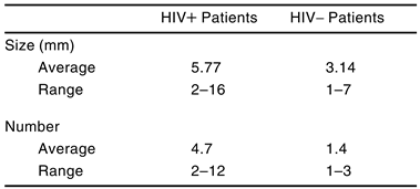
The other characteristics studied were also statistically significant. The correlation between HIV status and the number of warts had a P value of .0115. The HIV
+ group had, on average, more warts per individual than the HIV
− group. Moreover, HIV
+ subjects had larger warts, on average, than did HIV
− subjects (
P = .0358) (
Table 1).
There were no significant correlations between the CD4 levels of the HIV
+ individuals and the number, size, or clinical type of plantar verrucae. The CD4 levels of the HIV
+ individuals ranged from 0 to 500 cells/mm
3. Of the three parameters, clinical type of verrucae was the only one that was significant (
P = .0621) (
Table 2).
Discussion
Plantar verrucae in HIV+ individuals are, on average, more aggressive and severe than those found in HIV− individuals. The level of severity and aggressiveness can be determined through the three characteristics studied: size, number, and type of plantar verrucae. The present study found that the plantar verrucae manifesting in HIV+ individuals were larger and more numerous, on average, than those found in HIV− individuals. The HIV+ group presented with all three clinical types of plantar verrucae, with some individuals having more than one type of plantar verrucae. The type of verrucae appearing significantly more frequently in HIV+ patients as compared with HIV− patients was the more resistant mosaic type. HPV-2 has been correlated with the development of mosaic warts. Mosaic warts were not found in any of the ten HIV− individuals in this study, but four of the seven HIV+ patients presented with mosaic warts. Possibly, HIV+ patients have a greater susceptibility to infection with HPV-2 than with other HPV types.
One limitation of these findings is that the warts were identified only through clinical determinations and no biopsies were used to confirm diagnosis and determine the type of HPV present in the lesion. Additionally, HIV diagnoses and CD4 counts were selfreported. Future studies involving larger populations may improve on the statistical analysis and results of this study.
The severity and aggressiveness of the plantar verrucae did not correlate with the CD4 levels of the HIV
+ individuals. If the general immunosuppressed state of an HIV
+ patient is responsible for the severity of the HPV infection, then one would expect such a correlation. This leads to questions about the pathogenesis of plantar verrucae in the HIV
+ patient. There may be interactions between the two viruses that cause an up-regulation of HPV. For example, cytokines may be released at the local level by HIV-infected lymphocytes to up-regulate the expression of HPV, or there may be interactions between products of the HIV genome with the regulatory sequences of HPV. Recent studies have suggested that the tat protein, a regulatory protein of the HIV genome, can bind and up-regulate the upstream regulatory region of HPV-16 [
7]. Clearly, the pathogenesis and possible viral interactions are areas in which future research is needed.
Conclusion
Although the results of this study are quite preliminary and the sample size is extremely small, there is reason to suspect that HIV+ people may be more susceptible than HIV− people to certain varieties of plantar verrucae. The authors conclude that further studies are warranted in order to determine what relationship, if any, exists among HIV+ status, compromised immune status as suggested by CD4 counts, and additional host factors in the formation of plantar verrucae. Furthermore, future studies could help identify the factors that predispose an individual to specific plantar verrucae types. Current studies related to the classification of viruses associated with the various types of verrucae may also be helpful in the future to resolve this intriguing question.
Acknowledgments
The American Podiatric Medical Association for financial support through its research grant program, 1995–1996 cycle; Jamie Shkolnik, PhD, for her contributions to the statistical analysis of the data; and all of the volunteers who participated in the patient survey and screening at the Castro Street Fair in San Francisco, including Sandra Burns, DPM, and the following students at the California College of Podiatric Medicine: Pilar Almy, DPM ’96, Jeffrey Hogge, DPM ’96, Martha Ajlouny, DPM ’97, Richmond Kilpatrick, DPM ’97, Joanne Lee, DPM ’97, Lise Pariseau, DPM ’97, Molina Rekhi, DPM ’97, Kathleen Ryan, DPM ’97, Cecily Tsuchiya, DPM ’97, Bryan Berghout (class of ’98), Brett Kesler (class of ’98), Phillip Larsen (class of ’98), Ryan Rushton (class of ’98), and Matthew Smith (class of ’99).
References
- FIELDS BN, KNIPE DM: “Human Immunodeficiency Viruses and Their Replication,” in Fields Virology, Vol 2, 2nd Ed, ed by BN Fields, DM Knipe, p 1529, Raven Press, New York, 1990.
- BREESE PL, JUDSON FN, PENLEY KA, ET AL: Anal human papillomavirus infection among homosexual and bisexual men: prevalence of type-specific infection and association with human immunodeficiency virus. Sex Transm Dis 22: 7, 1995.
- BRAUN L: Role of human immunodeficiency virus infection in the pathogenesis of human papillomavirus-associated cervical neoplasia. Am J Pathol 144: 209, 1994.
- CAUSSY D, GOEDERT JJ, PALEFSKY J, ET AL: Interaction of human immunodeficiency and papilloma viruses: association with anal epithelial abnormality in homosexual men. Int J Cancer 46: 214, 1990.
- BUNNEY M, BENTON C, CUBIE H: “Cutaneous Warts: Diagnosis and Behavior,” in Viral Warts: Biology and Treatment, 2nd Ed, p 75, Oxford Medical Publications, Oxford, England, 1992.
- KENYON E, LOVELAND L, KILPATRICK R, ET AL: Epidemiology of plantar verrucae in HIV-infected individuals. J Acquir Immune Defic Syndr Hum Retrovirol 17: 94, 1998.
- VERNON SD, HART CE, REEVES WC, ET AL: The HIV-1 tat protein enhances E2-dependent human papillomavirus 16 transcription. Virus Res 27: 133, 1993.
Table 1.
Comparison of Size and Number of Verrucae in
HIV+ and HIV− Patients
Table 1.
Comparison of Size and Number of Verrucae in
HIV+ and HIV− Patients
Table 2.
Type, Number, and Size of Plantar Verrucae in Relation to CD4 Count of HIV+ Patient
Table 2.
Type, Number, and Size of Plantar Verrucae in Relation to CD4 Count of HIV+ Patient
© 1998 American Podiatric Medical Association