Without research that generates new knowledge, a profession’s growth and development will cease. An era of computational medicine and the computational health scientist is beginning. Clinicians involved in research need a high level of computational skill. In particular, the development of sophisticated theories, using mathematical and computational techniques, is becoming increasingly important to our understanding of the intrinsically complex phenomena being discovered in many areas of contemporary medicine. Computational medicine includes the formulation of problems in mathematical terms; the solution of the resultant mathematical problems; the discussion, interpretation, and evaluation of the results of this analysis; the exploration of new ideas and areas of application; and the development of mathematical theories in areas that have not undergone systematic mathematical treatment.
Geometric Models
Foot problems caused by mechanical factors are common and not only limit locomotor mobility but indirectly have a harmful effect on human posture. If one can represent the wide variety of human foot structures by a corresponding set of configurations derived from a geometric model, one can establish a theoretical model that may help the clinician in the diagnosis and treatment of biomechanical disabilities.
In an article titled “Same Gene May Shape Face, Heart and Hands” that appeared in the New York Times of February 13, 1996, George Johnson tells of the discoveries of genetic mechanisms that change a person’s face, heart, and hands along with other parts of the body. He mentions Pfeiffer’s syndrome, which produces distortion of the skull and deformities of the thumbs and big toes; Greig’s syndrome, in which distortion of the head and face is accompanied by malformation of the hands and feet; Aarskog syndrome, which is marked by a round face with widespaced eyes and small hands and feet; and Rubinstein- Taybi syndrome, a form of mental retardation accompanied by features that include a small head with downward-slanting, wide-spaced eyes, a beaked nose, broad thumbs, and big toes. Thus foot morphology, along with structural changes in other parts of the body, may lead to the diagnosis of a syndrome and its functional implications. The use of carefully chosen geometric models to represent morphological components of the foot may help to uncover a new syndrome as well as recognize structural variations.
Conic curve, ellipsoid, and triangular models are geometric models presented in this article. These models are derived from coordinates obtained from radiographic landmarks and may help to classify biomechanical defects and foot types in a new and precise way in order to produce a better diagnosis of and treatment regimen for orthopedic conditions.
Materials and Methods
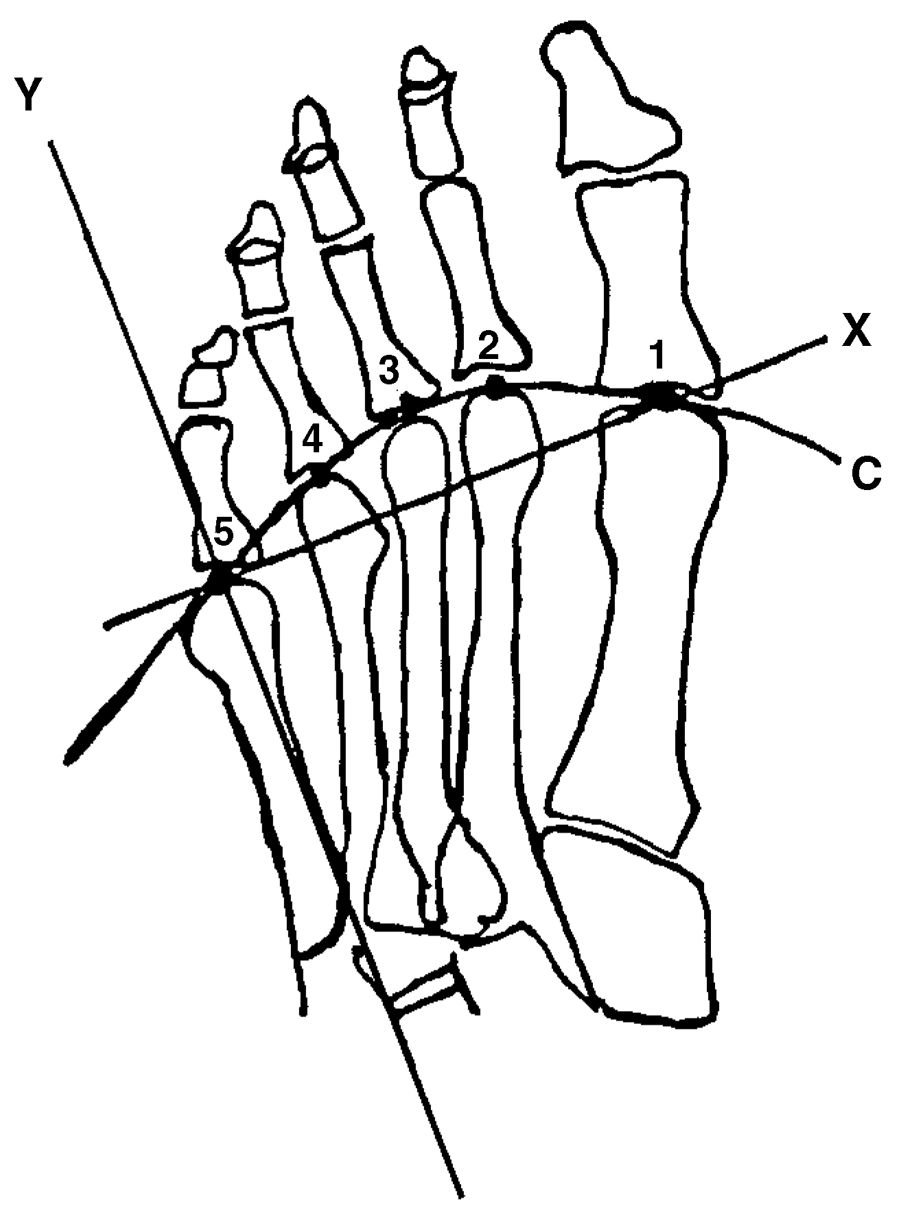
The conic curve and triangular models were derived from a relevant set of coordinates taken from weightbearing dorsoplantar and lateral radiographs. A line passing through the most distal aspect of the first and fifth metatarsal heads on the radiograph denotes the
x-axis; a line perpendicular to the
x-axis at the fifth metatarsal head denotes the
y-axis (
Figure 1). The origin (0,0) is located at the intersection of the two axes because the flow of forces moves from the fifth to the first metatarsal head. Each model is represented by an equation consisting of variables and parameters (arbitrary constants). As the numerical values of the parameters change, a different configuration of the model occurs. Thus there is a family of conic curves, a family of ellipsoids, and a family of triangles. It would take the establishment of a research project to apply the model to each foot (pathologic or nonpathologic) in the sample and determine its configuration and numerical value.
The ellipsoid model was derived from modified dimensions of length, height, and width that were mapped into a continuum of points by vectorization and projection.[
1] Each point represents a foot type.
Configurations and their numerical values based on these geometric models may lead to diagnostic precision because of proper refinement; may show degrees of disability by an ordering of numerical values; and may help in the formulation of a plan of treatment for the patient.
Conic Curve Model
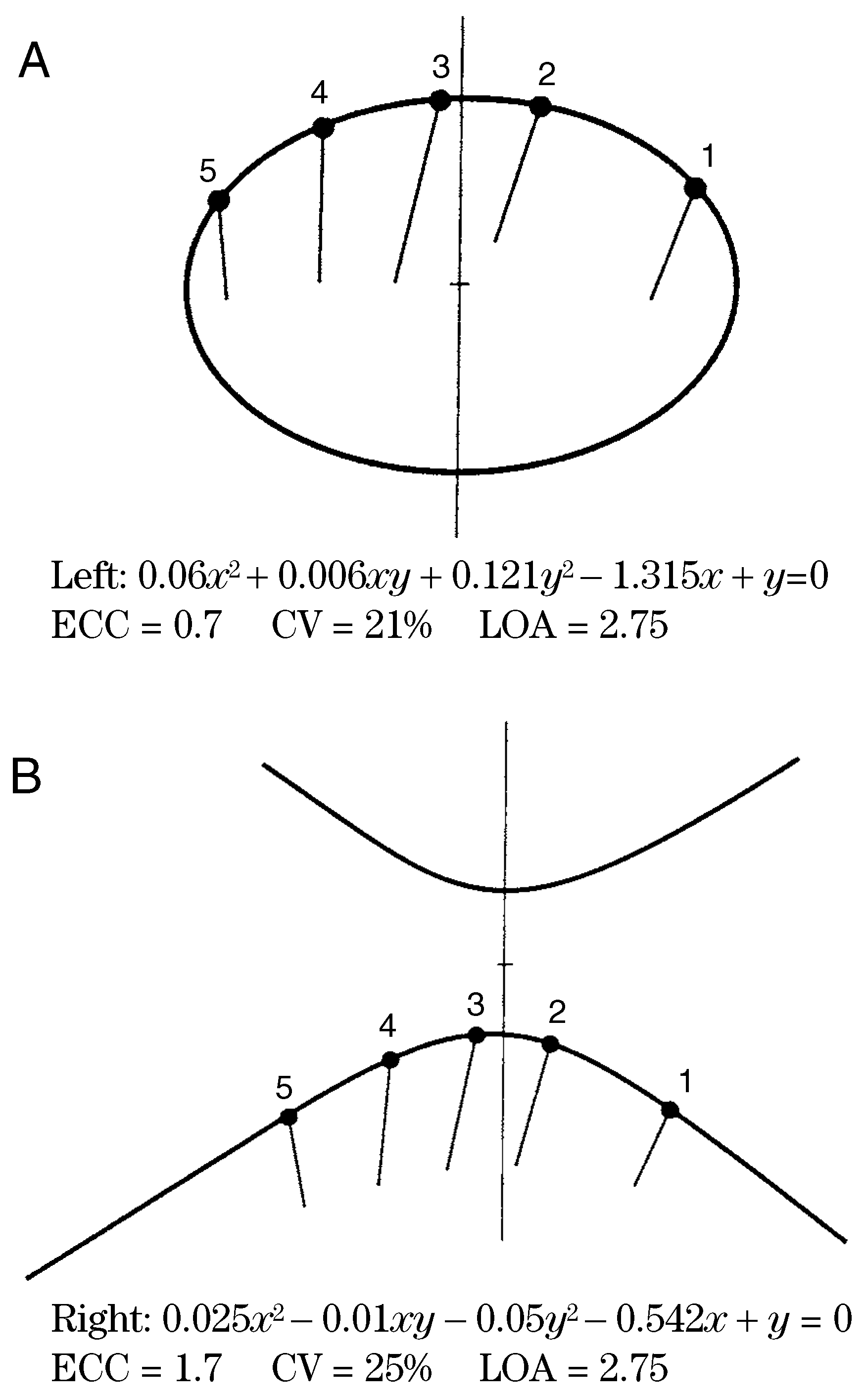
In the geometric model derived from the radiograph, each variation of the metatarsal arc consisting of the five metatarsal heads has a shape that corresponds to a unique conic curve, which in turn corresponds to the metatarsal length pattern. By a triangular scaling process, all metatarsal heads can be repositioned for comparable size while preserving shape.[
2] Thus each metatarsal arc is characterized by a configuration of unique shape and size (
Figure 1). This configuration can be quantified by using the eccentricity of the conic curve for the numerical value of shape and a pattern measure for the numerical value of size. The author [
3] has discussed the eccentricity measure for shape and is in the process of finding a similar measure for size. A future project will match forefoot disabilities with measures of shape and size and use these measures as a basis for choosing surgical intervention and prevention.
Ellipsoid Model
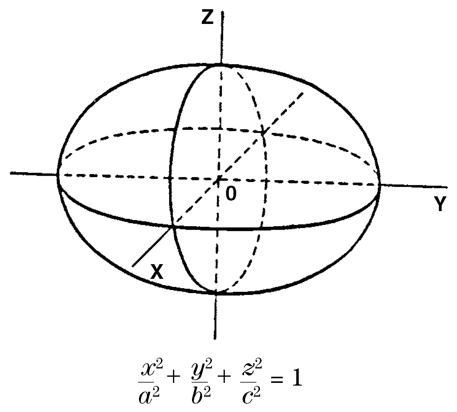
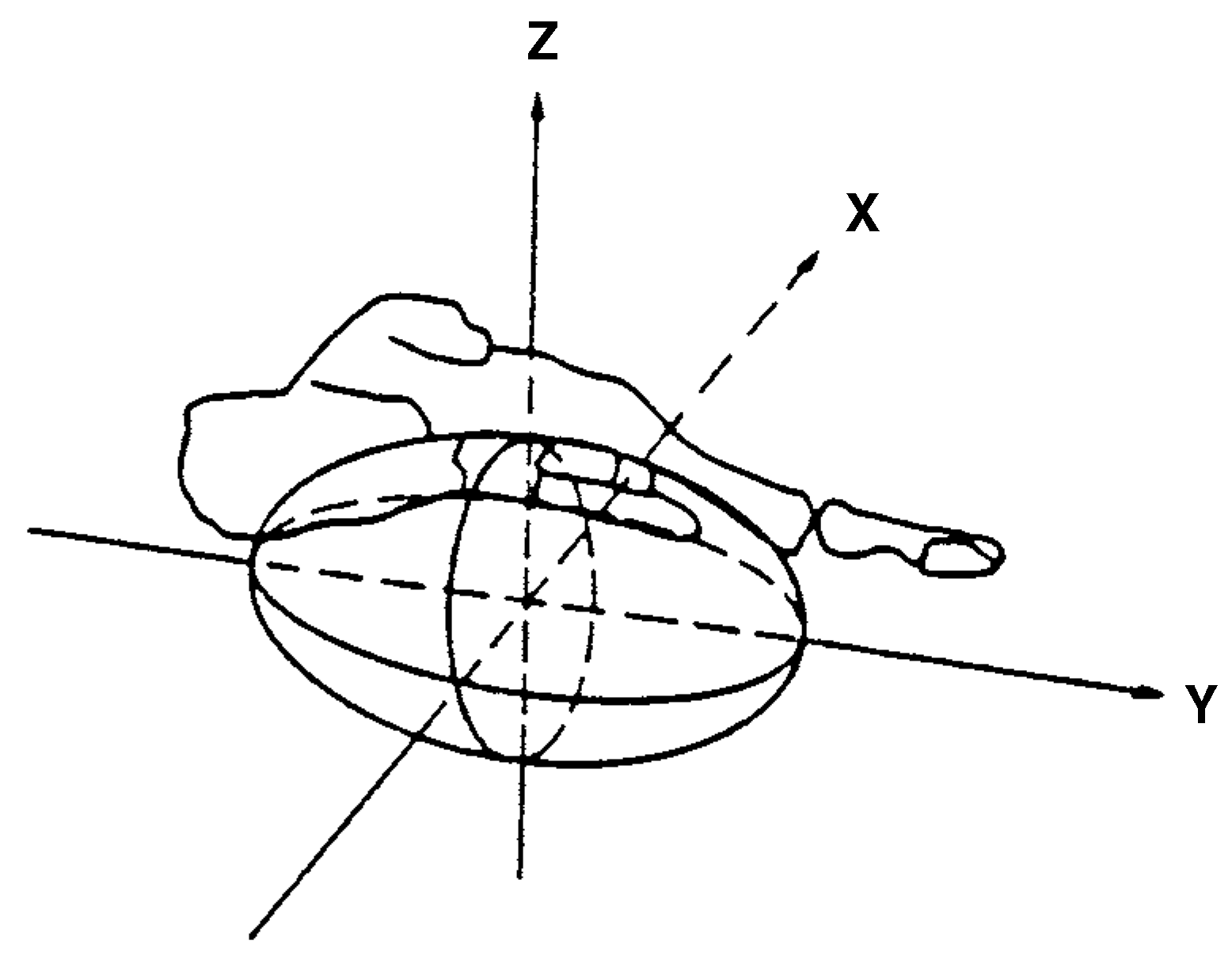
Based on anatomic, physiologic, and clinical considerations, the one-quarter, flexible ellipsoid was selected as the model of the foot (
Figure 2). Mathematical methods were used to classify human feet into a continuum of points representing the entire range of variations in foot configurations. There is an optimal point (foot) from which every other point is measured by a distance function. Each numerical deviation of a particular foot reflects the biomechanical quality of that foot. The mathematical details are found in an article by Demp.[
1] Again, future research is needed to correlate the entire range of different foot types, no matter how small that difference is, to measures of shape and size. A diagram and an equation of an ellipsoid are shown in
Figure 3.
Triangular Model
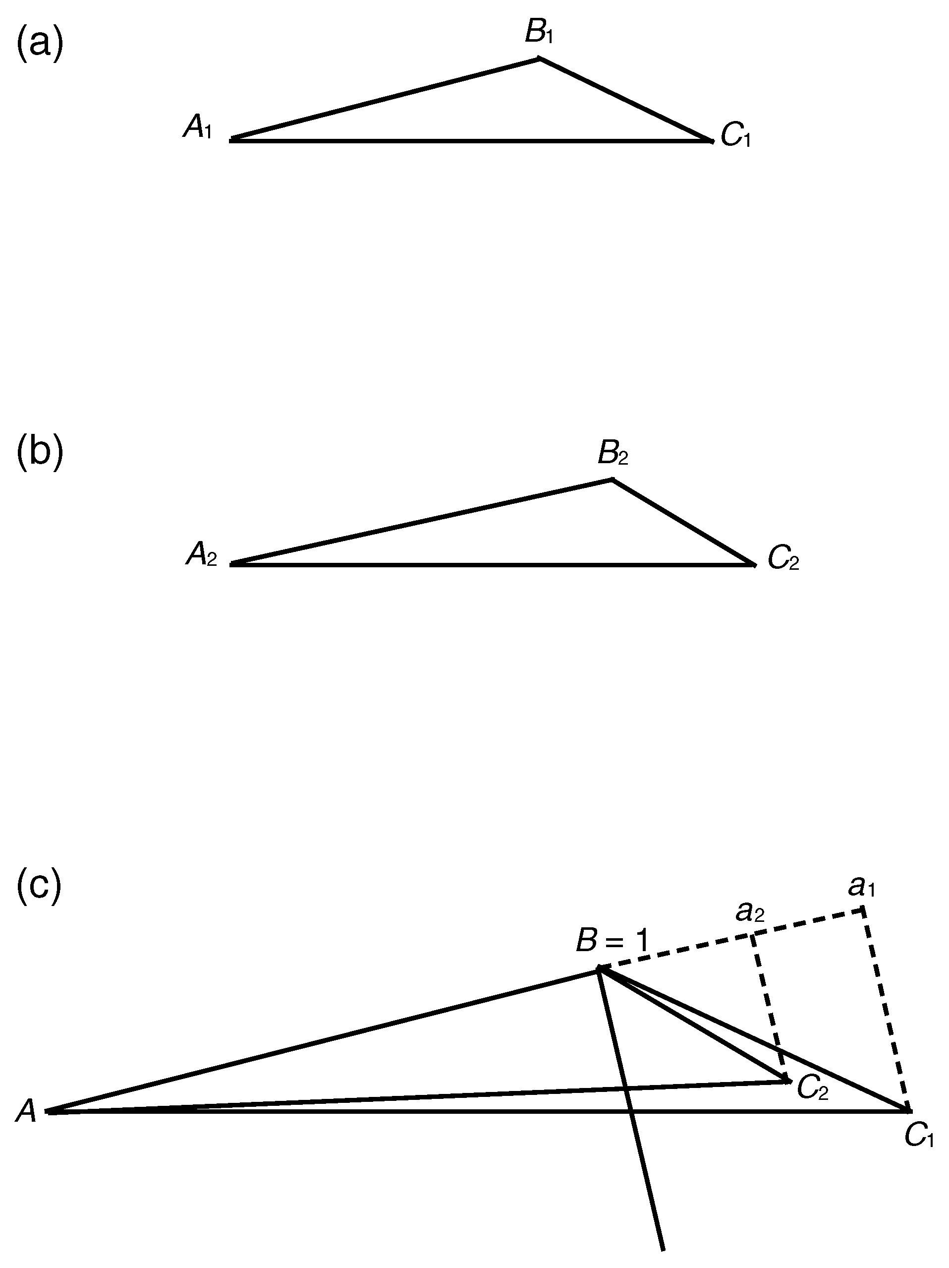
In seeking a more precise method to measure pathologic variations of hallux abducto valgus, a geometric model of a triangle was formed on a weightbearing dorsoplantar radiograph using the fifth, second, and first metatarsal heads as the vertices A, B, and C, respectively, and their respective coordinates (
x,
y) were determined. The triangle was standardized using the previously mentioned triangular rescaling process. A was moved to coordinates (0,0), B was moved to coordinates (1,0), and C was moved to the corresponding coordinates (
a,
b) so that the original shape of the triangle was preserved (
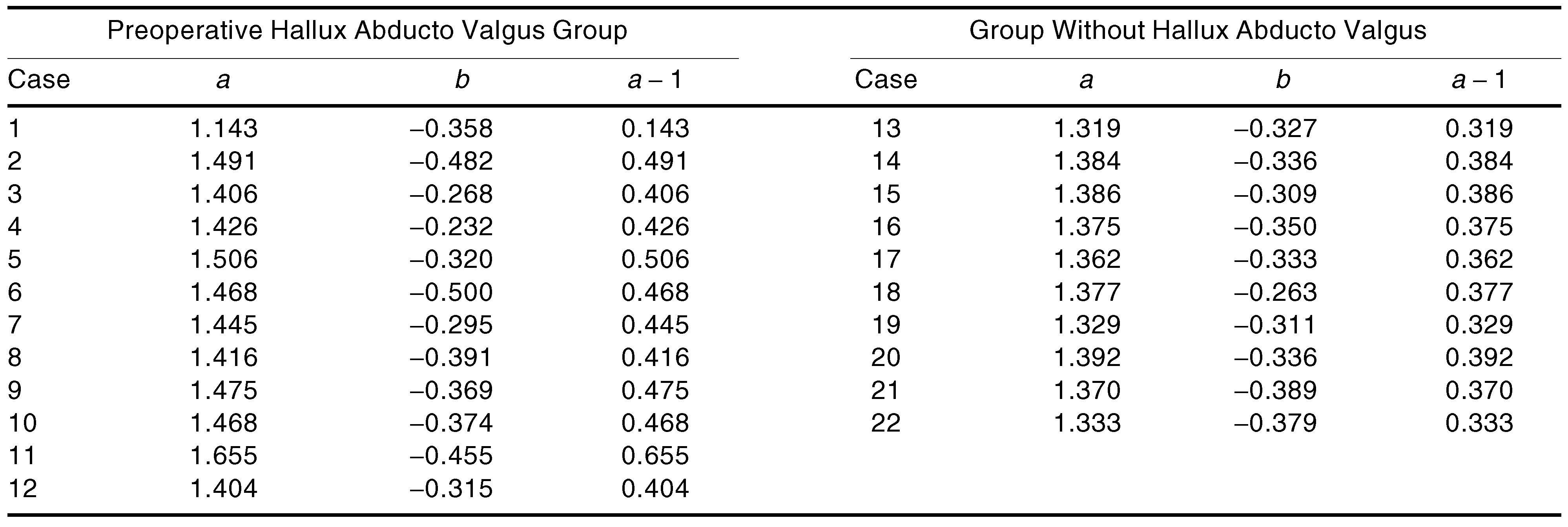
Figure 4). In this study, the preoperative radiographs of 12 feet that had undergone hallux abducto valgus surgery were used to determine the horizontal distance from
B to
C,
a − 1, which was taken as a measure. Such measures were compared with similarly obtained measures from feet considered not to have hallux abducto valgus. This small sample showed that the numerical value of 0.4 separated the feet with hallux abducto valgus from those without it (
Table 1). This information is expected to be a guide to help provide successful repositioning of the first metatarsal head. However, future research using a large sample is needed to validate this finding.
Rationale
A complete rationale for selecting the ellipsoid model can be found in a book chapter written by two podiatric surgeons, Henenfeld and Frankel. [
4] A conic curve model was selected for the following reasons: Every conic curve is uniquely determined by five points (most distal points of the five metatarsal heads). A conic curve gives a graphic representation of the metatarsal arc, and its equation corresponds to the metatarsal length pattern (
Figure 5). Also, every conic curve is associated with a unique numerical value called its eccentricity. The triangular model rationale was obtained by fixing the fifth and second metatarsal heads on a baseline of length 1, from (0,0) to (1,0). The first metatarsal head point can be positioned on coordinates (
x,
y) such that the shape of the triangle consisting of lines connecting (0,0), (1,0), and (
x,
y) is the same as the shape of the original triangle connecting the points of the fifth, second, and first metatarsal heads. By this standardization technique, the varying positions of the first metatarsal head point may discriminate between feet with hallux abducto valgus and those without it. Also, because of the location of the metatarsal heads, there will be a minimum of radiographic distortion, as the metatarsal heads will be in close contact with the radiographic film.
Discussion
The use of coordinates to construct the three geometric models presented here of the foot and to generate configurations that correspond to numerical values is a departure from the linear and angular measures that are used as data for the statistical techniques of discrimination and regression.5 Can one find geometric models of a static foot structure whose varying configurations more closely align with dynamic foot function than an analysis by linear and angular measures? The three geometric models discussed in this article are offered as a positive answer to that question, based on the following considerations:
- 1)
The representation of an anatomic structure by a geometric model allows changes in the anatomic structure to correspond to changes in the model’s configurations.
- 2)
The model’s configurations correspond to the entire range of structural variations that can be defined by numerical values.
- 3)
A model’s configurations stem from variations of its parameters.
- 4)
Each pathologic and nonpathologic structure corresponds to a unique single configuration or interval of configurations.
- 5)
There is minimal radiographic distortion for the conic curve and triangular models because the coordinates are taken at the metatarsal heads.
- 6)
Structural variations can be classified with unlimited refinement. The degree of refinement depends on diagnostic and therapeutic results.
Please note that the author has not proved the validity of these models. Rather, the author claims that these nonlinear models offer a numerical classification scheme that can be adjusted to any degree of refinement. The ability to assign a numerical value to every possible structural variation of the foot would produce a numerical classification that could be widely accepted for its clinical effectiveness, even though imperfectly valid. Further research should be conducted to validate this classification system.
Conclusion
A geometric model of an anatomic structure can be used to generate an infinite set of configurations by varying the model’s parameters. This configuration scheme is the basis for a new classification system that may be closely aligned to an individual’s foot function. A future research project is required to map biomechanical abnormalities into numerical configurations. An inverse mapping would show configurations of latent disabilities, thereby enhancing the practice of preventive medicine.