The image today of the typical South Florida podiatric medical patient might be that of an elderly, overweight person. Little has been documented, however, about actual demographic variability in podiatric patient populations. Even less is known about gender-specific, age-specific, and body size–specific associations with particular abnormalities of the foot, such as metatarsus adductus. Such scarcity of epidemiologic data could invite medical researchers to compare podiatric patients with less appropriate study cohorts. For instance, research in sports medicine suggests that there is no association between the dominant limb and laterality of running injuries [
1]. Yet podiatric patients and trained athletes may not share the same patterns and risks of foot disorder, so inferences about broader patient populations based on athletic-injury data can be misleading.
The goal of this study was fourfold: 1) to examine statistical distributions of sex, age, and body size in a diverse podiatric patient population; 2) to test the hypothesis that laterality is not associated with side of complaint; 3) to estimate the population prevalence of metatarsus adductus deformity; and 4) to examine the relationships among sex, age, body size, laterality, and metatarsus adductus deformity. The results may provide new insight into the demographic profile of podiatric patients and their risks of injury or disease.
Review of the Literature
Theories on Development of Laterality and Limb Dominance
Further complicating the discussion of handedness and footedness is the general lack of agreement in a long history of debate over factors that influence limb preference. For example, Plato (4th century BC) discussed the role of social factors and education in development of right-hand dominance (as cited in Hacaen and De Ajuriaguerra [
9]):
Of believing that in all our actions, there is a natural difference between right and left; this is for the hands, because, for the feet and the lower parts, no difference is visible in their behavior, and is only for the hands that the stupidity of nurses and mothers has made us all one-armed, so to speak. Indeed, the natural aptitude of the two arms is the same, and it is ourselves that have made them unequal and who do not use them as we should.
Thus Plato argued that limb dominance is acquired during development.
There is evidence to support this ancient view. Typically, hand preference does not become noticeable until a child is approximately 2 years of age and is largely established by 3 years of age [
10,
11]. Gabbard [
12,
13,
14] argued that while genes may play a role, it is behavioral influences that ultimately determine the phenotype; these studies indicated that, compared with handedness, right-foot preference was not as common, and mixed-footedness (no preference for either foot) was more prevalent with right-handedness. This is because during the developmental period there are fewer behavioral factors, such as practice and cultural pressure, to influence footedness. Gabbard [
12] found no correlation between foot dominance and motor skill in 4-year-olds, suggesting a lack of foot preference in this age group. Between the ages of 8 and 11, however, there is a greater shift toward right-footedness (unsupported foot, or kicking foot); foot preference tends to remain stable past this age [
15] Hart and Gabbard [
16] found evidence that foot preference in college students for complex tasks (involving limitation of visual cues) is less likely affected by neurodevelopment early in life and more likely affected by the specific context (eg, dim lights, closed eyes) of the task.
Alternatively, some authors believe that the development of limb dominance is influenced more by genetic endowment. Bakan et al [
17] stated that people are genetically programmed for right-handedness, with left-handedness manifesting itself only because of hypoxia during parturition. They also suggested that right-handers are genotypically right-foot dominant. Previc [
18], in an extensive review of laterality data, concluded that cerebral lateralization evolved millions of years ago in early, bipedal hominids, and that lateralization results from asymmetrical, prenatal development of the ear and labyrinth. This follows from the observation that the human fetus tends to lie predominantly on its left side along the anterior uterine wall during the final trimester, thus affecting otolith development and balance. Footedness in left-handers, however, is harder to predict, as it relates to the extent of neurologic damage during birth [
17,
19]. Most proponents of the inheritance theory support a model in which right-footedness follows right-handedness but left-footedness is less strongly associated with left-handedness. For example, Didia and Nyenwe [
6] found that 92% of right-handers were right-footed, while 52% of left-handers were left-footed. A study by Peters and Durding [
20] also suggested that right-footedness is associated with right-handedness but leftfootedness is not associated with left-handedness. However, Previc [
18] reviewed correlation data that showed relatively strong congruency (right-handed with right-footed, and left-handed with left-footed) in tasks involving voluntary leg flexion (eg, kicking a ball), but relatively weak congruency in tasks involving leg extension (eg, standing on one leg, stepping on a stool).
Materials and Methods
Nearly 400 patients from three of the teaching clinics of the Barry University School of Graduate Medical Sciences were surveyed between September 1995 and August 1996. Bilingual Spanish/English questionnaires were completed by the patients (assisted occasionally by office staff members). The data requested included age, sex, height, weight, handedness, side of complaint, and reason for visit. Not all patients provided all of the information requested: for example, age was reported on only 378 of the questionnaires, sex was reported on only 371 of the questionnaires, and both height and weight were reported on only 337 of the questionnaires. The self-reported information on the foot complaint was recorded simply as right foot, left foot, or both; the nature of the complaint was recorded, but not quantified or analyzed in any way.
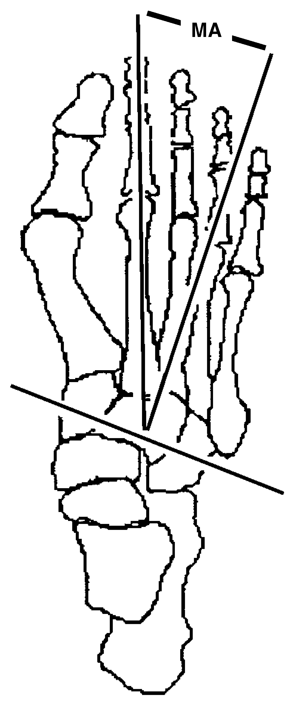
Figure 1.
Measurement of the metatarsus adductus angle (MA).
Figure 1.
Measurement of the metatarsus adductus angle (MA).
The body height and weight values were used to calculate the body-mass index (BMI), which is the weight in kilograms divided by the square of the height in meters. In this simple measure of body size, persons with relatively high BMI values tended to be relatively robust in build, while persons with relatively low BMI values tended to be thin. The BMI has been shown to correlate significantly with patterns of mortality in both men [
28] and women [
29]. It has also been used to examine crude and age-specific correlations between mortality and body size [
30,
31]. One of the largest BMI data sets available in the literature was assembled in the Cardiovascular Health Study conducted by Diehr et al. [
32] The sample proportions of BMI values from Diehr et al were used as population parametric values in the present study to compare the sample BMI values in the South Florida podiatric medical patients.
Radiographs were available for 55 patients. These were assessed for metatarsus adductus angle. The method of evaluation used was measurement of the angle formed by the bisection of the second metatarsal and a perpendicular to the bisection of the lesser tarsus (
Figure 1). A patient was categorized as having metatarsus adductus deformity if this angle exceeded 15°.
Statistical Analyses
All statistical methods used in this study were obtained from Sokal and Rohlf [
33], and all tests used the α = 0.05 level as indicating statistical significance. Correlations among quantitative variables (age, weight, height, BMI, and metatarsus adductus angle) were examined by calculation of Pearson product correlation coefficients. Associations among qualitative (nominal) variables were examined by contingency chi-square analysis. Mean metatarsus adductus angles were compared among samples (male versus female, metatarsus adductus present versus absent) with comparison of means and analysis of variance (ANOVA).
Results
Sex, Age, and Body-Mass Index
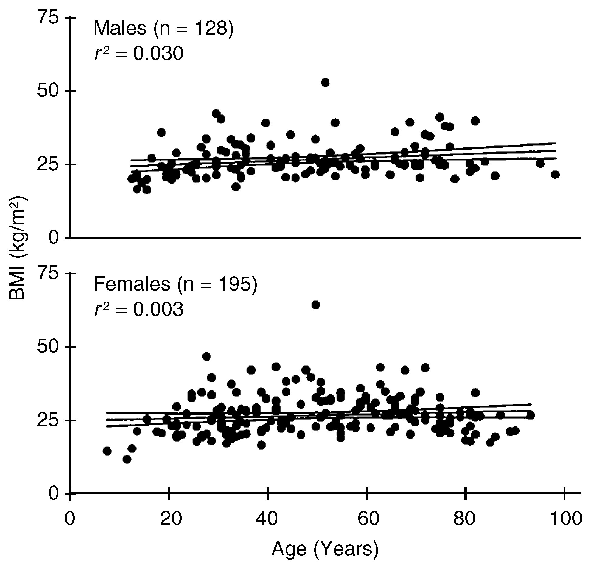
The distributions of age and BMI values for males and females are shown in
Figure 2. The age range for males was 5 to 97 years (median age, 48), and the age range for females was 3 to 94 years (median age, 49). Most of the adult males and females were within normal ranges of body weight (65 to 100 kg for males, and 45 to 82 kg for females). For all patients, weight and height showed a significant positive correlation (
r = 0.48;
df = 335;
P < .01). There was a significant correlation between BMI and age in the sample males (
r = 0.17; n = 128;
P < .01), but not in the sample females (
r = 0.05; n = 195;
P = .07). (Only 195 of the 222 females and 128 of the 149 males reported all of the components necessary for BMI analysis.)
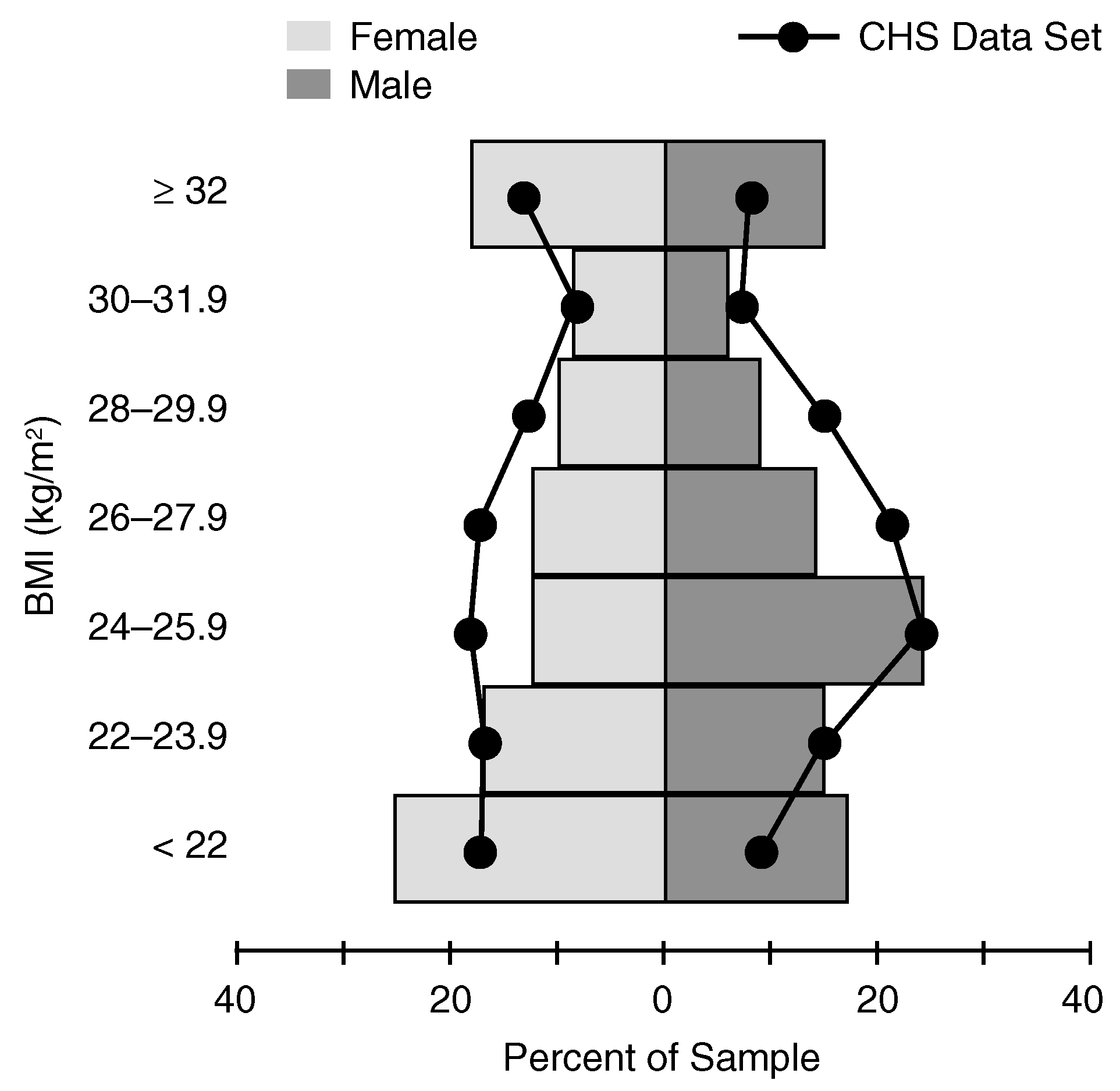
A comparison of distributions for BMI values between the South Florida podiatric medical patients and the Cardiovascular Health Study (CHS) data (Diehr et al [
32]) is shown in
Figure 3. There was a significant difference between the distribution of BMI values of men in the CHS data and that of the South Florida male podiatric patients (contingency χ
2 = 20.5;
df = 6;
P < .01). There were relatively more of the very low values (BMI < 22) and relatively more of the very high values (BMI ≥ 32) among the South Florida male patients, but relatively fewer moderately high BMI values (26 to 30). The same pattern appeared for the female podiatric patients (contingency χ
2 = 102.1;
df = 6;
P < .001).
Figure 2.
Correlation of age and BMI (kg/m2) according to sex. The line is the best-fit regression for the relationship (boundary curves represent 95% confidence interval estimate for the regression line); r2 = coefficient of determination.
Figure 2.
Correlation of age and BMI (kg/m2) according to sex. The line is the best-fit regression for the relationship (boundary curves represent 95% confidence interval estimate for the regression line); r2 = coefficient of determination.
Figure 3.
Frequency histograms for BMI (kg/m
2) sample distributions according to sex. Solid curves represent distributions of BMI values from the Cardiovascular Health Study (Diehr et al [
32]).
Figure 3.
Frequency histograms for BMI (kg/m
2) sample distributions according to sex. Solid curves represent distributions of BMI values from the Cardiovascular Health Study (Diehr et al [
32]).
A minority—though still a considerable proportion— of the South Florida patients was categorized as obese. Roughly 40% (78/195) of the sample females had BMI values of 27 or above, while 44% (56/128) of the sample males had BMI values of 26 or above.
Laterality and Side of Complaint
Ninety-three percent of the patients identified themselves as right-handers, 6% as left-handers, and 1% as ambidextrous. Thirty-one percent of the patients reported a complaint about the right foot, 37% about the left foot, and 32% about both feet.
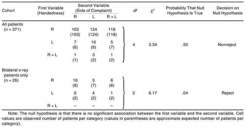
The associations between handedness and side of complaint are shown in
Table 1. There was no significant association between the two for the entire sample. However, within the subsample of bilateral x-ray patients, there was a significant association. There was no association between sex and handedness (χ
2 = 3.25;
df = 2;
P = .20), or between sex and side of complaint (χ
2 = 0.03;
df = 2;
P = .99).
Discussion
The age distributions in South Florida podiatric medical patients may be compared with those of broader populations. In 1995, 12% of the US population was elderly (≥65 years of age), but 19% of Florida residents were elderly [
34](p33); Florida held the highest state ranking in 1995 in proportion of residents who are elderly. In comparison, 28% of the sample of podiatric patients who reported age in this study (105/378) were 65 or older, suggesting that a disproportionately large number of elderly Florida residents seek podiatric care.
Do the elderly seek other sorts of medical care in similar proportions? Roughly 16% of all US residents seeking general medical attention in hospital outpatient visits in 1994 were elderly [
34](p131). An obvious factor to consider here is the fact that only 5% of the South Florida podiatric patients were under 15 years of age (17/378), while 20% of the 1995 US cohort seeking general medical attention in outpatient settings were under 15 years of age. On the basis of these data, it seems fair to characterize the podiatric population of South Florida as having a relatively upwardly skewed age distribution.
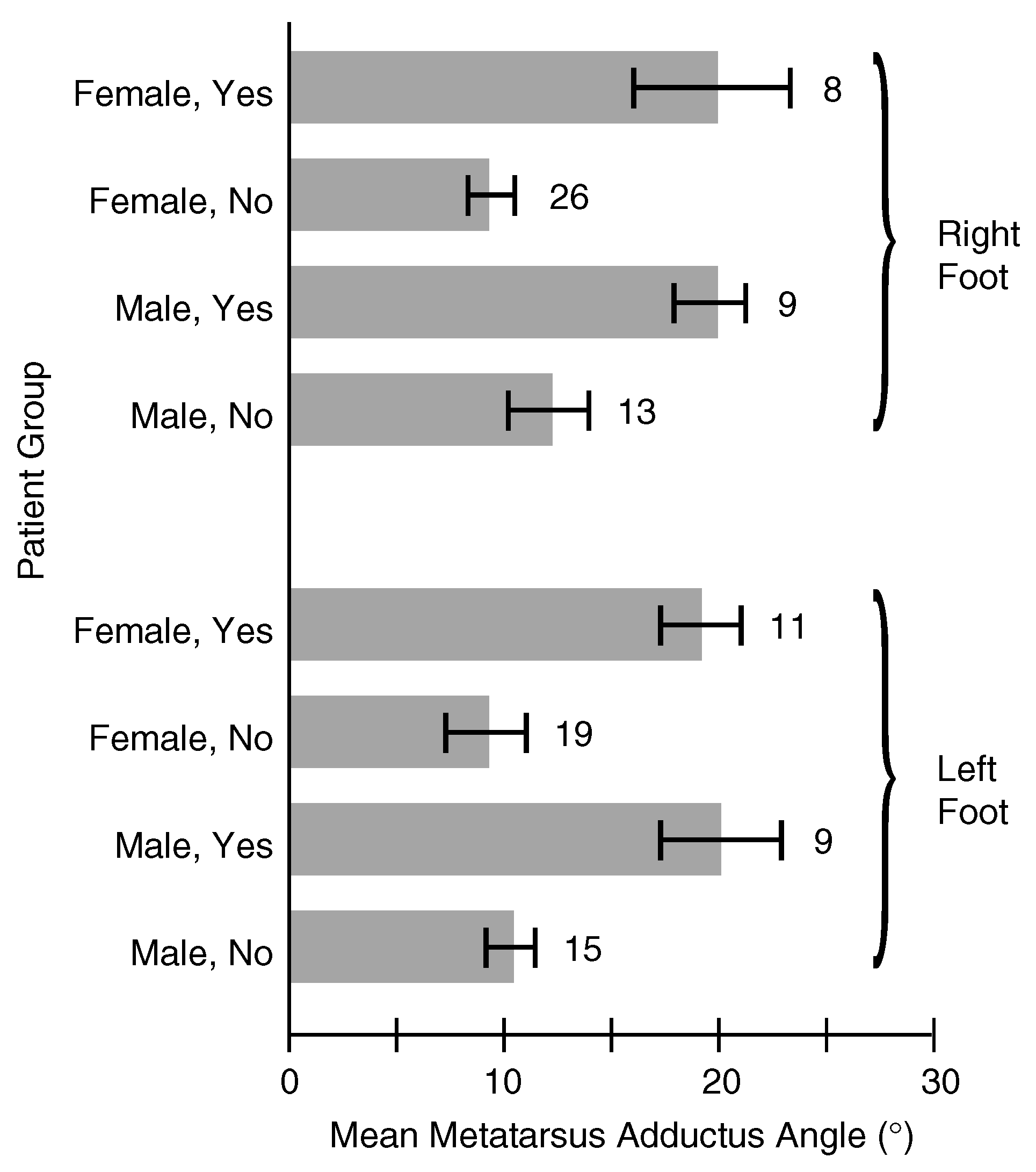
Figure 4.
Mean metatarsus adductus angle (with 95% confidence interval estimate of the mean) according to foot and sex (yes = metatarsus adductus present; no = metatarsus adductus absent). Numbers indicate number of x-ray patients per group.
Figure 4.
Mean metatarsus adductus angle (with 95% confidence interval estimate of the mean) according to foot and sex (yes = metatarsus adductus present; no = metatarsus adductus absent). Numbers indicate number of x-ray patients per group.
The BMI data suggest that most of the South Florida podiatric patients did not tend toward obesity (BMI ≥ 26 in adult men, [
28] BMI ≥ 27 in adult women [
29]). Most male and female patients were found close to the best-fit line in the age
versus BMI correlation. Foot disorders associated with obesity would appear to be not widely prevalent in the South Florida population, although this conclusion is based on indirect evidence (
Figure 3).
The South Florida sample did contain a larger than expected proportion of right-handed patients (93%
versus 89% reported in earlier literature [
4,
5]). Because the data were self-reported, there could have been confusion among some patients regarding the survey questions. It may be possible to attribute the discrepancy to misunderstanding among older patients about right- or left-handedness. Some of the middle-aged and elderly patients in the sample may have actually been left-handed but developed writing habits in elementary schools that stressed right-handedness despite preference for the left hand; there are no data available to test this hypothesis.
The data in this study do not support the prediction of Bodine [
5] of an association between the dominant limb and side of complaint. Neither do they support the hypothesis that metatarsus adductus is twice as prevalent in the left foot as in the right foot [
23]. It is possible that the significant association seen in the subsample of bilateral x-ray patients was a spurious or noncausal association, due perhaps to small sample size. In order to determine whether the association was spurious, larger samples of x-rays would be needed. The x-ray data obtained in this study did not necessarily represent a random sampling of all podiatric patients. The decision of whether to take x-rays was generally left to the discretion of the examining physician. When an x-ray of the right foot was taken, the physician sometimes asked for an x-ray of the left foot as well, even if the patient had not complained of a left-foot problem.
There is no evidence that laterality is associated with foot pathology, although the nature of the specific foot complaint could be open to interpretation. The patients were not grouped into complaint categories, such as trauma, vascular disorder, dermal disorder, and so on, because the self-reported data tended to be vague or incomplete. For example, complaints were reported often as “pain in right foot” or “swollen ankle.” In order to address the issue of disorder-specific associations, physicians’ assessments and diagnoses would have to be examined; this was beyond the scope of the current study.
Conclusion
In 1995–1996, 60% of South Florida podiatric patients were female. The demographic profile of the patients was skewed toward middle-aged and elderly individuals, perhaps more so than in any other US geographic region. These patients tended to be of average body size and build, although roughly 41% of the patients were obese. There was no evidence that general foot pathology was associated statistically with limb dominance. Metatarsus adductus deformity was relatively common among patients who received x-rays (prevalence, 62%), although its prevalence among all podiatric patients was unknown.